A COMPARATIVE STUDY OF SMALL INCISION
CATARACT SURGERY WITH
TRABECULECTOMY AND EXTRA CAPSULAR
CATARACT EXTRACTION WITH
TRABECULECTOMY IN EFFICACY OF
REDUCTION IN INTRA OCULAR PRESSURE
DISSERTATION SUBMITTED FOR
MS DEGREE (BRANCH III) OPHTHALMOLOGY
SEPTEMBER 2006
DEPARTMENT OF OPHTHALMOLOGY MADURAI MEDICAL COLLEGE AND GOVERNMENT RAJAJI HOSPITAL MADURAI.
CERTIFICATE
This is to certify that the dissertation entitled “A COMPARATIVE
STUDY OF SMALL INCISION CATARACT SURGERY WITH TRABECULECTOMY AND EXTRA CAPSULAR CATARACT EXTRACTION WITH TRABECULECTOMY IN EFFICACY OF REDUCTION IN INTRA OCULAR PRESSURE” presented herewith
by Dr. V.P SENTHILKUMAR to the faculty of Ophthalmology, The Tamilnadu Dr. M.G.R. Medical University, Chennai in partial fulfillment of the requirement for the award of M.S. degree in Ophthalmology is a bonafide work carried out by him under my direct supervision and guidance.
DECLARATION
I, DR.V.P.SENTHILKUMAR, solemnly declare that the dissertation
titled “A COMPARATIVE STUDY OF SMALL INCISION CATARACT
SURGERY WITH TRABECULECTOMY AND EXTRA CAPSULAR
CATARACT EXTRACTION WITH TRABECULECTOMY IN
EFFICACY OF REDUCTION IN INTRA OCULAR PRESSURE” has
been prepared by me.
This is submitted to The Tamil Nadu Dr.M.G.R. Medical University,
Chennai, in partial fulfillment of the requirements for the award of M.S Degree
Examination (Branch III) Ophthalmology to be held in SEPTEMBER 2006 .
Place: Madurai.
Date :
ACKNOWLEDGEMENT
I am deeply indebted to DR. R. GITA RAMANI M.S., D.O,
Professor and Head of the Department of Ophthalmology, Madurai medical College, Madurai for the able guidance, inspiration and encouragement she has rendered at every stage of this study.
I express my heartfelt gratitude to DR. R. UNNAMALAI M.S.,
DO, Additional Professor & Chief in the Department of Ophthalmology, Madurai Medical College for her valuable advice and help in carrying out this study.
I acknowledge with gratitude the guidance and persistent
encouragement given to me by my Assistant professors
DR.G.S. SRINIVASAN M.S., DO, DR.T. BADRINARAYANAN M.S., DO, DR.A.R. ANBARASI M.S., DO, AND DR. K. SIVAKUMAR MS.,
My sincere thanks to DR. R. SARASWATHI MS. Dean, Madurai
Medical College, Madurai for permitting me to utilize the clinical materials of the hospital.
I would like to thank my patients, friends and colleagues & family
CONTENTS
TITLE PAGE NO
PART I
1) Introduction 1
2) Review of literature 3
3) Anatomy 8
4) Evolution of cataract surgery 16
5) Evolution of combined surgery 18
6) Goals of treatment 21
7) Indications for surgical management 22
8) Surgical Procedure 23
9)Bleb characteristics 33
10) Complications 36
PART-II
11) Aim of the study 45
12) Materials and methods 46
13) Results and observation 51
14) Discussion 62
15) Conclusion 66
ABBREVIATIONS
AC - ANTERIOR CHAMBER
ECCE - EXTRA CAPSULAR CATARACT EXTRACTION
HM - HYPER MATURE CATARACT
ICCE - INTRA CAPSULAR CATARACT EXTRACTION
IMC - IMMATURE CATARACT
IOL - INTRA OCULAR LENS
IOP - INTRA OCULAR PRESSURE
MC - MATURE CATARACT
PACG - PRIMARY ANGLE CLOSURE GLAUCOMA
PCIOL - POSTERIOR CHAMBER INTRA OCULAR LENS
PHACO - PHACOEMULSIFICATION
POAG - PRIMARY OPEN ANGLE GLAUCOMA
P - PROBABILITY
PXF - PSEUDO EXFOLIATION
SACG - SECONDARY ANGLE CLOSURE GLAUCOMA
SICS - SMALL INCISION CATARACT SURGERY
TN - TENSION
INTRODUCTION
Glaucoma is a chronic progressive optic neuropathy caused by a group
of ocular conditions, which lead to damage of the optic nerve with loss of
visual function. Most common risk factor is raised intra ocular pressure.
Glaucoma is a group of diseases characterized by visual field changes
and progressive optic neuropathy for which raised Intra ocular pressure is a
risk factor.
Glaucoma is the leading cause of irreversible blindness throughout the
world; glaucoma coexisting with cataract is commonly prevalent among
elderly patients. The frequent coexistence has led to many ideas as to how best
to control glaucoma and at the same time improve the vision. Many successful
designs of combined operation have been tried in last 10 years such as ECCE,
ICCE and SICS, IOL implantation combined with new filtering procedures.
Glaucoma triple procedures can provide good visual rehabilitation and
IOP control in cases of co-existing cataract and primary open angle glaucoma.
that, when combined with trabeculectomy, one could expect visual
performance and intraocular pressure control as good as that with two separate
procedures.
This study is undertaken to compare the efficacy of reduction in IOP
between extra capsular cataract extraction with trabeculectomy and small
REVIEW OF LITERATURE
Literature search was performed from Medline using key words small
incision cataract surgery with trabeculectomy and extra capsular cataract
extraction with trabeculectomy
From the literature very few articles are seen comparing ECCE with
trabeculectomy and SICS with trabeculectomy.
Neumann et al20 used two groups, each consisting of 23 patients and
performed combined procedure in group-1 without intra ocular lens and
combined procedure with intra ocular lens in group-2. The pressure reduction
was almost the same in both groups.
Edwards8 studied 59 eyes that underwent combined Cataract extraction
and trabeculectomy. The mean age of the patient included in the study was 69
years with a range of 23 to 87 years. The visual outcome was good and IOP
reduction was achieved in 91.5% at 6 months and he stated that combined
Tord Jerndall11 performed combined trabeculectomy and cataract
extraction on 17 eyes of 16 patients. The mean intra ocular pressure
preoperatively was 30 mm Hg and postoperative IOP was 16 mm Hg. He
concluded that the combined technique is no more traumatic than an ordinary
extraction.
Jhons et al12 performed cataract extraction with trabeculectomy in 37 eyes
of 29 patients with an average follow-up of 23.7 months. Average preoperative
IOP was 23.5 mm Hg and postoperative IOP was 16.22 mm Hg. No greater
incidence of complication was evident as compared to trabeculectomy. So it
confirmed that combined procedure was a safe and valid procedure.
Bruce shields29,30 selected 73 cases of combined cataract and glaucoma
and performed cataract extraction alone in one group of patients and a two stage
procedure of filtering surgery followed by cataract extraction in second group of
patients and combined procedure in third group of patients. The author prefers
the two-stage procedure as an effective procedure.
and fewer complications, combined procedure is good for coexisting glaucoma
and cataract.
Mammalis et al17 performed a retrospective study of 212 eyes of 174
patients who underwent Phacoemulsification, Intra ocular lens implantation and
trabeculectomy with follow up of 26 months. The mean preoperative IOP of 23.1
mm Hg decreased postoperatively to 15.9 mm Hg. Hence he concluded
phacoemulsification, intra ocular lens implant and trabeculectomy yield
excellent results.
Percival24 conducted a study of 34 glaucoma eyes by performing triple
procedure and concluded that satisfactory control of glaucoma was achieved in
each case and 91% of eyes improved in visual acuity to 6/12 or better and hence
concluded that triple procedure is safer.
Nielson21 performed combined surgery in 36 cases of which 10 underwent
trabeculectomy by method 1 and 26 by method 2. Method 1 was conventional
trabeculectomy and in method 2 incision was started over ciliary body and
cleavage made forward until the scleral spur, where the anterior chamber was
procedure. There was no significant difference in visual rehabilitation between
the methods. The major complication was anterior chamber bleeding.
Simmons et al31 performed ECCE with IOL with trabeculectomy in 75
cases who had preoperative mean IOP of 19.3 mm Hg and used an average of
2.3 antiglaucoma medications. Follow up was done on day 1 and 2, 6, 12
months. Postoperative average IOP was 3.8 mm Hg lower than the preoperative
value, which is significant. Filtering bleb started gradually disappearing during
12 months. Hyphema was found to be more in limbal-based flap.
Wishart et al42 conducted a prospective study of 34 eyes, which
underwent phacoemulsification, intra ocular lens implant and trabeculectomy
and followed for a period of one year. The results were compared with those
obtained retrospectively of the previous 34 extra capsular extractions with intra
ocular lens with trabeculectomy performed by the same surgical team. This
study showed phacogroup had earlier visual rehabilitation, less postoperative
astigmatism, improved long term control of intra ocular pressure and less
postoperative complications.
patients (68.87%) were predominant in the study. At the end of one year the IOP
control in both the group was similar (phaco group: 14.77.ECCE group: 14.04).
The study concluded that phaco triple has the advantages like early post
operative IOP control, early visual rehabilitation and lower astigmatic shift.
DETAILS OF ANATOMY RELEVANT TO
SURGICAL PROCEDURE
Limbus is the most important transition zone at the corneoscleral
junction; most glaucoma surgical incisions are placed at limbus, the success of
surgery is dependent on the accuracy of the incision with relation to the limbus.
GROSS ANATOMY
The limbus is the transition zone between the cornea and sclera. On the
inner surface of the limbus is an indentation, the scleral sulcus, which has a
sharp posterior margin, the scleral spur and a sloping anterior wall that extends
to the peripheral cornea.
The trabecular meshwork, bridges the scleral sulcus and converts it into
a tube, called schlemm’s canal and where the meshwork inserts into the
Schlemm’s canal is connected by intrascleral channels to the episcleral
veins. The trabecular meshwork, schlemm’s canal and the intra scleral
channels comprise the main route of aqueous humor outflow.
The ciliary body attaches to the sclera and creates a potential space, the
supraciliary space between itself and the sclera.
On cross section, the ciliary body has the shape of a right angled
triangle and the ciliary processes. (the actual site of aqueous production)
occupy the innermost and the anterior-most portion of this structure, extending
back for approximately 2mm in the region called pars plicata .The ciliary
processes consists of approximately 70 radial ridges (major ciliary processes)
and equal number of smaller ridges (minor ciliary processes).
The posterior 4mm of the ciliary body, the pars plana (orbicularis
ciliaris) has a flattened inner surface and joins the choroid at the ora serrata.
The Iris inserts into the anterior side of the ciliary body. The lens is
suspended from the ciliary body by zonules and separates the vitreous
The Iris separates the aqueous compartment into posterior and an
anterior chamber and the angle formed by the Iris and cornea is called the
SURGICAL ANATOMY
ANTERIOR LIMBUS
On the external surface of the eye, anterior boundary of the limbus is the
termination of Bowman’s membrane, 0.5mm anterior to the insertion of the
conjunctiva and Tenon’s capsule. This is the corneolimbal junction (or)
anterior limbus.
The conjunctiva inserts more anteriorly in the superior and inferior
quadrants. So the limbus is wider in these quadrants, ranging between 1 and
1.5mm and gradually tapers in the nasal and temporal quadrants where the
range is 0.3 to 0.5mm. Hence during performing filtering surgery surgical
incisions at 12’o clock position have an advantage of wider area.
The conjunctiva and tenons capsule cover the limbus, the adhesions
between the conjunctiva and tenons capsule are moderately firm and sharp
dissection is required to dissect between these two structures.
The adhesion between the tenons capsule and the underlying limbus and
POSTERIOR LIMBUS
This is also known as sclerolimbal junction. It is identified as the
junction between opaque white sclera posteriorly and bluish grey translucent
limbus anteriorly.
This helps to identify the location of deeper structure of the anterior
chamber angle. The scleral spur is located just posterior to the sclero limbal
junction and schlemm’s canal found just anterior to this landmark.
In trabeculectomy a circumferential incision at the corneolimbal
junction enters the anterior chamber just in front of the trabecular meshwork by
extending the incision posteriorly to expose the anterior chamber angle
structures and excised along the scleral spur.
VESSELS
Besides the large arteries and veins in the rectus muscle insertions,
variable arrays of episcleral vessels are present in the perilimbal area. Most of
the bleeding encountered during preparation of conjunctival flaps comes from
avoided to prevent shrinkage. Cautery of the conjunctival edges should also be
avoided to prevent possible wound leak.
IRIS AND CHOROID
Normal iris vessels usually do not bleed after iridectomy. Bleeding from
iris new vessels may be massive, often when intra ocular pressure is suddenly
lowered. Bleeding from the anterior edge of the ciliary body or the posterior
part of deep scleral excision may be substantial but will often stop with simple
compression.
The choroid includes the choroidal capillaries immediately external to
the Bruch’s membrane. The arterial blood flow into the choriocapillaries is
from the branches of the short posterior ciliary arteries through perpendicularly
oriented arterioles. Shearing of these arterioles during surgical manipulation
(or) distortion of the eye wall explains the intra operative choroidal
ROUTES OF DRAINAGE OF AQUEOUS HUMOR
Following combined surgery (trabeculectomy followed by cataract
surgery) the drainage of aqueous humor occurs through one (or) more of the
following routes.
1. Filtration through outlet channels in scleral flap
2. Filtration through connective tissue substance of scleral flap
3. Filtration around margins of the scleral flap
4. Filtration through the cyclodialysis cleft (if tissue dissected posterior to
scleral spur)
5. Through cut ends of the schlemm’s canal.
Aqueous humor, which drains through any of the above channel, filters
through the sub conjunctival tissue and then through the conjunctiva diffuses
into the tear film. Also the vascular conjunctival tissue may absorb a portion
of the aqueous.
In the absence of a well-defined filtering bleb aqueous drains through
the lymphatic vessels, normal aqueous vessels and atypically incorporated
PATHOPHYSIOLOGY
After surgical trauma to the conjunctiva, episcleral and iris blood
vessels, leakage of plasma proteins and blood cells occur. Subsequently on
exposure to the tissue factors, a clot with fibrin fibronectin matrix is formed.
Inflammatory cells, new capillaries and fibroblasts migrate into this. The
inflammatory cells eventually degrade the fibrin fibronectin matrix. Later
fibronectin, collagen and glycosaminoglycans form a fibrovascular granulation
tissue. Gradually a dense collagenous subconjunctival scar with scattered
EVOLUTION OF CATARACT SURGERY
From pioneer Susruta’s couching to Kelmans phaco emulsification,
cataract surgery has witnessed a phenomenal progress.
1000 BC – 1745 AD couching
Susruta practiced this; the sclera is pierced with a lancet and blunt
instrument to depress the lens.
1745-1865AD –primitive extracapsular extraction
- Jacques Daniel first performed the procedure
- Albright Von Graefe (1828-1870) improved with
development of knife that created better wound apposition.
- Samuel Sharp – Expressed cataractous lens.
- Williams (1869) first to use corneal suture.
20 th century – intra capsular cataract
Sharp and Pagen Stecher – delivered the lens in toto by pressure
counter pressure method.
Intraocular lens implantation
- In 1949 Harold Ridley proved visual rehabilitation by
intraocular lens.
Conventional ECCE
After IOL implantation started, ECCE techniques improved with
improvement of instruments and the use of microscope.
Phacoemulsification
Kelman (1970) described this technique where ultrasonic vibrations are
EVOLUTION OF COMBINED SURGERY
The history of the evolution of combined surgery, deals mainly with the
various changes that anti glaucoma surgery underwent and also the changes that
overtook cataract surgery in later times.
In 1830, Mackenzie described sclerotomy and paracentesis.
In 1847, Crichett incarcerated an Iris wick and termed it iridodesis.
In 1856, Von Graefe, believed that excision of Iris reduced aqueous
secretion and performed sector iridectomy, it was followed as a standard
procedure for next 10-15 years where filtering bleb was thought as an
undesirable complication (in 20% of cases).
In 1869, it was De Wecker who understood the role of the filtering
cicatrix and emphasized, that an artificial pathway through the wall of the eye
was a way for aqueous to drain and that iridectomy could not be a cure in all
cases.
In 1906, La Grange performed sclerecto iridectomy, which acted as
In 1906 again Holth developed, Iridenclesis, a processes of incarceration
of pillars of Iris tissue, in the limbal wound, there by functioning as wicks.
In 1909 Elliot described limbal trephination.
In 1924 Preziosi pioneered the technique of thermal sclerostomy, which
involves, thermal cautery of scleral wound edge, with gradual entry into the
anterior chamber.
In 1958, Schie modified thermal sclerostomy by adding peripheral
iridectomy.
Watson in 1970 modified it by suggesting only a limbal-based partial
thickness scleral flap.
The advantages of partial thickness filtration surgery were that the rate of
aqueous outflow was controlled, so there was less postoperative complication
and the fall of intraocular pressure was controlled and predictable.
In 1962 Iliff and Hass pioneered the procedure of posterior lip
All the above procedures, described so far are full thickness filtering
procedures, in that the aqueous in anterior chamber is in direct contact with the
sub conjunctival space without any guard. While decrease in intraocular pressure
following surgery was excellent.
These procedures were often complicated by excessive aqueous filtration
that leads to prolonged flat anterior chamber associated with corneal
decompensation, synechiae formation, filtering blebs become very thin and are
prone to rupture resulting in endophthalmitis.
HISTORY OF PARTIAL THICKNESS PROCEDURES
Sugar pioneered partial thickness flap filtration surgery in 1967. It was
Cairns in 1968, who popularized the partial thickness filtration surgery.
Trabeculectomy with a scleral flap that was hinged posteriorly in the sclera (or)
GOALS OF TREATMENT
The treatment of glaucoma traditionally has been lowering the intra ocular
pressure, although the reduction (or) controlling does not normally preserve the
visual field in glaucoma patients. The continued progression of glaucomatous
optic neuropathy even after control of intra ocular pressure suggests that pressure
independent factors do play a role in the development of glaucoma.
Further several factors, which contribute to optic nerve damage, are
hypersensitive to intra ocular pressure such that even though it is within the
normal range the neurons still die. The ultimate goal is not only risk factors
management but also protection of optic nerve and preservation of visual fields.
The current practice is to estimate the pressure level below which further
damage to optic nerve is unlikely to occur (target pressure) and then aim to keep
the intra ocular pressure consistently below this level. In the average point
clinician should aim for a pressure of about 20% below the initial untreated
pressure. Patient having glaucoma along with cataract should be cleared of from
INDICATIONS FOR SURGICAL MANAGEMENT
1. Documented visual loss and optic nerve head damage despite maximum
tolerated medical therapy and laser treatment that threatens vision.
2. Anticipated progressive damage experienced in the same eye (or) fellow
eye that indicates that the current course will lead to loss of vision (or)
intolerably high intraocular pressure.
3. Medication failure due to ineffectiveness, intolerance, poor compliance.
4. Intraocular pressure that is high enough to place the future health of the
optic nerve at significant risk. This pressure will differ dramatically
depending on the condition of the optic nerve and patients prior history.
5. Family history of glaucoma.
6. It can be combined with cataract procedure if there is borderline intra
ocular pressure control, advanced damage (or) history of postoperative
IOP increase in the fellow eye.
DETAILS OF SURGICAL PROCEDURE
There are basically three choices in surgically dealing with coexisting
cataract and glaucoma
- Cataract extraction alone
- Trabeculectomy alone with cataract surgery later and
- Combined cataract surgery and trabeculectomy
Since this study is following up results of Trabeculectomy with
extracapsular cataract extraction and trabeculectomy with small incisional
cataract surgery the procedure of these two will be dealt as part
I. Extra capsular cataract extraction with trabeculectomy
II. Small incision cataract surgery with trabeculectomy
ANAESTHESIA
By peribulbar anaesthesia with 5ml of 2% lignocaine with 50IU/ml
hyaluronidase and 1:200000 adrenaline, given at two different sites with 23
gauge needle.
One at the lower orbital margin about lateral one third and medial two
third of about 3ml and other medial to supraorbital ridge about 2ml. Ocular
ECCE WITH TRABECULECTOMY
PROCEDURE
After separating the lids using a universal eyelid speculum a 4-0 silk
superior rectus bridle suture is applied.
CONJUNCTIVAL FLAP
A Limbal (or) Fornix based conjunctival flap taken with blunt tipped
scissors and conjunctiva is handled with a non toothed forceps to avoid button
holing of the conjunctiva and scarring which would jeopardize a good bleb.
A Limbal based conjunctival flap is preferred over Fornix based as it helps
in tighter wound closer and better retention of aqueous.
SCLERAL FLAP DISSECTION
The region of sclera, planned for preparation of flap is cauterized with a
wet field cautery to reduce bleeding, the flap is 1/2 to 1/3 scleral thickness,
equilateral triangle shaped and hinged at the limbus.
The flap is extended well past the limbus into the cornea, so that at least
1mm of bluish gray zone of the limbus is exposed.
CREATION OF INNER SCLEROSTOMY
The anterior chamber is entered with a blade just behind the hinge of the
scleral flap, and the incision is widened with scissors to about 4mm.
Radial incisions are then extended posteriorly on the either end of the
initial incision, posteriorly parallel to the first one there by completing the
rectangular sclerostomy.
This block of tissue 4mm by 1mm is reflected and excised with scissors
along the scleral spur. The block of tissue removed contains brownish trabecular
tissue.
PERIPHERAL IRIDECTOMY
After the inner sclerostomy has been made a peripheral iridectomy is
routinely done. Care should be taken to make a broad peripheral iridectomy, to
EXTRACAPSULAR CATARACT EXTRACTION AND PCIOL
IMPLANTATION
Partial thickness corneoscleral groove made at midlimbal line along the
edge of scleral flap from 10 ‘o clock to 2’o clock position.
Through the inner sclerostomy wound using a suitably bent 26 G tipped
needle, canopener anterior capsulotomy done, after protecting the corneal
endothelium with sufficient amount of viscoelastic substances in the anterior
chamber.
The corneoscleral incision is completed along the pre-placed groove using
a corneal scissors, taking care not to injure the scleral flap. The nucleus is
delivered by pressure –counter pressure technique using lens expressor and
vectis.
Cortical wash out is then carried out with simcoe cannula attached to
sterile ringer lactate solution. Using a lens holding forceps, IOL is implanted in
the capsular bag with the help of Mc phersons forceps. Then the IOL is dialed
WOUND CLOSURE
The triangular scleral flap is closed with 10-0 nylon suture 1 interrupted
suture is applied to the apex of the triangle and 2 interrupted sutures are applied
to the base of the triangular flap.
The corneoscleral wound is then closed with interrupted 10-0 nylon
sutures in a radial manner, the corneoscleral bite ratio being 1:1. The
conjunctival flap is closed with 10-0 silk sutures or with cautery to achieve
watertight approximation.
POST OPERATIVE CARE
Topical steroids are used to reduce the scar formation of the filtering bleb
SMALL INCISION CATARACT SURGERY WITH
TRABECULECTOMY
Small incision cataract surgery allows a dramatic reduction in the incision
size compared to that for an extracapsular procedure.
The size of the incision depends on the size of the lens to be placed. In
using Polymethyl Methacrylate lens the incision may be reduced to 5-7 mm, when
using foldable lenses it is reduced up to 3-3.5mm.
The technique for a combined surgery is of two types.
Performing phacoemulsification through a superior scleral incision, which
is later, converted to a trabeculectomy flap.
Performing two procedures at different sites with a superior
trabeculectomy and temporal clear corneal cataract extraction.
The later has an advantage of decreased manipulation of the conjunctival
and scleral flaps. This may decrease the level of conjunctival inflammation and
PROCEDURE
THE CONJUNCTIVAL FLAP
Fornix based flap is easier and useful in scarred conjunctiva.
Limbal-based flap is having lesser chance of post-operative leakage and leads to a
better-formed bleb in the early postoperative period. Disadvantage of it is making
cataract extraction field cumbersome.
The width of the conjunctival incision should be approximately 2mm
larger than the anticipated scleral incision.
Wet field cautery can be used to control bleeding. Achieving haemostasis
is important because subconjuctival blood can obstruct filtration and lead to
scarring.
SCLERAL INCISION
A three stage scleral tunnel is created with a straight incision
approximately 2mm posterior to the limbus. The initial incision is vertical. The
second stage of the incision involves tunneling in a horizontal plane into the
The length of the incision should be equal to the insertion width of a
Polymethyl Methacrylate IOL.
THE PUPIL MANAGEMENT
Glaucoma patients present with small pupil resulting from long-term
miotic use. If the pupil measures less than 4mm after maximally dilated we can
use the following methods for dilatation.
Pupils dilated with two kuglan hooks, pulling pupils 180 degrees apart by
push (or) pull, holding the hooks in maximum stretch for a few seconds.
Multiple equally spaced partial thickness sphincterotomies can be made
with long handle vannas scissors.
Use of high viscosity viscoelastic.
THE CAPSULOTOMY
A 360-degree continuous capsulorhexis is preferable as it aids in capsular
fixation of the lens.Performed with a 26 gauge bent needle (or) capsulorhexis
forceps.
NUCLEUS DELIVERY AND PCIOL IMPLANTATION
- Hydro dissection of lens cortex from capsule.
- Nucleus is rotated and made out to the anterior chamber.
- Nucleus is delivered through the scleral wound with help of
irrigating vectis (or) by visco expression.
- Polymethyl Methacrylate lens is implanted in the capsular bag.
THE TRABECULECTOMY28
After placement of the IOL, the scleral tunnel is modified into a
trabeculectomy flap. One edge of the tunnel is transected with keratome creating
a flap.
The Kelly’s descemet punch is then used to remove clear cornea and
trabecular meshwork from the posterior lip of scleral tunnel. Another option is to
A portion of the basal iris is removed with the vannas scissors creating a
peripheral iridectomy
SCLERAL WOUND CLOSURE
The scleral flap is closed with permanent sutures (or) with releasable
sutures .10 – 0 nylon is used close the sclera.
CONJUNCTIVAL FLAP CLOSURE
The conjunctiva can be secured to episclera with one interrupted suture at
each end of the conjunctival incision.8-0 (Or) 9-0 vicryl used, since it does not
require removal post operatively.
POSTOPERATIVE CARE
Patient should initially be taken off all antiglaucoma medication and
started on a steroid –antibiotic drop. Cycloplegics for all cases up to 4-6 weeks
to maintain anterior chamber.
If pressure is high (or) if the bleb is low due to under filtration, aqueous
can be released by depressing the posterior lip of the flap with a sterile cotton tip
BLEB CHARACTERISTICS
PHYISIOLOGY
Filtration surgery creates a fistula between the anterior chamber and the
subconjunctival space.
CHARACTERISTICS
The appearance of filtering bleb is an important factor in evaluating the
outcome of glaucoma filtering surgery. The successful outcome usually
demonstrates a reasonable bleb with minimal engorgement of conjunctival
vessels during the first week. The bleb tends to become more localized in second
and third weeks. By the end of the first month the bleb is well established and
moderately diffuse, the bleb will gradually become less hyperemic and at three
months will be well established with small microcysts visible on the conjunctival
surface.
TYPE I
Bleb has a thin and polycystic appearance resulting from trans
conjunctival flow of aqueous. It is associated with good filtration.
TYPE II
Bleb is flat, thin and diffuse with a relatively avascular appearance in
comparison to the surrounding conjunctiva. This is also indicative of good
filtration.
TYPE III
Bleb is non-filtering due to the subconjunctival fibrosis. It is flat and not
associated with microcystic spaces, contains engorged blood vessels on its
surface.
TYPE IV
Encapsulated bleb (tenon’s cyst) is a localized, highly elevated dome
shaped cyst like cavity of hypertrophied tenon’s capsule with engorged blood
FACTORS RELATED TO BLEB FAILURE
AGE
Vigorous healing response has been at least partly attributed to for poor
success of filtering surgery.
RACE
Vigorous wound healing response in blacks account for increased rate of
failure in bleb.
EXTERNAL FACTORS
Fibroblast proliferation, hence Tenon’s capsule should be removed to
prevent fibrosis.
INTRAOCULAR FACTORS
Blockage of filtration site by prolapse of lens, iris, vitreous and
ciliary body.
Inadequate sclerostomy to establish communication with the
anterior chamber.
Fibrous and cellular response associated with ocular inflammation
decreases the success of glaucoma filtering surgery.
COMPLICATIONS
INTRAOPERATIVE
Tearing or buttonholing of conjunctival flap
The conjunctiva may be inadvertently torn during the procedure. It can be
sutured with a mattress or running suture. Alternatively tissue adhesives or light
bipolar cautery may be used.
Haemorrhage
Commonest cause is episcleral bleeding. Minimal cauterization along the
bleeding vessel will stop it. Inadvertent cutting of ciliary body can cause severe
bleeding. Alternative is sustained pressure or large air bubble in AC. A choroidal
or expulsive haemorrhage is a devastating complication. Management is
immediate closure of the fistula.
Choroidal Effusion
Can occur in eyes with prominent episcleral vessels as in Sturge Weber
syndrome.
Indicated by sudden shallowing of AC intra operatively or rotation of
Vitreous Loss
Adequate vitrectomy should be done with vitrectomy instrument
Stripping of Descemet membrane
Large air bubble can be used to reposition it.
Tearing of scleral flap
It can be sutured with 10-0 nylon mattress sutures or if the flap is very
thin it can be replaced by a donor sclera
Lens Injury
Very rare.
EARLY POST OPERATIVE COMPLICATIONS
Shallow and Flat Anterior Chamber
This can lead to peripheral anterior synechiae, corneal endothelial
damage, cataract & hypotony associated maculopathy if it is not resolved soon.
Divided into 3 grades depending on severity.
Grade 1: Peripherally flat AC with iridocorneal touch but preservation of
anterior chamber in pupillary space.
Grade 2: Greater apposition between mid iris and cornea but some space
Grade 3: AC flat with complete contact of iris and pupillary space with
posterior surface of cornea.
Gr: 1& 2 almost always resolve with time and cycloplegia.
Gr 3 is a medical emergency and usually requires surgical intervention; if
not resolved in 1-2 days.
Flat Anterior Chamber with Hypotony
A Seidel test should be performed with 2% fluorescein over the bleb. If
there is aqueous leak, fluorescein will be diluted by the escaping aqueous, which
will be seen as bright green with cobalt blue filter.
Sometimes multiple punctate staining areas (sweating) may be seen which
later forms hole and show positive test.
Scleral Flap Leakage
Signs: Well formed bleb, low IOP, negative Seidel test.
Can be avoided by tight suturing of the flap with releasing sutures or
Bleb Leakage.
Signs: Flat bleb, low IOP & positive Seidel test.
Most common cause is inadequate suturing of conjunctiva. or inadvertent
button hole. Management includes atropine cycloplegia and topical & systemic
aqueous suppressants, pressure patching, large soft contact lens, collagen shield,
Simmons shell, compression sutures etc.
Full thickness hole may require conjunctival advancement to hood
existing bleb or free conjunctival patch autograft with removal of existing bleb.
Post operative hypotony is often associated with choroidal effusions or
detachments.
Indications for surgery are persistent kissing choroids or Grade 3 flat AC
damaging corneal endothelium.
AC is maintained with balanced salt solution or viscoelastic and supra
FLAT ANTERIOR CHAMBER IN NORMOTENSIVE AND
HYPERTENSIVE EYES
Pupillary Block
Signs are high IOP, flat bleb, negative Seidel test & iris bombe with
incomplete or non patent iridectomy.
Patency of peripheral iridectomy is confirmed. Laser or incisional
iridectomy should be done if necessary.
Ciliary Block Glaucoma (Malignant glaucoma)
Rare, but serious complication. Aqueous is blocked at the pars plicata of
ciliary body & is misdirected to circulate into or behind vitreous. Presents with
Shallow AC, high IOP, absent bleb & negative Seidel test.
Initially topical mydriatics, steroids, topical and systemic antiglaucoma
medications are used to reduce IOP.
Verify or create patent iridectomy or posterior capsulotomy in
Suprachoroidal haemorrhage
Rare. More often seen in aphakia, vitrectomised eyes, large eyes with
pathologic myopia or congenital glaucoma. Characterized by sudden severe pain
with loss of vision in first week.
If aqueous suppressants and hyperosmotics fail to lower IOP in 5 days,
sclerostomy and drainage is indicated.
Rarely intra operative expulsive supra choroidal haemorrhage can occur.
Then limbal incision should be closed immediately and posterior sclerostomy
should be performed over the presumed site of bleeding and blood should be let
out from the supra choroidal space.
Hyphema
Bleeding may be from iris, anterior ciliary body, or corneoscleral wound.
Usually subsides without intervention. Activities must be restricted. Protective
eye shield should be worn.
Intraocular infection
Risk factors include use of antimetabolites, myopia, thin bleb with leak,
presence of releasable sutures, concurrent upper respiratory tract infection and
blebs located in the inferior limbus.
Can be differentiated into
Blebitis: infection does not involve vitreous
Bleb will be milky white (stage 1) along with anterior uveitis (stage 2)
and normal red reflex.
Endophthalmitis: infection penetrating to the vitreous with impaired red reflex.
Blebitis respond to intensive antibiotic therapy with good visual outcome.
Smears and cultures have to be made from lids, conjunctiva and filtering blebs.
Staphylococcus and Streptococcus account for half of the culture positive
organisms. When hypopyon is present fluid for culture should be obtained from
AC and vitreous.
Endophthalmitis should be managed with vitreous biopsy and intra vitreal
antibiotics.
Sympathetic ophthalmia
Filtration failure
It can occur at any time from weeks to years after surgery. Management
depends on the cause.
Subconjunctival fibrosis is the most common cause.
Iris, vitreous, clot, ciliary process or lens plugging sclerostomy site.
Retained visco elastic substance
Imperforate Descemet membrane
Scleral flap too tightly sutured
Ciliary or pupillary block
MANAGEMENT OPTIONS.
• Digital compression through lower lid for 5-10 seconds.
• Focal compression under topical anesthesia with a moist sterile cotton
bud at the edge of the scleral flap.
• Suture manipulation with releasable sutures or argon laser suture lysis
within 2 weeks.
• Needling in an encapsulated bleb
• Subconjunctival injection of 5-fluorouracil, 5mg,10mm away from the
bleb.
LATE POST OPERATIVE COMPLICATIONS
Overhanging Filtering Blebs
Reduced by argon laser or excision and suturing
Spontaneous hyphema
Hypotony and ciliochoroidal detachment
Others
Corneal astigmatism. Usually meridian passing through the
trabeculectomised site has the strongest curvature. Making scleral flap at 12’O
clock can reduce this.
Corneal epithelial toxicity is usually mild and can be managed
AIM OF THE STUDY
To compare the efficacy of reduction in IOP between small incision
cataract extraction and posterior chamber intra ocular lens implantation with
trabeculectomy and extra capsular cataract extraction and posterior chamber
intra ocular lens implantation with trabeculectomy in cases of glaucoma
MATERIALS AND METHODS
Prospective, randomized and comparative type of study involving 50 eyes of
50 patients with glaucoma and cataract undergoing small incision cataract
surgery and posterior chamber intraocular lens implantation with
trabeculectomy and extra capsular cataract extraction and posterior chamber
intraocular lens with trabeculectomy in Government Rajaji Hospital,
Madurai Medical College, Madurai.
Duration of study
This study was done for a period of 1 year from January 2004 to January
2005, The follow up of the patients was done for a period of 12 months with
follow ups on 2nd week, 4th week, 6th week, 3rd month, 6th month and 12th
month.
Inclusion criteria
All cases of primary open angle glaucoma, primary angle closure glaucoma
and secondary angle closure glaucoma where in IOP was not controlled by
maximum medical therapy to less than 21 mm Hg having visually
Exclusion criteria
Eyes with other types of glaucoma
Traumatic cataract
Complicated cataract
Subluxation of lens
Fundus pathology affecting visual acuity
Patients with corneal opacity
Eyes with prior filtering surgery
Methodology
Among the patients who were seen during the recruitment period in the
ophthalmology department of Government Rajaji Hospital, we selected 50
eyes of 50 patients who satisfied our criteria to undergo surgery.
All the patients enrolled in our study received a standard ophthalmological
examination which included recording of best-corrected visual acuity, which
was determined by subjective and objective refraction with standardized
Evaluation of anterior segment of both eyes was done using slit lamp
biomicroscopy to evaluate the extent of cataract and glaucoma and special
attention was taken to note any abnormalities like corneal edema ,scarring or
opacity .
Preoperative baseline IOP and in all follow-up visits IOP were recorded for
all eyes with Schiotz indentation tonometer.
IOP ideally has to be below 25 mm Hg before surgery with antiglaucoma
medication and pilocarpine was stopped 1 week before surgery. If
pre-operative IOP was above 30 mm Hg. Surgery was planned after IV
mannitol.
The status of angle was determined by performing gonioscopy with
goldmann 3 mirror gonio lens. Angle grading was done by Shaffers grading
system. Other features of angle like PXF, hyperpigmentation of trabecular
meshwork and Sampoelesi line were also noted.
with special emphasis on zonular dialysis, posterior capsular rent, vitreous
loss and inability to implant IOL.
Postoperative examination was done after 24 hours and the status of bleb,
corneal edema, iritis, anterior chamber depth, hypotony and other
complications like IOL instability were noted.
The standard post operative medication given were ciprofloxacin (0.3 %)
and dexamethasone (0.1%) topically administered 6 times a day for one
week and then tapered over a period of 6 to 8 weeks.1% cyclopentolate is
given twice daily for 15 days.
After 48 hours vision with pinhole was recorded and IOP was recorded after
15 days.
Detailed slit lamp examination was done on day one, 3 weeks, 6 weeks, 3
months, 6 months and 12 months post operative period. During each visit
visual acuity, IOP by schiotz tonometer and bleb characteristics were noted
and recorded carefully. Objective and subjective refraction done at the end
Computer analysis of data was done utilizing the software - Epidemiological
Information Package 2002 developed by the Centers for Disease Control
and Prevention-Atlanta for World Health Organization.
Percentage, mean, standard deviation and ‘p’ values were calculated using
this package.
Chi Square test was done to find out significance of relationship between
both groups. Since the variances were not homogenous, kruskul-wallys test
RESULTS AND OBSERVATION
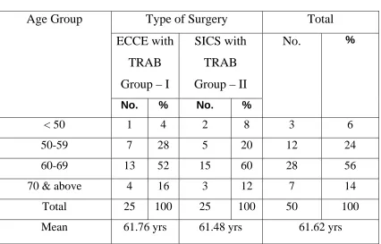
[image:57.612.98.524.154.427.2]AGE DISTRIBUTION
Table 1
Type of Surgery Total
ECCE with
TRAB
Group – I
SICS with
TRAB
Group – II Age Group
No. % No. %
No. %
< 50 1 4 2 8 3 6
50-59 7 28 5 20 12 24
60-69 13 52 15 60 28 56
70 & above 4 16 3 12 7 14
Total 25 100 25 100 50 100
Mean 61.76 yrs 61.48 yrs 61.62 yrs
The mean age of the patients in the study was 61.62 years, group I
(ECCE) mean age is 61.76 years and group II (SICS) mean age is 61.48 years
(p=0.9456) there is no statistically significant difference in the age composition
of the patients undergoing the two types of surgeries. Of the 50 patients
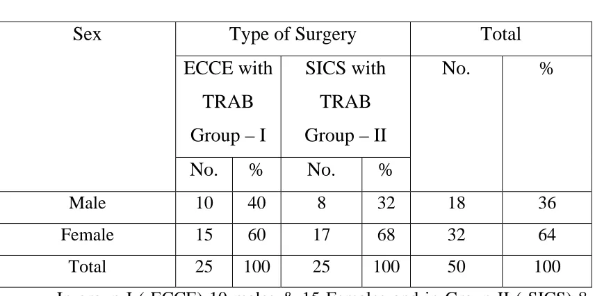
SEX DISTRIBUTION
Table 2
Type of Surgery Total
ECCE with
TRAB
Group – I
SICS with
TRAB
Group – II Sex
No. % No. %
No. %
Male 10 40 8 32 18 36
Female 15 60 17 68 32 64
Total 25 100 25 100 50 100
In group I ( ECCE) 10 males & 15 Females and in Group II ( SICS) 8
males & 17 Females which was statistically not significant (P=0.7683).
LATERALITY
Table 3
Type of Surgery Total ECCE with
TRAB Group – I
SICS with TRAB Group - II Operated eye
No. % No. %
No. %
Left 10 40 11 44 21 42
Right 15 60 14 56 29 58
The group I (ECCE) had 10 left eyes and 15 right
eyes and group II (SICS) had 11 left eyes and 14
right eyes and (P=1.000) there is no significant
[image:58.612.94.535.390.635.2]DIAGNOSIS
Table 4
Type of Surgery Total ECCE with
TRAB Group – I
SICS with TRAB Group - II Diagnosis
No. % No. %
No. %
PACG 2 8 2 8 4 8
POAG 16 64 15 60 31 62
POAG+PXF 2 8 3 12 5 10
SACG 5 20 5 20 10 20
Total 25 100 25 100 50 100
We included 31 (62%) patients with POAG, 5 (10%) patients with
POAG with PXF, 4 (8%) patients with PACG and 10 (20%) patients with
SACG in the study.25 patients underwent SICS with trabeculectomy and 25
patients underwent ECCE with trabeculectomy.
PRE OPERATIVE IOP
Table 5
Type of Surgery Pre operative IOP
ECCE with TRAB Group - I
SICS with TRAB Group - II
Mean 33.58 29.24
The mean preoperative IOP of the 25 patients in the group (I) was 33.58
and group (II) was 29.24. There was (P=0.1095) no statistically significant
difference between the two groups regarding IOP distribution in the
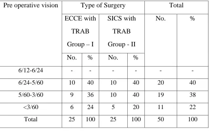
[image:59.612.136.459.451.562.2]PRE OPERATIVE VISION Table 6
Type of Surgery Total
ECCE with
TRAB
Group – I
SICS with
TRAB
Group - II Pre operative vision
No. % No. %
No. %
6/12-6/24 - - - -
6/24-5/60 10 40 10 40 20 40
5/60-3/60 9 36 10 40 19 38
<3/60 6 24 5 20 11 22
Total 25 100 25 100 50 100
In our patients 10 (40%) of them in both groups were having
preoperative vision of 6/24 – 5/60, 9 (36%) of them in Group I (ECCE) and 10
(40%) of them in Group II (SICS) having vision of 5/60 – 3/60, while 6 of them
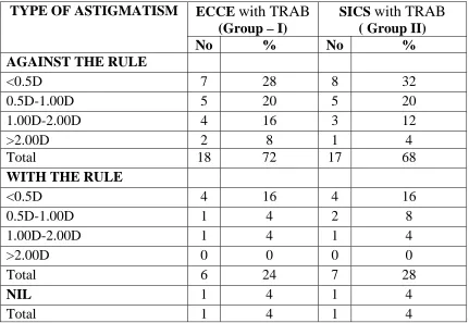
PREOPERATIVE ASTIGMATISM
Table – 7
ECCE with TRAB
(Group – I)
SICS with TRAB
( Group II) TYPE OF ASTIGMATISM
No % No % AGAINST THE RULE
<0.5D 7 28 8 32
0.5D-1.00D 5 20 5 20
1.00D-2.00D 4 16 3 12
>2.00D 2 8 1 4
Total 18 72 17 68
WITH THE RULE
<0.5D 4 16 4 16
0.5D-1.00D 1 4 2 8
1.00D-2.00D 1 4 1 4
>2.00D 0 0 0 0
Total 6 24 7 28
NIL 1 4 1 4
Total 1 4 1 4
In our study preoperative astigmatism of against the rule was found in 18
(72%) patients of ECCE group and 17 (68%) patients of SICS group, of which
<0.5D, 0.50D-1.00D, 1.00D-2.00D, >2.00D distribution between ECCE and
SICS group were (7,5,4,2) patients and (8,5,3,1) patients respectively with the
rule astigmatism were about 6 (24%) patients of ECCE group and 7 (28%)
patients of SICS group, of which <0.5D, 0.50D-1.00D, 1.00D-2.00D, >2.00D
distribution between ECCE and SICS group were (4,1,1,0) patients and
LENS PATTERN
Table 8
Type of Surgery Total ECCE with
TRAB Group – I
SICS with TRAB Group - II Lens
No. % No. %
No. %
MC 8 32 7 28 15 30
IMC 14 56 16 64 30 60
HM 3 12 2 8 5 10
In our patients the lens pattern of immature cataract, mature
cataract and hypermature cataract in Group I (ECCE) and Group II
(SICS) were (14, 8, 3) and (16, 7, 2) patients respectively.
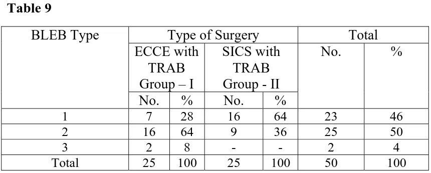
BLEB TYPE Table 9
Type of Surgery Total ECCE with
TRAB Group – I
SICS with TRAB Group - II BLEB Type
No. % No. %
No. %
1 7 28 16 64 23 46
2 16 64 9 36 25 50
3 2 8 - - 2 4
Total 25 100 25 100 50 100
The types of bleb in group I was 28% of type I, 64% of type II and 8% of type III
bleb, in group II was 64% of type I, 36% are of type II, (p=0.0232) there is
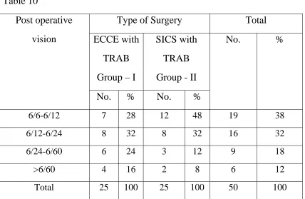
[image:62.612.94.524.414.587.2]POST OPERATIVE VISION
Table 10
Type of Surgery Total
ECCE with
TRAB
Group – I
SICS with
TRAB
Group - II Post operative
vision
No. % No. %
No. %
6/6-6/12 7 28 12 48 19 38
6/12-6/24 8 32 8 32 16 32
6/24-6/60 6 24 3 12 9 18
>6/60 4 16 2 8 6 12
Total 25 100 25 100 50 100
The postoperative vision was presented as 6/6 –6/12 in group I was 28%
and group II was 48% ,6/12-6/24 was 32% and 32% respectively, 6/24-6/60
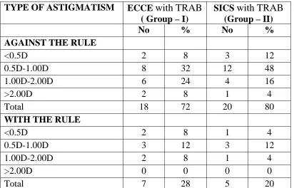
POST OPERATIVE ASTIGMATISM
Table - 11
TYPE OF ASTIGMATISM ECCE with TRAB
( Group – I)
SICS with TRAB
(Group – II)
No % No % AGAINST THE RULE
<0.5D 2 8 3 12
0.5D-1.00D 8 32 12 48
1.00D-2.00D 6 24 4 16
>2.00D 2 8 1 4
Total 18 72 20 80
WITH THE RULE
<0.5D 2 8 1 4
0.5D-1.00D 3 12 3 12
1.00D-2.00D 2 8 1 4
>2.00D 0 0 0 0
Total 7 28 5 20
In our study the post operative astigmatism of against the rule was found
in 38 (76%) patients, of which ECCE had 18 (72%) and SICS had 20 (80%)
patients respectively. The respective <0.5D, 0.50D-1.00D, 1.00D-2.00D,
>2.00D distribution in ECCE group and SICS group were (2,8,6,2) and
(3,12,4,1) patients respectively in our patients the astigmatism of with the rule
in ECCE group and SICS group in the post operative period were 28% and 20%
respectively.
Our study shows against the rule astigmatism was more frequent than
with the rule astigmatism. In our patients the astigmatism of <1.00 D in ECCE
and SICS group were 15(60%) and 19 (76%), which shows SICS group had
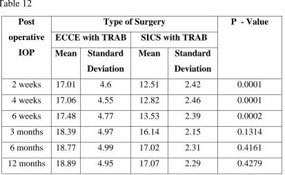
POST OPERATIVE IOP
Table 12
Type of Surgery
ECCE with TRAB SICS with TRAB
Post operative
IOP Mean Standard
Deviation
Mean Standard Deviation
P - Value
2 weeks 17.01 4.6 12.51 2.42 0.0001
4 weeks 17.06 4.55 12.82 2.46 0.0001
6 weeks 17.48 4.77 13.53 2.39 0.0002
3 months 18.39 4.97 16.14 2.15 0.1314
6 months 18.77 4.99 17.02 2.31 0.4161
12 months 18.89 4.95 17.07 2.29 0.4279
The mean postoperative IOP in group I (ECCE) and group II (SICS) in 2
weeks follow-up was 17.01 and 12.51 (p=0.001), 4 weeks follow-up was 17.06
and 12.82 (p=0.0001), 6 weeks follow up was 17.48 and 13.53 (p=0.0002), 3rd
month was 18.39 and 16.14 (p=0.1314), 6th month was 18.77 and
17.02(p=0.4161) and 12th month was 18.89 and 17.07(p=0.4279).
The post operative tension is lower in group II (SICS) than in group I
(ECCE) at all periods of followup (from 2 weeks to 12 months) but the
difference is significant only upto 6 weeks. After 6 weeks there is no
statistically significant difference in the post-operative tension between group I
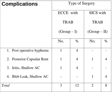
POSTOPERATIVE COMPLICATIONS
Table 13
Type of Surgery
ECCE with
TRAB
(Group – I)
SICS with
TRAB
(Group – II)
Complications
No. % No. %
1. Post operative hyphema
2. Posterior Capsular Rent
3. Iritis, Shallow AC
4. Bleb Leak, Shallow AC
1 1 1 - 4 4 4 - - 1 - 1 - 4 - 4
Total 3 12 2 8
In post operative period the complications in group I was 12% of which
postoperative hyphema in 1 patient, posterior capsule rent in one patient and
iritis with shallow anterior chamber in one patient . In group II the
ASSOCIATED SYSTEMIC DISEASES
Table – 14
ECCE with
TRAB
(Group – I)
SICS with
TRAB
(Group – II)
Systemic
Diseases
No. % No. %
1.Diabetes 2.Hypertension 3. Asthma 2 1 1 8 4 4 1 1 2 4 4 8
Total 4 16 4 16
The associated systemic diseases were equal in both groups (16%),2
diabetics, 1 hypertensive, 1 Asthmatic in Group I (ECCE) and 1 diabetic, 1
DISCUSSION
Glaucoma triple procedure was controversial in the past because of their
greater surgical trauma and success rate. With respect to IOP control was less
than what could be expected when cataract extraction and trabeculectomy were
performed separately.
SICS offers greater possibility of performing a simultaneous
trabeculectomy with better IOP control, less surgical trauma and achieving
good success rate.
In our study the mean age of patients was 61.76 in group I and 61.48 in
group II (p=0.9451) shows no statistically significant difference.
Also in our study the sex distribution was not statistically significant
(p=0.7685), the male and female distribution was 10, 15 in group I (ECCE) and
8, 17 in group II (SICS) respectively.
In our study we included the patients with POAG, PACG, POAG+PXF AND
SACG, of which POAG predominates with 72%, of which group I (ECCE) had
18 (72%) patients and group II (SICS) had 18 (72%) patients and SACG
comes next with 20% of which group I (ECCE) had 5 (20%) patients and group
II (SICS) had 5 (20%) patients , and PACG comes next with 8% of which group
I (ECCE) had 2 (8%) patients and group II (SICS) had 2 (8%) patients
respectively, the distribution of the type of glaucoma was almost similar in both
the groups.
The lens pattern also shows similarity in both the groups, the distribution
of lens pattern of IMC, MC, HM in group I (ECCE) and group II (SICS) were
(56%, 32%, 12%)and (64%, 28%, 8%).
In our study the average pre operative IOP was 33.58 in group I (ECCE)
and 29.24 in group II (SICS), which shows (p=0.1095) no statistically
significant difference between the two groups.
Hence in our study both the groups are having almost similar distribution
of age, sex, laterality, lens pattern and the type of glaucoma .
In this study, the follow up in the 2nd week shows there was a significant
IOP control (p=0.0001) in SICS group (12.51) as compared to ECCE group
(17.01).
Also significant IOP control in the 4th week (p=0.0001) and 6th week
(p=0.0002) follow up in SICS group (12.82,13.53) than ECCE group
(17.06,17.48) respectively.
In our study the postoperative IOP control in the two groups (ECCE and
SICS) at 3rd month, 6th month and 12th month were (18.39, 16.14, [p=0.1314]) ;
(18.77, 17.02, [p=0.4161]) ; (18.89, 17.07, [p=0.4279]) respectively, there was
no statistically significant difference between the two groups at 3rd month, 6th
month and 12th month respectively. These results were similar to the results of
the study of P. Sathyan et al23.
In study conducted by Wishart et al42 shows phaco group had improved
long-term control of IOP than ECCE group, which is same with our study but
our study shows that no statistically significant difference exists between the
In our study the type 1 bleb is about 28% in ECCE group and 64% in
SICS group. While type 2 bleb is 64 % in ECCE, 36% in SICS group. Only 2
cases showed non filtering bleb and it belongs to ECCE group. Our study shows
statistically significant difference in bleb type (p=0.0232) between SICS and
ECCE group.
In our study out of 25 eyes of SICS group 19 eyes (76%) had < 1.00 D
astigmatism, while 16 eyes (60%) in ECCE group had < 1.00 D astigmatism,
which shows SICS group had better astigmatic stabilization.
Also our study shows against the rule astigmatism is more common in
postoperative patients (76%).
In this study there is a better visual rehabilitation in SICS group than
ECCE group as in the study of Wishart et al42 and study by Percival et al24.
In this study the postoperative complications are minimal, SICS group
CONCLUSION
Our study shows small incision cataract surgery with trabeculectomy
has better efficacy in reduction of IOP than extra capsular cataract extraction
with trabeculectomy.
Also small incision cataract surgery with trabeculectomy gives better
IOP control, better visual results and early visual rehabilitation, good filtering
bleb and lesser postoperative complications than extra capsular cataract
extraction with trabeculectomy.
Since our study is a short term of one year follow up only, further studies
BIBLIOGRAPHY
1) Addick EM, Quigley HA, Green WR, et al. Histological characteristic of
filtering blebs in glaucomatous eyes. Arch ophthalmology. 1983; 67:655.
2) Becker Shaffer. Diag & Therapy of glaucoma 7th Edition page 92-100.
3) Cantor LB, Mantravadi A, Wundunn D, et al. Morphological
classification of filtering blebs after glaucoma filtering surgery: The
Indiana Bleb appearance grading scale. J. Glaucoma 2003; 12:266.
4) Chary MR, chena, Lee DA. Basic science and clinical aspects of wound
healing in glaucoma filtering surgery J. pharmacol therapy 1998;14:75.
5) Das JC, Sharma P, Zia chaudhri MG, Shared Bhaniji. A comparative
study of small incision cataract trabeculectomy avoiding Tenons capsule
with conventional trabeculectomy. Ophthalmic surg. laser 2002;
33:30-36.
6) Doughal Jhonson MD, ECCE with IOL implantation & trabeculectomy.
The combined procedure with IOL.Glaucoma 1990; 30:209.
7) Durcan FJ, Cioffi GA, Van Beskirk EM, revision of failed filtering
blebs, J. glaucoma 1992; 1:2- 6.
8) Edwards RS. Trabeculectomy combined with cataract extraction a follow
9) Gerg FM. Phacoemulsification & Modified trabeculectomy from
managing combined cataract surgery & glaucoma. Glaucoma 1992;
18:362-365.
10) Harritz LM. Combined surgery for cataract & glaucoma. Curr opin
ophthal 1993; 4: 73- 78.
11) Jerndal T, Lundstorm M Trabeculectomy combined with cataract
extraction. Am. Jr. 1976; 81:227-23.
12) Jhons GE, Laydes WE, combined trabeculectomy and cataract
extraction .AM. Jr. ophthal 1979; 88: 973-981.
13) Kanski –clinical ophthalmology 5th edition page 192-268.
14) Koch PS. Structural analysis of cataract incision, construction. J.
Cataract refraction surgery 1991; 17:661-667.
15) Kooner KS, Cooksey JC, perry P, Zimmerman TJ, Intraocular pressure
following ECCE, phacoemulsification and PC-IOL implantation
ophthalmic surgery 1988; 19:643-6.
16) Long Staff S, warland RPL, Mazover A, Hitchny RA, Glaucoma Triple
procedure; Efficacy of IOP control and visual outcome Opthal Surgery
1990; 21:786-793.
17) Mammalis W, Lohner S, Rand AN, Grandoll AS: combined
18) Mc cartney DL, Memmen JE, Stack WJ, et al. The efficacy and safety
of combined trabeculectomy, cataract extraction and IOL implantation
Ophthalmology 1988; 95:754-62.
19) Menezo JL, Maldonado MJ, Munoz G, Cisneros AL. combined
procedure for glaucoma and cataract; a retrospective study J. cataract
refract surgery 1994; 20:498-503.
20) Neumann R, Zalish .M, Oliver M, Effect of intraocular lens
implantation on combined ECCE with trabeculectomy, a comparative
study. Br J ophthal 1988; 72:742-745.
21) Nielsen PJ Combined small incision cataract surgery &
Trabeculectomy. A prospective study with 1 year of follow up
ophthalmic surgery. Lasers 1997; 28:21-29.
22) Obstbaum AS. Combined surgery for glaucoma and J cataract refract
surgery 1992; 18:539.
23) P.sathyan et al-combined surgery-phaco trab Vs ecce trab as a primary
procedure; TNOA 2001; 42:15-18.
24) Percival SPB. glaucoma triple procedure of ECCE, PCIOL And
Trabeculectomy. Br.J.Opthalmol 1985:69: 99-102.
25) Savage JA, Thomas JV, Belcher CD III, simans RJ, ECCE and PCIOL
in glaucoma eyes, ophthalmology 1985; 92:1506-16.
27) Seah SK, prata JA, Mineckler DS, et al.visual recovery after
trabeculectomy J. glaucoma 1995; 4:228-234.
28) Shashi kapoor.phaco surgery and foldable IOLs.
29) Shield MB. Combined cataract extraction & glaucoma surgery.
Ophthalmology 1982; 89:231-237.
30) Shield MB. Another reevaluation of combined cataract surgery &
glaucoma surgery. AM J ophthal 1993; 115: 806-810.
31) Simmons ST, Litooff D, Nicholas DA, sherwod MB and Spaeth GL
ECCE and PCIOL implantation combined with trabeculectomy in
patients with glaucoma. Am J.Ophthal.1987; 104:465-470.
32) Skarpic C, Parousis P, Gnad HD, Menapace R, Trabeculectomy and
intraocular lens implantation: a combined procedure J cataract refract
surgery 1987; 13:39-42.
33) Skuta GL, Parrish RK II, wound healing in glaucoma filtering surgery.
Surv ophthalmology, 1987; 32:149-170.
34) Spaeth GL. Management of patient with cataract and glaucoma.
Ophthalmic surgery 1980; 11:780-783.
35) Stevart WC, Crinkely CMC, Carlson AN. results of trabeculectomy
combined with phaco versus trabeculectomy combined with ECCE with
36) Steward WC, crinkley CMC, Carlson AN. Results of combined
phacoemulsification and trabeculectomy in patients with elevated
preoperative Intra ocular pressure. J.glaucoma 1994; 4:164-698.
37) Text book of glaucoma M Bruce Shield MD, 4th edition.
38) The Glaucoma –glaucoma therapy, 2nd edition volume 3 Robert Rich,
M Bruce Shields, Theodre krupin.
39) Vesti E filtering blebs: follow up of trabeculectomy. Ophthalmic
surgical lasers. 1993; 24: 249-255.
40) Wedrich A, Menapace R, Radax U, et al. Combined small incision
cataract surgery and trabeculectomy – technique and results .Int ophthal
1992; 16:409-14.
41) Wedrich A, Menapace R, Radax U, Papanos. P. Long term results of
combined trabeculectomy and small incision cataract surgery. J.cataract
refractive surgery.1995; 21:49-54.
42) Wishart Pk, Austin MU combined cataract extraction and
trabeculectomy phacoemulsification compared with extra capsular
technique ophthalmic surgery 1993; 24:814-821.
43) Writmer R, Rohon JW, combined cataract glaucoma option. Trans.
COMPARISON OF ECCE WITH TRABECULECTOMY AND
SICS WITH TRABECULECTOMY IN EFFICACY OF
REDUCTION IN IOP
PROFORMA
Name Age Sex
Address
OP NO: IP NO:
DOA DOS DOD
PRESENTING COMPLAINTS
DURATION
• Defective Vision
• Pain
• Redness
• Field Defects
• Watering
• Blurring
• Haloes
• Others
HISTORY OF PAST ILLNESS
DURATION
• Hypertension
• Diabetes Type 1/ Type 2
• Myopia