0022-538X/97/$04.0010
Copyrightq1997, American Society for Microbiology
Importance of a c-Myb Binding Site for Lymphomagenesis
by the Retrovirus SL3-3
ANGEL NIEVES,1LAURA S. LEVY,2ANDJACK LENZ1*
Department of Molecular Genetics, Albert Einstein College of Medicine, Bronx, New York 10461,1
and Tulane University School of Medicine, New Orleans, Louisiana 701122
Received 8 August 1996/Accepted 21 October 1996
All murine leukemia viruses (MuLVs) and related type C retroviruses contain a highly conserved binding site for the Ets family of transcription factors within the enhancer sequences in the viral long terminal repeats (LTRs). The T-cell lymphomagenic MuLV SL3-3 (SL3-3) also contains a c-Myb binding site adjacent to the Ets site. The presence of this Myb site distinguishes SL3 from most other MuLVs. We tested the importance of these two sites for the lymphomagenicity of SL3-3. Mutation of the Ets site had little effect on viral pathoge-nicity, as it only slightly extended the latency period to disease onset. In contrast, mutation of the Myb site strongly inhibited pathogenicity, as only a minority of the inoculated mice developed tumors in the two mouse strains that were tested. All tumors that were induced by either mutant appeared to be lymphomas, and no evidence for reversion of either mutation was detected. The effects of the Ets and Myb site mutations on transcriptional activity of the SL3 LTR were tested by inserting the viral enhancer sequences into a plasmid
containing the promoter region of the c-mycgene linked to a reporter gene. Mutation the Myb site almost
eliminated enhancer activity in T lymphocytes, while mutation of the Ets site had smaller effects. Thus, the effects of the enhancer mutations on transcriptional activity in T cells paralleled their effects on viral lymphomagenicity. The absence of the c-Myb site in the LTR enhancer of the weakly lymphomagenic MuLV, Akv, likely contributes to the low pathogenicity of this virus relative to SL3-3. However, Moloney MuLV also lacks the Myb site in its LTR, although it induces T-cell lymphomas with a potency similar to that of SL3-3. Thus, it appears that SL3-3 and Moloney MuLV evolved genetic determinants of T-cell lymphomagenicity that are, at least in part, distinct.
Murine leukemia viruses (MuLVs) are retroviruses that in-duce various types of hematopoietic neoplasias in mice, includ-ing T- and B-cell lymphomas, erythroleukemia, and myeloid leukemia. Recombination studies using molecular clones of MuLVs established that the viral long terminal repeats (LTRs) contain major genetic determinants of viral leukemogenicity (6, 7, 10, 13, 17, 20, 21). Specifically, a portion of the U3 region of the LTR that functions as a transcriptional enhancer plays a major role in determining the type of leukemia that the virus causes and the potency of the virus. Often these viral LTR enhancers are present as tandem repeats about 50 to 100 nucleotides in length (14). They evolved to be recognized by a set of transcription factors in the target cell for disease and appear to play multiple roles in the process of leukemogenesis (4, 11, 27, 32). One key step that the viral LTR enhancers affect is the transcriptional activation of proto-oncogenes when a provirus is integrated adjacent to such gene (27). For example, the c-myc gene is utilized as a common integration site in 15 to 20% of T-cell lymphomas induced by MuLVs (22, 27, 29, 31, 34). Since many of these proviruses are found in the opposite transcriptional orientation relative to c-myc, it appears that the mechanism of its deregulation or activation is by enhancer insertion. Genetic studies indicated that the ability to activate c-myc depended on the nature of the viral LTR enhancer (27). Specifically, in lymphomas induced by a variant MuLV with a single base pair mutation in the LTR, the tumor cell genomes contained LTRs with the original mutations, reversions, or putative second-site suppressor mutations. However, the
pro-viruses adjacent to c-myc always contained reversions or puta-tive suppressor mutations. Thus, viral LTR enhancers in the provirus adjacent to c-myc apparently must have an appropri-ate structure to be capable of activating the proto-oncogene.
SL3 is an MuLV that causes T-cell lymphomas. Multiple transcription factor binding sites have been identified in the SL3 enhancer (Fig. 1). Within each of the 72-bp tandem re-peats that comprise the SL3 enhancer, there are adjacent bind-ing sites for c-Myb and Ets-1. The Ets site, also called the LVb site, is conserved in all MuLVs and can bind multiple members of the Ets family of transcription factors (14, 25, 36, 38). How-ever, the Myb site was identified only in the LTRs of SL3 and the very similar Gross passage A virus (20, 23, 43). On either side of these two sites, there is a binding site for core binding factor (CBF), which is also called AML1, polyomavirus en-hancer-binding protein 2, and SL3 enhancer factor 1 (SEF1) (2, 18, 30, 39). The CBF site adjacent to the Ets site is called core I, while the one next to the Myb site is termed core II. The core I element of SL3 is similar to the core elements of other MuLVs (14). The core II element, like the c-Myb site, is unique to SL3 and the Gross passage A virus (20, 40).
Enhancer elements differ in their importance for viral patho-genicity. Mutations in the conserved core I element of SL3 significantly reduced viral lymphomagenicity (15, 27). How-ever, alteration of the unique core II element had little effect on pathogenicity unless core I was also mutated, in which case the virus was even less lymphomagenic than the core I mutant (15). Mutation of the core element of another T-cell lym-phomagenic virus, Moloney MuLV (Mo-MuLV), also signifi-cantly reduced viral lymphomagenicity (37). Moreover, in the case of Mo-MuLV, mutations in the enhancer core element also altered the tissue specificity of disease from T-cell lym-phomas to erythroleukemias (37). Thus, the conserved core
* Corresponding author. Mailing address: Department of Molecular Genetics, Albert Einstein College of Medicine, 1300 Morris Park Ave., Bronx, NY 10461. Phone: (718) 430-3715. Fax: (718) 430-8778. E-mail: lenz@aecom.yu.edu.
1213
on November 9, 2019 by guest
http://jvi.asm.org/
element is an important determinant of viral tumorigenicity. Similarly, mutation of the Ets site of Mo-MuLV also reduced the lymphomagenic potential of the virus and resulted in a fraction of the resulting tumors being erythroleukemias (37).
In this report, we tested the importance of the c-Myb and Ets sites for the lymphomagenicity of SL3. It was of interest to determine whether the Ets site, which is highly conserved in MuLVs (14), was crucial for pathogenicity. It was also of in-terest to determine whether the relatively unique c-Myb site was important or whether, like the unique core II element, it had only modest effects on virally induced disease.
MATERIALS AND METHODS
Cell lines.SL3H is a T-lymphoma cell line derived from a spontaneous thymic lymphoma of an AKR/J mouse. L691-6 is a T-lymphoma cell line from a radia-tion-induced thymic lymphoma of a C57L mouse. Jurkat is a human T-lympho-blast cell line from a tumor secondary to thymic lymphoT-lympho-blastic lymphoma. NIH
3T3 is a mouse embryo fibroblast cell line. SL3H and Jurkat cell lines were maintained in RPMI 1640 supplemented with either 10% fetal calf serum or fetal bovine serum, 100 U of penicillin per ml, 10 mg of streptomycin per ml, and 2 mM glutamine. The L691 cell line was grown in Dulbecco’s modified Eagle medium with the same supplements mentioned above. NIH 3T3 cells were also grown in Dulbecco’s modified Eagle medium and the same supplements, with the difference that 10% calf serum was added to supplement the medium. All cells were grown at 378C in 100% humidity and 7.5% CO2.
Mutagenesis of LTR plasmids.Reporter plasmids containing viral LTRs linked to the chloramphenicol acetyltransferase (CAT) gene have been previ-ously described (3, 24, 27, 35). These plasmids contain the U3 and R sequences of the viral LTRs that extend from the PstI site near the 59end of the viral LTR to a SmaI site in the middle of the R region. These sequences include the viral promoter and the tandem 72-bp repeats that comprise the viral enhancer. Mu-tations were generated in the Ets-1 and c-Myb binding sites in the SL3 enhancer by using a PCR-based strategy (Fig. 1). The plus-strand PCR primer, 59-AGTA TCCTCGAGGCTGCAGTAACGCCATTTTGCAAGGCAT-39, was homolo-gous to sequences near the 59 end of the U3 region of the SL3 LTR. The minus-strand primers contained the corresponding mutated bases in the Ets-1 or c-Myb site. The sequences of the minus-strand primers used were 59-ATGGTG TCGTTAGCGGTCTGGGGACCATC-39(Ets-1) and 59-ATCCTGTCACCAG CGGTCTGGGGACCATCTGT-39(c-Myb), where the underlined nucleotides identify the mutations. Since the minus-strand primers hybridized to sequences within the 72-bp tandem repeats, this approach yielded PCR products containing either one or two 72-bp units. Those with two 72-bp repeats were a mixture of products with the mutation either in both 72-bp units or only in the downstream repeat unit. The optimal PCR conditions to increase the percentage of products with mutations on both 72-bp repeats were empirically determined to be an initial incubation at 948C for 2 min, followed by 30 cycles of 1 min at 948C and 2 min at 728C. After PCR, the products were digested with XhoI, which cuts at the 59end. The 39ends of these products were flush ends corresponding to an
EcoRV site within the SL3 enhancer. The 213-bp pieces (XhoI to EcoRV) were
then isolated by electrophoresis in 2% agarose gels and used to replace the corresponding wild-type sequences in a plasmid containing the SL3 LTR. Com-plete viral genomes containing a single viral LTR linked to the viral gag, pol, and
env sequences were generated by the insertion of a 7.7-kbp BssHII-to-EcoRI
fragment as previously described (27). All of the final products were sequenced to verify the presence of mutations on both repeats and that no other mutations were generated during the PCR. Mutagenesis of the core elements, core I and core II, was previously described (44).
Transcriptional activity of each of the mutations was analyzed by inserting the viral enhancer sequences into a plasmid that contained the c-myc promoters linked to the CAT gene. Construction of a plasmid (Myc-CAT) that contained 534 bp of 59flanking sequences, exon 1, and 8 bp of intron 1 from c-myc linked to the CAT gene was previously described (8). Generation of Myc-CAT plasmids that contained the LTR enhancer sequences of the viruses, SL3, Akv, and the core I mutant, SAA, 534 bp upstream of exon 1 was previously described (8). Plasmids containing the Ets-1, c-Myb, and core II mutations were constructed by substituting a 325-bp PstI-to-DraI fragment that contained each of the enhancer mutations for the wild-type sequences in the SL3-Myc-CAT plasmid.
Tumorigenicity assays.Infectious virus particles were generated from the single-LTR plasmid clones as previously described (27). Briefly, the 8.2-kbp viral genomes containing the mutations were excised from the plasmid vector by digestion with PstI. These were ligated with T4 DNA ligase to form concatemers that contained two LTRs, each with the identical mutation. Ten micrograms of ligated DNA was transfected by using Lipofectin (GIBCO BRL) into NIH 3T3 mouse fibroblasts that were plated at a density of 106cells per 60-mm2plate 24 h before transfection. Cells were passaged 1:10 every third day. Supernatants from transfected cells were collected and tested for the presence of reverse transcrip-tase activity (12). Virus stocks were harvested after reverse transcriptranscrip-tase activity reached a maximum level, 2 to 3 weeks after transfection. Viral titers were determined by XC plaque assay on fresh NIH 3T3 cells (33). The presence of the mutations in the virus stocks was confirmed by direct sequencing of PCR prod-ucts from NIH 3T3 cells infected with each virus, using the approach described below.
Newborn NIH/Swiss and AKR/J mice (,1.5 days old) were injected intraperi-toneally with 0.1 ml of virus (103
to 104
XC PFU). Diseased mice were sacrificed by CO2anesthesia and necropsied. Animals were examined for gross appearance of malignant lymphomas, which include enlargement of thymus, spleen, periph-eral and/or mesenteric lymph node, and liver. Diagnosis of lymphoma was con-firmed by the presence of T-cell receptorb-chain (TCRb) rearrangements upon Southern blot analysis of tumor DNA.
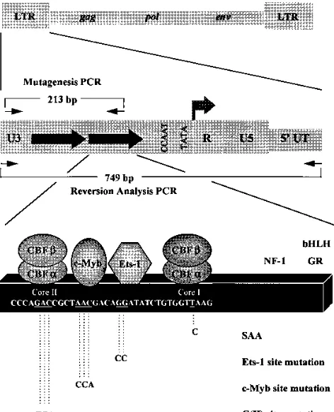
[image:2.612.62.298.71.362.2]Reversion analysis of enhancer mutations.Proviral LTRs were amplified from tumor DNAs by PCR using a primer pair at positions2475 to2442 (59-TTCA TAAGGCTTAGCCAGCTAACTGCAG-39) and 274 to 247 (59-ACACACACT CTCCC-39) relative to the viral transcriptional initiation site (Fig. 1) as previ-ously described (27). PCR conditions were a 30-cycle program of 1 min at 948C, 1 min at 648C, and 2 min at 728C. PCR products were resolved by electrophoresis through either a 2% agarose or a nondenaturing 5% polyacrylamide gel. Indi-vidual bands were excised, and DNA was isolated by using QIAGEX (Qiagen). The isolated fragments were then directly sequenced by using Sequenase (Am-ersham) with a minus-strand primer from positions2150 to2171 (59-TTGAA FIG. 1. Organization of the SL3 LTR and positions of mutations. The top
diagram shows the positions of the LTRs in the viral genome. The middle diagram shows the organization of the 59LTR. The large arrows represent the tandem 72-bp repeats in the U3 region. The arrow at the U3-R boundary indicates the initiation site and direction of transcription. The small arrows show the positions of the primers used for PCR; those above the diagram were used in the mutagenesis procedure, and those below the line were used to amplify the 59LTR from proviruses in infected cells and tumors. The bottom diagram shows the organization of one 72-bp enhancer repeat of SL3. Binding sites for various transcription factors are indicated, and the core elements where CBF binds are marked. Geometrical figures are used to indicate the factors that are relevant to this study. Positions of sites for other factors are also shown. These are a site for NF-1, a site for the glucocorticoid receptor (GR), and an E box that binds basic helix-loop-helix (bHLH) transcription factors (5, 9, 28, 36). Part of the sequence of the enhancer is shown within the box, and the positions of the mutations are underlined. Sequences of the mutated sites are shown below the line. The mutations were introduced simultaneously into both of the 72-bp repeats in the viral LTRs. The single base pair mutation of the core I site to the sequence of Akv virus is indicated as SAA, the name previously given to a mutated SL3 virus that contained this mutation in both 72 bp repeats (27). C(II) indicates core II. UT, untranslated region.
on November 9, 2019 by guest
http://jvi.asm.org/
ACTGTTGTTGTTTTAGC-39) or a plus-strand primer from positions2382 to
2364 (59-ACAAGGAAGTACAGAGAGGC-39) relative to the transcription initiation site.
Analysis of TCRband IgH gene rearrangements.Isolation of genomic DNA from tumors and Southern blot analysis of tumor DNA were performed as previously described (1). Organization of the TCRblocus was examined by using the 86T5 probe, a 600-bp EcoRI fragment of murine TCRbcDNA (16). Orga-nization of the immunoglobulin heavy-chain (IgH) locus was examined by using the p2-1 probe, a 700-bp XbaI-EcoRI fragment containing sequences immedi-ately 39to the murine IgH J region (41).
CAT assays.The DEAE-dextran method was used for transfection of cell lines as described before (3, 35). A total of 5.03106
cells were pelleted and resus-pended in 1 ml of TD (25 mM Tris-HCl [pH 7.4], 0.7 mM Na2HPO4, 5.1 mM KCl, 137 mM NaCl) containing 250 mg of DEAE-dextran per ml, 5mg of reporter plasmid DNA, and 1 mg of a Rous sarcoma virus LTR-luciferase plasmid used as internal control. This mixture was incubated at room tempera-ture for 15 min. Then 5 ml of medium supplemented with serum was added, and the cells were incubated for 20 min at 378C. Cells were then pelleted, resus-pended in 5 ml of medium supplemented with serum, and harvested 48 h later. Protein concentrations were determined, and aliquots of lysates were used to perform CAT assays and luciferase assays as previously described (44). CAT activity was quantified by calculating the percentage of chloramphenicol that was acetylated by using PhosphorImager analysis of thin-layer chromatography plates. CAT activities were normalized to luciferase activities for each sample. Multiple trials were performed for each experiment, and the means were calcu-lated and plotted.
RESULTS
Generation of SL3 viruses mutated in the Myb and Ets sites. To test the importance of the Myb and Ets binding sites for lymphomagenicity of SL3, point mutations were introduced simultaneously into these sites in each of the 72-bp repeats in the SL3 enhancer. Figure 1 shows the organization of the SL3 LTR and the sequences of the binding sites for three transcrip-tion factors, CBF, c-Myb, and Ets-1, within a portranscrip-tion of the 72-bp repeats. The mutations introduced at each site were previously were shown to disrupt binding of the cognate factor to DNA, except for the single base pair mutation in one of the CBF binding sites, core I, which induces only a twofold reduc-tion in CBF binding (23, 37, 39, 40, 43). Infectious virus was generated by transfection of the full-length genome into NIH 3T3 fibroblasts. The spread of infectious virus particles in the cultures was monitored by the presence of reverse transcrip-tase activity in culture supernatants. All SL3 enhancer mutant viruses exhibited reverse transcriptase activity comparable to that of wild-type SL3 and yielded similar titers in XC plaque assays.
To confirm the integrity of the LTR mutations in the viral stocks, the enhancers of proviruses in cellular DNA from in-fected NIH 3T3 fibroblasts were amplified by PCR analysis and
analyzed by DNA sequencing. Primers that hybridized to
se-quences at the 59 end of U3 and to the 59 untranslated
se-quence of viral DNA (Fig. 1) were used to amplify proviral 59
LTR sequences. These primers distinguished SL3 and the LTR mutants from endogenous MuLVs. The PCR products were resolved on 2% agarose gels. A 749-bp fragment was the pre-dominant PCR product, indicating that viruses with two en-hancer repeats predominated in the viral stocks, although PCR products corresponding to LTRs with one or three 72-bp units were also detected. This finding is consistent with previous evidence that these viruses exist as quasispecies with variable numbers of enhancer repeats (27). PCR-amplified fragments were directly sequenced to confirm the presence of the muta-tions. Both the Myb and Ets site mutations were maintained. Tumor induction by viruses with mutated enhancer
se-quences.Virus stocks generated for each enhancer mutation
were tested for tumorigenicity by injection of newborn mice of two different mouse strains, NIH/Swiss (Fig. 2) and AKR/J (Table 1). Parallel controls were performed with wild-type SL3 and a previously described mutant virus, SAA (27), that has a 1-bp mutation in the core I element (Fig. 1). Since AKR/J mice have a high spontaneous incidence of T-cell lymphomas that first appear about 7 months after birth, these mice were main-tained only until they reached the age of 7 months.
The Ets site mutant induced disease in both NIH/Swiss (Fig. 2) and AKR/J (Table 1) mice, with only a modest increase in the latent period to disease onset. The increase in the mean latent period in each strain was significant in a Student t test
(P50.0018 for NIH/Swiss and P50.023 for AKR/J). The Ets
mutant induced disease with a mean latent period that was
significantly shorter than that of the core I mutant, SAA (P5
0.029 for NIH/Swiss and P50.045 for AKR/J). We conclude
that mutation of the Ets site of SL3 had a modest but statis-tically significant effect on the tumorigenicity of SL3.
In contrast, mutation of the Myb site almost eliminated the ability of SL3 to induce tumors. Only 3 of 11 NIH/Swiss mice developed tumors following inoculation with the Myb mutant (Fig. 2). Curiously, all three developed the tumors at about 7 months of age. Only one of eight AKR/J mice developed a tumor before 7 months of age (Table 1). Thus, mutation of the Myb site eliminated much of the lymphomagenicity of SL3. The Myb mutation reduced tumorigenicity to a greater extent than the core I mutation did in both mouse strains (Fig. 2; Table 1). This result indicates that the Myb site in the LTR enhancer is crucial for the pathogenicity of this virus.
LTR structures of tumor-derived proviruses.It was
impor-tant to test the stability of the viral Ets and Myb mutations during the process of tumorigenesis. Previous data showed that the majority of mice that developed lymphomas following in-oculation with the core I mutant virus, SAA, contained provi-ruses with reversions of the original mutation (27). To analyze the LTR structures of proviruses in tumors, genomic DNA was purified from leukemic spleens and thymuses and used as a
template for PCR amplification of proviral 59 LTRs as
[image:3.612.58.297.69.211.2]previ-ously described (27). Products were analyzed from 10 of the
[image:3.612.316.556.660.727.2]FIG. 2. Tumor induction by viral enhancer mutants in NIH/Swiss mice. Par-allel controls were performed with SL3 and the core I mutant, SAA. Surviving mice that were inoculated with the Myb mutant were maintained for a total of 1.5 years, with no additional tumors detected.
TABLE 1. Viral tumorigenicity in AKR/J mice
Virus No. of mice inoculated
No. of mice with tumor by 8 mo
Avg latency (days)
SL3 12 12 71
Myb mutant 8 1 186
Ets mutant 6 6 91
SAA 8 8 109
on November 9, 2019 by guest
http://jvi.asm.org/
Ets induced tumors and three of the Myb mutant-induced tumors. Multiple products were detected in all of the tumors. As previously described (27), these consisted of LTRs that contained variable numbers of 72-bp enhancer repeat units. No amplified products were detected in parallel control experiments performed with tissues from uninfected mice. The PCR products from the tumors were excised from the gels and directly sequenced.
Sequence analysis demonstrated that all integrated provi-ruses from mice inoculated with the Ets and Myb mutant viruses maintained the original point mutations. No reversions were apparent. The sequences of the remainder of the en-hancer regions were also intact, indicating that no potential suppressor mutations were present. Thus, viruses that retained the Ets or Myb mutations were the predominant forms in the tumors.
Characterization of the types of tumors induced by the Myb
and Ets mutant viruses.SL3 appears to induce strictly T-cell
lymphomas. Mo-MuLV also induces T-cell lymphomas. How-ever, mutation of the Ets site in Mo-MuLV resulted in eryth-roleukemias in about 25% of inoculated mice (37). Therefore, it was important to test whether mutation of the Ets and Myb sites altered the tissue target for transformation by SL3. T-cell lymphomas induced by SL3 generally involve an enlargement of the thymus, and most show evidence of rearrangement of a
TCRb locus. In about 70% of SL3-induced lymphomas, the
thymus, spleen, and lymph nodes are grossly enlarged, al-though in about 30% of the tumors, the enlargement of the thymus is less than fivefold relative to age-matched controls. All mice inoculated with the Ets-1 mutant virus showed gross pathologies typical of thymic lymphomas that included en-larged thymus, spleen, and peripheral and mesenteric lymph
nodes. We performed an analysis of TCRb gene
rearrange-ments on DNA from six of the Ets mutant-induced tumors.
TCRbgene rearrangements were seen in all six (Fig. 3). Thus,
it appears that the tumors induced by the Ets mutant are T-cell lymphomas.
Tumors developed in four mice that were inoculated with the Myb mutant. Gross pathologies of the four tumors were similar in that the spleens were greatly enlarged but little to no thymic enlargement was evident. DNA was obtained from
three of the tumors and screened for TCRbrearrangements.
Figure 3 shows that one of the two tumors in NIH/Swiss mice
had a TCRbrearrangement. The tumor induced by the Myb
mutant in an AKR mouse also showed a TCRb
rearrange-ment. The NIH/Swiss tumor without a detectable TCRb
rear-rangement was also tested for a rearrear-rangement of the IgH D-J region. A rearrangement was detected (data not shown). Thus, all three of the examined tumors induced by the Myb mutant were lymphoid, and at least two were T-cell lymphomas.
Correlation between viral lymphomagenicity and
transcrip-tional activity on the c-myc promoter. In about 20% of the
T-cell lymphomas induced by MuLVs such as SL3 and Molo-ney, the c-myc gene is a common proviral integration site. Evidence exists that the SL3 LTR enhancer must have an appropriate structure to be able to activate c-myc during the process of lymphomagenesis (27). In addition, comparison of the SL3 and Akv LTR enhancers for the ability to activate transcription from the two major c-myc promoters in plasmids that contained the c-myc promoters linked to the CAT re-porter gene showed that the Akv enhancer was almost unable to activate them in T cells (8). In contrast, the SL3 enhancer activated the c-myc promoters as much as 50-fold in some T-cell lines (8). The near lack of activity of the Akv enhancer on c-myc in T cells was consistent with the hypothesis that this virus is not lymphomagenic in T cells, at least in part because it cannot sufficiently activate c-myc and, possibly, other proto-oncogenes (8, 27). Therefore, it was of interest to investigate the effects of c-Myb and Ets-1 mutations on the ability of the SL3 LTR enhancer to activate the c-myc promoters.
The enhancer regions of the LTRs of Ets and Myb mutants were inserted into a plasmid containing the first exon and
59-flanking sequences of the c-myc gene linked to the CAT
reporter gene (Fig. 4A). This plasmid contained the two major promoters of c-myc, P1 and P2. Enhancer sequences were placed in the opposite transcriptional orientation in relation to c-myc, as this is the manner in which they are situated in most of the tumors where a provirus is integrated adjacent to c-myc. Activities of the mutated enhancers were compared to those of the wild-type SL3 and Akv enhancers by transfection into cul-tured T cells (Fig. 4B).
As previously shown (8), the SL3 LTR enhancer strongly stimulated transcription from the c-myc promoters compared to the activity of the c-myc promoters alone (Fig. 4B). In contrast, enhancer sequences of nonleukemogenic Akv gave little to no activation. Mutation of the c-Myb binding site greatly reduced the ability of the SL3 enhancer to activate the c-myc gene promoters. Interesting, the levels of transcription were reduced to almost the same levels seen with the Akv enhancer. Thus, mutation of the Myb site strongly reduced both the lymphomagenicity of SL3 and the capacity of the viral LTR enhancer to activate the c-myc promoters.
Mutation of the Ets-1 binding site had less of an effect on c-myc promoter activity (Fig. 4B). In the mouse T-cell lines SL3H and L691, the Ets site mutation reduced transcription twofold or less. In the human T-cell line Jurkat, it resulted in about a fivefold decrease in activity. The Ets mutation had less of an effect on transcription than the single base pair mutation in the enhancer of the core I mutant virus, SAA. Mutation of the core II site had no effect on activity in any of the cell lines. Thus, mutation of the Ets site resulted in only a modest de-crease in lymphomagenicity of SL3 and had less of an effect on transcription from the c-myc promoters than mutations in the Myb or core I sites. These findings show that the mutations that most inhibited the ability of the viral enhancer to activate the c-myc promoters also caused the greatest reduction in viral lymphomagenicity.
[image:4.612.83.272.70.196.2]SL3 is integrated adjacent to c-myc in about 15% of the lymphomas induced by this virus (27). We tested whether the Ets and Myb site mutants of SL3 were integrated adjacent to
FIG. 3. Southern blot analysis of TCRbrearrangements in tumors induced by the Ets and Myb mutants. Six different tumors induced by the Ets mutant in NIH/Swiss mice are numbered 1 through 6. Two different tumors induced by the Myb mutant in NIH/Swiss mice and one tumor induced in an AKR/J mouse are also shown. Negative control DNAs from livers of uninfected mice of the cor-responding strains are indicated by2. DNA samples were digested with HpaI, and Southern blots were hybridized to 86T5, a cDNA clone for the murine TCRb
locus (16).
on November 9, 2019 by guest
http://jvi.asm.org/
c-myc by Southern blotting as previously described (27). None of the tumors examined in this study (Fig. 2; Table 1) tained a provirus within the 20-kbp EcoRI fragment that con-tains the c-myc gene (28a). However, the number of tumors generated in this study was too low to determine whether the mutation of the Myb or Ets site in fact disrupted the ability of the virus to utilize c-myc in the process of lymphomagenesis.
DISCUSSION
Mutation of the Myb site drastically reduced the lymphoma-genicity of SL3, while mutation of the Ets site had only a modest, though statistically significant, effect. Previous studies showed that the core I site was also very important for lym-phomagenicity of SL3 whereas the core II site had only modest effects (15, 27). Thus, the Myb and core I sites appear to be the most important sites in the SL3 LTR enhancer for viral lym-phomagenicity. The Ets and core II sites appear less important quantitatively, although both do affect viral lymphomagenicity. Interestingly, no correlation between the relative impor-tance of a particular site for lymphomagenicity of SL3 and its conservation among MuLVs was evident. The Myb and core II sites that are just upstream of the Ets and core I sites appear to be unique to SL3 and the very similar Gross passage A virus (20, 23). Presumably, this was due to the addition of these novel sites into the genome of a precursor virus that lacked them. The acquisition of one of the unique sites, the Myb site, was more crucial for viral lymphomagenicity than the other, since mutation of core II had only modest effects on tumori-genic potential (Fig. 2 and reference 15).
Likewise, the highly conserved sites in the viral enhancers differed in their importance for viral lymphomagenicity. The Ets (LVb) site and the adjacent core element, core I in SL3, are present in all MuLVs (14), although some sequence vari-ation has occurred within them, particularly in the core ele-ment (14). The core I site was previously shown to be impor-tant for lymphomagenicity by SL3 (15, 27). However, mutation of the Ets site had only a minor effect. These results argue that it is not possible to predict the relative importance of particular transcription factor binding sites for viral lymphomagenicity based on evolutionary conservation.
Endogenous ecotropic MuLVs like Akv are at best weakly leukemogenic (19). Akv is the likely evolutionary precursor of SL3 (20). The Myb site is not present in the LTR enhancer of Akv (Fig. 5). Thus, the acquisition of the Myb site was crucial in the derivation of highly lymphomagenic SL3 from Akv. It was previously demonstrated that the single base pair change between the core I element of SL3 and the core element of Akv was important for acquisition of pathogenicity (27). In addition, the appearance of the core II element in SL3 also slightly increased pathogenicity (15). Thus, three changes in transcription factor binding sites, a mutation in one CBF bind-ing site (core I), the addition of a c-Myb bindbind-ing site, and the gain of a second CBF binding site (core II), contributed to the development of highly lymphomagenic SL3. Conversely, the absence of these sites is correlated with the minimal pathoge-nicity of the endogenous ecotropic MuLVs.
Both SL3 and Mo-MuLV induce T-cell lymphomas. The LTR enhancers of both viruses contain homologous Ets (LVb) and adjacent core (core I in SL3) sites (Fig. 5). Thus, the cellular transcription factors that appear to be responsible for the ability of SL3 and Mo-MuLV to cause T-cell lymphomas are, in part, shared. However, the Myb site is not present in the LTR enhancer of Mo-MuLV. Thus, the highly T-cell lym-phomagenic properties of SL3 and Mo-MuLV appear to be
FIG. 4. (A) Organization of the sequences within the LTR-Myc-CAT plas-mids. The arrows in the viral enhancer indicate the 59-to-39orientation of the en-hancer on the viral coding strand. P1 and P2 indicate the initiation sites and direction of transcription from the two major promoters of the mouse c-myc gene. (B) Relative transcriptional activities of the SL3 and mutated viral enhancers. Activities were tested in three different T-cell lines, SL3H, L691, and Jurkat. Each experiment was performed four times, and the means were calculated. Activities are shown normalized to that of the SL3 enhancer in each cell line. The number above each bar indicates the activity. Error bars indicate 1 standard de-viation. The identities of the enhancers are shown underneath the horizontal axis of each graph. Myc indicates the plasmid with no viral enhancer. C(II), core II.
on November 9, 2019 by guest
http://jvi.asm.org/
due to the evolution of transcription factor binding sites that are, in part, different.
Mo-MuLV contains a second Ets binding site, called the LVc site, that is situated downstream of the core element and is not found in either SL3 or Akv (Fig. 5). However, mutation of the LVc site had no effect on the lymphomagenicity of Mo-MuLV (37). Besides the Ets (LVb) and core elements, two other sites were found to be important for lymphomagenicity of Mo-MuLV, the NF-1 site and the glucocorticoid response element (GRE) (37). The mutation in the GRE also presum-ably disrupted an overlapping site that likely binds basic helix-loop-helix (bHLH) factors including SEF2 and ALF1 (9, 28). The NF-1 and the GRE/bHLH sites appear to be conserved in MuLVs including SL3 and Akv. Thus, it is uncertain what distinguishes highly T-cell lymphomagenic Mo-MuLV from weakly pathogenic Akv. It would also be interesting to know whether the NF-1 and GRE/bHLH sites are important for T-cell lymphomagenicity by SL3.
Cotransfection studies with transcription factors showed that CBF could not activate transcription through the con-served core element of SL3 and Mo-MuLV unless a heterol-ogous transcription factor was bound to a neighboring site. In the case of Mo-MuLV, the activity of CBF required the bind-ing of a member of the Ets family of factors to the Ets (LVb) site (38, 42). In the case of SL3, CBF required either a factor at the Ets site or the binding of c-Myb to the Myb site (43). It is interesting that the cooperative stimulation of transcription by CBF and c-Myb did not require an intact Ets site (43). Thus, the appearance of the Myb site in SL3 may have reduced the
necessity for binding of an Ets family member. This is consis-tent with modest effect of the Ets site mutation on the lym-phomagenicity of SL3 seen here. An alternative speculation to explain the limited importance of the Ets site in SL3 is that DNA binding by an Ets family member might be less affected by the mutation of the Ets site when both CBF and c-Myb are bound at the flanking sites that are present in this virus.
It is interesting to note the correlation between the reduc-tion in transcripreduc-tional activity and the reducreduc-tion in viral lym-phomagenicity. Mutations that had the greatest effect on the ability of the viral enhancer to activate the c-myc promoters also had the greatest effect on viral lymphomagenicity. This is consistent with the idea that the transcriptional activity of the LTR enhancers determines viral lymphomagenicity. It is curi-ous that although the relationship between effects on transcrip-tional activity and viral lymphomagenicity is proportranscrip-tional, it does not appear to be linear. It is also interesting that the Myb site mutation reduced the activity of the SL3 enhancer to the same level as that of the Akv enhancer on the c-myc promoters. The inability of the LTR enhancer of the virus with Myb site mutation to activate c-myc promoters could certainly contrib-ute to its greatly decreased lymphomagenicity. It remains to be tested whether these mutations show similar effects on the promoters of other proto-oncogenes.
ACKNOWLEDGMENTS
We thank Eleanore Kim, Patricia Lobelle-Rich, Ari Zaiman, Joseph Pantginis, and Kathleen Comer for helpful advice and assistance with some of the experiments.
This work was supported by Public Health Service grants CA44822 and CA57337 to J.L. J.L. was the recipient of a Hirschl-Caulier career scientist award. Core facilities for oligonucleotide synthesis and PhosphorImager analysis were supported by Public Health Service Cancer Center grant CA13330.
REFERENCES
1. Athas, G., B. Choi, S. Prabhu, P. Lobelle-Rich, and L. S. Levy. 1995. Genetic determinants of feline leukemia virus-induced multicentric lymphomas. Vi-rology 214:431–438.
2. Bae, S. C., Y. Yamaguchi-Iwai, E. Ogawa, M. Maruyama, M. Inuzuka, H. Kagoshima, K. Shigesada, M. Satake, and Y. Ito. 1993. Isolation of PEBP2aB cDNA representing the mouse homolog of human acute myeloid leukemia gene, AML1. Oncogene 8:809–814.
3. Boral, A. L., S. A. Okenquist, and J. Lenz. 1989. Identification of the SL3-3 virus enhancer core as a T-lymphoma cell-specific element. J. Virol. 63:76– 84.
4. Brightman, B. K., A. Rein, D. J. Trepp, and H. Fan. 1991. An enhancer variant of Moloney murine leukemia virus defective in leukemogenesis does not generate mink cell focus-inducing virus in vivo. Proc. Natl. Acad. Sci. USA 88:2264–2268.
5. Celander, D., and W. A. Haseltine. 1987. Glucocorticoid regulation of mu-rine leukemia virus transcription elements is specified by determinants within the viral enhancer region. J. Virol. 61:269–275.
6. Chatis, P. A., C. A. Holland, J. W. Hartley, W. P. Rowe, and N. Hopkins. 1983. Role for the 39end of the genome in determining disease specificity of Friend and Moloney murine leukemia viruses. Proc. Natl. Acad. Sci. USA 80:4408–4411.
7. Chatis, P. A., C. A. Holland, J. E. Silver, T. N. Frederickson, N. Hopkins, and J. W. Hartley.1984. A 39end fragment encompassing the transcriptional enhancers of nondefective friend virus confers erythroleukemogenicity on Moloney leukemia virus. J. Virol. 52:248–254.
8. Comer, K. A., H. L. Morrison, and J. Lenz. Induction of transcripts from both major promoters of c-myc by the long terminal repeat enhancer of a T-cell lymphomagenic retrovirus. Submitted for publication.
9. Corneliussen, B., A. Thornell, B. Hallberg, and T. Grundstro¨m.1991. Helix-loop-helix transcriptional activators bind to a sequence in glucocorticoid response elements of retrovirus enhancers. J. Virol. 65:6084–6093. 10. DesGroseillers, L., and P. Jolicoeur. 1984. The tandem direct repeats within
the long terminal repeat of murine leukemia viruses are the primary deter-minant of their leukemogenic potential. J. Virol. 52:945–952.
11. DesGroseillers, L., E. Rassart, and P. Jolicoeur. 1983. Thymotropism of murine leukemia virus is conferred by its long terminal repeat. Proc. Natl. Acad. Sci. USA 80:4203–4207.
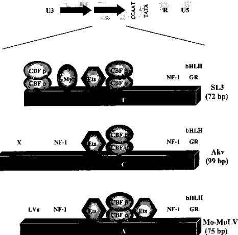
[image:6.612.63.296.71.302.2]12. Goff, S., P. Traktman, and D. Baltimore. 1996. Isolation and properties of FIG. 5. Comparison of the SL3, Akv, and Mo-MuLV enhancers. The
orga-nization of one enhancer repeat unit is shown for each virus below the general organization of the viral LTRs. Geometric figures are used to represent c-Myb, the Ets family of factors, and the subunits of CBF. Two Ets binding sites in Mo-MuLV are shown. The one to the left of the CBF binding sites is also known as the LVb site, and the one to the right is also called the LVc site (36, 38). Sites for NF-1, the glucocorticoid receptor (GR), and bHLH factors are shown. X in Akv indicates a binding activity of unknown identity (26). LVa in Mo-MuLV also indicates a binding activity of unknown identity (36). The positions of the ho-mologous core elements in the three viruses are indicated by the single nucleo-tides within the boxes. These represent the positions where the three core elements, TGTGGTTA in SL3, TGTGGTCA in Akv, and TGTGGTAA in
Mo-MuLV (20, 36), differ.
on November 9, 2019 by guest
http://jvi.asm.org/
Moloney murine leukemia virus mutants: use of a rapid assay for release of virion reverse transcriptase. J. Virol. 38:239–248.
13. Golemis, E., Y. Li, T. N. Fredrickson, J. W. Hartley, and N. Hopkins. 1989. Distinct segments within the enhancer region collaborate to specify the type of leukemia induced by nondefective Friend and Moloney viruses. J. Virol. 63:328–337.
14. Golemis, E. A., N. A. Speck, and N. Hopkins. 1990. Alignment of U3 se-quences of mammalian type C viruses: identification of highly conserved motifs and implications for enhancer design. J. Virol. 64:534–542. 15. Hallberg, B., J. Schmidt, A. Luz, F. S. Pedersen, and T. Grundstro¨m.1991.
SL3-3 enhancer factor 1 transcriptional activators are required for tumor formation by SL3-3 murine leukemia virus. J. Virol. 65:4177–4181. 16. Hedrick, S. M., D. I. Cohen, E. A. Nielsen, and M. M. Davis. 1984. Isolation
of cDNA clones encoding T cell-specific membrane-associated proteins. Nature 308:149–153.
17. Ishimoto, A., M. Takimoto, A. Adachi, M. Kakuyama, S. Kato, K. Kakimi, K. Fukuoka, T. Ogiu, and M. Matsuyama.1987. Sequences responsible for erythroid and lymphoid leukemia in the long terminal repeats of Friend mink cell focus-forming and Moloney murine leukemia viruses. J. Virol. 61:1861– 1866.
18. Kamachi, Y., E. Ogawa, M. Asano, S. Ishida, Y. Murakami, M. Satake, Y. Ito, and K. Shigesada.1990. Purification of a mouse nuclear factor that binds to both the A and B cores of the polyomavirus enhancer. J. Virol. 4808–4819. 19. Lawrenz-Smith, S. C., A. C. Massey, D. J. Innes, and C. Y. Thomas. 1995. Pathogenic determinants in the U3 region of recombinant murine leukemia viruses isolated from CWD and HRS/J mice. J. Virol. 68:5174–5183. 20. Lenz, J., D. Celander, R. L. Crowther, R. Patarca, D. W. Perkins, and W. A.
Haseltine.1984. Determination of the leukemogenicity of a murine retrovi-rus by sequences within the long terminal repeat. Nature 308:467–470. 21. Li, Y., E. Golemis, J. W. Hartley, and N. Hopkins. 1987. Disease specificity
of nondefective Friend and Moloney murine leukemia viruses is controlled by a small number of nucleotides. J. Virol. 61:693–700.
22. Li, Y., C. A. Holland, J. W. Hartley, and N. Hopkins. 1984. Viral integration near c-myc in 10–20% of MCF 247-induced AKR lymphomas. Proc. Natl. Acad. Sci. USA 81:6808–6811.
23. LoSardo, J., A. Nieves, and J. Lenz. 1996. Unpublished results.
24. LoSardo, J. E., A. L. Boral, and J. Lenz. 1990. Relative importance of elements within the SL3-3 virus enhancer for T-cell specificity. J. Virol. 64:1756–1763.
25. Manley, N. R., M. O’Connell, W. Sun, N. A. Speck, and N. Hopkins. 1993. Two factors that bind to highly conserved sequences in mammalian type C retroviral enhancers. J. Virol. 67:1967–1975.
26. Morrison, H. L., H. Y. Dai, F. S. Pedersen, and J. Lenz. 1991. Analysis of the significance of two single-base-pair differences in the SL3-3 and Akv virus long terminal repeats. J. Virol. 65:1019–1022.
27. Morrison, H. L., B. Soni, and J. Lenz. 1995. Long terminal repeat enhancer core sequences in proviruses adjacent to c-myc in T-cell lymphomas induced by a murine retrovirus. J. Virol. 69:446–455.
28. Nielsen, A. L., N. Pallisgaard, F. S. Pedersen, and P. Jorgensen. 1996. Basic helix-loop-helix proteins in murine type C retrovirus transcriptional regula-tion. J. Virol. 68:5638–5647.
28a.Nieves, A., and J. Lenz. Unpublished data.
29. O’Donnell, P. V., E. Fleissner, H. Lonial, C. Koehne, and A. Reicin. 1985. Early clonality and high-frequency proviral integration into the c-myc locus in AKR leukemia. J. Virol. 55:500–503.
30. Ogawa, E., M. Maruyama, H. Kagoshima, M. Inuzuka, J. Lu, M. Satake, K. Shigesada, and Y. Ito.1993. PEBP2/PEA2 represents a family of transcrip-tion factors homologous to the products of the Drosophila runt gene and the human AML1 gene. Proc. Natl. Acad. Sci. USA 90:6859–6863.
31. Reicin, A., J.-Q. Yang, K. B. Marcu, E. Fleissner, C. F. Koehne, and P. V. O’Donnell.1986. Deregulation of the c-myc oncogene in virus-induced thy-mic lymphomas of AKR/J thy-mice. Mol. Cell. Biol. 6:4088–4092.
32. Rosen, C. A., W. A. Haseltine, J. Lenz, R. Ruprecht, and M. W. Cloyd. 1985. Tissue selectivity of murine leukemia virus infection is determined by long terminal repeat sequences. J. Virol. 55:862–866.
33. Rowe, W. P., W. E. Pugh, and J. W. Hartley. 1970. Plaque assay techniques for murine leukemia viruses. Virology 12:1136–1139.
34. Selten, G., H. T. Cuypers, M. Zijlstra, C. Melief, and A. Berns. 1984. In-volvement of c-myc in MuLV induced T-cell lymphomas in mice: frequency and mechanisms of activation. EMBO 3:3215–3222.
35. Short, M. K., S. A. Okenquist, and J. Lenz. 1987. Correlation of leukemo-genic potential of murine retroviruses with transcriptional tissue preference of the viral long terminal repeats. J. Virol. 61:1067–1072.
36. Speck, N. A., and D. Baltimore. 1987. Six distinct nuclear factors interact with the 75-base-pair repeat of the Moloney murine leukemia virus en-hancer. Mol. Cell. Biol. 7:1101–1110.
37. Speck, N. A., B. Renjifo, E. Golemis, T. Fredrickson, J. Hartley, and N. Hopkins.1990. Mutation of the core or adjacent LVb elements of the Moloney murine leukemia virus enhancer alters disease specificity. Genes Dev. 4:233–242.
38. Sun, W., B. J. Graves, and N. A. Speck. 1995. Transactivation of the Moloney murine leukemia virus and T-cell receptorb-chain enhancers by cbf and ets requires intact binding sites for both proteins. J. Virol. 69:4941–4949. 39. Thornell, A., B. Hallberg, and T. Grundstro¨m.1988. Differential protein
binding in lymphocytes to a sequence in the enhancer of the mouse retro-virus SL3-3. Mol. Cell. Biol. 8:1625–1637.
40. Thornell, A., B. Hallberg, and T. Grundstro¨m.1991. Binding of SL3-3 enhancer factor 1 transcriptional activators to viral and chromosomal en-hancer sequences. J. Virol. 65:42–50.
41. Weaver, D., F. Constantini, T. Imanishi-Kari, and D. Baltimore. 1985. A transgenic immunoglobulin mu gene prevents rearrangements of endoge-nous genes. Cell 42:117–127.
42. Wotton, D., J. Ghysdael, S. Wang, N. A. Speck, and M. J. Owen. 1994. Cooperative binding of ets-1 and core binding factor to DNA. Mol. Cell. Biol. 14:840–850.
43. Zaiman, A. L., and J. Lenz. 1996. Transcriptional activation of a retrovirus enhancer by CBF (AML1) requires a second factor: evidence for cooperat-ivity with c-Myb. J. Virol. 70:5618–5629.
44. Zaiman, A. L., A. F. Lewis, B. E. Crute, N. A. Speck, and J. Lenz. 1995. Transcriptional activity of core binding factora(AML1) andbsubunit on murine leukemia virus enhancer cores. J. Virol. 69:2898–2906.