Copyright © 2001, American Society for Microbiology. All Rights Reserved.
Establishment of New Transmissible and Drug-Sensitive
Human Immunodeficiency Virus Type 1 Wild Types due to
Transmission of Nucleoside Analogue-Resistant Virus
ANTHONY
DERONDE,
1* MAAIKE
VANDOOREN,
1† LIA
VAN DERHOEK,
1DENISE BOUWHUIS,
1ESTHER
DEROOIJ,
1BOB
VANGEMEN,
2ROB
DEBOER,
3ANDJAAP GOUDSMIT
1Department of Human Retrovirology, Academic Medical Center, University of Amsterdam,
1105 AZ Amsterdam,
1Primagen, 1105 BA Amsterdam,
2and Theoretical Biology,
Utrecht University, 3584 CH Utrecht,
3The Netherlands
Received 30 May 2000/Accepted 14 October 2000
Sequence analysis of human immunodeficiency virus type 1 (HIV-1) from 74 persons with acute infections
identified eight strains with mutations in the reverse transcriptase (RT) gene at positions 41, 67, 68, 70, 215,
and 219 associated with resistance to the nucleoside analogue zidovudine (AZT). Follow-up of the fate of these
resistant HIV-1 strains in four newly infected individuals revealed that they were readily replaced by sensitive
strains. The RT of the resistant viruses changed at amino acid 215 from tyrosine (Y) to aspartic acid (D) or
serine (S), with asparagine (N) as a transient intermediate, indicating the establishment of new wild types.
When we introduced these mutations and the original threonine (T)-containing wild type into infectious
molecular clones and assessed their competitive advantage in vitro, the order of fitness was in accord with the
in vivo observations: 215Y < 215D
ⴝ
215S
ⴝ
215T. As detected by real-time nucleic acid sequence-based
amplification with two molecular beacons, the addition of AZT or stavudine (d4T) to the viral cultures favored
the 215Y mutant in a dose-dependent manner. Our results illustrate that infection with nucleoside
analogue-resistant HIV leads in newly infected individuals to mutants that are sensitive to nucleoside analogues, but only
a single mutation removed from drug-resistant HIV. Such mutants were shown to be transmissible, stable, and
prone to rapid selection for resistance to AZT or d4T as soon as antiretroviral therapy was administered.
Monitoring of patients for the presence of new HIV-1 wild types with D, S, or N residues at position 215 may
be warranted in order to estimate the threat to long-term efficacy of regimens including nucleoside analogues.
Since 1987, when antiviral drug therapy of human
immuno-deficiency virus type 1 (HIV-1) infection started with the use of
zidovudine (AZT), drug-resistant viral mutants have rapidly
emerged (3, 12). Due to the error-prone reverse transcriptase
(RT), viral mutants are generated during every replication
cycle (16). The mutational pathway is in essence the result of a
stochastic process in which a single mutation appears more
frequently than double or multiple mutations (13). A newly
formed viral mutant that has a higher replicative capacity than
its parent will overgrow the parent. This process of selection
results in the appearance of mutants with the highest fitness or
replicative capacity in their environment (7). When an
antiret-roviral drug is present, mutants with the highest level of
resis-tance to the drug will have an advantage. As soon as antiviral
therapy is stopped, the original virus, which is usually well
adapted to an environment without the drug, will win the
competition with the resistant viruses (1). However, a different
situation occurs when a drug-resistant virus is transmitted to a
previously uninfected and untreated person. Separated from
the original drug-sensitive virus and lacking this dominant
competitor in the new drug-free environment, the
drug-resis-tant virus will serve as a new starting point for the evolution of
HIV-1. The process does not necessarily lead back to a virus
with a genotype identical to that of the original, drug-sensitive
virus, but the phenotype (i.e., replicative capacity) will adapt to
be optimally fit in the new drug-free environment (7).
Of 74 new HIV-1 infections that occurred in the period 1992
to 1999, 8 were caused by viruses with mutations at positions
associated with AZT resistance. After their transmission to
new hosts, those with the resistance-conferring T215Y
muta-tion in the RT gene showed rapid evolumuta-tion to
better-replicat-ing, more-fit viruses that acquired novel residues at amino acid
215 of RT. The novel evolutionary pathway led to a viral
phenotype adapted to a drug-free environment and having a
replicative capacity similar to that of the original wild type.
There was evidence that the new viruses were transmissible
and rapidly converted by a single mutation to a drug-resistant
phenotype when suboptimal antiviral therapy was introduced.
MATERIALS AND METHODS
Study population.The Amsterdam cohort studies (ACS) have monitored a cohort of homosexual men and a cohort of intravenous drug users, with quarterly screening of participants for the presence of the HIV-1 antigen and antibodies. New HIV-1 infections are identified by a seroconversion to the HIV-1 antigen and/or antibody with confirmation by Western blotting and HIV-1 viral load assay. In addition, patients admitted to the clinic of the Academic Medical Center (AMC) in Amsterdam with symptoms of suspected acute HIV-1 infection have been examined. We studied a total of 74 new infections identified by ACS and AMC between 1992 and 1999. Of these 74 new HIV-1 infections, 8 had RT mutations that are associated with AZT-resistance (see below for the sequencing method). Mutations specifically associated with RT inhibitors other than AZT (zalcitabine [ddC], didanosine, stavudine [d4T], lamivudine (3TC), nevirapine
* Corresponding author. Mailing address: Department of Human
Retrovirology, Academic Medical Center, University of Amsterdam,
Meibergdreef 15, 1105 AZ Amsterdam, The Netherlands. Phone:
31.20.566.8571. Fax: 31.20.566.9080. E-mail: [email protected].
† Present address: Primagen, 1105 BA Amsterdam, The
Nether-lands.
595
on November 9, 2019 by guest
http://jvi.asm.org/
and other nonnucleoside RT inhibitors) were not found. Of the 28 new HIV-1 infections identified during or after 1996, the year of the introduction of protease inhibitors, none involved viruses that had mutations that confer resistance to
protease inhibitors. Sequences of the 5⬘end of the RT gene with mutations
associated with AZT resistance were deposited in GenBank (see below for sequence numbers).
Viral load and CD4 counts.Viral load was determined by using the NucliSens
assay of Organon Teknika BV (Boxtel, The Netherlands). CD4⫹cells in blood
were counted by standard flow cytometry using a FACScan flow cytometer (Becton Dickinson, San Jose, Calif.) and commercially available monoclonal antibodies (Becton Dickinson).
Screening for resistance-conferring mutations.Viral RNA was isolated from
200l of serum drawn from each of the 74 subjects at the first point of
sero-conversion to the HIV-1 antigen or antibody by using the method described by
Boom et al. (2). Part of the HIV-1polregion including the part of the RT gene
encoding the amino terminus was amplified by RT-PCR, essentially according to the protocol described by Nijhuis et al. (18). To facilitate accurate two-stranded sequence analysis of the amino terminus-encoding part of the RT gene, we adapted the nested PCR in the Nijhuis protocol by amplifying two overlapping
fragments instead of one. The 5⬘fragment was amplified using primer
SP6-RT19new,ATTTAGGTGACACTATAGCACCTGTCAACATAATTGGAAG
(SP6 sequence is in italics; HXB-2 nucleotides [nt] 2491 to 2512), and T7-B-SEQ,
TAATACGACTCACTATAGGGAATATTGCTGGTGATCCTTTCCA (T7
se-quence is in italics; HXB-2 nt 3030 to 2006). The 3⬘fragment was amplified with
primers SP6-C-SEQ,ATTTAGGTGACACTATAGGTATACTGCATTTACCA
TACC (SP6 sequence is in italics; HXB-2 nt 2927 to 2947), and T7-ET10,TAA
TACGACTCACTATAGGGCTGCCAGTTCTAGCTCTGCTTC (T7 sequence is in italics; HXB-2 nt 3462 to 3441). Both strands of the nested PCR fragments were directly sequenced using the SP6 and T7 primer sequences. Sequencing was
performed withTaqdye primers (Applied Biosystems, Foster City, Calif.) and
the ThermoSequenase fluorescence-labeled primer cycle sequencing kit (Amer-sham International, Little Chalfont, England). The sequence products were analyzed on an automatic sequencer (Applied Biosystems DNA sequencer model 373A stretch or 377). The sequence of the RT gene was screened at positions associated with drug resistance as described by Hirsch et al. (10). For new infections caused by viruses with mutations at positions associated with AZT resistance, serum samples drawn after the seroconversion point were analyzed for changes compared to the seroconversion sample. Mixtures of different viral RT sequences were quantified by assessing the ratio between the relevant peak areas of the signals of the corresponding nucleotides on the electropherogram using the sequences of both strands. In cases identified during or after 1996, the year of introduction of protease inhibitors in The Netherlands, we sequenced the protease gene as well as the RT gene. The protease gene was amplified according to the procedure described by de Jong et al. (5). Nested PCR was performed by
using primers SP6-A-SEQ,ATTTAGGTGACACTATAGGAGCCAACAGCCC
CACCAG (SP6 sequence is in italics; HXB-2 nt 2149 to 2167) and T7-A-SEQ,
TAATACGACTCACTATAGGGTAAAGAAGACAGTTACCG (T7 sequence is in italics; HXB-2 nt 2639 to 2621). Sequence analysis was performed as above for the RT gene.
Infectious molecular clones.Biological clones of the virus of patient 4 were prepared by a limiting-dilution series of human peripheral blood mononuclear cells (PBMC) derived from a sample taken 1 month after seroconversion (14). Viral RNA was isolated from the culture supernatants of individual biological clones. The complete RT gene of the virus was reverse transcribed and amplified (5) but by using primers ET44 (GACATTTATCACAGCTGGCTAC; antisense;
nt 4338 to 4359 of HIVHXB2CG; GenBank accession no. K03455) and 5⬘
-PROT-OUT (GAGCCAACAGCCCCACCAG; nt 2149 to 2167; sense) in the RT reaction and first PCR and by using primers ET42 (CTGTTGACTCAGAT TGGTTGCACTTTAAATTTTCC; nt 2517 to 2551; sense) and ET17 (AGGT GGCAGGTTAAAATCAACTAGCCATGCCATTGCTCTCC; nt 4319 to 4285; antisense) in the nested PCR. The PCR product was cloned in plasmid pCR-2.1 using a TA vector system (InVitrogen BV, Groningen, The Netherlands).
Se-quence analysis of the 5⬘end of the RT gene was performed, and a clone with the
T215Y mutation (AZT resistant) was chosen. The insert was subcloned in the
EcoRI site of vector pUC19 to remove the flankingBstXI sites, creating p4-Y.
PCR-mediated mutagenesis of amino acid 215 of RT was performed essentially as described by de Jong et al. (5) except that primer ET10 (CTGCCAGTTCT AGCTCTGCTTC; nt 3462 to 3441) and the sense mutagenesis primer for the first PCR were used, whereas primer ET07 (GGAAGTTCAATTAGGAATA CC; nt 2812 to 2832) and the antisense mutagenesis primer were used for the second PCR. The two PCR products were combined, and a third PCR was performed using primers ET10 and ET07. The resulting PCR product was
digested withBstXI and cloned into aBstXI-digested p4-Y. Mutagenesis primers
ET47 (GAAGTGGGGATTCGACACACCAGACAAAAAAC; nt 3170 to 3209; sense; codon 215 is underlined) and ET48 (GTTTTTTGTCTGGTGTGT CGAATCCCCACTTC; nt 3209 to 3170; complementary) were used to intro-duce the 215 aspartic acid (GAC codon) mutation; ET49 (GAAGTGGGGAT TCTCCACACCAGACAAAAAAC; sense) and ET50 (GTTTTTTGTCTGGT GTGGAGAATCCCCACTTC; complementary) were used to introduce the 215 serine (TCC codon) mutation; ET51 (GAAGTGGGGATTCACCACACCAGA CAAAAAAC; sense) and ET52 (GTTTTTTGTCTGGTGTGGTGAATCCCC ACTTC; complementary) were used to introduce the 215 threonine (ACC co-don) wild-type mutation. Sequencing was performed to verify that the exchanged
BstXI fragment contained the introduced mutations. By this procedure, we
created plasmids p4-D, p4-S, and p4-T encoding the indicated (one-letter code) amino acids at the 215 position.
At week 154, viral RNA was isolated from a sample drawn from patient 4, and RT-PCR was performed as described above using primers ET42 and ET10; the product was cloned in the TA cloning vector pCRII (InVitrogen BV). Sequence analysis showed that individual clones were representative of the quasispecies that contained an S68G change relative to the p4-Y clone. In addition, these clones had naturally occurring variations at amino acids 39 and 135 (T39A,
I135T). The 3⬘end of one of these clones (p4-68G) starting from theSspI site
(HXB-2 nt 3025) was replaced by the 3⬘end of p4-Y or p4-D creating p4-Y-68G
and p4-D-68G, respectively. The infectious molecular clone in which RT genes derived from patient 4 were recombined was pHIV-Lai, lacking its RT gene
(pLAI-⌬RT). The RT gene deletion was obtained by PCR-mediated
mutagen-esis. Briefly, the deletion was constructed in a subclone of HIV-Lai containing
theApaI-NcoI fragment by using primers ET40 (TGACCGCGGAAAATTTA
AAGTGCAAC; nt 2550 to 2532; antisense;SacII site underlined) and ET23
(AGTCCGCGGAGAGCAATGGCTAGT; nt 4286 to 4300; sense;SacII site
underlined) and primers 5⬘and 3⬘ofApaI andNcoI sites, respectively. This
created molecular clone pLAI-⌬RT with aSacII site replacing the RT gene.
Recombinant viruses with a recipient-derived RT gene.To obtain recombinant viruses, C33A cells (human cervix carcinoma cell line) were seeded in 24-well plates and grown to 80% confluence in 1 ml of RPMI 1640 medium (Life Technologies, Breda, The Netherlands) supplemented with 10% fetal calf serum
and antibiotics. Cotransfection of 300 ng of pLAI-⌬RT and 200 ng of the purified
RT gene-containingEcoRI fragment of p4-Y, -D, -S, -T, -Y-68G, or -D-68G was
performed using the TFX-50 reagent protocol (Promega Benelux BV, Leiden, The Netherlands). One day after transfection, 200,000 MT2 cells per well were added, and C33A and MT2 cells were cocultured. After 2 days, the MT2 cells
including medium were transferred to a 25-cm2flask, with the addition of fresh
medium and MT2 cells. Cells were monitored for the formation of syncytia, and the supernatant containing the recombinant virus was harvested after the spread
of syncytia throughout the culture. The virus was frozen into aliquots at⫺70°C.
The virus titer (50% tissue culture infective dose [TCID50]) was determined by
limiting-dilution titration on MT2 cells. Resistance to antiviral drugs was assayed by VIRCO (Edegem, Belgium) (9).
Competition experiments were performed by mixing a total amount of 1,000
TCID50of the recombinant viruses in different ratios. These virus mixtures were
used to infect 2 ml of phytohemagglutinin-stimulated PBMC (2⫻106cells/ml),
which were maintained in a six-well plate in RPMI 1640 medium supplemented with 10% fetal calf serum and interleukin-2 (6). Twice a week, cells were
pas-saged, and 10l of the culture was transferred to 2 ml of a new culture of 2⫻
106PBMC/ml. Similarly, 500,000 MT2 cells were infected with 1,000 TCID
50and
maintained in 1 ml of RPMI medium supplemented with 10% fetal calf serum in
a 24-well plate. When syncytia in the MT2 cells had formed, 5l of the culture
was transferred to 1 ml of a new culture composed of 1 ml of medium containing 500,000 MT2 cells. Competition experiments were performed in the absence or presence of antiviral drugs (i.e., AZT, d4T, or ddC). At each passage, samples from the supernatant from which to isolate viral RNA for sequence analysis were drawn. Viral RNA was isolated according to the method of Boom et al. (2). The RNA was directly sequenced as described above. The ratios between the differ-ent recombinant viruses were determined from the direct sequence by assessing the ratios between the relevant peak areas of the signals on the electropherogram corresponding to the nucleotides encoding amino acid 215 of RT. Alternatively, the ratios between the different viruses were determined by using real-time nucleic acid sequence-based amplification (NASBA) with molecular beacons, as described in the next section.
Quantification of mutant mixtures using real-time NASBA and molecular beacons.RNA was isolated from 100l of culture supernatant according to the
method of Boom et al. (2). The nucleic acids were eluted in 50l of water, and
5l of the eluate was used as input for amplification by NASBA. NASBA was
performed by using the basic kit of Organon Teknika supplemented with primers 215-P2 GACTTAGAAATAGGGCAGCA (HXB-2 nt 2705 to 2724) and 215-P1
on November 9, 2019 by guest
http://jvi.asm.org/
AATTCTAATACGACTCACTATAGGGGTTCATAACCCATCCAAAGGAAT GGA (T7 sequence in italics; HXB-2 nt 2831 to 2806) and the molecular beacons
(20) RT 215GAC, ccgactcTCGACACACCAGACAAAAAACgagtcgg (FAM,
dabcyl), and RT 215TAC, ccgactcTCTACACACCAGACAAAAAACgagtcgg
(6-ROX, dabcyl) (stem sequence is in lowercase; HXB-2 nt 2772 to 2792; position 215 [underlined, boldface] is either a GAC [D] or TAC [Y] codon). The two primers and two beacons were each added to a final concentration of 200 nM. Fluorescence was measured in real time on a Fluoroscan Ascent (Labsystems Oy, Helsinki, Finland). The assays were calibrated by mixing different quantities of in vitro-synthesized RNA containing the GAC or TAC position 215 codon. To synthesize the RNA, PCR fragments were generated from p4-D-68G and p4-Y-68G by using primers AATTCTAATACGACTCACTATAGGG (T7 site located in the plasmid) and ET10 (HXB-2 nt 3462 to 3441). These PCR fragments were used to produce RNA containing the GAC or TAC position 215 codon by using T7 RNA polymerase (Amersham Pharmacia Biotech Benelux, Roosendaal, The Netherlands) in an in vitro transcription reaction. Under our reaction conditions
with 105molecules of in vitro-synthesized RNA per reaction, the beacon specific
for the GAC codon did not react with the RNA containing the TAC codon and vice versa. In vitro-synthesized RNA containing the GAC and TAC codons was mixed at different percentages (0, 2, 4, 10, 33, 50, 67, 90, 96, 98, and 100%).
Repeated (n⫽5) experiments showed that quantification of the mixtures using
real-time NASBA could be achieved until 4% of either the TAC or GAC variant (see Fig. 4A and B). Below 4% of the variant, the variant could be observed but not quantified. Quantification of mixtures by real-time NASBA and direct se-quencing were compared for the competitions in the presence of AZT (see Fig.
4C). The methods gave comparable percentages (⫾10%) of a variant mixture,
provided that direct sequencing was performed using the ET dye primer on the ABI automatic sequencers and not the big-dye techniques on the ABI machines. However, when a variant was present in small amounts (less than 10%), quan-tification by direct sequencing was no longer reliable due to background signals. Real-time NASBA was broader in dynamic range; it could reliably quantify a variant at a level as low as 4% and could detect a variant at a level as low as 1%.
Fitness calculation.Selection coefficients were calculated by a novel method involving the estimation of the total viral replication during the observation period (16a). The method can be used both for steady-state viral populations, to which most previously used methods apply (8), and for decreasing or expanding viral populations, such as those observed, for example, in cell cultures. Briefly, the model is derived as follows. Because mutations in the RT are expected to mainly influence the replication rate of a mutant virus and to hardly influence its
half-life, we write dW/dM⫽rW⫺ ␦W and dM/dt⫽r(1⫹s)M⫺ ␦Mfor the
dynamics of wild-type virusWand mutant virusM. Hereris the (wild-type)
replication rate (which may change over time [t]), 1/␦is the generation time, and
sis the classical coefficient of selection. Because this model combines the
dy-namics of productively infected cells and free virions, the generation time should
be about 2 days (11). The same model has been used before for estimatingsfrom
in vivo data with approximately steady-state viral loads (8). One conventionally
defines the frequency of the mutant genotype by the equationP⫽M/(W⫹M),
and hence 1⫺pis the frequency of the wild-type virus. Ifris a constant, one
writes the solutionsW(t)⫽W(0)e(r⫺ ␦)tandM(t)⫽M(0)e[r(1⫹s)⫺ ␦]tso thats
is computed from the logarithms of the genotype ratios (H⫽M/W⫽p/[1⫺p])
at time 0 and timet, i.e.,s⫽ln[H(t)/H(0)]/rt.
Thus, for estimating the relative fitness one generally needs to know the ratio
of the genotype frequencies and the total replication,rt.rmay vary over time,
however. This requires the integration of the replication rate over the
experi-ment. Fortunately,scan still be estimated by the simple formula (16a)s⫽ln
[H(t)/H(0)]/{ln[W(t)/W(0)]⫹ ␦t}, which is available at http://www-binf.bio.uu.nl
/⬃rdb/fitness.html. If the total viral loads and the percentages of wild-type virus
at times 0 andtare known, the website equation can be used to compute the
change in the wild-type virus load,W(t)/W(0), and the change in the ratio of the
genotype frequencies,H(t)/H(0), to gives.His undefined when the percentage
of mutant virus is estimated as 0 or 100%. Whenever possible, we therefore ignored such data points. However, in a few instances (patients 2 and 4) no alternative data points were available. In such instances, we took 1 or 99% for the genotype frequency, which was a conceivable assumption (see Table 1 for the example of patient 1). For the fitness calculations for the viral culture we took the cumulative p24 value as the viral load, i.e., a p24 value of 1,000 pg/ml after a 1:100 dilution of a viral culture was 100,000 pg/ml, allowing us to keep a dilution factor of 1 in the formula on the website.
We calculatedsover the interval between samples 1 and 6 (Table 1) (using 99
and 1% for the percentages of virus withYat position 215 [215Y virus] in
samples 1 and 6, respectively); the fitness difference between 215Y and 215N was 2.8%. Corresponding fitness differences were 2.3, 3.3, 2.6, 2.5, and 3.0% for intervals between samples 2 and 5, 2 and 4, 2 and 3, 3 and 4 (all of which used detected percentages of 215Y virus), and 2 and 6 (which used 1% 215Y virus for
sample 6), respectively. The one-samplettest of the SSPS, version 8.0, statistical
package was used to calculate the mean and the 95% confidence interval. We concluded that 215N had a relative fitness of 103% (95% confidence interval: 102
to 104%), with 215Y fitness⫽100%.
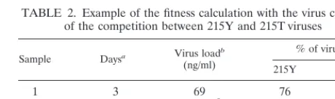
We calculatedsover the interval between samples 1 and 4 for the virus culture
of the competition between 215Y and 215T viruses (Table 2); the fitness differ-ence between 215Y and 215T was 5.1%. Corresponding fitness differdiffer-ences were 5.4, 6.0, 4.7, and 4.9% for intervals between samples 2 and 4, 3 and 4, 1 and 3, and 2 and 3, respectively. We concluded that 215T had a relative fitness of 105%
(95% confidence interval: 104 to 106%), with 215Y fitness⫽100%.
Nucleotide sequence accession numbers.Accession numbers for viral RT gene sequences obtained in this study were as follows: patient 1 (H0671), AF265569; patient 2 (I6056), AF265572; patient 3 (H0095), AF265570; patient 4 (I7052), AF265571; patient 5 (H0137), AF265568; patient 6 (I3234), AF265573; patient 7 (M12690), AF265574; patient 8 (2202575), AF265567.
RESULTS
[image:3.612.53.294.92.183.2]In vivo evolution of viruses with a mutated RT gene in the
absence of an antiviral drug.
Among 74 new HIV-1 infections
occurring between 1992 and 1999, 8 were found to be due to
viruses that have mutations in the RT gene that are associated
with AZT resistance. At seroconversion, the corresponding
mutations in RT were M41L and T215Y (patients 1, 2, and 4);
T215Y (patient 3); D67N, K70R, and T215F (patient 5);
D67N, K70R, and K219Q (patient 6); M41L and the T215D
mutation, which involved an unusual amino acid not seen
as-sociated with AZT resistance (patient 7); and K70R (patient
8). The K70R mutation in the virus infecting patient 8 could be
the result of a natural polymorphism, although in the absence
of AZT the K70R variant rarely becomes the dominant variant
in the viral quasispecies (17). The eight infections were
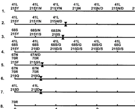
mon-itored until antiviral therapy (Fig. 1). Infections 1 to 5, which
all were found at seroconversion to have an AZT
resistance-conferring mutation at amino acid 215 of RT (either 215Y or
TABLE 1. Example of the fitness calculation with
data from patient 1
Sample Daysa Virus loadb
(copies/ml)
% of virus:
215Y 215N
1
47
400
100
0
2
299
400
80
20
3
392
230
33
67
4
517
620
9
91
5
614
7,200
7
c80
c6
694
1,400
0
100
aDays after seroconversion.
bRNA load in serum.
cThe mixture contained 13% 215D virus.
TABLE 2. Example of the fitness calculation with the virus culture
of the competition between 215Y and 215T viruses
Sample Daysa Virus loadb
(ng/ml)
% of virus:
215Y 215T
1
3
69
76
24
2
10
4.0
⫻
10
565
35
3
24
1.1
⫻
10
1141
59
4
35
4.5
⫻
10
1423
77
aDays after infection.
bCumulative p24 in culture medium.
on November 9, 2019 by guest
http://jvi.asm.org/
[image:3.612.312.552.639.710.2]215F) showed evolution in their RT genes. Since infections 1 to
4 could be monitored for at least 1 year before antiviral therapy
was given, the evolution of the RT gene, and in particular the
evolution at amino acid 215 of RT, was analyzed in these cases
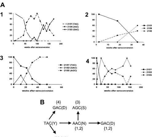
(Fig. 2A, 1 to 4).
In patient 1 at seroconversion, the RT of the infecting
vi-ruses had the M41L and T215Y mutations conferring AZT
resistance. While the codon at position 41 of the RT gene did
not change, amino acid codon 215 changed from TAC,
encod-ing tyrosine, to AAC, encodencod-ing asparagine. The 215N virus had
completely replaced 215Y within 100 weeks (2 years) after
seroconversion. Over the next 50 weeks, the AAC codon at
position 215 was gradually replaced by a GAC codon encoding
aspartic acid, which remained the dominant species thereafter.
In patient 2 at seroconversion, the M41L and T215Y
muta-tions were likewise present and the mutants evolved as in
patient 1, but on a far smaller time scale. During the 36 weeks
that patient 2 was monitored 215Y evolved to a mixture of
215N and 215D, in which 215D eventually became the
domi-nant species, a process that took 150 weeks in patient 1.
In patient 3, RT had only the T215Y mutation at
serocon-version, with codon 215 evolving from TAC (215Y) to AGC,
encoding serine (215S), although at week 11 GAC (215D) was
transiently observed. Unlike the other mutations we studied,
which involve a one-nucleotide change within the codon 215,
the change from TAC to AGC involved a two-nucleotide
change, and no obvious intermediate was detected. However,
in this infection, the 215 AAC codon may have been transiently
formed as an intermediate to the 215 AGC codon encoding
serine. A notable mutation was observed at codon 68 of the RT
gene, which changed from wild-type AGC (serine) to a mixture
of AGA and AAC (asparagine).
In patient 4, RT had the M41L and T215Y mutations at
seroconversion. At week 3 after seroconversion, the 215 TAC
(Y) codon was already being replaced by the 215 GAC (D)
codon, with full replacement observed at week 20. At week 37,
we detected a mixture of the 215 GAC codon and the 215 TCC
codon (S), in which the relative abundances of the two
fluctu-ated until week 150, when the last sample was drawn before
therapy began. The changes to an aspartic acid and to a serine
at position 215 of RT required only a single mutation, from the
TAC codon to GAC and TCC codons, respectively. Analogous
to what was observed in patient 3, the RT gene of patient 4
showed a change at position 68. In this case, however, the
wild-type 68 AGC codon (serine) changed to a GGC codon
encoding glycine (68G). At week 120, wild-type 68S had been
completely replaced by 68G, which persisted thereafter. An
overview of the mutational pathways in the different patients is
shown in Fig. 2B.
The rate of RT evolution in these cases varied considerably.
In patient 1, replacement of 215Y by 215N and subsequently by
215D took place over a time period of more than 150 weeks.
Concurrent with the replacement of 215Y by 215N, the viral
load increased from below 1,000 copies/ml to 10,000 copies/ml,
and it increased further to 30,000 copies/ml when 215D
be-came the dominant species. During this period, the CD4 T-cell
counts remained stable and high at approximately 1,000/
l. In
patient 2, replacement of 215Y by 215D occurred within less
than 40 weeks, with a viral load between 30,000 and 100,000
copies/ml and CD4 T-cell counts hovering between 400 and
600/
l. In patient 3, replacement of 215Y by 215S occurred
after 40 weeks; the viral load declined from 300,000 copies/ml
at seroconversion to 10,000 to 30,000 copies/ml from week 30
to 55 and CD4 T cells remained at 200 to 400/
l. In patient 4,
replacement of 215Y by 215D occurred within 25 weeks, with
a viral load that declined from above 100,000 copies/ml at
seroconversion to about 25,000 copies/ml at week 100 and with
CD4 T-cell counts varying from approximately 300 to 600/
l.
In all three patients who had been infected with a virus in
which the RT gene contained both the 41L and 215Y
muta-tions, the dominant viral species that emerged contained an
RT with the 41L and 215D mutations. Patient 7 was infected
with a virus containing an RT with these 41L and 215D
mu-tations, indicating that a virus with these mutations is
trans-missible.
[image:4.612.56.291.70.261.2]In vivo fitnesses of the newly emerged viruses with
muta-tions in the RT gene.
The increased replicative capacity, or
fitness, of the newly emerged viruses with changes in their RT
genes was expressed relative to the replicative capacity of the
215Y variant at seroconversion, which was arbitrarily set at
100%. The fitnesses of the variants newly formed in an
envi-ronment without an antiviral drug were calculated. In patient 1
the available serum samples allowed the calculation of the
fitness of the 215N variant, which was 103% (95% confidence
interval: 102 to 104%) of that of 215Y in an environment
without an antiviral drug. By the same method, the fitness of
the 215S variant (patient 3) was calculated to be 106% (103 to
109%) of that of 215Y and the fitness of the 215D variant was
calculated to be 107 (patient 2) or 110% (patient 4) (96 to
124%) of that of the 215Y variant. By way of comparison, the
S68G change observed in the RT of patient 4 resulted in a
relative fitness of the 68G viruses of 101% compared to that of
the 68S virus, which was set at 100%. To give an impression of
the competition between the new variants and the
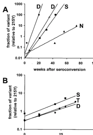
AZT-resis-tant 215Y, we plotted the fractions (relative to 215Y) of the
FIG. 1. In vivo evolution of transmitted viruses with drug
resis-tance-conferring mutations in the absence of antiretroviral therapy.
Indicated are the positions and amino acids (minor species in italics) in
which the viruses differ from the wild type at positions known to be
involved in drug resistance (10). Our subjects were monitored until
antiviral therapy.
on November 9, 2019 by guest
http://jvi.asm.org/
different variants in the viral population (on a log scale) against
the elapsed time after seroconversion (Fig. 3A).
In vitro fitnesses of viruses differing at amino acid 215 of
RT.
In vivo, if HIV-1 isolates found in two different samples
drawn from a patient show genetic differences, they differ
usu-ally at multiple positions, of which only a few may significantly
contribute to a change in replicative capacity. To examine the
significance of RT amino acid 215 for replicative capacity,
viruses that differed only at this amino acid were constructed.
In the RT isolated from patient 4, containing the M41L and
T215Y mutations, only the 215 position was changed by in vitro
mutagenesis to either 215D, 215S, or to 215T, the wild-type
amino acid present before the introduction of AZT. The
[image:5.612.58.554.72.519.2]wild-type 215T was found also in the viruses of the donor of patient
4 before he received AZT and resistant viruses developed. The
various RT genes were recombined into an HIV-1 Lai
back-ground, creating viruses that differed only at amino acid 215 of
RT. Competition between the resulting viruses was observed in
PBMC in the absence of an antiviral drug. In that environment,
the 215Y virus was the least fit virus. The relative fitnesses of
all the viruses were calculated. The in vitro relative fitnesses of
215D, 215S, and 215T compared to that of 215Y (100%) were
104 (95% confidence interval: 102 to 106%), 107 (104 to
109%), and 105% (104 to 106%), respectively (Fig. 3B).
Com-petition among the 215D and the 215S or 215T viruses showed
them to be equally fit, i.e., having a fitness difference of less
FIG. 2. In vivo evolution at amino acid 215 of RT. (A) Relative abundances of viruses with the indicated amino acids at position 215 of RT
after transmission of AZT-resistant viruses. The evolution of transmitted viruses is shown in patients 1 to 4. All start with 215Y and evolve to 215D
(patients 1, 2, and 4) and/or 215S (patients 3, and 4), with 215N observed as an intermediate (patients 1 and 2). (B) In vivo mutational pathways
as observed (patients 1, 2, and 4) or hypothesized (patient 3) at amino acid 215 of RT after transmission of AZT-resistant viruses.
on November 9, 2019 by guest
http://jvi.asm.org/
than 1%. The order of in vitro fitness of the viruses differing
only at amino acid 215 was 215Y
⬍
215D
⫽
215S
⫽
215T,
corresponding to the in vivo-observed order.
Characterization of the viruses after antiviral therapy by
using real-time NASBA.
Patient 1 was not treated because his
CD4 counts remained stable and high at approximately 1,000
CD4 cells/
l. Patients 2 and 3 were treated with a combination
of protease and RT inhibitors (indinavir plus ritonavir plus d4T
plus 3TC and indinavir plus d4T plus 3TC), and their viral
loads declined to below detection level (
⬍
400 copies/ml).
Pa-tient 4 was treated at 160 weeks after seroconversion with RT
inhibitors d4T and ddC. No change in the viral load was
ob-served between week 154 (pretherapy) and week 170 (after
therapy), indicative of the development of resistance to the
antiviral drug. In this case, the only observed mutation in RT
was at amino acid 215, which was changed from a mixture of
215D and 215S to 215Y by a single mutation (GAC and/or
TCC to TAC). Phylogenetic analysis of the RT genes of the
viruses found before and after therapy showed that the
post-therapy 215Y virus was derived from viruses present before
therapy but not present at transmission. The newer viruses
shared 215Y with the transmission virus but had, in addition,
mutations at positions 68 (S68G), 39 (T39A), and 135 (I135T).
To examine whether the change of environment caused by the
therapy with d4T and ddC was involved in the selection of a
viral population with the 215D- or 215S-to-215Y mutation,
viruses that differed only at amino acid 215 of RT were studied.
The viruses tested had either 215D or 215Y with 68G (present
directly before and after therapy) or 68S (present directly after
transmission). By using competition experiments with MT2
cells, the replicative capacities of the viruses were compared at
increasing concentrations of AZT, d4T, or ddC. To facilitate
the analysis of the competition experiments, we developed a
real-time NASBA with two molecular beacons differing at one
nucleotide. The dynamic range of quantification and the
throughput of the real time NASBA are higher than those in
sequence analysis, which generally cannot quantify mixtures
with less than 10% of a variant. The molecular beacons, among
which the GAC beacon contained a FAM label and the TAC
beacon contained a ROX label, were able to discriminate
vi-ruses differing at the 215 codon of the RT gene (Fig. 4A). The
real-time NASBA was able to quantify mixtures containing as
little as 4% of either the GAC- or TAC-containing virus (Fig.
4B) and to detect the GAC- or TAC-containing virus in a
mixture in which they composed no more than 1%. Using the
real-time NASBA, the analysis of the competition experiments
showed that the background of the viruses with the 68S or 68G
mutation (either those directly after transmission or those
present at therapy) did not influence the phenotype. However,
the 215D-to-215Y change was crucial to obtain a replicative
advantage in the environment with both AZT and d4T (Fig.
4C). The replicative advantage of a virus with a 215Y codon in
the presence of AZT or d4T varied in a dose-dependent
man-ner. The replicative advantage of 215Y virus could be
ex-pressed as an average fitness difference of 5 and 11% in the
presence of 45 and 170 nM AZT, respectively (0.5 and 2 times,
respectively, the 50% inhibitory concentration [IC
50] of a
sen-sitive virus), and 2 and 28% in the presence of 3 and 12
M
d4T, respectively (0.5 and 2 times, respectively, the IC
50of a
sensitive virus), indicating that the replicative advantage of
215Y increased with higher drug concentrations. At 1.5 and 4.5
M (0.5 and 1.5 times, respectively, the IC
50of a sensitive
virus), ddC did not appear to influence the relative replicative
capacities of the viruses. In classical phenotypic resistance
as-says, the 215Y virus is
⬎
100-fold more resistant to AZT and
2-fold more resistant to d4T than the wild type and sensitive to
ddC, whereas the 215D and 215S viruses are sensitive to AZT,
d4T, and ddC.
DISCUSSION
[image:6.612.80.267.70.342.2]Our study indicates that between 1992 and 1999, 10 to 15%
of new HIV-1 infections in Amsterdam, The Netherlands, were
caused by HIV-1 strains with mutations in the RT gene at
positions that are associated with AZT resistance. We found
no mutations associated with resistance to other antiretroviral
drugs, including the protease inhibitors introduced in 1996.
When the HIV-1 strains with drug resistance-conferring
mu-tations are transmitted, a new environment without antiviral
drugs in a new host is encountered. After transmission of
usually a small amount of virus, the wild-type strains from
which the drug-resistant strains were derived are lost from the
FIG. 3. In vivo and in vitro relative fitnesses of viruses marked by
their amino acids at position 215 of RT. (A) Fitness plot of viruses
differing at amino acid 215 (N, S, and D), which was derived from the
observed replacement of 215Y by viral variant 215N, 215S, or 215D in
the four patients (two of them had a 215D). The data points
corre-spond to the observed viral mixtures in the patients (Fig. 2A). Shown
are the rates (weeks after seroconversion) at which and to what extent
(on a log scale) 215Y virus is replaced by a different variant. (B)
Fitness plot of viruses differing only at amino acid 215 of RT, which
competed in PBMC in the absence of an antiretroviral drug. Shown are
the rates (number of days of culture) at which and to what extent (on
a log scale) variants 215D (
}
), 215S (
■
), and 215T (
F
) replaced 215Y.
When 215D was competed with 215T or 215S, 215D was found to be
as fit as 215T or 215S.
on November 9, 2019 by guest
http://jvi.asm.org/
(memory of the) quasispecies and thereby are excluded from
competition in the new environment (19). The separation of
individual viruses from the remainder of competing viruses by
bottleneck transmission is reminiscent of the founding of an
island population and may lead to new directions in evolution
(15). In the new, drug-free host, viruses that have an increased
replicative capacity or fitness compared to that of their
drug-resistant parent will be selected. Although the replicative
prop-erties of the viruses we studied will most likely be determined
by more than one gene, we focused on the
resistance-confer-ring RT gene. We found that the drug-resistance-conferresistance-confer-ring
RT amino acid 215Y in viruses changed after transmission, but
not to the wild-type threonine. Instead, novel amino acids at
position 215 were observed (see also reference 21). Single
mutations in the TAC codon (215Y) gave rise to AAC
(aspar-agine), GAC (aspartic acid), or TCC (serine). In addition, in
one of the new infections an AGC codon (serine, 215S) was
formed by a two-step mutation. Selection occurred for the
fittest variant, the 215N virus being less fit than the 215D or
215S virus. Compared to the transmitted AZT-resistant 215Y
virus the 215N viruses were 3% more fit, the 215S viruses were
6% more fit, and the 215D viruses were 7 to 10% more fit. Our
calculations assumed a constant viral generation time;
how-ever, variation among the respective infections could affect the
relative fitness values. In this respect, patient 1, with an initially
low viral load and slow replacement of the different variants, is
illustrative. Nevertheless, the order of viral fitness is
215-ty-rosine
⬍
215-asparagine
⬍
215-aspartic acid
⫽
215-serine. In
vitro experiments in a drug-free environment showed that
wild-type viruses with 215-threonine were as fit as the 215D or 215S
viruses and fitter than the 215Y viruses. Threonine (ACC
co-don; 215T) can be formed by a two-step mutational process
from the TAC codon (215Y). Such a two-step mutant,
gener-ated by stochastic mutational processes, will usually be formed
later than one-step mutants such as 215D (GAC) and 215S
(TCC). The principle of competitive exclusion could explain
why we observed no 215T viruses in the new infections started
with AZT-resistant viruses, since an equally fit variant will not
FIG. 4. In vitro competitions of 215Y and 215D viruses in the absence and presence of antiviral drugs as analyzed by using real-time NASBA.
(A) Representative experiment in which relative fluorescence units (RFU) are shown as a function of time when RNA mixtures (total of 10
5molecules) of 215D and 215Y (ranging from a 25:1 to a 1:25 ratio) were assayed by real-time NASBA using GAC (FAM-labeled) and TAC
(ROX-labeled) molecular beacons indicative of 215D and 215Y, respectively. (B) Calibration curve derived from panel A. The log function of the
slope of the increase of fluorescence in time is proportional to the relative abundances of 215D and 215Y in a sample, which can be inferred from
the known mixtures. (C) Competition between 215Y and 215D viruses in the presence of increasing concentrations of d4T (left) or AZT (right).
Viruses containing RT with 215Y and 215D competed in MT2 cells in the presence of 0, 3, or 12
M d4T or 0, 45, or 170 nM AZT. The ratio
of 215Y/215D virus in the supernatant at passage was determined by using real-time NASBA.
on November 9, 2019 by guest
http://jvi.asm.org/
replace an existing virus that already dominates the population
(4). In this group, the phenotype of a 215D or 215S virus will
to a large extent be the new wild type. This is most clearly
shown in the virus of patient 7, in which the 41L and 215D
mutations appeared directly at seroconversion, strongly
point-ing at the transmissibility of the 215D virus.
When antiviral therapy is applied, the environment changes
once yet again, and under conditions of suboptimal therapy in
which sufficient residual viral replication can occur, a new
selection for the most fit variant takes place. For patient 4, a
single mutation (GAC or TCC to TAC) within the codon of
amino acid 215 of RT appeared sufficient to confer a decisive
replicative advantage in the presence of d4T and ddC. By using
a newly developed real-time NASBA with a high throughput
and dynamic quantification range, in vitro competitions with
increasing concentrations of AZT or d4T showed that the
change from 215D to 215Y by a single nucleotide mutation
conferred the replicative advantage. Apparently, the
AZT-re-sistant genotype of the virus of patient 4 is also advantageous
in the presence of d4T. This finding shows that although the
transmitted AZT-resistant viruses that have adapted to the
drug-free environment closely resemble the 215T wild type in
their phenotypes, their genotype retains many characteristics
of the transmitted AZT-resistant 215Y virus.
Our observations confirm the high plasticity of the genome
of HIV-1 and in particular of the RT gene. Transmission from
a treated host to an untreated host immediately results in
adaptation of the virus to its new, drug-free environment. Such
adaptation becomes manifest as a fixation of a 215D or 215S
mutation instead of reversion to the wild type of the
pre-therapy era: 215T. These new wild types share AZT sensitivity
with the old wild type, 215T. However, the new wild types are
only one step away from 215Y and 215Y is therefore expected
to arise rapidly upon administration of antiretroviral therapy
when therapy is suboptimal. As we have observed, the newly
established wild types can be transmitted. The monitoring of
new wild types with D, S, or N at position 215 may thus be
warranted in order to estimate the threat to long-term efficacy
of a therapy regimen that includes nucleoside analogues.
ACKNOWLEDGMENTS
We thank M. Bakker and S. Jurriaans for providing serum samples;
K. Lindenberg and R. Coutinho of the Amsterdam Municipal Health
Service for patient data; V. Benes and H. Voss of EMBL for initial
help with sequencing; B. Hemmelder and J. Maas for technical
assis-tance; L. Phillips for editorial assisassis-tance; B. Berkhout, M. Cornelissen,
and V. Lukashov for discussion and ideas; and the participants of the
ACS for their cooperation over many years.
This research was supported by grants 1311 and 1317 of the Dutch
AIDS Foundation.
REFERENCES
1.Albert, J., J. Wahlberg, J. Lundeberg, S. Cox, E. Sandstrom, B. Wahren, and M. Uhlen.1992. Persistence of azidothymidine-resistant human
immunode-ficiency virus type 1 RNA genotypes in posttreatment sera. J. Virol.66:5627–
5630.
2.Boom, R., C. J. Sol, M. M. Salimans, C. L. Jansen, P. M. Wertheim-van Dillen, and J. van der Noordaa.1990. Rapid and simple method for
purifi-cation of nucleic acids. J. Clin. Microbiol.28:495–503.
3.Boucher, C. A., E. O’Sullivan, J. W. Mulder, C. Ramautarsing, P. Kellam, G. Darby, J. M. Lange, J. Goudsmit, and B. A. Larder.1992. Ordered appear-ance of zidovudine resistappear-ance mutations during treatment of 18 human
immunodeficiency virus-positive subjects. J. Infect. Dis.165:105–110.
4.Clarke, D. K., E. A. Duarte, S. F. Elena, A. Moya, E. Domingo, and J. Holland.1994. The Red Queen reigns in the kingdom of RNA viruses. Proc.
Natl. Acad. Sci. USA91:4821–4824.
5.de Jong, J. J., J. Goudsmit, V. V. Lukashov, M. E. Hillebrand, E. Baan, R. Huismans, S. A. Danner, J. H. ten Veen, F. de Wolf, and S. Jurriaans.1999. Insertion of two amino acids combined with changes in reverse transcriptase containing tyrosine-215 of HIV-1 resistant to multiple nucleoside analogs.
AIDS13:75–80.
6.de Ronde, A., B. Klaver, W. Keulen, L. Smit, and J. Goudsmit.1992. Natural HIV-1 NEF accelerates virus replication in primary human lymphocytes.
Virology188:391–395.
7.Domingo, E., and J. J. Holland.1997. RNA virus mutations and fitness for
survival. Annu. Rev. Microbiol.51:151–178.
8.Goudsmit, J., A. de Ronde, E. de Rooij, and R. de Boer. 1997. Broad spectrum of in vivo fitness of human immunodeficiency virus type 1
sub-populations differing at reverse transcriptase codons 41 and 215. J. Virol.71:
4479–4484.
9.Hertogs, K., M. P. de Bethune, V. Miller, T. Ivens, P. Schel, A. van Cau-wenberge, E. C. van den Eynde, V. van Gerwen, H. Azijn, M. van Houtte, F. Peeters, S. Staszewski, M. Conant, S. Bloor, S. Kemp, B. Larder, and R. Pauwels.1998. A rapid method for simultaneous detection of phenotypic resistance to inhibitors of protease and reverse transcriptase in recombinant human immunodeficiency virus type 1 isolates from patients treated with
antiretroviral drugs. Antimicrob. Agents Chemother.42:269–276.
10. Hirsch, M. S., B. Conway, R. T. D’Aquila, V. A. Johnson, F. Brun-Vezinet, B. Clotet, L. M. Demeter, S. M. Hammer, D. M. Jacobsen, D. R. Kuritzkes, C. Loveday, J. W. Mellors, S. Vella, and D. D. Richman.1998. Antiretroviral drug resistance testing in adults with HIV infection: implications for clinical
management. International AIDS Society—USA Panel. JAMA279:1984–
1991.
11. Ho, D. D., A. U. Neumann, A. S. Perelson, W. Chen, J. M. Leonard, and M. Markowitz.1995. Rapid turnover of plasma virions and CD4 lymphocytes in
HIV-1 infection. Nature373:123–126.
12. Kellam, P., C. A. Boucher, and B. A. Larder.1992. Fifth mutation in human immunodeficiency virus type 1 reverse transcriptase contributes to the de-velopment of high-level resistance to zidovudine. Proc. Natl. Acad. Sci. USA
89:1934–1938.
13. Keulen, W., C. Boucher, and B. Berkhout.1996. Nucleotide substitution patterns can predict the requirements for drug-resistance of HIV-1 proteins.
Antivir. Res.31:45–57.
14. Koot, M., A. B. van’t Wout, N. A. Kootstra, R. E. de Goede, M. Tersmette, and H. Schuitemaker.1996. Relation between changes in cellular load, evolution of viral phenotype, and the clonal composition of virus populations in the course of human immunodeficiency virus type 1 infection. J. Infect.
Dis.173:349–354.
15. Losos, J. B., T. R. Jackman, A. Larson, K. Queiroz, and L. Rodriguez-Schettino.1998. Contingency and determinism in replicated adaptive
radi-ations of island lizards. Science279:2115–2118.
16. Mansky, L. M., and H. M. Temin.1995. Lower in vivo mutation rate of human immunodeficiency virus type 1 than that predicted from the fidelity of
purified reverse transcriptase. J. Virol.69:5087–5094.
16a.Maree, A. F. M., W. Keulen, C. A. B. Boucher, and R. J. De Boer.2000.
Estimating relative fitness in viral competition experiments. J. Virol.74:
11067–11072.
17. Na´jera, I., A´. Holguı´n, M. E. Quin˜ones-Mateu, M. A´. Mun˜oz-Ferna´ndez, R. Na´jera, C. Lo´pez-Galı´ndez, and E. Domingo.1995.polgene quasispecies of human immunodeficiency virus: mutations associated with drug resistance in
virus from patients undergoing no drug therapy. J. Virol.69:23–31.
18. Nijhuis, M., C. A. Boucher, and R. Schuurman.1995. Sensitive procedure for the amplification of HIV-1 RNA using a combined reverse-transcription and
amplification reaction. BioTechniques19:178–180.
19. Ruiz-Jarabo, C. M., A. Arias, E. Baranowski, C. Escarmis, and E. Domingo.
2000. Memory in viral quasispecies. J. Virol.74:3543–3547.
20. Tyagi, S., D. P. Bratu, and F. R. Kramer.1998. Multicolor molecular beacons
for allele discrimination. Nat. Biotechnol.16:49–53.
21. Yerly, S., A. Rakik, S. K. De Loes, B. Hirschel, D. Descamps, F. Brun-Vezinet, and L. Perrin.1998. Switch to unusual amino acids at codon 215 of the human immunodeficiency virus type 1 reverse transcriptase gene in
seroconvertors infected with zidovudine-resistant variants. J. Virol.72:3520–
3523.
on November 9, 2019 by guest
http://jvi.asm.org/