Polyploid genome evolution : a thesis presented in partial fulfillment of the requirements for the degree of Doctor of Philosophy in Plant Biology, Institute of Fundamental Sciences, Massey University, Palmerston North, New Zealand
174
0
0
Full text
(2) POLYPLOID GENOME EVOLUTION A thesis presented in partial fulfillment of the requirements for the degree of. Doctor of Philosophy in Plant Biology. Institute of Fundamental Sciences Massey University Palmerston North New Zealand. Tina Sehrish 2014.
(3)
(4) ABSTRACT Genome duplication is a major force influencing plant genome evolution. Many plant species have shown multiple rounds of whole genome duplications in the past. Duplicated genes show variable rate of retention, silencing, subfunctionalization and neofunctionalization which are pronounced outcomes of genome duplication. This thesis addresses polyploid genome evolution focusing on the genetic and epigenetic consequences of genome duplication. Tragopogon dubius, T. pratensis and T. porrifolius (diploid progenitors) and their polyploids T. miscellus and T. mirus were employed as an ideal system to examine the outcomes of polyploidy. An investigation of cytonuclear coordination in T. miscellus polyploids showed a maternal influence which was evident from the biased retention and expression of the maternally inherited homeolog of rbcS possibly to facilitate its interaction with the maternally derived rbcL in independently formed T. miscellus natural polyploids. The second study involved the genetic characterization of synthetic T. miscellus and T. mirus polyploids in the context of their relationship with each other. Results showed the presence of the same multilocus genotypes reported previously in natural T. miscellus and T. mirus and also suggested that there are certain genetic rules to the formation of polyploids; that is, only some progenitor genotypes are successful in producing polyploids. In the third study, a comparative transcriptome analysis of the reciprocally formed synthetic and natural T. miscellus polyploids was conducted. This study demonstrated additivity in the expression of progenitor orthologs of floral identity genes in reciprocally formed T. miscellus polyploids, suggesting other genetic factors are responsible for the differing inflorescence and flora morphologies in T. miscellus. The fourth study explored the epigenetic consequences of polyploidy. The DNA methylation status of homeologous loci previously reported to be silenced in T. miscellus natural polyploids was investigated. This study revealed silencing of two out of five homeologous loci by DNA methylation, suggesting. i.
(5) other mechanisms may be responsible for silencing of the remaining three homeologous loci. In short, collectively these studies significantly contribute to our knowledge of polyploid genome evolution in Tragopogon in particular and in plants in general.. ii.
(6) ACKNOWLEDGEMENTS This thesis marks the end of my journey of perusing a Doctoral degree. Though only my name appears on the cover of this dissertation, many people have contributed to its production and completion. I owe my deepest gratitude to Almighty Allah Who has complete knowledge about everything, for blessing me a smart brain and enabling me achieving my big goal of life i.e. PhD. I couldn’t see Him but I’ve felt him everywhere and in every difficult moment of my life, standing to guide and help me. I would never have been able to finish my dissertation without the excellent guidance, patience, kind help and a great support of my supervisor Dr. Jennifer A. Tate, an always welcoming, smiling and my favourite personality. I always found her there whenever I came across a research question, need technical assistance in planning an experiment and/or to discuss results or even share personal experiences of expecting and raising a baby along with research. I have been amazingly fortunate to have an advisor who gave me the freedom to explore on my own and at the same time the guidance to keep me on the right track. I feel proud in saying that I was no less than a lucky person for working with such a motivated, encouraging and a great scientist. Dr. Vaughan Symonds, my co-adviser, has also been a great help throughout my degree and I am fan of his critical but insightful comments and constructive criticisms at different stages of my research. It was a great opportunity to work with him who made understanding of population genetics and all the involved experimentations and their results a trivial thing for me. I take this opportunity to sincerely acknowledge the Higher Education Commission (HEC) Pakistan, for supporting my stay and studies at Massey University, New Zealand. Massey University exclusively Institute of Fundamental Sciences (IFS) deserves my sincere expression of thanks for providing highly equipped lab facilities, qualified staff and friendly & research conducive environment. I am indebted to all my lab fellows especially Cindy Skema for taking me through high quality RNA extractions needed for MiSeq & HiSeq RNA sequencing; Prashant Joshi for technical and lab help and Rebecca Bloomer for her moral and academic support throughout my PhD candidature. I find no words to express my gratitude to my parents for their love, care, time, patience, moral & financial support and encouragement to do more and more. Whatever I am today is the result of continuous support, dedication and prayers of my parents. I can’t pay you back for the care which you have taken for my daughter “Ayesha” up to first nine months after her birth. I am highly grateful to Muhammad Faisal, my husband and also a Ph.D scholar was the greatest motivation for my higher studies. His intellectual thoughts, love for science and encouragement always helped me to achieve my dream of life. I appreciate his patience and permission to leave him for the first year of my studies in New Zealand. I find no one deserving for dedication this dissertation more than my cuddly, fairy and the loveliest daughter “Ayesha Bint e Sehrish Faisal” Who couldn’t enjoy the warmth of her mother for first nine months for being away from me and missing my company, love, care, and proper time that she deserved.. iii.
(7) TABLE OF CONTENTS. ABSTRACT .................................................................................................................................................. i ACKNOWLEDGEMENTS ...................................................................................................................... iii TABLE OF CONTENTS .......................................................................................................................... iv LIST OF FIGURES................................................................................................................................. viii LIST OF TABLES ...................................................................................................................................... x LIST OF ABBREVIATIONS ................................................................................................................... xi CHAPTER ONE.......................................................................................................................................... 1 1. Introduction ....................................................................................................................................2 1.1 Polyploidy .................................................................................................................................... 2 1.2 Types of polyploids .................................................................................................................. 2 1.3 Chromosomal pairing behavior of polyploids ............................................................... 3 1.4 Formation of polyploids......................................................................................................... 4 1.5 Phenotypic consequences of polyploidy ......................................................................... 5 1.6 Genetic and epigenetic consequences of polyploidy .................................................. 6 1.6.1 Change in genome size.......................................................................................... 7 1.6.2 Gene loss .................................................................................................................... 8 1.6.3 Changes in the transcriptome and proteome .............................................. 9 1.6.4 DNA methylation.................................................................................................. 11 1.6.5 Transposon activation, small RNAs and RNAi .......................................... 12 1.7 Synthetic allopolyploid lines as a useful resource .................................................... 13 1.8 Tragopogon as a study system ......................................................................................... 15 1.9 Thesis chapters ...................................................................................................................... 19 1.9.1 Cytonuclear coordination in T. miscellus polyploids (Chapter 2) ..... 19 1.9.2 Exploring genetic structure of T. mirus and T. miscellus synthetic polyploids (Chapter 3) .................................................................................................. 19 1.9.3 Comparative analysis of floral transcriptomes (Chapter 4) ............... 20 1.9.4 DNA methylation: A gene silencing mechanism post-polyploidization (Chapter 5) ........................................................................................................................ 20 CHAPTER TWO ..................................................................................................................................... 21 2. Biased paternal genomic loss and maternal expression of rbcS-1 homeologs in Tragopogon miscellus (Asteraceae) allopolyploids: insight into cytonuclear compatibility ................................................................................................................ 22 2.1 Abstract ..................................................................................................................................... 22 2.2 Introduction............................................................................................................................. 23 2.3 Materials and Methods ........................................................................................................ 26 2.3.1 Plant material ........................................................................................................ 26 iv.
(8) 2.3.2 DNA and RNA extraction................................................................................... 26 2.3.3 Primer design, PCR and sequencing of rbcL and rbcS-1 ....................... 27 2.3.4 Genomic and cDNA CAPS analysis ................................................................ 29 2.3.5 5ʹ Genome walking and 3’ RACE of rbcS-1 ................................................. 30 2.3.6 Prediction of rbcS-1 gene structure .............................................................. 32 2.3.7 Homeolog-specific RT-PCR .............................................................................. 32 2.4 Results ....................................................................................................................................... 34 2.4.1 rbcS gene family ................................................................................................... 34 2.4.2 Characterization of rbcS-1 in Tragopogon ................................................. 34 2.4.3 Divergence between rbcS-1 and rbcL homeologs in the diploids and their pattern of retention in T. miscellus................................................................ 35 2.4.4 Expression of rbcS-1 homeologs in T. miscellus polyploids ................. 38 2.5 Discussion................................................................................................................................. 41 2.5.1 Characterization of rbcS-1 in Tragopogon diploid species .................. 41 2.5.2 Genomic loss and expression of rbcS-1 homeologs biased towards the maternal parent in T. miscellus polyploids ........................................................... 42 2.6 Acknowledgements .............................................................................................................. 46 2.7. Supplementary material ................................................................................................ 47. CHAPTER THREE ................................................................................................................................. 55 3. Genetic characterization of synthetic Tragopogon polyploids using microsatellite markers ...................................................................................................................... 56 3.1 Abstract ..................................................................................................................................... 56 3.2 Introduction............................................................................................................................. 57 3.3 Materials and Methods ........................................................................................................ 62 3.3.1 Plant material ........................................................................................................ 62 3.3.2 Microsatellite PCR ............................................................................................... 63 3.3.3 Comparison of F1, S0 and S1 synthetics with natural Tragopogon polyploids .......................................................................................................................... 64 3.3.4 Microsatellite data analysis ............................................................................. 66 3.3.5 Exploration of multilocus genotypes in the synthetic polyploids .... 67 3.4 Results ....................................................................................................................................... 68 3.4.1 Amplification efficiency and diversity of microsatellite markers ..... 68 3.4.2 Occurrence of multilocus genotypes in the synthetic polyploids ..... 69 3.4.3 Genetic structure of the synthetic polyploids ........................................... 70 3.4.3.1 T. miscellus ........................................................................................................70 3.4.3.2 T. mirus ...............................................................................................................72 3.5 Discussion................................................................................................................................. 77 3.5.1 Genetic structure of synthetic polyploids at microsatellite level...... 77 v.
(9) 3.5.2 Genetic contribution of parental diploids into synthetic polyploid lineages ............................................................................................................................... 78 3.5.3 Implication of genetic variation present in synthetic polyploids on the genetic changes observed in the synthetics ......................................................... 80 CHAPTER FOUR .................................................................................................................................... 83 4. Comparative analysis of floral transcriptomes ................................................................... 84 4.1 Abstract ..................................................................................................................................... 84 4.2 Introduction............................................................................................................................. 84 4.3 Materials and methods ........................................................................................................ 89 4.3.1 Plant Material ........................................................................................................ 89 4.3.2 RNA extraction ...................................................................................................... 91 4.3.3 RNA quantification and quality control ...................................................... 91 4.3.4 RNA Sequencing ................................................................................................... 92 4.3.5 Data analysis .......................................................................................................... 92 4.4 Results ....................................................................................................................................... 93 4.4.1 Divergence between parental species ......................................................... 93 4.4.2 Expression of floral development genes in Tragopogon diploids and polyploids .......................................................................................................................... 95 4.4.2.1 Expression of A-class genes .......................................................................95 4.4.2.2 Expression of B-class genes........................................................................96 4.4.2.3 Expression of C-class genes ........................................................................98 4.4.2.4 Expression of E-class genes ........................................................................99 4.4.2.5 Floral symmetry genes .................................................................................99 4.5 Discussion............................................................................................................................... 100 4.5.1 Divergence among parental species ...........................................................101 4.5.2 Transcript abundance or expression of floral identity genes ..........101 4.5.2.1 Transcript abundance for A-class genes ............................................ 101 4.5.2.2 Transcript abundance for B-class genes ............................................ 102 4.5.2.3 Transcript abundance for C- and E-class genes............................... 103 4.5.2.4 Floral symmetry genes .............................................................................. 104 CHAPTER FIVE .................................................................................................................................... 116 5. Gene silencing via DNA methylation in naturally occurring Tragopogon miscellus (Asteraceae) allopolyploids ...................................................................................... 117 5.1 Abstract ................................................................................................................................... 117 5.2 Introduction........................................................................................................................... 117 5.3 Materials and methods ...................................................................................................... 119 5.3.1 Plant material ......................................................................................................119 5.3.2 Bisulfite conversion ..........................................................................................119 vi.
(10) 5.3.3 Amplification and sequencing of genomic and bisulfite-converted DNA ....................................................................................................................................120 5.3.4 Genome walking .................................................................................................120 5.4 Results and Discussion ...................................................................................................... 121 5.5 Supplementary material and methods........................................................................ 126 5.5.1 Principle of bisulfite conversion ..................................................................126 5.5.2 Cloning of BS-converted sequences............................................................126 5.5.3 Genome walking protocols ............................................................................126 CHAPTER SIX ....................................................................................................................................... 133 6. General discussion ...................................................................................................................... 134 6.1 Conclusions and future perspectives ........................................................................... 136 Bibliography......................................................................................................................................... 138. vii.
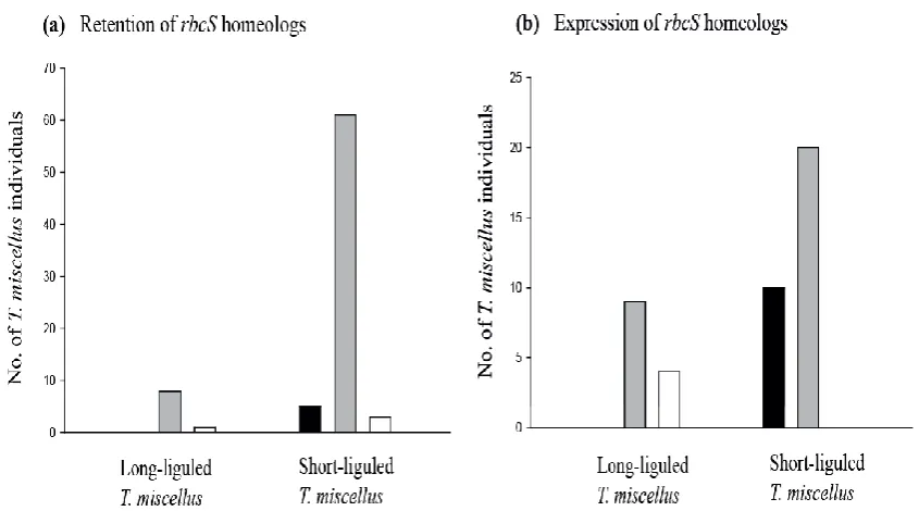
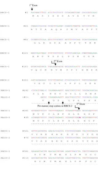
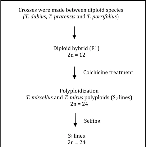
(11) LIST OF FIGURES Fig. 2.1 rbcS-1 gene structure and locations of SNPs. The structure of the rbcS1 gene is shown at the top with both coding regions and non-coding regulatory elements indicated. Locations for SNPs between T. dubius and T. pratensis homeologs have been scaled along the length of the rbcS-1 gene. ............................................................................................................................ 33 Fig. 2.2 CAPS analysis showing additivity and expression of rbcS-1 homeologs. (a) Genomic DNA CAPS results for six representative samples and (b) cDNA CAPS results for four representative samples of naturally occurring T. miscellus polyploids along with representative diploid parents, T. dubius and T. pratensis. Chromatograms belong to the same polyploid samples (from top to bottom) as in the gel photo below (from left to right). Chromatograms show sequence polymorphisms at the third SNP in exon 1 for both genomic DNA and cDNA sequences of the natural polyploids. “L” following the polyploid name denotes the longliguled form, and “S” denotes the short-liguled form. ........................................... 37 Fig. 2.3 Graphical illustration of retention and expression of parental rbcS-1 homeologs in T. miscellus (short and long liguled) natural polyploids. Bar charts show (a) retention and (b) expression of parental rbcS-1 homeologs in T. miscellus polyploids. Black, grey and white colours of the bars correspond to retention/expression of T. pratensis rbcS-1 homeolog, both progenitor rbcS-1 homeologs and T. dubius rbcS-1 homeolog respectively. Short liguled and long liguled individuals are represented with separate bars. .................................................................................... 38 Fig. 2.4 Homeolog-specific RT-PCR of rbcS-1. (a) T. dubius and (b) T. pratensis homeolog-specific RT-PCR results are shown for 11 T. miscellus natural polyploids with representative diploid parents, T. dubius and T. pratensis. Six individuals of the allotetraploid T. miscellus (indicated by an asterisk (*) showed genomic loss of one parental fragment (T. dubius homeolog was lost in five short-liguled polyploids, and the T. pratensis homeolog was lost in one long-liguled Pullman individual 2605-28). The remaining five natural polyploids show expression biased towards one of the parents. “L” following the polyploid name denotes the longliguled form, and “S” denotes the short-liguled form. ........................................... 40 Fig. S2.1 cDNA-CAPS for TDF-85 as a control to check equal expression of parental copies in T. miscellus polyploids showing biased maternal expression for rbcS-1.......................................................................................................... 52 Fig. S2.2 Alignment of rbcS-1 and rbcS-2 cDNA sequences of Tragopogon dubius along with protein translation for both genes. Arrows indicate position of exons and premature stop codons. ........................................................ 53 Fig. 3.1 Formation of synthetic polyploids. .............................................................................. 62 Fig. 3.2 Neighbor-net of T. miscellus synthetic polyploids. (a) Based on all microsatellite loci. (b) Based on only T. dubius loci and includes their T. dubius parents and generic types named as G1, G2 and G3 based on multilocus genotype I, II and III. Each split is corresponding to the cluster of individuals having distinct multilocus genotype found in the natural polyploid populations. ....................................................................................... 71 Fig. 3.3 Neighbor-net of T. mirus synthetic polyploids. (a) Based on all microsatellite loci (b) Based on only T. dubius loci and include their T. viii.
(12) dubius parents and generic types named as G1, G2 and G3 based on multilocus genotype I, II and III. Each split corresponds to the cluster of individuals having distinct multilocus genotypes. ................................................. 73 Fig. 3.4 (a) Neighbor-net of T. miscellus polyploids along with their T. pratensis parents. .................................................................................................................................... 74 Fig. 4.1 Diagrammatic illustration of the ABC model. .......................................................... 86 Fig. 5.1 Sequence polymorphisms between the diploid parents (Tragopogon dubius and T. pratensis) were used to determine if there is homeologspecific silencing in T. miscellus allopolyploids. (a) Diagrammatic illustration of the expected chromatogram peaks for genomic and bisulfite-converted sequences when un-methylated or methylated in allopolyploid T. miscellus. This example shows silencing of the T. dubius homeolog. (b) Chromatograms of TDF-44 indicating the position of a methylated CpG adjacent to a polymorphic site (red box) in T. miscellus compared to the diploids. (c) Chromatograms from S18 showing an unmethylated CpG site in T. miscellus (black box) and the location of a polymorphic site between parental copies (red box). Red, blue, green and yellow colors of the chromatogram correspond to A, C, T and G, respectively.BS-converted=bisulfite-converted. .................................................. 124. ix.
(13) LIST OF TABLES Table 2.1 rbcS-1 primers designed in this study. ................................................................... 27 Table 2.2 Naturally occurring individuals of Tragopogon miscellus that showed bias in the retention and expression of parental rbcS-1 homeologs. A dash (-) indicates that we were not able to study a particular individual for both retention (genomic DNA) and expression (cDNA). ............................... 39 Table S2.1 List of natural and synthetic polyploids (T. miscellus) and diploid parents (T. dubius and T. pratensis) examined. Data are summarized from genomic DNA and cDNA sequencing, genomic and cDNA CAPS, and homeolog-specific RT-PCR. Note: Letters “D” and “P” correspond to the diploid parents T. dubius and T. pratensis, respectively. A ‘D’ or a ‘P’ indicates that only one parental homeolog was detected in genomic DNA or expressed. P>D indicates that the T. pratensis homeolog showed higher relative expression than the T. dubius rbcS-1 homeolog in the T. miscellus individual and vice versa for D>P. ............................................................. 47 Table S2.2 Transcription factor binding sites in rbcS-1 promoter region as determined by Plant Promoter Analysis Navigator (PlantPAN). ...................... 51 Table 3.1 Crossing information and number of F1, S0 and S1 synthetic polyploid lines examined in the study. ............................................................................................ 65 Table 3.2 Occurrence of the multilocus genotypes in the synthetic polyploids. ....... 67 Table 4.1 List of diploid and polyploid samples. ................................................................... 90 Table 4.2 List of MADS-box orthologs from other species of Asteraceae that were used as reference genes. ........................................................................................ 94 Table 4.3 Total read count for Tragopogon transcripts mapping to ABC genes for each of the diploid parents and T. miscellus polyploid................................ 106 Table 4.4 Percentages of parental transcripts in Tragopogon diploid and polyploids orthologous to ABC class genes from other groups of Asteraceae. .......................................................................................................................... 108 Table 5.1 Individual plants used in the study and their methylation status for the genes studied; silencing data from Tate et al. (2006) and Buggs et al. (2009).............................................................................................................................. 122 Table S5.1 List of primers ............................................................................................................ 128. x.
(14) LIST OF ABBREVIATIONS A AG AS AP1 bp BSConverted °C CAPS cDNA CTAB Contig CYC C DEF dNTP DNA EDTA EST F1 FUE gDNA GLO g G HS ID Indel Kb ♀. MADS Mb m NA µg µl µM ml mM MYA min M ng NUE ♂. PI. Adenosine AGAMOUS Antisense APETALA1 Base Pair Bisulfite converted Degrees celcius Cleaved amplified polymorphic sequence Complementary DNA Cetyl trimethylammonium bromide Contiguous sequence CYCLOIDEA Cytosine DEFICIENS Deoxynucleoside 5'-triphosphate Deoxyribonucleic acid Ethylenediaminetetraacetic acid Expressed sequence tags First hybrid generation Far upstream elements Genomic DNA GLOBOSA Gram Guanine Homeolog specific Identification number Insertion or deletion Kilobase Maternal parent MCM1, Agamous, Deficiens, Serum response factor (family of transcription factors with conserved DNA binding site) Megabase Messenger RNA Microgram Microliter Micromolar Milliliter Millimolar Million years ago Minute(s) Molar Nanogram Near upstream elements Paternal parent PISTILLATA xi.
(15) PCR PLACE PVP 3’ RACE RNA RNase rRNA RuBisCO rbcL rbcS RIN S0 S1 S SEP SNP Spp. T TDF TE TSS U UTR U WGD. Polymerase chain reaction Plant cis-acting regulatory DNA elements Polyvinyl pyrrolidone Rapid amplification of cDNA ends Ribonucleic acid Ribnuclease Ribosomal RNA Ribulose-1,5-bisphosphate carboxylase oxygenase Ribulose bisphosphate carboxylase large subunit Ribulose bisphosphate carboxylase small subunit RNA integrity number First generation of synthetic polyploids Second generation of synthetic polyploids sense SEPALLATA Single nucleotide polymorphism Species Thymine Transcript derived fragment Transposable elements Transcription start site Unit(s) Un-translated region Uracil Whole genome duplication. xii.
(16) CHAPTER ONE. 1.
(17) 1. Introduction. 1.1 Polyploidy Polyploidy is the heritable state of having more than two complete sets of chromosomes. The majority of polyploids possess an even number of sets of chromosomes, with tetraploidy being the most common (Jiao et al., 2011; Chester et al., 2012; Wendel et al., 2012; Madlung, 2013). Polyploids are frequent among plants and also common among fish and amphibians (Mable et al., 2011). Polyploidy in plants was first reported by Hugo de Vries on Oenothera lamarkiana mut. Gigas (Onagraceae), which was determined to be a tetraploid (Vaughan, 1906; Lutz, 1907). Polyploidization has been inferred to occur at least once in the evolutionary history of most species (Blanc & Wolfe, 2004a). The study of eukaryotic genomes is offering astounding evidence of the evolutionary potential of polyploids: many sequenced genomes exhibit evidence of polyploid ancestry (Soltis & Soltis, 1995; Chen et al., 2007; Soltis et al., 2009). In this chapter, I present a review of polyploidy, focusing on the genetic and epigenetic consequences of polyploidy observed in various plant polyploids.. 1.2 Types of polyploids Polyploids are characterized by their chromosomal composition and their mode of formation. Several different types of polyploids have been described including autopolyploids,. segmental. allopolyploids,. true. or. genomic. polyploids. and. autoallopolyploids (Stebbins, 1950; reviewed in Tate et al., 2005). Primarily, two types of polyploids occur: autopolyploids and allopolyploids. Typically, autopolyploids are formed within populations of the same species (same origin), and allopolyploids result from interspecific hybridization (diverse origin) (Ramsey & Schemske, 1998; Comai, 2005a). Segmental polyploids fall between auto- and allopolyploids because they are formed within. 2.
(18) a species, but have partial homology of their chromosomes, which exhibit both multivalent and bivalent formation; for instance B1B1 × B2B2 → B1B1B2B2. Autoallopolyploids possess both auto- and allopolyploid-derived chromosome sets, such as AAAA × BB → AAAABB (Stebbins, 1947; Swaminathan, 1954; Sybenga, 1996; Ramsey & Schemske, 1998). Aneuploids possess either an extra chromosome or are missing a chromosome resulting in a different haploid number (Myers & Hill, 1940). The term paleopolyploid refers to ancient polyploids that have been diploidized over time and the term neopolyploid refers to newly formed allo- and autopolyploids (Wolfe, 2001; Ramsey & Schemske, 2002).. 1.3 Chromosomal pairing behavior of polyploids Pairing of chromosomes during meiosis varies according to the type of polyploid. In some allopolyploids, homologous chromosomes from one genome type preferentially pair with each other at metaphase I, resulting in the formation of bivalents. While autopolyploids have more than one pair of homologous chromosomes, so pairing can take place either between two. randomly (non-preferentially) selected homologous. chromosomes (forming bivalents) or between more than two homologous chromosomes resulting in multivalent formation (Sybenga, 1996; Hauber et al., 1999; Wu et al., 2001). In polyploids, pairing affinity also depends on the degree of chromosomal homology; identical homologous chromosomes tend to pair preferentially compared to less similar chromosomes (Doyle, 1963; Benavente & Orellana, 1991). During meiosis, homologous chromosomes physically link by synapsis and recombination to ensure accurate segregation. This meiotic recombination results in either cross-overs (reciprocal exchange of DNA fragments between pairing partners) or gene conversions (unidirectional transfer of DNA fragments from one non-sister chromatid to another) (Bhalla & Dernburg, 2008). Homologous recombination (recombination between homologous chromosomes) occurs more commonly compared to homeologous recombination (recombination between distinct but related chromosomes). In allopolyploids, homeologous recombination can. 3.
(19) lead to chromosomal rearrangements and hence genetic variability needed for adaptation (Pinto et al., 2005; Nicolas et al., 2007; Modliszewski & Willis, 2014).. 1.4 Formation of polyploids In general, polyploids result from mitotic or meiotic mishaps, such as failure of chromosomes to segregate, resulting in the production of either polyploid somatic tissues on a normally diploid plant or meiotic nuclear restitution during gamete formation results in unreduced gametes (2n) giving rise to polyploid plants. The latter is considered the more common mechanism of polyploid formation (Harlan & Dewet, 1975; Thompson & Lumaret, 1992; Brownfield & Kohler, 2011). Somatic doubling is a rarely reported mechanism of polyploid formation, which is known to form mixoploid chimeras in meristemic tissue of sporophytes. For example, the development of a tetraploid shoot was observed on a diploid F1 hybrid produced between Mimulus nelsoni and M. lewissii (Hiesey et al., 1971). Somatic doubling is known to occur in non-meristematic plant tissues as well, where cells can initiate new growth in tumors or wounds and are an important source of new shoots (D’amato, 1952). Somatic doubling also occurs in zygotes or young embryos producing polyploid sporophytes. There is very little information available on the frequency of somatic doubling in plants and none of the effects of interspecific hybridization are known on its occurrence (reviewed in Ramsey & Schemske, 1998). The more common mechanism of polyploid formation is by gametic non-reduction during micro- and mega-gametogenesis. The process results in the formation of 2n gametes, containing the complete somatic chromosome number. These 2n gametes are produced by meiotic events, such as cytological alterations associated with first division restitution (FDR) and second division restitution (SDR) (Ramanna, 1979; Carputo et al., 2000). Formation of 2n pollen results from the disorientation of spindle fibres at metaphase II or abnormal cytokinesis, while 2n egg production is caused by the absence of. 4.
(20) cytokinesis after telophase II, but the absence of the first and second meiotic divisions has also been reported (Werner & Peloquin, 1991; Barcaccia et al., 2003). Most reports on unreduced gametes are based on pollen because it is easy to identify compared to unreduced eggs that are much harder to study (Ramsey & Schemske, 1998). The formation of polyploid embryos may occur by the union of unreduced (2n) gametes (Bretagnolle & Thompson, 1995; Comai, 2005a; De Storme & Geelen, 2013). In addition to genetic factors, there are also environmental factors that can stimulate unreduced gamete formation; these include rapid changes in temperature (heat or cold treatment), x-rays, UV light, dehydration and infections. For example, Rosa plants were exposed to different temperature regimes. Extreme temperatures were associated with abnormal meiosis, resulting in disorientation of spindles at telophase II and an increase in the ploidy level of pollen grains (Pecrix et al., 2011). Similarly, Brassica allopolyploids showed an increase in the frequency of unreduced gamete formation at cold temperatures (Mason et al., 2011). Hence, adverse conditions could facilitate polyploid formation in the wild (Sax, 1936).. 1.5 Phenotypic consequences of polyploidy Allopolyploidy is one of the major forces involved in plant speciation, resulting from the union of two or more diverse, but generally closely associated, genomes into the same nucleus by hybridization. The accumulation of increased genetic variation in allopolyploids is possible by gene redundancy, which provides the likelihood of generating novel functional diversity between homeologous genes and genomes (Adams & Wendel, 2005b; Soltis et al., 2009; Madlung, 2013). Polyploidy can have instant phenotypic consequences, for instance, enlarged cell size and organ size, and occasionally better vigour and increased biomass (Soltis & Soltis, 1995; Comai et al., 2000; Balao et al., 2011). The emergence of these new phenotypes in polyploids possibly involves modifications in gene expression (Osborn et al., 2003; Paun et. 5.
(21) al., 2011). Polyploids often show novel phenotypes or show increased variation compared to their parents (Ramsey & Schemske, 2002). According to an evolutionary or ecological perspective, polyploid events may be observed as a stimulus for novel phenotypic changes. For example, studies revealed changes in Brassica for a number of environmentally critical phenotypic characters, including flowering time (Pires et al., 2004b), leaf morphology, and seed set (Doyle et al., 2008). Some of these characters, like drought tolerance, flowering time, pest resistance, apomixis, and increased biomass, could permit polyploids to adapt to new places and environments or increase their probability to be utilized in agriculture (Fawcett et al., 2009; Van Laere et al., 2011; Martin & Husband, 2012; Hannweg et al., 2012).. 1.6 Genetic and epigenetic consequences of polyploidy The duplication and merger of distinct genomes in one nucleus can lead to considerable genetic and epigenetic restructuring of the duplicated genomes. The genes duplicated as a result of allopolyploidy (homeologs) have a number of evolutionary outcomes. Three potential fates are expected for these duplicated genes: (I) both copies are preserved and stay functional, (II) one copy maintains the actual function whereas the other copy is silenced or lost, or (III) subfunctionalization occurs when the two copies deviate such that each copy contributes only a part of the original gene function, or neofunctionalization may occur in which one copy attains a novel function (Ramsey & Schemske, 1998; Lynch & Conery, 2000; Edger & Pires, 2009; Roulin et al., 2013). Genetic. changes. post-polyploidization. comprise. chromosomal. reshuffling,. translocations, gene loss, concerted evolution of rDNA repeats, and/or transcriptomic changes (Kovarik et al., 2004; Anssour et al., 2009; Buggs et al., 2010b; Jackson & Chen, 2010; Malinska et al., 2010; Buggs et al., 2012a; Chester et al., 2012; Tang et al., 2012; Ma et al., 2013). Epigenetic changes involve DNA methylation, histone modifications, deacetylation, microRNAs and prions (Halfmann & Lindquist, 2010; Vanyushin & Ashapkin,. 6.
(22) 2011; Lee & Shin, 2012). These genetic and epigenetic modifications can occur instantly in the first generation after polyploidization or during many generations after polyploid formation (Madlung et al., 2002; Adams & Wendel, 2005a; Chantret et al., 2005; Skalicka et al., 2005; Otto, 2007; Doyle et al., 2008; Hegarty & Hiscock, 2008; Chague et al., 2010; Flagel & Wendel, 2010; Dong & Adams, 2011b; Hu et al., 2013; Madlung, 2013). Genetic and epigenetic outcomes of polyploidy studied in various plant genera are as follows:. 1.6.1 Change in genome size Presumably, polyploids are expected to be additive of their progenitor genomes but deviation from additivity in genome size is one of the significant consequences of a polyploidization event. Although additivity in genome size has been observed in newly formed natural and synthetic polyploids (Liu & Wendel, 2002; Pires et al., 2004a; Russell et al., 2013), patterns of non-additivity in polyploid genome size have been found in older and long-established polyploids (Gossypium, Kadir, 1976; Vigna, Parida et al., 1990; Nicotiana, Leitch et al., 2008). Some polyploids have been described with an increased quantity of DNA relative to their progenitors (Jakob et al., 2004; Leitch et al., 2008), but the overall trend in polyploid genome evolution in angiosperms is towards genome downsizing (loss of DNA following polyploidization), most probably to reduce genetic instability caused by genetic redundancy and any phenotypic effects of having larger nucleus and cell size. C values (Quantity of DNA in the gametic nucleus) in some polyploids were less than predicted (Parida et al., 1990; Leitch & Bennett, 2004; Castro et al., 2012; Wong & Murray, 2012; Duchoslav et al., 2013; Li et al., 2013). Predominantly, loss of repetitive DNA clusters like Ty3-gypsy and Ty1-copia retroelements have caused reduction in the predicted genome size in Nicotiana polyploids (Renny-Byfield et al., 2011; Renny-Byfield et al., 2013). In addition to changes in the DNA sequence, epigenetic mechanisms are also associated with transposon elements and are involved in genome restructuring and downsizing in Spartina and Dactylorhiza (Parisod et al., 2009; Paun et al., 2010).. 7.
(23) 1.6.2 Gene loss Loss of one set of duplicated genes (homeologs) has been widely reported in polyploid species (Kashkush et al., 2002b; Mun et al., 2009; Tate et al., 2009a; Buggs et al., 2012a; Buggs et al., 2012b; Mlinarec et al., 2012; Akhunov et al., 2013). Synthetic allopolyploids of Brassica showed extensive loss of parental DNA fragments at the F5 generation (Song et al., 1995). In a similar study of 49 independently resynthesized Brassica lines, homeolog losses were observed in the S0 generation (Lukens et al., 2006). Moreover, in successive generations, a number of sequence losses occurred because of homeologous recombination leading to non-reciprocal translocations (Pires et al., 2004b). Recently, Xu et al. (2012) revealed genetic changes involving deletions and insertions of novel fragments in two independently generated sets of Brassica napus synthetic allopolyploids.. Sequence. loss. (loss. of. anonymous. DNA. fragments). following. polyploidization has also resulted in phenotypic variation in wheat. Elimination of parental fragments was illustrated in synthetic wheat allopolyploids and their relatives (Feldman et al., 1997; Liu et al., 1998), both instantaneously after formation of the polyploid (synthetic allotetraploids, Shaked et al., 2001; Kashkush et al., 2002b), and in generations afterwards (synthetic allohexaploids, Ma et al., 2004; Ma & Gustafson, 2008). In many cases, DNA fragments appear to have been lost via homeologous recombination in the synthetic allopolyploids (Shaked et al., 2001; Kashkush et al., 2002a; Gaeta & Pires, 2010). Extensive loss of the duplicated genes may be responsible for significant variation among related plant species (Paterson et al., 2004; Pellicer et al., 2010). One of the stimulating features of variable retention of duplicated genes involves the pattern of sequence elimination versus survivorship (Liu & Davis, 2011). Considering the functional importance of retained duplicates, it is proposed that the probability of duplicate retention was related to the number of functional interactions between the gene products. Dosage sensitive genes which are involved in various regulatory networks (like transcription factors) are retained significantly compared to other non-functional DNA sequences (Udall. 8.
(24) & Wendel, 2006; Edger & Pires, 2009; Severin et al., 2011; Wang et al., 2011; De Smet & Van De Peer, 2012). For instance, in Arabidopsis, genes that were involved in regulatory networks such as signal transduction and transcription remain duplicated, while others involved in DNA repair were reduced to single copy (Blanc & Wolfe, 2004b; Chapman et al., 2006). In contrast to the above pattern, in Asteraceae, genes associated with structural components and cellular organization were reported to be preferentially retained while genes associated with transcription and regulatory pathways were considerably underrepresented (Barker et al., 2008). The chromosomal position of conserved versus lost genes, including the level to which retained genes are grouped together, is also of interest. For instance, in Brassica napus, the rate of sequence loss has been shown to increase with increasing genetic distance from the centromere (Nicolas et al., 2012).. 1.6.3 Changes in the transcriptome and proteome Generally, it is assumed that polyploids will additively express their paternal genes but polyploidy events have significant effects on duplicate gene expression, with up- or down-regulation of one of the parental homeologs commonly resulting. Advances in highthroughput technologies have made it possible to analyze evolutionary outcomes of genome duplication events at the level of the transcriptome and proteome (De Smet & Van De Peer, 2012). Absolute comparisons between transcriptome and proteome profiles are not possible because they are not equivalent to each other due to the involvement of various post-transcriptional and post-translational modifications (Gygi et al., 1999). Because the prediction of proteome expression from mRNA expression is not possible, the analysis of both transcriptome and proteome would improve our understanding of polyploid genome evolution and adaptation. One of the most significant findings with regard to gene expression in polyploids is the unequal contribution of homeologous genes to the transcriptome, as shown in cotton (Adams et al., 2003b). Adams et al. (2003) showed that 10 out of 40 homeologs from the A and D genomes of allotetraploid cotton demonstrate biased expression, including various. 9.
(25) examples of reciprocal silencing among neighbouring floral whorls. This illustration of unequal contributions of duplicated genes to the transcriptome has been confirmed and extended in a number of later studies. For instance, Hovav et al. (2008) examined homeolog ratios for around 1400 gene pairs throughout the growth of the cotton “fiber.” Biases in homeologous expression were extended to the temporal variation across developmental phases (Doyle et al., 2008; Hovav et al., 2008; Chaudhary et al., 2009; Grover et al., 2012). Hegarty et al. (2005) observed substantial variation in the expression levels of various genes within diploid, allohexaploid and triploid Senecio species. Analysis of the transcriptome of synthetic polyploids of Arabidopsis have shown alterations in gene expression that were associated with modified DNA methylation patterns (Yu et al., 2010). Recently, analysis of leaf transcriptomes of F1 hybrids, synthetic and natural Gossypium polyploids demonstrated considerable up- and down-regulation of gene expression and biases in expression towards one of the parental genomes, suggesting various regulatory and epigenetic interactions arising through transcriptome networks post-polyploidization (Dong & Adams, 2011a; Yoo et al., 2013). In wheat, Nicotiana and Brassica, RNA sequencing revealed structural and functional modifications in the duplicated genes owing to neo/subfunctionalization mechanisms (Pont et al., 2011; Bombarely et al., 2012; Higgins et al., 2012) Koh et al. (2012) examined the proteome of F1 hybrids, synthetic and natural Tragopogon mirus polyploids and its diploid progenitors (T. dubius and T. porrifolius) and reported expression changes after hybridization and polyploidization events and also found proteins with novel expression. Moreover, proteome analysis of early generations (F1-F4) of Brassica napus synthetic polyploids showed non-additive repatterning of their protein expression profile. However, this non-additivity did not involve expression profiles of housekeeping genes involved in regulatory networks. Hence, after polyploid formation gene silencing is a continued phenomenon and can be activated at any generation (Kong et al., 2011).. 10.
(26) 1.6.4 DNA methylation Allopolyploidy. frequently. causes. unpredicted. deviations. from. expected. transcriptomic additivity, which may be the result of extensive epigenetic alterations. DNA methylation is one of those epigenetic modifications causing silencing of duplicated genes by methylation of the promoter or coding sequences of the genes, thereby repressing transcription by inhibiting binding of the transcription factors to the promoter (Finnegan et al., 1998; Salmon et al., 2005; Salmon & Ainouche, 2010). DNA methylation is the addition of a methyl group at position 5 of pyrimidine ring of a cytosine residue (Finnegan et al., 1998; Chan et al., 2005; Vanyushin, 2006). Cytosine methylation is important for genomic stability and is involved in genomic imprinting, transposon silencing and epigenetic regulation of gene transcription (Martienssen & Colot, 2001; He et al., 2011; Vanyushin & Ashapkin, 2011; Ji & Chen, 2012). In wheat, cytosine methylation has been observed as an instant response to genome duplication (Shaked et al., 2001). Similarly, Lukens et al. (2006) reported cytosine methylation in resynthesized Brassica napus allopolyploids. A high level of DNA methylation in Brassica oleracea has been proposed to be associated with genome plasticity and a high level of phenotypic variability (Salmon et al., 2008). Madlung et al. (2002) observed that the phenotypic instability of synthetic Arabidopsis polyploids was due to an irregular methylation status causing an altered rate of transcription resulting in both gene silencing and gene activation, with the latter caused by de-methylation. Verhoeven et al. (2010) reported de novo methylation in triploid dandelion lineages post-polyploidization. DNA methylation is proposed to generate phenotypic variation and thereby could facilitate the adaptation of naturally occurring polyploids (Richards et al., 2008). Genome wide mapping of DNA methylation in Arabidopsis thaliana revealed the widespread prevalence of DNA methylation in intergenic regions and repetitive sequences, while limited occurrence in the regulatory regions such as promoter, thus serving to maintain genomic integrity (Zhang et al., 2006; Weber & Schuebeler, 2007; Zilberman et al., 2007). Melamed-Bessudo and Levy (2012) studied the. 11.
(27) role of DNA methylation in chromosomal recombination by employing decreased DNA methylation (ddm1) mutant lines in Arabidopsis. They reported that a decrease in DNA methylation increases the rate of meiotic crossing over in the euchromatic regions, but not in heterochromatin, suggesting a repressive role of DNA methylation in meiotic recombination. In Oryza sativa, the methylation level (the proportion of methylated CpG sites) of the coding regions of genes was estimated in five plant tissues for different gene categories including transposable element-like genes, duplicated genes and singleton genes (Wang et al., 2013b). They reported a low level of methylation associated with high levels of expression, in contrast to a high level of methylation associated with low levels of expression suggesting genome-wide methylation is related to duplicate gene expression and determines the survivorship of duplicated genes (Wang et al., 2013b).. 1.6.5 Transposon activation, small RNAs and RNAi Transposable elements (TEs) are discrete fragments of DNA that can transpose or ‘jump’ around the genome from one site to another unrelated site. Their random insertions in genomic sequences can mutate host DNA thereby affecting gene expression. TEs play a key role in restructuring genomes following allopolyploidization (Kidwell, 2005; Parisod et al., 2010). Analysis of various allopolyploid systems has shown structural, functional and epigenetic modification by transposons during allopolyploidization (Kraitshtein et al., 2010; Parisod et al., 2010; Yaakov & Kashkush, 2011; Zhang et al., 2013). For example, transposon activation has been observed in wheat allopolyploids. The activation of some transposable elements can lead to silencing of neighbouring downstream genes (Kashkush et al., 2003; Domingues et al., 2012). For instance, activation of the Wis transposon resulted in inactivation of the downstream purB gene, which is involved in softening of endosperm of the wheat kernal (Kashkush et al., 2003; Chen et al., 2010). After polyploid formation, epigenetic remodelling of the genome also occurs through various regulatory pathways such as RNA interfence (RNAi). RNAi helps in overcoming the gene redundancy and maintaining genomic integrity post-polyploidization (Lawrence & Pikaard, 2003). During. 12.
(28) the process of RNAi, small interferring RNA (siRNA) molecules of 21-24 nucleotides are produced from double stranded endogenous RNAs which are formed by back-folding to form hair-pin structures. These siRNA unwind to form single-stranded siRNA molecules which integrate into RNA induced silencing complex (RISC). Then they bind to a complementary site of the target mRNA sequence and induce its cleavage by argonaute proteins, resulting in the repression of gene expression of the target mRNA (Mocellin & Provenzano, 2004). Ha et al. (2009) proposed that the loss of siRNA in F1 hybrids of Arabidopsis was related to genomic instability, while non-additive expression of miRNA and siRNA in the Arabidopsis allopolyploids was associated with chromatin maintenance, vigor and adaptation. Small RNAs play significant roles in many biological processes, including the regulation of gene expression, developmental timing, the maintenance of genomic integrity and defence against invasive nucleic acids (e.g., viruses and transposons) (Cam, 2010; Ketting, 2011; Ng et al., 2012; Feng & Guang, 2013).. 1.7 Synthetic allopolyploid lines as a useful resource Many polyploid species originated several million years ago such as wheat (13 mya), (Brandon, 2002), cotton (5-15 mya) (Wendel & Cronn, 2003) and Brassica (~20 mya) (Lagercrantz, 1998). Employing plant systems of different ages is important particularly to separate mechanisms that might describe the earliest stages of polyploid formation from those processes that are accountable for longer-term evolutionary variation (Adams & Wendel, 2004; Levy & Feldman, 2004; Otto, 2007). Moreover, crop polyploids, which are most often studied, have been subjected to artificial selection at some point during their history, which could be an argument for studying natural polyploids (Buggs, 2008). Natural allopolyploids that were formed in the last century, like Senecio cambrensis (Ashton & Abbott, 1992), Spartina anglica (Baumel et al., 2001), Tragopogon miscellus and Tragopogon mirus (Ownbey, 1950), offer a closer look into a variety of genome changes. 13.
(29) and gene expression differences occurring upon allopolyploidization (Adams, 2007). However, synthetically made polyploids are excellent models for examining the instant outcomes of polyploidization, which cannot be shown in the corresponding natural systems and they offer insights into mechanisms that take place immediately upon formation (Adams & Wendel, 2005a). Because the progenitors of a synthetic polyploid are known with certainty, it can be specifically resolved whether widespread genome changes take place immediately after synthesis of the polyploid and if so, then the timing and mechanisms of genome changes can be determined (Song et al., 1995). Recently, a number of synthetic polyploids of various plant species have been developed by interspecific hybridizations between the diploid progenitors and then chromosome doubling of the F1 hybrids (Brassica, Song et al., 1995; Arabidopsis, Comai et al., 2000; Cotton, Adams et al., 2003a; Wheat, Ma & Gustafson, 2008; Tragopogon, Tate et al., 2009b; Miscanthus, Chae et al., 2012; Rice, Wang et al., 2013a). Recent molecular studies of newly synthesized wheat (Triticum spp.) have shown sequence loss, gene expression changes, methylation, rearrangement of transposable elements and chromosomal instability after F1 hybrid and allopolyploid formation (Ma & Gustafson, 2008; Yaakov & Kashkush, 2011; Yang et al., 2011b). Investigation of synthetic polyploids of Arabidopsis have determined that genes duplicated by polyploidy (homeologs) can be silenced instantly or soon after polyploidy (Comai et al., 2000). Madlung et al. (2005) found meiotic instability, transposon activation and chromosomal rearrangements in newly formed synthetic polyploids of Arabidopsis. Similarly, chromosomal rearrangements were reported by Pontes et al. (2004). Arabidopsis synthetic polyploids exhibited considerable structural genomic modifications including deletion of a major portion of the upper arm of chromosome II (Beaulieu et al., 2009). Synthetic allopolyploids of Senecio have shown gene silencing and alterations in the transcriptome (Hegarty et al., 2006a). A synthetic autotetraploid of Aegilops tauschii demonstrated alterations in the phenotype associated with modified DNA methylation pattern (Zeng et al., 2012). Similarly, phenotypic variation. 14.
(30) was observed in the synthetic polyploid of Miscanthus (Chae et al., 2012). Synthetic polyploids of Nicotiana have also demonstrated considerable morphological, genetic and genomic changes post-polyploidization (Anssour et al., 2009). Rice synthetic polyploids showed superiority in growth and seed set compared to parents and genomic in situ hybridization (GISH) showed some chromosomal translocations between parental genomes (Wang et al., 2013a). On the other hand, synthetic polyploids of Tragopogon miscellus have not showed any evidence of gene silencing in the seven homeologs studied at the S1 generation (Buggs et al., 2009).. 1.8 Tragopogon as a study system Tragopogon is an ideal system for studying allopolyploidy. The genus has about 150 species found in Eurasia (Ownbey, 1950). Three diploid (2n = 2x = 12) species (Tragopogon dubius, T. pratensis, and T. porrifolius) were established in eastern Washington State, USA, and neighbouring Idaho in the early 1900s. Since their introduction, these species have recurrently produced two allopolyploid (2n = 4x = 24) species: T. mirus resulting from T. dubius × T. porrifolius and T. miscellus produced by T. dubius × T. pratensis. Natural T. mirus is produced only with T. porrifolius as the maternal parent, while T. miscellus is produced reciprocally in nature (Ownbey, 1950; Ownbey & Mccollum, 1954; Soltis et al., 1995a; Soltis & Soltis, 1999). Ownbey and McCollum (1954) applied conventional cytogenetic approaches to karyotype the six pairs of chromosomes in the diploid species of Tragopogon. In addition to studying morphological features, they also identified considerable chromosomal variation (e.g., terminal knobs and secondary constrictions) within different populations of diploid species to understand the multiple origins of the two newly formed allopolyploids. Considerable work has ben done to identify independent origins of these polyploid species (Soltis & Soltis, 1989; Soltis & Soltis, 1991; Soltis et al., 1995a; Soltis & Soltis, 1999). Most recently Symonds et al. (2010) evaluated the recurrent formations and independent. 15.
(31) origins of T. mirus and T. miscellus polyploids using progenitor-specific microsatellite markers, specifically to determine the genetic contribution of the parental species into each independently formed polyploid species. Considering only the T. dubius loci (common parent of both polyploids), three multilocus genotypes (containing five loci) were found in natural population, which likely represents the historical population structure of T. dubius at the time of polyploid formation. A lack of gene flow between populations was also reported, representing potential reproductive barriers between polyploid lineages. Kovarik et al. (2005) examined rDNA loci of Tragopogon including internal and external transcribed spacer (ITS and ETS) in independently formed allopolyploids from various natural populations and diploid progenitor species. This study reported concerted evolution of rDNA repeats with T. dubius rDNA loci lost more frequently in both polyploids (Kovarik et al., 2005). Pires et al. (2004a) compared genome sizes of independently formed T. mirus and T. miscellus from various populations and found additivity in genome size in T. mirus polyploids between parental genome sizes, while two populations of T. miscellus showed genome downsizing. Moreover, no evidence of major genomic rearrangement was observed when these T. mirus and T. miscellus polyploids were analyzed using four chromosomal markers by fluorescent in situ hybridization (FISH). Ownbey and McCollum (1953) tried to resynthesize T. mirus and T. miscellus and generated a diploid F1 generation, but they were not able to produce allopolyploid plants. Moreover, as natural T. miscellus is produced reciprocally from T. pratensis and T. dubius, Ownbey and McCollum were only able to effectively synthesize one F1 hybrid with T. pratensis as the maternal parent. More recently, these allopolyploids (Tragopogon mirus and T. miscellus) were resynthesized by Tate et al. (Tate et al., 2009b) using several individuals of the parental diploid species. Several studies have shown loss of homeologs in Tragopogon allopolyploids. Early studies by Roose and Gottlieb (1976) and Soltis et al. (1995b) identified loss of genetic fragments using allozyme markers. Tate et al. (2006) identified loss of homeologous. 16.
(32) fragments in two natural populations (Pullman and Moscow) of T. miscellus using cDNA– amplified fragment length polymorphism (AFLP), along with genomic and cDNA cleaved amplified polymorphic sequence (CAPS) analyses. The suggested mechanism for loss of homeologous loci was by recombination of the parental homeologs. Additivity of parental fragments for the same loci studied for the T. miscellus polyploids was found in synthetic F1 hybrids between T. pratensis and T. dubius, demonstrating that these genomic changes are associated with polyploidization rather than hybridization (Tate et al., 2006a). Another study of homeologous loci in ten natural populations of T. miscellus revealed homeolog losses in independently formed populations suggesting homeolog losses are potentially responsible for genome downsizing, but some homeologs were retained consistently (Tate et al., 2009a). Similarly, frequent loss of homeologous loci in natural T. mirus polyploids has been observed by Koh et al. (2010). Buggs et al. (2009) analysed patterns of sequence loss in 10 sets of homeologs in five natural populations of Tragopogon miscellus and 44 synthetic allopolyploids from the S1 generation of the same species. They found that none of the genes examined showed immediate loss or silencing in the re-synthesized first allopolyploid generation of T. miscellus. Buggs et al. (2009) suggested that loss and silencing of some homeologs begin in natural populations within 40 generations or less after the whole-genome duplication event. In this study, only a few loci were examined in T. miscellus and no changes were observed in gene expression in the S1 generation, which appears to be in contrast to studies showing gene loss and silencing in other resynthesized allopolyploids (e.g. Adams et al., 2003b; Hegarty et al., 2005; Lukens et al., 2006). Recently, using Sequenom MassARRAY iPLEX genotyping, loss of clustered duplicated genes postpolyploidization was reported in natural T. miscellus populations by Buggs et al. (2012a) suggesting that repeated reduction in the duplicated genomes occur after whole genome duplication events. Moreover, those missing genes were corresponding to the genes lost after ancient whole genome duplications in the family Asteraceae and lost clusters were mostly associated with dosage sensitive genes. Hence, the loss of homeologous loci is a. 17.
(33) frequent phenomenon in Tragopogon as has been shown in other polyploid genera (Feldman et al., 1997; Kashkush et al., 2002a; Lukens et al., 2006). Chromosomal rearrangements were also commonly observed in Tragopogon polyploids post-polyploidization. Some multivalent formations were noticed by Ownbey (1950) in natural populations of T. mirus and T. miscellus. Lim et al. (2008) also observed multivalent formation at the S0 and S1 generation in synthetic T. mirus individuals. Moreover, they found loss of rDNA loci along with homeologous translocations in genomic in situ hybridization analyses. Later, extensive and recurrent chromosomal variation, including intergenomic translocations and aneuploidy were also observed in individuals from six populations of T. miscellus (Chester et al., 2012) and recently karyotype restructuring was also reported in T. miscellus allopolyploids (Chester et al., 2013). Evolution of rDNA loci has also been studied for both Tragopogon mirus and Tragopogon miscellus natural and synthetic polyploids. Uniparental losses of rDNA loci were reported with losses more frequent in natural polyploids than in synthetic polyploids. T. dubius rDNA loci are lost more frequently than either of the other parents, T. pratensis or T. porrifolius. Moreover, T. dubius rDNA loci were expressed at a higher level in spite of their reduced copy number (Matyasek et al., 2007; Malinska et al., 2010; Malinska et al., 2011). Polyploid formation involves considerable modification at the level of transcriptome and proteome, but as mentioned earlier, these are not directly correlated. Koh et al. (2012) analyzed the proteome of F1 hybrids of Tragopogon mirus, as well as natural and synthetic polyploids with the progenitors, T. dubius and T. porrifolius. Out of 476 proteins identified, 68 proteins showed quantitatively differential expression in T. mirus. Differential expression of 32 proteins was associated with hybridization (as observed in F1 hybrids), 22 protein changes were associated with polyploidization (observed in S1 synthetic polyploids) and 14 changes occurred since the natural formation of T. mirus suggesting hybrization may have a greater influence on genome evolution than gene duplication.. 18.
(34) 1.9 Thesis chapters The research presented in this thesis examined the genetic and epigenetic consequences of polyploidization in Tragopogon allopolyploids, with an emphasis on Tragopogon miscellus. A brief overview and hypothesis for each project follows:. 1.9.1 Cytonuclear coordination in T. miscellus polyploids (Chapter 2) The nuclear genome is biparentally inherited in hybrids and allopolyploids while cytoplasmic genomes (chloroplast and mitochondria) are typically maternally inherited. Cytonuclear coordination between the nuclear and cytoplasmic genomes is required for genomic stability and could be best studied in reciprocal hybrids and polyploids (Levin, 2003; Chen, 2007a). Naturally occurring and reciprocally formed T. miscellus allopolyploids present an ideal system to study this cytonuclear coordination. In this study, using the Riboluse-1, 5-Bisphosphate Carboxylase oxygenase (Rubisco) system, cytonuclear coordination between the maternally inherited rbcL (encoded by chloroplast genome) subunit and the biparentally inherited rbcS (encoded by nuclear genomes) subunit was examined in natural and synthetic Tragopogon miscellus allopolyploids. We hypothesized that the maternal homeolog of rbcS would be expressed in the T. miscellus polyploids to coordinate with the maternally inherited cytoplasmic subunit rbcL. For this purpose, the nuclear subunit rbcS was characterized in the diploid progenitors, T. dubius and T. pratensis, to determine, relative retention and expression of rbcS homeologs in T. miscellus polyploids.. 1.9.2 Exploring genetic structure of T. mirus and T. miscellus synthetic polyploids (Chapter 3) The genetic structure of natural polyploids was studied by Symonds et al. (2010) using progenitor-specific microsatellite loci. Symonds et al. (2010) reported a repeated pattern of three multilocus genotypes in both T. mirus and T. miscellus polyploids. Based on that study, here we hypothesized that patterns of polyploid formation follow genetic. 19.
(35) ‘rules’. To address this hypothesis, the genetic structure of synthetic T. mirus and T. miscellus polyploids was examined and compared to natural polyploids. Here, we looked for that same pattern of multilocus genotypes and also discovered novel genotypes in the synthetic polyploids.. 1.9.3 Comparative analysis of floral transcriptomes (Chapter 4) Tragopogon miscellus has formed reciprocally exhibiting short-liguled (T. dubius♂ × T. pratensis♀) and long-liguled (T. dubius♀ × T. pratensis♂) forms (Soltis & Soltis, 1989). As these morphologies were repeated in synthetic polyploids, here we hypothesize a maternal influence to the formation and variation in inflorescence/floral morphology. To determine genetic factors that might be controlling these floral morphologies, floral transcriptomes were studied from diploid parents and reciprocally formed natural and synthetic T. miscellus polyploids. RNA sequencing data (transcriptome) were analyzed to compare variation in the transcriptome between natural and synthetic T. miscellus polyploids to evaluate transcript abundance and up- or down-regulation of progenitorspecific floral development genes post-polyploidy.. 1.9.4 DNA methylation: A gene silencing mechanism post-polyploidization (Chapter 5) Gene silencing of parental homeologs have been previously reported in Tragopogon miscellus polyploids (Tate et al., 2006a; Buggs et al., 2009). DNA methylation is one of the epigenetic mechanisms that results in gene silencing. We hypothesized that DNA methylation would be responsible for the silencing of previously reported homeologous loci in Tragopogon miscellus polyploids. To investigate this, the pattern of CpG methylation was analyzed for those previously studied five homeologous loci [S2, S3, S8, S18 and TDF44 (Tate et al., 2006a; Buggs et al., 2009)] in several individuals of natural T. miscellus polyploids using comparative bisulfite sequencing.. 20.
(36) CHAPTER TWO. This chapter is in preparation as: Sehrish T, Symonds VV, Soltis DE, Soltis PS, and Tate JA. Biased paternal genomic loss and maternal expression of rbcS homeologs in Tragopogon miscellus (Asteraceae) allopolyploids: insight into cytonuclear compatibility using Rubisco. In prep for: New Phytologist.. 21.
(37) 2. Biased paternal genomic loss and maternal expression of rbcS-1 homeologs in Tragopogon miscellus (Asteraceae) allopolyploids: insight into cytonuclear compatibility. 2.1 Abstract . Ribulose-1,5-bisphosphate carboxylase/oxygenase (Rubisco), which consists of a chloroplast-encoded large subunit (rbcL) and a nuclear-encoded small subunit (rbcS), was used to examine cytonuclear interactions in naturally occurring and synthetic Tragopogon miscellus (Asteraceae) allotetraploids (including reciprocally formed individuals). Tragopogon miscellus formed recently (~80 years ago) and repeatedly via hybridization and whole-genome doubling.. . Genome-specific single nucleotide polymorphisms (SNPs) in the diploid progenitors (T. dubius and T. pratensis) were used to identify patterns of inheritance, retention, and expression of rbcL and the duplicated rbcS-1 gene copies (homeologs) in 25 synthetic and 78 natural T. miscellus individuals.. . All allopolyploids inherited the maternal rbcL copy. Both parental homeologs of rbcS-1 were retained and expressed in all synthetic and most natural individuals of T. miscellus. Genomic loss of one homeolog was apparent in ten natural polyploids, with retention of the maternal rbcS-1 copy more frequent. Eight natural individuals retained both homeologs, but showed biased expression, always in favor of the maternal rbcS-1 copy.. . Cytonuclear coordination occurs relatively early following polyploidization, but it does not appear immediately upon formation in Tragopogon miscellus. Moreover, the pattern of biased maternal retention and expression of rbcS-1 homeologs is repeated across independently formed populations and in reciprocally formed individuals, indicating repeatability of enhanced cytonuclear interactions.. 22.
(38) 2.2 Introduction Allopolyploidy is a major force in plant speciation and results from the union of two or more diverse, but generally closely related, genomes by hybridization (Soltis & Soltis, 2009; Te Beest et al., 2012). Genomic data indicate that all angiosperms may be regarded as polyploid, if paleopolyploid events are taken into account (Soltis et al., 2009; Yang et al., 2011a). Allopolyploid genomes experience both instant (immediately after the duplication event) and long-term evolutionary changes, which may involve a variety of genetic. and. epigenetic. interactions. leading to. genome. alteration,. regulatory. incompatibilities, chromosomal abnormalities, and reproductive failures (Comai et al., 2000; Hegarty et al., 2006b; Chen, 2007b; Gaeta et al., 2007; Ma & Gustafson, 2008; Xiong et al., 2011; Chester et al., 2012; Feldman et al., 2012; Yoo et al., 2013). Polyploidy has been considered a driver of modifications in gene function, potentially resulting in any of three fates of the duplicated genes (homeologs): (I) both copies are preserved and stay functional, (II) one copy maintains the original function whereas the other copy is silenced, or (III) the two copies diverge such that each copy contributes only a part of the original gene function (subfunctionalization), or one copy attains a novel function (neofunctionalization) (Lynch & Conery, 2000; Prince & Pickett, 2002; Blanc & Wolfe, 2004b; Conant & Wolfe, 2008; Doyle et al., 2008; Edger & Pires, 2009; Roulin et al., 2013). In newly formed hybrids and allopolyploids, coordination between the maternally inherited cytoplasmic (chloroplast and mitochondrial) and the biparentally inherited nuclear genomes is required to facilitate genomic stability (Chen, 2007b). Most protein complexes consisting of multiple subunits have proteins encoded by both nuclear and organellar genomes (Rodermel et al., 1988). For example, the enzyme Riboluse-1, 5bisphosphate carboxylase/oxygenase (Rubisco) is involved in carbon fixation in the Calvin cycle (Staswick, 1994): structurally, it is multi-meric, commonly composed of eight large subunits encoded by the chloroplast gene rbcL and eight small subunits encoded by the nuclear rbcS (Miziorko & Lorimer, 1983; Clegg, 1993). Depending on the species, rbcS is. 23.
Figure


+7


Related documents
European Journal of Taxonomy 167 1–40 http //dx doi org/10 5852/ejt 2015 167 ISSN 2118 9773 www europeanjournaloftaxonomy eu 2015 Hughes M et al This work is licensed under a
European Journal of Taxonomy 325 1–22 https //doi org/10 5852/ejt 2017 325 ISSN 2118 9773 www europeanjournaloftaxonomy eu 2017 Lehr E et al This work is licensed under a Creative
European Journal of Taxonomy 316 1–27 https //doi org/10 5852/ejt 2017 316 ISSN 2118 9773 www europeanjournaloftaxonomy eu 2017 Olesen J et al This work is licensed under a Creative
Fore wing 7.6 mm long, 2.8 mm wide, apparently hyaline except for black pterostigma, 1.7 mm long, 0.23 mm wide, narrow and elongate, situated well distal of cell [1M]; pterostigma
The size of the first elongated segment CH9 seems constant, about 2.5–3 longer than any short anterior segment CH1–CH8; the relative size of following elongated segments is
Taxonomic history of Cocconeis molesta Kütz., Cocconeis diaphana W.Sm., Cocconeis dirupta W.Greg.. and allied taxa Cocconeis
European Journal of Taxonomy 200 1–45 http //dx doi org/10 5852/ejt 2016 200 ISSN 2118 9773 www europeanjournaloftaxonomy eu 2016 Huber B A et al This work is licensed under a
Collum with three transverse rows of setae: 6+6 anterior, 4+4 intermediate and 6+6 posterior; a small lateral incision at about midway; caudal corner very broadly rounded,
