1
COVID-19: role of the Interferons
Claudio G. Gallo 1, Sirio Fiorino2, Giovanni Posabella3, Donato Antonacci,4 Antonio
Tropeano5, Emanuele Pausini6, Carlotta Pausini7, Tommaso Guarniero8, Marco Zancanaro9
1 Infectious disease Specialist, Physician ([email protected])
2 Internal Medicine Unit, Budrio Hospital Azienda USL, Bologna, Italy ([email protected]) 3 Sport Medicine Specialist, Bologna, Italy ([email protected])
4 Medical Science Department, “Casa Sollievo della Sofferenza” Hospital, San Giovanni Rotondo (FG)
5 Dentist ([email protected])
6 Biologist, Bologna, Italy ([email protected]) 7 Physician ([email protected])
8 Bachelor of Science ([email protected]) 9 Sport Physician ([email protected])
Corresponding Author
Claudio G. Gallo, MD [email protected]
Abstract
COVID-19 disease, caused by the SARS-CoV2 virus, is a potentially fatal disease that represents a serious public health and economic problem worldwide. The SARS-CoV2 virus infects the lower respiratory tract and can cause pneumonia in humans. ARDS is the leading cause of death in COVID-19 disease. One of the main characteristics of ARDS is the cytokine storm, an uncontrolled systemic inflammatory response resulting from the release of pro-inflammatory cytokines and chemokines and growth factors, by immune cells. The other important aspect of the disease is represented by the involvement of the vascular organ that undergoes endothelitis. Hyperinflammation and endothelitis contribute in various ways to trigger coagulation disorders with diffuse micro thrombotic and thromboembolic phenomena. Lastly, multiple organ failure may occur (MOF). Since so far there is no approved treatment, there is an urgent need to reposition known treatments, considered safe, to be included in trials. Naturally produced interferons represent the body's first line of defense against viruses. Pharmacological forms, obtained by means of genetic recombination techniques, have long been approved and used to treat numerous pathologies. Interferons are divided into three families, within which some subfamilies are distinguishable. Only IFN-II comprises a single isoform which has completely different aspects and functions. The IFN I and III, however, each comprise different subfamilies (17 subfamilies the IFN-I and 4 subfamilies the IFN-III), share many aspects, representing the body's first antiviral response, but play different roles. The use of IFNs has been studied in two severe hCoV (Human Coronavirus) diseases, closely related to COVID-19 disease, such as SARS and MERS. Numerous in vitro and in vivo studies have been conducted, often in combination with other antivirals. The results have been controversial. The positive results in vitro and in experimental animals were often not replicable in humans. The possible positioning of these molecules in the right window of therapeutic opportunity requires that the complex dialogue between IFN, inflammasome, cytokines, pro-inflammatory chemokines, growth factors and barrier function be shed light.
Keywords: COVID-19; SARS CoV-2; IFN-a; IFN-b; IFN-l.
Introduction
of the virus, two pathways are activated: the antiviral one (mediated by IFNs) and the inflammatory one (inflammasome and NFkB). The inflammatory pathway involves a cascade of signals that leads to induction, mediated by the transcription factor NF-kB, of cytokines and pro-inflammatory chemokines (IL-1-b, IL-6, TNF-a, IL-18, IL-8, IL-17, MCP-I). The antiviral route involves the production, mediated by IRF3 and IRF7, of IFN-I and IFN-III 29. IFNs represent the first major line
of defense against viruses. Interferons (IFNs) are divided into three families, within which some subfamilies are distinguishable. Only IFN-II includes a single isoform 33:
o IFNs-I (a,b,e,k,w);
o IFN-II (g);
o IFNs-III o IFN l (l1, l2, l3, l4).
The first step in interferon-mediated response to viruses is the recognition of molecular models associated with pathogens (PAMPs), represented by specific viral components such as viral RNA or DNA or viral intermediates1. PAMPs are detected by special molecules that function as sensors: PRRs
(Pattern Recognition Receptors), including Toll-like receptors (TLRs) and RIG-I-like receptors (RLRs) 4,5,6,7.
Several TLRs, including TLR 3, 7, 8, and 9, detect viral RNA and DNA in the endosome, whereas RLRs bind to viral RNA in the cytosol 8. RLRs comprise RIG-I, MDA5, and LGP2, all of which
contain an RNA helicase domain 9,10. The objectives are the control of viral replication in infected
cells, the activation and maintenance of the adaptive immune response for the eradication of the virus
2.
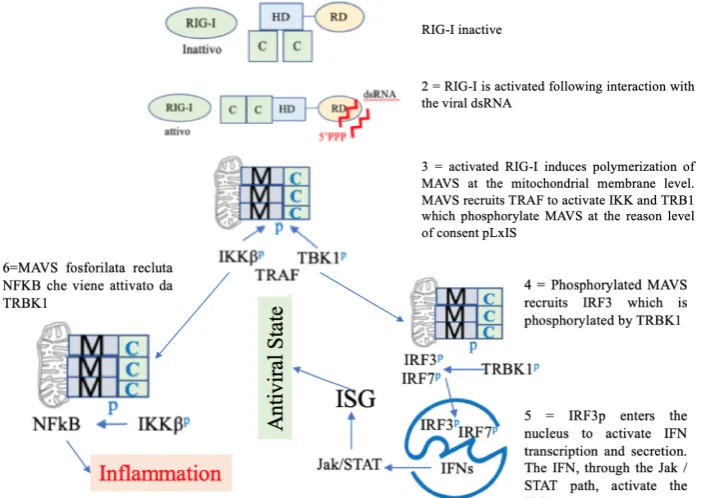
Figure 1. MAVS is an adapter that must be phosphorylated by IKK and TRB1 to recruit and authorize the phosphorylation of IRF3 by IKK and TRB1. After phosphorylation, IRF3 moves to the nucleus where it activates IFN transcription and secretion. The IFN, through the Jak/STAT signaling pathway, activates the ISG genes. RIG-I: retinoic acid-inducible gene-I-like receptors; HD: Helicase Domain, C=CARD: Caspase Recruitment Domain; RD: repressive Domain, ds: double strand RNA, M=MAVS Mitochondrial antiviral-signaling; TBK1p: TANK Binding Kinase 1; IKKp:
IκB kinas; TRAF: TNF-Receptor Associated Factors; IRF3: Interferon Regulatory Factor 3 IRF3p; NFkB: nuclear factor
Viral RNA, cytosolic DNA, and the bacterial cell wall component lipopolysaccharide activate signaling cascades through a number of pattern recognition receptor (PRR)–adaptor protein pairs, including RIG-I–MAVS, cGAS-STING, and TLR3/4-TRIF (TLR3/4, Toll-like receptors 3 and 4) 3.
The adaptor proteins MAVS, STING, and TRIF each activate the downstream protein kinase TBK1, which then phosphorylates the transcription factor interferon regulatory factor 3 (IRF3), which drives type I IFN production 3. Figure 1.
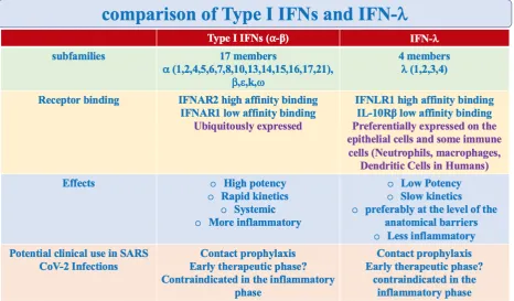
The IFNs-I and III have shared characteristics (activation paths, transcriptional programs) and distinct characteristics (receptors and functions) 11-22. Table 1.
Type I and III IFNs are genetically distinct, use different receptors, are induced by similar pathogen detection sensors and activate related antiviral, antiproliferative and immunomodulatory gene expression programs 33. The IFN-l represents the first border antiviral defense, the guardian of the
mucous barriers 34 (respiratory, gastro-enteric, urinary tract, etc.), minimizing harmful inflammatory
responses 32. Contrasts viral replication in epithelial cells at the entry point, limiting the spread from
the upper respiratory tract to the lung 34. In addition, it protects the mucous barrier thanks to the
stimulation of adaptive immunity 33,35-38. Finally, it dampens inflammation 34,39,40 and the harmful
effects due to the activation of neutrophils 34,40,41 protecting the integrity of the barrier.
The IFN-l has unique characteristics, thanks to its ability to counteract the viral invasion at the level of the penetration site and simultaneously curb inflammation. Its action is restricted according to the distribution of specific receptors at the level of mucous barriers.
Table 1. Comparison of Type I IFNs and IFN-l. Although their signaling pathways and transcriptional responses share some aspects, other characteristics distinguish type I IFNs and IFN-l: (1) the type I IFN family is larger, comprising 17 members, compared to the 4 members of the IFN type III in humans (2) Type I IFNs and IFN-l bind to distinct receptors. The type I IFNs receptor (IFNAR) is expressed ubiquitously. Instead, the IFN-l receptor (IFNLR) is preferentially expressedon epithelial cells and myeloid immune cells (mainly on neutrophils; also on macrophage and dendritic cells in Humans)24; (3) Although the genes activated by type I IFN and IFN-l are similar, differences in cell type specificity
Furthermore, its immunoregulatory actions are unique. While the IFN-I receptors (IFNAR1 and IFNAR2) are ubiquitous, the IFN-l high affinity receptor (IFNLR1) is restricted at the level of the epithelial cells of the barriers and to some immune cells, mainly the Neutrophils and, in humans, the Dendritic cells and Macrophages 34.
Type I IFN signaling can be deleterious because of its systemic pro-inflammatory effects 25. The
most powerful type I IFN response comes into play when local responses are insufficient.
Type I and III IFNs are both induced when viral infection is detected by PRR: RLR, such as RIG-I, MDA-5; TLR (TL3, 4, 7, 8, 9); CGAs. PRR signaling activates the transcription factors of the IRF family which, together with NFkB, promote the expression of IFNs.
The IFN secreted by the infected cells act on adjacent uninfected cells, inducing the activation of a powerful antiviral defense program (antiviral state), composed of hundreds of genes stimulated by interferon (ISG), which have the ability to interfere with any stage of viral replication. Adaptive immune responses are also initiated 25. The antiviral state consists of an intrinsic cellular condition of
virus resistance. Among the ISGs induced by IFNs there are other IFNs, which give rise to a positive feedback cycle of antiviral activity 23. It should be noted that among ISGs there is also the receptor
for SARS virus Cov-2 ACE2. Despite this powerful host antiviral strategy, some viruses, including the three most pathogenic coronaviruses (SARS CoV. MERS CoV and SARS CoV-2), are capable of causing severe infections, at least in part, due to the ability of the viruses to evade and suppress the response mediated by IFNs 42-44.
Viruses produce proteins capable of promoting immune evasion with various mechanisms: a) they prevent the ignition of IFNs genes; b) prevent transcription factors from entering the nucleus, blocking the activation of the antiviral genes stimulated by IFNs, c) block the actions of the antiviral genes 46.
Viruses are formed by a central nucleus, represented by genetic material (DNA or RNA), surrounded by proteins and a lipid shell. Proteins are necessary for viral reproduction, they bind the genetic material and participate in the formation of the outer shell. Some proteins have the function of obtaining the final proteins from other longer ones. Finally, some accessory proteins, also produced during SARS CoV and MERS infections, are dedicated to contrasting the host's IFNs-mediated response 26-28. In fact, the HCoV-229E virus that determines a more robust IFN-I response, does not
cause serious infections, unlike other Human coronavirus (HCoV), such as SARS CoV, SARS CoV-2 and MERs CoV26. In the epithelial cells that form the barriers, following viral infection, the genes
are turned on for the production of type I and III IFNs. The released IFNs signal to the same cells that produced them and to the adjacent cells, to turn on the antiviral genes (antiviral state) 45-47.
Therapeutic potential of two different families of IFNs: IFN I and IFN-l
SARS, caused by the SARS CoV virus, and MERS, caused by the MERS CoV virus, were the first two severe known hCoV diseases (Human Coronavirus). Both are closely related to COVID-19 disease, which recently appeared. The treatment of SARS and MERS with IFN-I has been extensively studied, in vitro and in vivo, alone or in combination with other antivirals48-53. The positive results
obtained in vitro or in laboratory animals were not usually replicable in vivo48. Therefore, studies on
the use of IFN I in severe hCoV (Human Cooronavirus) diseases were inconclusive. However, some lessons can be drawn from these studies. The SARS CoV-2 virus is more sensitive to IFN I than the SARs CoV virus 54,55.
The coronavirus genome encodes four main proteins: S protein (Spike), N protein (Nucleocapsid), membrane protein (M) and Envelope protein (E). Protein S is responsible for entry, binding to the ACE2 membrane receptor and allowing entry of the virus into the target cells (epithelial and endothelial cells). Various accessory proteins are also produced. Some of these (ORF3b and ORF6) block the response to the IFN 61,62.
phosphorylation of IRF3. In the SARS CoV-2 virus these proteins are truncated, losing their anti-interferon function 54. This could explain the higher sensitivity of the SARS CoV-2 virus to IFN I
than the SARS CoV virus 54. In addition, pretreatment with IFN I reduced the viral titre, configuring
a possible role in contact prophylaxis or early stage treatment. The Chinese guidelines indicate the spray formulation 56. This route of administration aims to target action mainly at the respiratory tract.
However, unlike the subcutaneous or intravenous route, the pharmacokinetics and pharmacodynamics of inhaled administration are unknown. Several clinical trials have been recorded Numerous studies have suggested that the innate immune response mediated by IFN-I is dysregulated in severe forms of coronavirus diseases. According to the studies, it was too long 92, too scarce 93,94,
untimely 60,95.
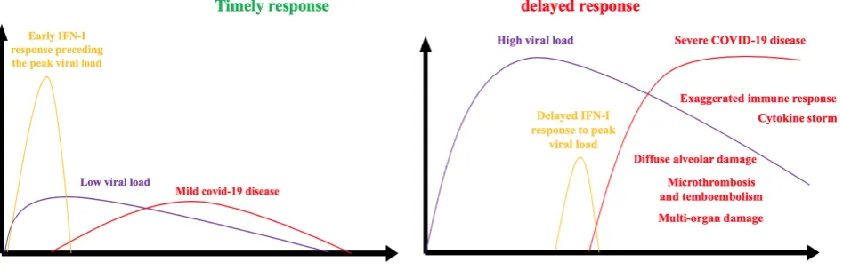
Studies in mice have shown that the timeliness of natural IFN-I production or exogenous administration makes the difference 102. Natural production or exogenous administration preceding
the peak of viral replication are protective. Delayed endogenous production or exogenous administration, compared to the peak of viral replication, become pathogeni 60,63. Figure 2.
Figure 2. Timeliness of the response mediated by the IFNs.
Delayed type I IFNs response promotes the apoptosis of T lymphocytes, the recruitment of inflammatory monocytes-macrophages at the lung level and the exaggerated production of inflammatory echemokines cytokines. Therefore, natural production or timely exogenous administration of IFN-I can prevent the exaggerated production of inflammatory cytokines and are protective. Delayed natural production or exogenous administration are pathogenic.
Studies have shown that in severe forms of COVID-19 disease, type I IFNs response is more compromised than mild or moderate cases 54,93,96.
Therefore, the results of type I IFNs administration depend on the phase in which it is used. At a very early stage it exerts the greatest positive effects. At a later stage, the effects obtained are harmful. In China, the guidelines recommend the administration of 5 million U. of IFN-a by inhalation, twice a day, in combination with Ribavirin 64,65. Despite the conflicting results obtained with SARS and
MERS treatment with type I IFNs, in vitro studies suggest a greater sensitivity of the SARS CoV-2 virus to type I IFNs.
Timeliness problems encountered with Type I IFNs have also recently been highlighted with the
IFN-l. A very recent study on mice has shown that even the IFN-l can have harmful effects on the barrier, favoring bacterial complications 96. While early administration of IFN lambda could protect against
the SARS CoV-2 virus infection 97,98 and increase the barrier function of intestinal epithelial cells and
endothelial cells 99-101, delayed exposure, compared to the viral peak, or when tissue damage has
already occurred, causes deleterious effects 96.
Peculiarities of the IFN- l in the antiviral response
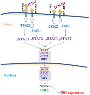
IFN-l and IFN I are induced when viruses and their derivatives (PAMPs) are recognized by the appropriate sensors (PRRs). After being produced, IFNs act on target cells by activating shared signaling pathways. Figure 3.
Unlike IFN III, only IFN-l can activate JAK272,73. Activated JAKs recruit and phosphorylate the
transcription factor STAT1. Phosphorylated STAT1 migrates to the nucleus and, together with the transcription factor IRF9, activate the same antiviral transcriptional program 29,33,34,36,74,75. The action
on IFN target cells is mediated by specific receptors (IFNLR1/IFL10Rb). The expression of IFNLR1 is restricted to certain types of cells (epithelial cells of the respiratory tract, gastro-enteric, uro-genital lining, endothelial) 67,68,69 and some types of myeloid cells (especially neutrophils, but also dendritic
cells and macrophages) 24,37,40,41,77. In vitro studies 39,88 and in vivo 40,81,82 have shown that in the same
cells, at the barrier level, the antiviral response is mainly mediated by IFN-l, compared to type III IFN 39,40,81,81,88.
The most substantial differences between type I IFN and IFN-l concern their effects on the immune and inflammatory response. IFN I associates the deleterious effects of the inflammatory response with the powerful antiviral action. When their production is prolonged, beyond the peak of the viral load, type I IFNs contribute to the inflammatory cytokine and chemokine release syndrome, to the recruitment of inflammatory cells 85.86, favor cell-mediated adaptive immunity, inducing the cytotoxic
activity of T lymphocytes and Natual Killers (NK) cells 83,84.
Hence, Type I IFNs can contribute to the aggravation of the cytokine storm and cause damage to the mucous barriers and the endothelium.
In contrast, IFN-l modulates the inflammatory response and protects mucous barriers 34. In fact, IFN-l dampens the activity of neutrophils which, through the release of granules, the production and release of ROS (reactive oxygen species) 34,76, the formation of extracellular traps (NETs) 41,87,
seriously damage the tissues.
The recruitment of Neutrophils is very important, because these cells can form structures called extracellular traps (TRAPs or NETs) that trap pathogens but favor the formation of thrombi. In mice infected with influenza virus, Neutrophils respond to both Type I IFNs and IFN-l.
However, only type I IFNs stimulate the production and release of pro-inflammatory cytokines by neutrophils40, while IFN-l only induces ISG-mediated antiviral status 34.
Other cells and barriers are also responsive to the IFN-l (liver cells, endothelial cells, blood-brain barrier)34. Therefore, while IFN I, thanks to the ubiquitous expression of its receptors
(IFNAR1/IFNAR2), has systemic action, IFN-l has a more targeted sphere of action, at the level of anatomical barriers. The activated transcriptional program is superimposable, but the activity of the IFN I is faster and shorter, while that of the IFN-l is more sustained 29,34. Type I IFN carries out a
pro-inflammatory IRF1-mediated action, absent in IFN-l29,34,40,66.
The differential role of the different IFNs in the defense against the SARS CoV virus has been investigated in mice deprived of their respective receptors. In the Ifnlr1 -/- or Ifnra1 -/- mice, the ability to eliminate the virus was impaired. Even greater was the impairment in Ifnlr1 / and Ifnra1 -/- 67 mice with double Knockout. These results confirms that the activity of the two type I and III IFNs
is not redundant but additive.
The impairment, however, was even more severe in STAT1 -/-mice, in which, to the deficits of type I and III IFNs, was added the loss of IFN II signals (IFN ), emphasizing the role of IFN in the antiviral defenses 68.
On the upper respiratory tract, however, the action of the IFN-l is paramount. Ifnlr1 -/- mice, to which the influenza virus had been administered at sub lethal doses in the upper respiratory tract, eliminated a greater quantity of viruses and were more contagious, compared to Ifnra1 -/- mice.
Figure 3.Type I IFNs and and IFN-l act by binding to the respective receptors expressed on the surface of the target cells. Type I and type III IFNs receptors are heterodimers made up of two subunits: IFNAR1 and IFNAR2 (type I IFNs) and IFNLR1 and IL10Rβ (IFN-l) subunits, respectively. IFNs first bind a chain of high affinity receptors (IFNAR2 or IFNLR1), then recruit a chain of low affinity receptors (IFNAR1 or IL10Rβ), to create a competent ternary complex for signaling. Receptor dimerization activates TYK2 and JAK1 kinases, which phosphorylate STAT1 and STAT2. The phosphorylated STAT1 and STAT2 heterodimers complex with the IRF9 factor to produce the ISGF3 transcription factor. ISGF3 binds to ISREs and promotes the expression of hundreds of ISGs.
These results highlight the increased importance of IFN-l in controlling viral load in the upper respiratory tract and in contact prophylaxis. Instead, prophylactic intranasal administration of IFN-a
and IFN-b is necessary to block the spread of the virus in the lung 70..
The recent discovery that commensal bacteria that colonize the upper respiratory tract can maintain an antivral state, mediated by IFN-l, at lung level, sufficient to protect mice from influenza infection, supports the importance of IFN-l at the level of barriers 89.
These results highlight at least four relevant aspects. i) In mice with IFN-λ deficiency, adaptive immunity is compromised, both humoral (IgG1 and IgA reduction) and cellular immunity (CD8 + cytotoxic T lymphocyte reduction) ii) Although the cells of the adaptive immune system do not have IFN-λ receptors, this cytokine has effects on the adaptive immune response iii) IFN-λ shows a strong adjuvant activity on vaccines applied on the mucous membrane of the airways of mice iiii) the ability of IFN-λ to support adaptive immunity depends on a local factor that acts as a bridge.
In fact, the other routes of administration of the vaccine do not benefit from the IFN-λ. A new action and a potential new stimulating role have emerged for IFN-λ as an adjuvant to make vaccines against viruses with tropism for the respiratory tract more effective and safe.
The link between IFN-λ and adaptive respiratory immunity is represented by the thymic stromal lymphopoietin (TSLP). IFN-λ increases adaptive immune responses after intranasal immunization of mice via an indirect mechanism that involves the production of thymic stromal lymphopoietin (TSLP) in upper airway M cells. TSLP, in fact, stimulates the migration of dendritic cells (DC CD103+) from
the airways to the draining lymph nodes at the mediastinal level 90.91. Figure 4.
Figure 4.The cells of the adaptive immune system do not have IFN-l receptors. The effects of IFN-l on adaptive response depend on local stimulation of thymal stromal lymphopoietin (TSLP). TSLP, produced by lung M cells, acts by activating lung dendritic cells. M cells (microfold) reside in the follicle-associated epithelium of the lymphoid tissues of the mucosa. They are cells specialized in detecting luminal antigens to initiate the immune responses of the mucosa. 1) IFN-l-induced virus release stimulates TSLP production from M cells that, in turn, stimulates the migration of dendritic cells from airways to draining lymph nodes at the mediastinal level. 2) TSLP boosts adaptive immunity and increases the production of IgG1 and IgA immunoglobulins.
This results in the enhancement of the germinal center (GC) and an increased production of antigen specific IgG1 and IgA antibodies.
systemic inflammation; g) limits the recruitment and activation of neutrophils, blocking the formation of dangerous NETs favoring thrombotic phenomena.
Unlike the recombinant IFN I, already used in the clinic, the IFN-l has not yet been approved for any use. The peculiar characteristics of the IFN-l, targeted actions at the level of the barriers, sustained, without inflammatory effects, are fundamental for the rationale of its use in randomized clinical trials already underway. Efficacy and safety of use has already been demonstrated in studies of animals infected with the flu virus 39,40,71.
CONCLUSIONS
IFN-I response times, in relation to virus replication, can affect the evolution of SARS-CoV-2 infection. The combination of delayed production of IFN-I, with a rapid and robust replication of SARS-CoV2, favors the aggravation of the disease. In this scenario, interventions aimed at reducing the viral load are essential. Consistent with this hypothesis, older people, at a higher risk of severe COVID infections, present an impaired antiviral response mediated by IFNs, while retaining the ability to produce an inflammatory response. Therefore, the inability to mount an adequate and early antiviral response mediated by IFNs, on the one hand, the exaggerated inflammatory response, on the other, increase the susceptibility to severe forms of COVID-19.
IFN I has a faster and more powerful action, mainly effective on the lung, but it helps to exacerbate inflammation. Its role could be limited to the earlier stages of promoting the elimination of the virus. In the hyper inflammatory stages it should not be used. IFN-l acts at the level of anatomical barriers and its action is prominent on the upper respiratory tract. The IFN-l has the ability to prevent viral replication at the entry site, by limiting viral load and reducing contagiousness. In addition, it appears effective in preventing the spread of the virus to the lungs. It has no inflammatory effects and, therefore, its use could be very useful in the prophylaxis of contacts and in the early stages of the disease, to prevent the spread of the virus. IFNs, inserted in a personalized therapy perspective, could be useful in the early stages, before the viral peak, for prophylactic or therapeutic purposes, also associated with other antivirals. When the first signs of laboratory or clinical signs of progressive inflammation appear, the rationale for their use ceases. When the results of the ongoing randomized clinical trials are available, it will be possible to design a reasoned therapeutic strategy that meets the criteria of efficacy and safety.
Declaration of interests:
The authors declare that there are no conflicts of interest regarding the publication of this paper.
List of abbreviations:
ACE2: Angiotensin-Converting Enzyme 2 ARDS:Acute Respiratory Distress Syndrome cGAS: Cyclic GMP-AMP synthase
DAMPs: damage-associated molecular patterns IFNs: Interferons
IFNAR: interferon-α / β receptor IFNLR: Interferon Lambda Receptor
IL-1b: Interleukin 1b IL-6: Interleukin 6 IL-8: Interleukin 8
IL-10Rb: Interleukin 10 receptor beta IL-17: Interleukin 17
IKKB: inhibitor of κB kinase IRF: interferon regulatory factors ISGs: interferon-stimulated gene
ISRE: Interferon-sensitive response element JAKs: Janus kinases
MAVs: Mitochondrial antiviral-signaling protein MDA-5: Melanoma Differentiation-Associated protein 5 MCP-1: Monocyte Chemoattractant Protein-1
MERS: Middle East Respiratory Syndrome MOF: Multiple Organ Failure
NETs: Neutrophil Extracellular Traps NFkB: Nuclear factor-κB
ORF: Open Reading Frame
PAPMPs: pathogen-associated molecular patterns PRRs: Pattern recognition receptors
RLRs:RIG-I-like receptors
RIG-I: Retinoic acid-inducible gene I ROS: Reactive Oxygen Species
SARS: Severe Acute Respiratory Syndrome
STAT: signal transducer and activator of transcription STING: Stimulator of interferon genes
TBK1: TANK-Binding Kinase 1 TLSP: Thymic stromal lymphopoietin TYK2: Tyrosine kinase 2
TLRs: Toll-Like Receptors TNF-a: Tumor Necrosis Factor a TRAF: TNF receptor associated factors TRAPs: Neutrophil Extracellular Traps
Reference
1 Sehgal, P. B., L. M. Pfeffer, and I. Tamm, Interferon and its inducers, in: Chemotherapy of Viral InfectIOns (P. E.
Came and L. A. Caliguiri, eds.), pp. 205-312, Springer-Verlag, Berlin (1982).
2 O’Neill LA (2008) When signaling pathways collide: positive and negative regulation of toll-like receptor signal
transduction. Immunity 29: 12–20.
3 Rothlin CV, Ghosh S, Zuniga EI, Oldstone MB, Lemke G (2007) TAM receptors are pleiotropic inhibitors of the
innate immune response. Cell 131: 1124–1136.
4 Aman MJ, Tretter T, Eisenbeis I, Bug G, Decker T, et al. (1996) Interferon- alpha stimulates production of
interleukin-10 in activated CD4+ T cells and monocytes. Blood 87: 4731–4736.
5 Nozell S, Laver T, Patel K, Benveniste EN (2006) Meccanismo di inibizione mediata da IFN-beta dell'espressione del
gene IL-8 nelle cellule di astroglioma. J Immunol 177: 822–830.
6 Prinz M, Schmidt H, Mildner A, Knobeloch KP, Hanisch UK, et al. (2008) Le funzioni in vivo distinte e non
ridondanti dell'IFNAR sulle cellule mieloidi limitano l'autoimmunità nel sistema nervoso centrale. Immunità 28: 675– 686.
7 Abreu SL (1982) Soppressione dell'encefalomielite allergica sperimentale da parte dell'interferone. Immunol Commun
11: 1–7.
8 Billiau A (2006) Proprietà antinfiammatorie degli interferoni di tipo I. Ricerca antivirale 71: 108-116.
9 Guo B, Chang EY, Cheng G (2008) Il percorso di induzione IFN di tipo I limita l'infiammazione autoimmune mediata
da Th17 nei topi. J Clin Invest 118: 1680–1690.
10 Van Holten J, Reedquist K, Sattonet-Roche P, Smeets TJ, Plater-Zyberk C, et al. (2004) Il trattamento con interferone
beta ricombinante riduce l'infiammazione e rallenta la distruzione della cartilagine nel modello di artrite indotta da collagene dell'artrite reumatoide. Resistenza all'artrite Ther 6: R239–249.
11 Crouse, J., Kalinke, U., and Oxenius, A. (2015). Regulation of antiviral T cell responses by type I interferons. Nat.
Rev. Immunol. 15, 231–242.
12 Garcıa-Sastre, A. (2017). Dieci strategie di evasione dell'interferone da parte dei virus. Microbo ospite cellulare 22,
176–184.
13 Hoffmann, H.H., Schneider, W.M. e Rice, C.M. (2015). Interferoni e violette: una corsa agli armamenti evolutiva di
interazioni molecolari. Trends Immunol. 36, 124–138.
14 Ingle, H., Peterson, S.T. e Baldridge, M.T. (2018). Distinti effetti degli interferoni di tipo I e III sui virus enterici.
Virus 10, E46.
15 Kotenko, S.V., Gallagher, G., Baurin, V.V., Lewis-Antes, A., Shen, M., Shah, N.K., Langer, J.A., Sheikh, F.,
Dickensheets, H., and Donnelly, R.P. (2003). IFN-lambdas mediate antiviral protection through a distinct class II cytokine receptor complex. Nat. Immunol. 4, 69–77.
16 Lazear, H.M., Nice, T.J., and Diamond, M.S. 2015. Interferon-l: immune functions at barrier surfaces and beyond.
Immunity 43, 15–28.
17 Schoggins, J.W. (2018). Recent advances in antiviral interferon-stimulated gene biology. F1000Res. 7, 309..
19 Snell, L.M., McGaha, T.L., and Brooks, D.G. (2017). Type I Interferon in Chronic Virus Infection and Cancer.
Trends Immunol. 38, 542–557.
20 Sorgeloos, F., Kreit, M., Hermant, P., Lardinois, C., and Michiels, T. (2013). Antiviral type I and type III interferon
responses in the central nervous system. Viruses 5, 834–857.
21 Wack, A., Terczynska-Dyla, E., and Hartmann, R. (2015). Guarding the fron- tiers: the biology of type III interferons.
Nat. Immunol. 16, 802–809.
22 Wells, A.I., and Coyne, C.B. (2018). Type III Interferons in Antiviral Defenses at Barrier Surfaces. Trends Immunol.
39, 848–858.
23 Schoggins, J.W., and Rice, C.M. (2011). Interferon-stimulated genes and their antiviral effector functions. Curr. Opin.
Virol. 1, 519–525.
24 Kotenko, S.V., Rivera, A., Parker, D. e Durbin, J.E. (2019). IFN di tipo III: oltre la protezione antivirale. Semin.
Immunol. 43, 101303.
25 Ivashkiv, L.B., and Donlin, L.T. (2014). Regulation of type I interferon responses. Nat. Rev. Immunol. 14, 36–49.
26 Lim, Y.X., Ng, Y.L., Tam, J.P., and Liu, D.X. (2016). Human Coronaviruses: A Review of Virus-Host Interactions.
Diseases 4, 26.
27 Nelemans, T. e Kikkert, M. (2019). Viral Innate Immune Evasion e la patogenesi delle infezioni emergenti da virus
RNA. Virus 11, 961.
28 Totura, A.L., Whitmore, A., Agnihothram, S., Scha € fer, A., Katze, M.G., Heise, M.T. e Baric, R.S. (2015). La
segnalazione del recettore Toll-Like 3 tramite TRIF contribuisce a una risposta immunitaria innata protettiva alla grave infezione da coronavirus della sindrome respiratoria acuta. MBio 6, e00638-e15.
29 Park A, Iwasaki A. Type I and Type III Interferons - Induction, Signaling, Evasion, and Application to Combat
COVID-19. Cell Host Microbe. 2020;27(6):870-878.
30 Smits SL, de Lang A, van den Brand JM, et al. Exacerbated innate host response to SARS-CoV in aged non-human
primates. PLoS Pathog. 2010;6(2):e1000756. Published 2010 Feb 5.
31 Koyama S, Ishii KJ, Coban C, Akira S. Innate immune response to viral infection. Cytokine. 2008;43(3):336-341.
32 Iwasaki A, Pillai PS. Innate immunity to influenza virus infection. Nature reviews. Immunology. 2014 May;14(5):315-328.
33 Lazear HM, Schoggins JW, Diamond MS. Shared and Distinct Functions of Type I and Type III Interferons. Immunity. 2019 Apr;50(4):907-923.
34 Broggi A, Granucci F, Zanoni I. Type III interferons: Balancing tissue tolerance and resistance to pathogen
invasion. J Exp Med. 2020;217(1):e20190295.
35 Ye L., Schnepf D., and Staeheli P.. 2019c Interferon-λ orchestrates innate and adaptive mucosal immune responses.
Nat. Rev. Immunol. 19:614–625.
36 Andreakos E., Zanoni I., and Galani I.E.. 2019. Lambda interferons come to light: dual function cytokines mediating
antiviral immunity and damage control. Curr. Opin. Immunol. 56:67–75.
37 Koltsida O., Hausding M., Stavropoulos A., Koch S., Tzelepis G., Übel C., Kotenko S.V., Sideras P., Lehr H.A., Tepe
M., et al. . 2011. IL-28A (IFN-λ2) modulates lung DC function to promote Th1 immune skewing and suppress allergic airway disease. EMBO Mol. Med. 3:348–361.
38 Hemann E.A., Green R., Turnbull J.B., Langlois R.A., Savan R., and Gale M. Jr. 2019. Interferon-λ modulates
39 Davidson, S., McCabe, T. M., Crotta, S., Gad, H. H., Hessel, E. M., Beinke, S., Hartmann, R., & Wack, A. (2016).
IFNλ is a potent anti-influenza therapeutic without the inflammatory side effects of IFNα treatment. EMBO molecular medicine, 8(9), 1099–1112.
40 Galani I.E., Triantafyllia V., Eleminiadou E.-E., Koltsida O., Stavropoulos A., Manioudaki M., Thanos D., Doyle
S.E., Kotenko S.V., Thanopoulou K., and Andreakos E.. 2017. Interferon-λ mediates non-redundant front-line antiviral protection against influenza virus infection without compromising host fitness. Immunity. 46:875–890.e6.
41 K. Blazek, H. L. Eames, M. Weiss, A. J. Byrne, D. Perocheau, J. E. Pease, S. Doyle, F. McCann, R. O. Williams, I.
A. Udalova, IFN-λ resolves inflammation via suppression of neutrophil infiltration and IL-1β production. J. Exp. Med. 212, 845– 853 (2015)
42 García-Sastre A. Ten Strategies of Interferon Evasion by Viruses. Cell Host Microbe. 2017;22(2):176-184.
43 Lee, H., Chathuranga, K. & Lee, J. Intracellular sensing of viral genomes and viral evasion. Exp Mol Med51, 1–13 (2019).
44 Bonjardim CA, Ferreira PC, Kroon EG. Interferons: signaling, antiviral and viral evasion. Immunol Lett.
2009;122(1):1-11.
45 Samuel CE. Antiviral actions of interferons. Clin Microbiol Rev. 2001;14(4):778-809.
46 Levy DE, García-Sastre A. The virus battles: IFN induction of the antiviral state and mechanisms of viral
evasion. Cytokine Growth Factor Rev. 2001;12(2-3):143-156.
47 Assil, S.; Webster, B.; Dreux, M. Regulation of the Host Antiviral State by Intercellular Communications. Viruses2015, 7, 4707-4733.
48 Stockman LJ, Bellamy R, Garner P. SARS: systematic review of treatment effects. PLoS Med. 2006;3(9):e343.
49 Chan JF, Chan KH, Kao RY, et al. Broad-spectrum antivirals for the emerging Middle East respiratory syndrome
coronavirus. J Infect. 2013;67(6):606-616.
50 Sheahan TP, Sims AC, Leist SR, et al. Comparative therapeutic efficacy of remdesivir and combination lopinavir,
ritonavir, and interferon beta against MERS-CoV. Nat Commun. 2020;11(1):222. Published 2020 Jan 10.
51 Omrani AS, Saad MM, Baig K, et al. Ribavirin and interferon alfa-2a for severe Middle East respiratory syndrome
coronavirus infection: a retrospective cohort study [published correction appears in Lancet Infect Dis. 2015 Jan 15;211(2):13]. Lancet Infect Dis. 2014;14(11):1090-1095.
52 Morgenstern B, Michaelis M, Baer PC, Doerr HW, Cinatl J Jr. Ribavirin and interferon-beta synergistically inhibit
SARS-associated coronavirus replication in animal and human cell lines. Biochem Biophys Res Commun. 2005;326(4):905-908.
53 Loutfy MR, Blatt LM, Siminovitch KA, et al. Interferon alfacon-1 plus corticosteroids in severe acute respiratory
syndrome: a preliminary study. JAMA. 2003;290(24):3222-3228.
54 Lokugamage, K. G., Hage, A., Schindewolf, C., Rajsbaum, R., & Menachery, V. D. (2020). SARS-CoV-2 is sensitive to type I interferon pretreatment. bioRxiv : the preprint server for biology, 2020.03.07.982264.
https://doi.org/10.1101/2020.03.07.982264
55 Mantlo, E., Bukreyeva, N., Maruyama, J., Paessler, S., and Huang, C. (2020). Antiviral activities of type I interferons
to SARS-CoV-2 infection. Antiviral Res. 179, 104811.
56 Sallard, E., Lescure, F.-X., Yazdanpanah, Y., Mentre, F., and Peiffer-Smadja, N. (2020). Type 1 interferons as a
potential treatment against COVID-19. Anti- viral Res. 178, 104791.
57 Chan, J.F.W., Chan, K.H., Kao, R.Y.T., To, K.K.W., Zheng, B.J., Li, C.P.Y., Li, P.T.W., Dai, J., Mok, F.K.Y., Chen,
58 Dong, L., Hu, S., Gao, J., 2020. Discovering drugs to treat coronavirus disease 2019 (COVID-19). Drug Discov.
Ther. 14, 58–60
59 Hart, B.J., Dyall, J., Postnikova, E., Zhou, H., Kindrachuk, J., Johnson, R.F., Olinger, G.G., Frieman, M.B.,
Holbrook, M.R., Jahrling, P.B., Hensley, L., 2014. L'interferone-β e l'acido micofenolico sono potenti inibitori della sindrome respiratoria mediorientale coronavirus nei test basati su cellule. J. Gen. Virol. 95, 571–577.
60 Channappanavar, R., Fehr, A.R., Zheng, J., Wohlford-Lenane, C., Abrahante, J.E., Mack, M., Sompallae, R.,
McCray, P.B., Meyerholz, D.K., Perlman, S., 2019. IFN-I response timing relative to virus replication determines MERS coronavirus infection outcomes. J. Clin. Invest. 129, 3625–3639
61 Frieman, M., Yount, B., Heise, M., Kopecky-Bromberg, S.A., Palese, P., Baric, R.S., 2007. Severe acute respiratory
syndrome coronavirus ORF6 antagonizes STAT1 function by sequestering nuclear import factors on the rough endoplasmic reticulum/golgi membrane. J. Virol. 81, 9812–9824.
62 Kopecky-Bromberg, S.A., Martinez-Sobrido, L., Frieman, M., Baric, R.A., Palese, P., 2007. Severe acute respiratory
syndrome coronavirus open reading frame (ORF) 3b, ORF 6, and nucleocapsid proteins function as interferon antagonists. J. Virol. 81, 548–557.
63 Siddiqi, H.K., Mehra, M.R., 2020. COVID-19 illness in native and immunosuppressed states: a clinical-therapeutic
staging proposal. J. Heart Lung Transplant.
64 Dong, L., Hu, S., Gao, J., 2020. Discovering drugs to treat coronavirus disease 2019 (COVID-19). Drug Discov.
Ther. 14, 58–60.
65 Lu, H., 2020. Drug treatment options for the 2019-new coronavirus (2019-nCoV). Bioscience Trends.
66 Forero, A., Ozarkar, S., Li, H., Lee, C.H., Hemann, E.A., Nadjsombati, M.S., Hendricks, M.R., So, L., Green, R.,
Roy, C.N., et al. (2019). Differential Activation of the Transcription Factor IRF1 Underlies the Distinct Immune Responses Elicited by Type I and Type III Interferons. Immunity 51, 451–464.e6.
67 Mordstein, M., Neugebauer, E., Ditt, V., Jessen, B., Rieger, T., Falcone, V., Sorgeloos, F., Ehl, S., Mayer, D., Kochs,
G., et al. (2010). Lambda interferon renders epithelial cells of the respiratory and gastrointestinal tracts resistant to viral infections. J. Virol. 84, 5670–5677.
68 Mahlako ̃iv, T., Ritz, D., Mordstein, M., DeDiego, M.L., Enjuanes, L., Mu€ller, M.A., Drosten, C., and Staeheli, P.
(2012). Combined action of type I and type III interferon restricts initial replication of severe acute respiratory syn- drome coronavirus in the lung but fails to inhibit systemic virus spread. J. Gen. Virol. 93, 2601–2605.
69 Klinkhammer, J., Schnepf, D., Ye, L., Schwaderlapp, M., Gad, H.H., Hartmann, R., Garcin, D., Mahlako iv, T., and ̃
Staeheli, P. (2018). IFN-l prevents influenza virus spread from the upper airways to the lungs and limits virus transmission. eLife 7, 266.
70 Stanifer, M.L., Kee, C., Cortese, M., Triana, S., Mukenhirn, M., Kraeusslich, H.- G., Alexandrov, T., Bartenschlager,
R., and Boulant, S. (2020). Critical role of type III interferon in controlling SARS-CoV-2 infection, replication and spread in primary human intestinal epithelial cells. bioRxiv, 2020.04.24.059667.
71 Kim, S., Kim, M.-J., Kim, C.-H., Kang, J.W., Shin, H.K., Kim, D.-Y., Won, T.-B., Han, D.H., Rhee, C.S., Yoon,
J.-H., and Kim, H.J. (2017). The Superiority of IFN-l as a Therapeutic Candidate to Control Acute Influenza Viral Lung Infec- tion. Am. J. Respir. Cell Mol. Biol. 56, 202–212.
72 Odendall, C., E. Dixit, F. Stavru, H. Bierne, K.M. Franz, A.F. Durbin, S. Bou- lant, L. Gehrke, P. Cossart, and J.C.
Kagan. 2014. Diverse intracellular pathogens activate type III interferon expression from peroxisomes. Nat. Immunol. 15:717–726
73 Odendall, C., A.A. Voak, and J.C. Kagan. 2017. Type III IFNs are commonly induced by bacteria-sensing TLRs and
74 Kotenko SV. IFN-λs. Curr Opin Immunol. 2011;23(5):583-590.
75 Pervolaraki, K., M.L. Stanifer, S. Münchau, L.A. Renn, D. Albrecht, S. Kurzhals, E. Sen ́ıs, D. Grimm, J. Schro ̈
der-Braunstein, R.L. Rabin, and S. Boulant. 2017. Type I and Type III interferons display different de- pendency on mitogen-activated protein kinases to mount an antiviral state in the human gut. Front. Immunol. 8:459.
76 Broggi, A., Y. Tan, F. Granucci, and I. Zanoni. 2017. IFN-λ suppresses intestinal inflammation by non-translational
regulation of neutrophil function. Nat. Immunol. 18:1084–1093.
77 Espinosa, V., O. Dutta, C. McElrath, P. Du, Y.-J. Chang, B. Cicciarelli, A. Pitler, I. Whitehead, J.J. Obar, J.E. Durbin,
et al. 2017. Type III interferon is a critical regulator of innate antifungal immunity. Sci. Immunol. 2: eaan5357.
78 Sommereyns, C., S. Paul, P. Staeheli, and T. Michiels. 2008. IFN-lambda (IFN- lambda) is expressed in a
tissue-dependent fashion and primarily acts on epithelial cells in vivo. PLoS Pathog. 4:e1000017.
79 Herna ́ndez,P.P.,T.Mahlakõiv,I.Yang,V.Schwierzeck,N.Nguyen,F. Guendel, K. Gronke, B. Ryffel, C. Hoelscher, L.
Dumoutier, et al. 2015. Interferon-λ and interleukin 22 act synergistically for the induction of interferon-stimulated genes and control of rotavirus infection. Nat. Immunol. 16:698–707.
80 Pott, J., T. Mahlakõiv, M. Mordstein, C.U. Duerr, T. Michiels, S. Stockinger, P. Staeheli, and M.W. Hornef. 2011.
IFN-lambda determines the intestinal epithelial antiviral host defense. Proc. Natl. Acad. Sci. USA. 108: 7944–7949.
81 Galani, I.E., O. Koltsida, and E. Andreakos. 2015. Type III interferons (IFNs): Emerging master regulators of
immunity. Adv. Exp. Med. Biol. 850:1–15.
82 Mahlakõiv, T., P. Hernandez, K. Gronke, A. Diefenbach, and P. Staeheli. 2015. Leukocyte-derived IFN-α/β and
epithelial IFN-λ constitute a compart- mentalized mucosal defense system that restricts enteric virus infections. PLoS Pathog. 11:e1004782.
83 Stetson, D.B., and R. Medzhitov. 2006. Type I interferons in host defense. Immunity. 25:373–381.
84 Gonzalez-Navajas, J.M., J. Lee, M. David, and E. Raz. 2012. Immunomodula- tory functions of type I interferons.
Nat. Rev. Immunol. 12:125–135.
85 Trinchieri, G. 2010. Type I interferon: friend or foe? J. Exp. Med. 207: 2053–2063.
86 Davidson, S., S. Crotta, T.M. McCabe, and A. Wack. 2014. Pathogenic potential of interferon αβ in acute influenza
infection. Nat. Commun. 5:3864.
87 Chrysanthopoulou, A., K. Kambas, D. Stakos, I. Mitroulis, A. Mitsios, V. Vi- dali, I. Angelidou, M. Bochenek, S.
Arelaki, A. Arampatzioglou, et al. 2017. Interferon lambda1/IL-29 and inorganic polyphosphate are novel regulators of neutrophil-driven thromboinflammation. J. Pathol. 243: 111–122.
88 Crotta, S., S. Davidson, T. Mahlakõiv, C.J. Desmet, M.R. Buckwalter, M.L. Albert, P. Staeheli, and A. Wack. 2013.
Type I and type III interferons drive redundant amplification loops to induce a transcriptional signa- ture in influenza-infected airway epithelia. PLoS Pathog. 9:e1003773.
89 Kim, H.J., A. Jo, Y.J. Jeon, S. An, K.-M. Lee, S.S. Yoon, and J.Y. Choi. 2019. Nasal commensal Staphylococcus
epidermidis enhances interferon-λ-dependent immunity against influenza virus. Microbiome. 7:80.
90 Ye, L., Schnepf, D., Becker, J. et al. Interferon-λ enhances adaptive mucosal immunity by boosting release of thymic
stromal lymphopoietin. Nat Immunol20, 593–601 (2019).
91 Ye L, Ohnemus A, Ong LC, et al. Type I and Type III Interferons Differ in Their Adjuvant Activities for Influenza
Vaccines. Journal of Virology. 2019 Dec;93(23).
92 Cameron, M. J., Ran, L., Xu, L., Danesh, A., Bermejo-Martin, J. F., Cameron, C. M., Muller, M. P., Gold, W. L.,
D., Wilkinson, P., Greller, L. D., Somogyi, R., Humar, A., Kelvin, D. J. (2007). Interferon-mediated
immunopathological events are associated with atypical innate and adaptive immune responses in patients with severe acute respiratory syndrome. Journal of virology, 81(16), 8692–8706.
93 Blanco-Melo, D., Nilsson-Payant, B. E., Liu, W. C., Uhl, S., Hoagland, D., Møller, R., Jordan, T. X., Oishi, K., Panis,
M., Sachs, D., Wang, T. T., Schwartz, R. E., Lim, J. K., Albrecht, R. A., & tenOever, B. R. (2020). Imbalanced Host Response to SARS-CoV-2 Drives Development of COVID-19. Cell, 181(5), 1036–1045.e9.
94 Reghunathan R, Jayapal M, Hsu LY, et al. Expression profile of immune response genes in patients with Severe
Acute Respiratory Syndrome. BMC Immunol. 2005;6:2. Published 2005 Jan 18.
95 Zhao J, Wohlford-Lenane C, Zhao J, et al. Intranasal treatment with poly(I•C) protects aged mice from lethal
respiratory virus infections. J Virol. 2012;86(21):11416-11424.
96 Achille Broggi, Sreya Ghosh, Benedetta Sposito, Roberto Spreafico, Fabio Balzarini, Antonino Lo
Cascio, Nicola Clementi, Maria De Santis, Nicasio Mancini, FrancescaGranucci, Ivan Zanoni. Type III interferons disrupt the lung epithelial barrier upon viral recognition. bioRxiv 2020.05.05.077867.
97 L. Ye, D. Schnepf, P. Staeheli, Interferon-λ orchestrates innate and adaptive mucosal immune responses. Nat. Rev. Immunol. 19, 614–625 (2019).
98 Dinnon KH, Leist SR, Schäfer A, et al. A mouse-adapted SARS-CoV-2 model for the evaluation of COVID-19
medical countermeasures. Preprint. bioRxiv. 2020;2020.05.06.081497. Published 2020 May 7.
99 C. Odendall, A. A. Voak, J. C. Kagan, Type III IFNs Are Commonly Induced by Bacteria-Sensing TLRs and
Reinforce Epithelial Barriers during Infection. J. Immunol. 199, 3270–3279 (2017)
100 H. M. Lazear, B. P. Daniels, A. K. Pinto, A. C. Huang, S. C. Vick, S. E. Doyle, M. Gale Jr., R. S. Klein, M. S.
Diamond, Interferon-λ restricts West Nile virus neuroinvasion by tightening the blood-brain barrier. Sci. Transl. Med. 7, 284ra59 (2015)
101 F. Douam, Y. E. Soto Albrecht, G. Hrebikova, E. Sadimin, C. Davidson, S. V. Kotenko, A. Ploss, Type III
Interferon-Mediated Signaling Is Critical for Controlling Live Attenuated Yellow Fever Virus Infection In Vivo. mBio
8, e00819-17 (2017).
102 Retrospective Multicenter Cohort Study Shows Early Interferon Therapy Is Associated with Favorable Clinical