Journal of Inflammation Research 2018:11 253–264
Journal of Inflammation Research
Dove
press
submit your manuscript | www.dovepress.com 253
R E V I E W
open access to scientific and medical research
Open Access Full Text Article
Is there a link between inflammation and fatigue
in multiple sclerosis?
Moussa A Chalah1,2
Samar S Ayache1–3
1EA 4391, Excitabilité Nerveuse et Thérapeutique, Université Paris-Est-Créteil, Créteil, France; 2Service de Physiologie – Explorations Fonctionnelles, Hôpital Henri Mondor, Assistance Publique – Hôpitaux de Paris, Créteil, France; 3Neurology Division, Lebanese American University Medical Center, Rizk Hospital, Beirut, Lebanon
Purpose: Among autoimmune diseases of the central nervous system stands multiple sclero-sis (MS), which is characterized by demyelination, synaptopathy, and neurodegeneration. MS fatigue can affect up to 90% of patients and be very disabling, with a drastic impact on their quality of life. To date, the evaluation of MS fatigue has relied mainly on subjective scales, and actual therapeutic interventions are challenged by modest efficacy and numerous undesirable effects. Therefore, finding biomarkers of MS fatigue might help in optimizing evaluation and treatment strategies. The main objective here was to assess the relationship between MS fatigue and inflammatory or other immunomediated markers.
Methods: Research was conducted according to PRISMA guidelines. Computerized databases (ie, PubMed/Medline and Scopus) were consulted till February 2018 aiming to identify articles that addressed inflammation and MS fatigue. Studies in English and French published at any time were considered.
Results: A total of 27 studies matched the research criteria. Inconsistency existed regarding the relationship between fatigue and the orexin A system, hypothalamus–pituitary–adrenal axis, and cerebrospinal fluid inflammatory markers. As for peripheral markers, although there was scarcity in the available data, serum proinflammatory cytokines (ie, IL6, TNFα, and IFNγ) seem to be associated with MS fatigue. Finally, no link was found between MS fatigue and T-cell populations (ie, CD3+CD4+ T lymphocytes, regulatory T cells) or other peripheral markers of
inflammation (ie, CRP, erythrocyte-sedimentation rate, soluble ICAM1).
Conclusion: Future large-scale studies would benefit from comparing the relationship between fatigue and immune measures in patients with different disease phenotypes with and without disease-modifying drugs. With the subjective nature of fatigue scales, finding objective biomark-ers for fatigue would be of great help.
Keywords: pathophysiology, cytokines, interleukins, cerebrospinal fluids, inflammatory markers
Introduction
Among autoimmune diseases of the central nervous system (CNS) stands multiple sclerosis (MS), which is the second-most common cause of physical handicap in young individuals.1–3 Its pathophysiological hallmarks are demyelination, synaptopathy, and
neurodegeneration.4,5 Throughout the disease course, MS patients can experience
periods of acute symptom emergence separated by symptom-free intervals. This characterizes the relapsing–remitting (RR) MS phenotype, which usually converts to a secondary progressive phase where patients can experience steady clinical deterio-ration.3 Primary progressive MS represents a third disease phenotype, where patients
Correspondence: Moussa A Chalah Service de Physiologie – Explorations Fonctionnelles, Hôpital Henri Mondor, 51 Avenue de Lattre de Tassigny, Créteil 94010, France
Tel +33 1 4981 2694 Fax + 33 1 4981 4660 Email [email protected]
Journal name: Journal of Inflammation Research Article Designation: REVIEW
Year: 2018 Volume: 11
Running head verso: Chalah and Ayache
Running head recto: Inflammation and fatigue in multiple sclerosis DOI: http://dx.doi.org/10.2147/JIR.S167199
Journal of Inflammation Research downloaded from https://www.dovepress.com/ by 118.70.13.36 on 24-Aug-2020
For personal use only.
This article was published in the following Dove Press journal:
Dovepress
Chalah and Ayache
witness an evolutionary pattern of their disease from onset. Demyelination appears to be the fingerprint of the first type (ie, RRMS), whereas neurodegeneration/axonal loss seems to be the backbone of progressive types.5 While an
immuno-mediated attack by blood-borne autoreactive T lymphocytes would dictate the occurrence of demyelination, immunomedi-ated processes involving immune cells and soluble cytokines could also lead to excitotoxic changes and neurodegeneration, based on experiments involving the animal model of MS.5
Regardless of the disease phenotype, patients may experi-ence a panel of symptoms involving the sensory, motor, cere-bellar, emotional, cognitive, and behavioral domains. Among the frequently encountered symptoms, MS fatigue can affect up to 90% of patients and be very disabling, with a drastic impact on their quality of life.6 It is a challenging symptom
that is described by patients as “malaise”, “excessive tired-ness”, or “weakness” that seems to worsen throughout the day, as well as with hot and humid environments.4 From a
scientific perspective, MS fatigue is considered a multidimen-sional symptom with physical, cognitive, and psychosocial components. Among the available definitions, some authors consider fatigue a lack of physical and/or mental energy.4,6
For another group of authors, fatigue designates a failure to initiate and/or maintain physical or mental activities requiring self-motivation in the absence of or not related to physical or cognitive dysfunction.4,6
Ever since the original work of Freal et al in 1984,7 there
has been growing interest in understanding the pathophysi-ology of MS fatigue, especially the fact that this symptom remains difficult to be reported by patients and managed by physicians. To date, the evaluation of MS fatigue has relied mainly on subjective scales, such as the Modified Fatigue Impact Scale (MFIS), Fatigue Severity Scale (FSS), and Fatigue Scale for Motor and Cognitive Functions (FSMC), among others, and actual therapeutic interventions are chal-lenged by their modest efficacy in face of their numerous undesirable effects.6 From this perspective, understanding the
underlying mechanisms of this symptom might be of help in easing its evaluation and optimizing patient care. In a previ-ous work, we addressed the cerebral anatomical correlates of MS fatigue.4 Based on neuroimaging studies, pathological
findings were observed in a corticostriatothalamocortical loop that was linked to MS fatigue. These findings included regional gray- and white-matter pathologies, as well as abnormal patterns of brain activation. The inflammatory and immune medium might be implicated as well in the context of MS fatigue. Therefore, the main aim of the current work was to assess the role of MS-related central and peripheral
inflammation and immunomediated endocrine dysregulation in the development of this symptom.
Study selection
Research was conducted according to the PRISMA guide-lines.8 First, computerized databases that index peer-reviewed
journals (PubMed/Medline and Scopus) were consulted till the end of February 2018. The research aimed to identify articles that addressed the relationship between MS fatigue and inflammatory, immune, and endocrine factors. Studies that were published at any time in English and French were considered. The following research terms were combined: (“fatigue” OR “fatigue severity scale” OR “FSS” OR “Modi-fied Fatigue Impact Scale” OR “MFIS” or “Fatigue Scale for Motor and Cognitive Functions” or “FSMC”) AND (“multiple sclerosis” OR “MS”) AND (“inflammation” OR “inflammatory” OR “immune” OR “cytokine” OR “interleu-kin” OR “cerebrospinal fluid” OR “CSF” OR “lymphocytes” OR “blood cells” OR “endocrine”). In addition, both coau-thors independently checked the references of the articles obtained, aiming to obtain additional sources. The initial search identified 503 articles in PubMed/Medline and 258 articles in Scopus. After removal of duplicates and exclud-ing reviews, opinions, editorials, commentaries, viewpoints, and research articles involving healthy volunteers or patients with autoimmune diseases other than MS, 25 articles were retained. An additional two references were retrieved from the articles’ reference lists, yielding a total of 27 articles that were considered in the qualitative synthesis. These comprised information on MS fatigue and inflammatory or neuroendocrine markers and addressed the relationship between fatigue and hypothalamic function (two about the orexin A system, eight about the hypothalamic–pituitary– adrenal [HPA] axis), cerebrospinal fluid (CSF) markers (one about humoral and cellular CSF markers, one about CSF cytokines), serum-cytokine or blood-cell expression (15), or other peripheral inflammatory markers (three). In addition, six studies assessed changes in fatigue and cytokine profiles following exercise (four) or pharmacological (two) interven-tions. For the sake of this work, data of the selected studies are classified as central inflammation and neuroendocrine dysregulation and peripheral inflammation.
Central inflammation,
neuroendocrine dysregulation, and
MS fatigue
The exploration of inflammatory patterns within the CNS is possible by means of CSF analysis. However, the procedure
Journal of Inflammation Research downloaded from https://www.dovepress.com/ by 118.70.13.36 on 24-Aug-2020
Dovepress Inflammation and fatigue in multiple sclerosis
consists of performing lumbar puncture, a procedure that is not only difficult to perform but also traumatizing for patients. This explains the scarcity of existing literature in this field. Available works on CSF analysis and MS fatigue focused on studying inflammation-related neuroendocrine dysregulation, humoral and cellular components, and cytokine levels. The remaining literature employed serum and salivary hormonal tests to assess specific hypothalamic functions and their relationship with fatigue.
To start, the hypothalamus plays a role in controlling several homeostatic functions. Some researchers were interested in assessing the relationship between MS fatigue and CSF levels of orexin A (also known as hypocretin 1), a hypothalamic peptide involved in arousal, motivation, energy, and circadian rythm.9 In fact, consolidating night sleep and
keeping adequate daytime activity seem to be respectively promoted by low and high orexin A levels.10 Therefore, the
rationale behind these works lay in the fact that neuroinflam-mation, such as that seen in MS, may impact the orexin A system.11 As such, one can speculate that downregulation
of the latter system might happen in the course of MS and result in sleep disorders and/or fatigue. The first insight on orexin A-system status in MS derived from case reports on patients suffering from hypersomnia displaying low CSF levels of orexin A.12,13 Afterward, Papuć et al studied orexin A
levels in MS patients and healthy controls.14 In the absence of
group difference (MS vs healthy controls) with regard to this peptide, significant positive correlation was found between fatigue severity and orexin A levels in the whole patient group. Although this positive relationship was unexpected, the authors of this work hypothesized that this might have occurred due to the activation of endogenous compensatory mechanisms. One year later, Constantinescu et al were not able to replicate this correlation.15 Here, the authors found
neither a group difference in orexin A levels between MS patients and other patients with inflammatory and noninflam-matory neurological disease nor a correlation between orexin A levels and fatigue scores. Given the impact of daytime and season on orexin A levels,16 there is a good chance that
these two studies were performed in different seasons and/or at different times of the day, a finding that could provide an explanation for the difference in the reported results.
Besides regulating the orexin A system, the hypothalamus is involved in many axes, of which the most studied is the HPA axis. Facing physiological and stressful situations, the hypothalamus secretes the corticotropin-releasing hormone (CRH) to stimulate the activity of the anterior pituitary gland. The latter responds by producing the adrenocorticotropic
hormone (ACTH) which in turn activates the adrenal glands (ie, zona fasciculata) yielding cortisol production. Proin-flammatory cytokines can influence the activity of the HPA axis.17 This might provide an explanation for the HPA-axis
dysregulation that appears to occur in patients with MS.18–23
In fact, the majority of studies on this topic have shown a hyperactive HPA axis, with fewer reports suggesting a hypo-activity pattern. Few studies have assessed the relationship between MS fatigue and HPA-axis activity.
Heesen et al employed combined dexamethasone–CRH challenge24–26 and low-dose dexamethasone-suppression
tests.27 Both of these are used widely to measure
activ-ity of the HPA axis. The dexamethasone-suppression test consists of orally administering dexamethasone, a synthetic glucocorticoid, the night before blood sampling, in order to check the suppression of cortisol production (which is the normal physiological reaction). The combined dexametha-sone–CRH-challenge test resembles the first, but CRH is also given intravenously the day of blood sampling and blood withdrawn at regular intervals to determine plasma levels of ACTH and cortisol levels at different times. In their four studies, the authors did not detect any significant association between MS fatigue and HPA-axis activity.24,25 In line with
these results, Akcali et al employed a more comprehensive neuroendocrine evaluation that included plasma levels of ACTH, cortisol, and other pituitary products, namely corticotropin-like intermediate-lobe peptide (CLIP), which is an ACTH variant, and melanocyte-stimulating hormone (α-MSH, β-MSH, γ-MSH), produced in the anterior pituitary gland28 and previously found to be implicated in chronic
fatigue syndrome (ie, α-MSH).29 Although abnormal HPA
measures were observed in MS patients compared to healthy controls (ie, higher ACTH, cortisol and α-MSH and lower CLIP levels among patients), these measures did not differ between fatigued and unfatigued MS patients, suggesting the absence of any relationship between HPA-axis activity and MS fatigue. Conversely, a third study by Gottschalk et al employed combined dexamethasone–CRH-suppression tests and found significantly higher ACTH plasma levels
among fatigued compared to unfatigued counterparts.30
The discrepancy in the results of the aforementioned works might have resulted from differences in clinical character-istics and treatments of the MS cohorts studied. Contrarily to Heesen et al24–27 and Akcali et al,28 who mostly enrolled
patients receiving disease-modifying drugs, Gottschalk et al30
recruited drug-naïve patients. Here, it is worth noting that immunotherapy may impact cytokine-expression levels and thus might influence HPA-axis activity.31,32
Journal of Inflammation Research downloaded from https://www.dovepress.com/ by 118.70.13.36 on 24-Aug-2020
Dovepress
Chalah and Ayache
In addition to the previously mentioned studies, Powell et al and Gold et al focused on the assessment of the cor-tisol awakening response (CAR) in MS patients, using a salivary test.27,33 CAR is a spike in serum cortisol around
30–45 minutes after awakening, and is crucial for sustaining normal circadian rhythm and wakefulness.34 In the former
trial, baseline fatigue scores, but not those obtained at the same day of CAR testing, were correlated with CAR.33 In
the latter, CAR did not predict MS fatigue, as per regression-analysis results.27
Among the other adrenal products stands dehydroepian-drosterone (DHEA) and its sulfated ester (DHEAS). Low DHEA and DHEAS levels have been linked to fatigue in some autoimmune diseases, such as systemic lupus erythe-matosus and rheumatoid arthritis.35,36 In the only available
study addressing serum levels of DHEA and DHEAS in MS patients, lower levels of both components were detected in fatigued compared to unfatigued patients.37 Interestingly,
these results support those of an earlier pharmacological study, in which fatigue improvement was obtained follow-ing DHEA hormone replacement.38 However, the results of
the latter work should be interpreted with caution, mainly because of its nonrandomized design. These preliminary findings warrant further research on this matter.
In addition to studies on HPA axis and MS fatigue, some researchers were interested in evaluating humoral, cellular, and other immune CSF markers. For instance, Biberacher
et al included an exploratory and a validation phase that contained several evaluations. Of interest, they assessed the relationship between fatigue and several cellular and humoral CSF markers.39 No correlation was found between fatigue
scores and any of the CSF markers. More interestingly, fatigue scores tended to correlate negatively with CSF CD4:CD8 ratio in the discovery group and correlate positively with the former ratio in the validation group. However, the multivari-ate model failed to detect associations between fatigue and CSF parameters in either group (exploratory vs validation). A recent work aimed to understand the relationship between MS-fatigue and CSF-interleukin levels, particularly IL6 and IL8. While IL6 took part in innate and adaptive immune responses, including differentiation of T helper 17 cells, IL8 was mainly implicated in innate immunoresponses and had cytokine- and chemokine-like functions. In this work, Brenner et al documented a significant correlation between fatigue scores and IL6 levels.40 This relationship was only
seen among patients not receiving MS treatment. This might explain the absence of association in Biberacher et al,39 where
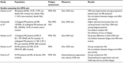
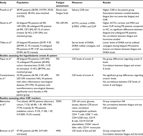
>75% of patients were under MS therapies. Another difference between the studies lies in their methodological approach which consisted of cellular and humoral markers in the first versus interleukins in the second. The different clinical and demographic characteristics between the cohorts might pres-ent a third plausible explanation for the inconsistency in their results. Table 1 provides a summary of these studies.
Table 1 Studies assessing the relationship between multiple sclerosis fatigue and central inflammatory or neuroendocrine markers
Study Population Fatigue
assessment
Measures Results
Studies assessing the HPA axis
Heesen et al24 40 patients (8 RR, 19 SP, 13 PP, sex details NA, treated, but details NA); 11 HCs (sex-matched, details NA)
MFIS, FSS Dex–CRH test HPA-axis hyperactivity among progressive patients (but not RR patients)
No correlation between fatigue and HPA-axis activity
Gottschalk et al30
15 fatigued MS patients (all RR, 12F/3M); 16 unfatigued MS patients (all RR, 10F/6M); 19.4% of whole cohort treated
FSS, MFIS, VAS
Dex–CRH test Higher adrenocorticotropin (but not cortisol) levels in the Dex–CRH test among fatigued patients
Correlation analysis NA No influence of sex on fatigue Heesen et al25 15 fatigued MS patients (6 RR, 8
SP, 1 PP, 9F/6M, 66.7% treated); 15 unfatigued MS patients (11 RR, 2 SP, 2 PP, sex-matched, 9F/6M, 60% treated)
MFIS, FSS Dex-CRH test No group difference in Dex-CRH test Correlation between fatigue and HPA-axis activity
Heesen et al26 50 MS patients (27 RR, 23 SP, 29F/21M, 48% treated)
MFIS Dex–CRH test Group comparison NA
No correlation between fatigue and HPA-axis activity
Gold et al27 44 MS patients (all RR, all female, 59% treated)
MFIS, FSS Dexamethasone-suppression test, salivary CAR test
Group comparison NA
Dexamethasone-suppression test and CAR data did not predict fatigue
(Continued)
Journal of Inflammation Research downloaded from https://www.dovepress.com/ by 118.70.13.36 on 24-Aug-2020
Dovepress Inflammation and fatigue in multiple sclerosis
Study Population Fatigue
assessment
Measures Results
Powell et al33 38 MS patients (38 RR, 31F/7M, 39.5% untreated); 38 HCs (sex-matched, 31F/7M)
Eleven-item Fatigue Scale
Salivary CAR test Higher CAR in the patient group Association between recalled baseline fatigue (but not same-day fatigue) and CAR
Akcali et al28 26 fatigued MS patients (all RR, 14F/12M); 28 unfatigued MS patients (all RR, 15F/13M); 87.1% of cohort treated; 26 HCs (13F/13M); sex-matched groups
FSS, NFI-MS ACTH, cortisol, α-MSH, β-MSH, γ-MSH, and CLIP
Higher ACTH, cortisol, and MSH and lower CLIP among MS patients compared to HC; no significant differences between fatigued and unfatigued MS patients No correlation between fatigue and hormones studied
Téllez et al37 29 fatigued MS patients (20 PP, 9 SP, 20F/9M, 51.7% treated); 9 unfatigued MS patients (5 PP, 4 SP, sex-matched, 5F/4M, 66.7% treated)
FSS Serum levels of DHEA, DHEA–sulfate conjugate, and cortisol
Lower levels of DHEA and its sulfated conjugate among fatigued MS patients Inverse correlation between fatigue and DHEA levels
Studies assessing the hypothalamic orexin A system Papuć et al14 28 fatigued MS patients (15F/13M);
10 unfatigued MS patients (6F/4M); cohort characteristics 25 RR, 13 SP, all untreated; 15 HCs (8F/7M); sex-matched groups
FSS CSF levels of orexin A No group differences regarding orexin A levels
Correlation between fatigue and orexin A levels
Constantinescu et al15
34 MS patients (26 RR, 2 SP, 6PP, 22F/12M, treatment NA); 24 patients with other inflammatory neurological diseases (7F/17M); 42 patients with noninflammatory neurological diseases; significantly more females in MS-patient group
FSS CSF levels of orexin A No significant group differences regarding orexin1 levels
No correlations between CSF levels of orexin A and fatigue
Studies assessing other CSF markers Biberacher
et al39
Two phases: 68 MS patients (discovery cohort, 7 CIS, 60 RR, 1 SP, 49F/19M, 77.9% treated); 81 MS patients (validation cohort, 9 CIS, 71 RR, 1 SP, 51F/30M, 75.3% treated)
FSMC CSF cell count, glucose, lactate, albumin CSF:serum ratios, intrathecal immunoglobulin synthesis, CD4+ T cells, CD8+ T cells,
CD4+/CD8 ratio, CD19+
B cells, CD19+CD138+
plasmablasts, CD56+ natural
killer cells, CD14+ monocytes
Group comparison NA
No correlations between fatigue and any CSF parameter
Brenner et al40 47 MS patients (all RR, 31F/16M, untreated)
MFIS CSF levels of IL6 and IL8 Group comparison NA
Correlation between fatigue and IL6 levels
Abbreviations: ACTH, adrenocorticotropic hormone; CAR, cortisol awakening response; CIS, clinical isolated syndrome; CLIP, corticotropin-like intermediate-lobe peptide; CSF, cerebrospinal fluid; Dex–CRH, dexamethasone–corticotropin-releasing hormone-suppression; DHEA, dehydroepiandrosterone; F, female; FSS, Fatigue Severity Scale; FSMC, Fatigue Scale for Motor and Cognitive Functions; HCs, healthy controls; HPA, hypothalamic–pituitary–adrenal; M, male; MFIS, Modified Fatigue Impact Scale; MS, multiple sclerosis; MSH, melanocyte-stimulating hormone; NA, not available, NFI, Neurological Fatigue Index; PP, primary progressive; RR, relapsing–remitting; SP, secondary progressive; VAS, visual analog scale.
Table 1 (Continued)
Peripheral inflammation and MS
fatigue
Immunodysregulation constitutes the core of the disease process in MS.41–43 The role of peripheral inflammation in the
development of MS fatigue has been considered in few immu-nological studies that assessed serum levels of cytokines, cytokine-producing cells, or other inflammatory markers. The first evidence on this matter dates back to 1990.44 In a series
of eight fatigued MS patients, the authors assessed serum levels of IL2 and its soluble receptor, an interleukin that was suggested to intervene in CNS demyelination in MS and was previously found to be associated with the disease state in an animal MS model. The authors reported that in all patients, the variables studied were below the level of sensitivity of the test used (enzyme-linked immunosorbent assay [ELISA]), and thus denied the association between MS fatigue and
Journal of Inflammation Research downloaded from https://www.dovepress.com/ by 118.70.13.36 on 24-Aug-2020
Dovepress
Chalah and Ayache
IL2 levels. Afterward, Flachenecker and colleagues applied real-time polymerase chain reaction (RT-PCR) to compare the expression (mRNA) of circulating proinflammatory (ie, TNFα, IFNγ) and anti-inflammatory cytokines (IL10) in fatigued and unfatigued MS patients.45 TNFα expression was
heightened among fatigued patients, with no group differ-ences regarding IFNγ and IL10 expression. The role of TNFα has also been suggested in some studies that documented a decrease in TNFα levels following exercise therapy, a finding that was paralleled by an improvement in MS fatigue.46–48
In a similar way to these studies, the same cytokines were assessed in two trials by Heesen et al.25,49 ELISA was
employed in both works. In the first, only fatigued MS patients were recruited and compared to healthy controls at baseline following a cognitive task that assessed psychologi-cal stress.49 No group difference was observed at baseline
with regard to cytokine levels but following the cognitive task the MS group had relatively diminished IFNγ response compared to the healthy group. No significant correlations were observed between fatigue scores and cytokine levels. In the second study, the authors recruited two groups of MS patients with and without fatigue and obtained positive find-ings.25 That is to say that, compared to unfatigued patients, the
fatigued ones had higher proinflammatory cytokines (TNFα and IFNγ), with no group differences observed with regard to the anti-inflammatory cytokine (IL10). A correlation was also found between fatigue scores (MFIS and FSS) and TNFα and IFNγ levels. The difference between the studies might lie in differences in the study populations, where the first considered only fatigued patients, whereas the second also considered an unfatigued patient group.
The same group of authors performed a third study that highlighted the role of IFNγ-producing CD8+ T cells as the only significant predictor of fatigue scores.27 The
contribu-tion of IFNγ to the pathophysiology of MS fatigue was also assessed by Pokryszko-Dragan et al.50 Using flow cytometry,
they studied the production of this cytokine by CD3+CD4+ T lymphocytes. Heightened IFNγ production was observed among fatigued patients compared to unfatigued patients and healthy controls, a finding that also tended to correlate with fatigue scores (FSS and MFIS). However, when multiple regression analysis was run, fatigue scores were not linked to IFNγ. The role of interferon signaling was also highlighted in a pilot study by Mulero et al.51 Here, compared to healthy
controls, fatigued MS patients had significant activation in genes participating in the systemic interferon response.
Another important factor in the context of MS is IL17 which appears to be implicated in glutamate-mediated
excitotoxicity and thus may explain the potential link between inflammation and neurodegeneration in MS.52 The
relation-ship between fatigue and IL17 levels has been addressed by three works that yielded inconsistent outcomes, with two studies confirming such a correlation47,53 and one denying it.56
Other circulating cytokines were the subject of a study by Malekzadeh et al, who compared serum levels of a panel of proinflammatory (IL1β, IL2, IL6, IL8, IL12p70, IL17, TNFα, and IFNγ) and anti-inflammatory cytokines (IL4, IL5, IL10, and IL13) in fatigued and unfatigued MS patients using an electrochemiluminescence-based multiplex immunoassay.53 In the absence of group differences with
regard to these variables, a significant correlation was found between IL6 levels and fatigue scores. Of interest, the IL6 levels were found to diminish in another work following the administration of antifatigue pharmacological therapies such as amantadine and pemoline, a finding that was paralleled by fatigue improvement.54
An additional study by Akcali et al evaluated serum levels of TNFα, IL10, and other interleukins (IL1β, IL2, and IL35) in MS patients and healthy controls.28 Compared to healthy
controls, the only group difference was observed with regard to IL35 and IL2, which were higher in the patient group. However, neither was there a difference between fatigued and unfatigued patients nor was there any correlation between fatigue scores and the markers considered.
Again, inconsistency in results across studies might have been related to cohort characteristics and sample-size difference, but also to other plausible factors. For instance, levels of pro/anti-inflammatory markers can fluctuate dur-ing the disease course and disease-modifydur-ing therapies can impact cytokine expression.28,55 To overcome this limitation,
recent studies by Alvarenga-Filho et al enrolled drug-naïve MS patients.47,56 Here, higher IL6 and TNFα levels were
observed among fatigued patients,47 and fatigue scores
were correlated with IL6 and TNFα levels47,56 and tended
to correlate with IFNγ levels.47 Another factor to consider
is differences in methods adopted in measuring cytokine levels. This is obviously illustrated with regard to IFNγ. In reality, IFNγ was found to be unrelated to fatigue when using RT-PCR45 and a multiplex kit53 and associated or
tended to associate with fatigue when using ELISA25,56 and
flow cytometry.50 As such, these tests seem to have different
sensitivity/specificity profiles, and this would hamper the possibility of drawing formal conclusions from the exist-ing literature. Moreover, even when usexist-ing ELISA, results might vary between in vivo and in vitro approaches. The best documentation of this variability can be found in the
Journal of Inflammation Research downloaded from https://www.dovepress.com/ by 118.70.13.36 on 24-Aug-2020
Dovepress Inflammation and fatigue in multiple sclerosis
studies on IL17 (ie, serum-cytokine levels in vivo47,53,56
versus stimulated cytokines production in vitro47,56). In fact,
one of these works simultaneously adopted both approaches, but only documented a significant correlation between fatigue and in vitro IL17 production.56 In addition,
differ-ences in statistical approaches might explain differdiffer-ences in results reported. While positive studies IFN-γ employed group-comparison and correlation analysis,25,47,50,56 studies
that failed to demonstrate this relationship adopted group comparison without correlation analysis45 or multiple
regression analysis.53
Given the role of T cells in the pathophysiology of MS, few works have addressed the relationship between circulat-ing T-cell populations and MS fatigue and failed to document any association. Fatigued and unfatigued MS patients did not differ with regards to the amount of IFNγ-producing
CD3+CD4+ T lymphocytes in one study50 or the number of
leukocyte and lymphocyte subsets including regulatory T cells and its suppressive function in another study.57
Finally, three studies included markers of peripheral inflammation in the assessment of MS fatigue. In the first, Giovannoni et al failed to demonstrate any relationship between fatigue and serum (ie, CRP, soluble ICAM1) or urinary (daily urinary neopterin excretion measured over 2 weeks) markers.58 Similarly, in a second study by
Flache-necker et al, the erythrocyte-sedimentation rate, a marker of systemic inflammation, did not differ between fatigued and unfatigued MS patients.45 Finally, in a third study by
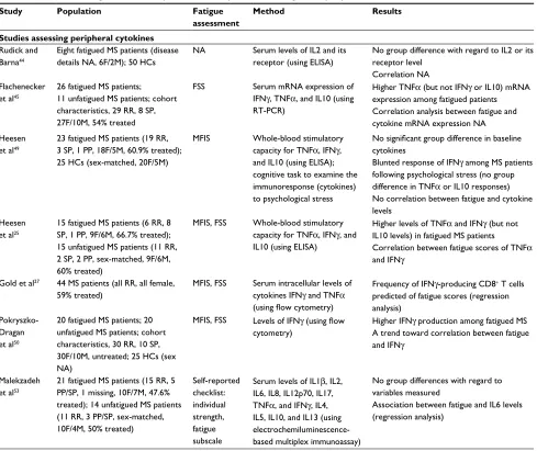
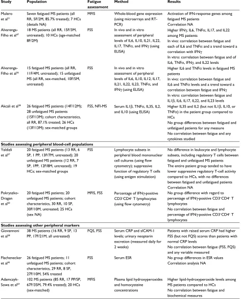
Adamczyk-Sowa et al, no correlation was found between MS fatigue and plasma lipid hydroxyperoxides or homocysteine concentration, which are markers of oxidation.59 Table 2
summarizes these studies.
Table 2 Studies assessing the relationship between multiple sclerosis fatigue and peripheral inflammation
Study Population Fatigue
assessment
Method Results
Studies assessing peripheral cytokines Rudick and
Barna44
Eight fatigued MS patients (disease details NA, 6F/2M); 50 HCs
NA Serum levels of IL2 and its receptor (using ELISA)
No group difference with regard to IL2 or its receptor level
Correlation NA Flachenecker
et al45
26 fatigued MS patients; 11 unfatigued MS patients; cohort characteristics, 29 RR, 8 SP, 27F/10M, 54% treated
FSS Serum mRNA expression of
IFNγ, TNFα, and IL10 (using RT-PCR)
Higher TNFα (but not IFNγ or IL10) mRNA expression among fatigued patients Correlation analysis between fatigue and cytokine mRNA expression NA Heesen
et al49
23 fatigued MS patients (19 RR, 3 SP, 1 PP, 18F/5M, 60.9% treated); 25 HCs (sex-matched, 20F/5M)
MFIS Whole-blood stimulatory capacity for TNFα, IFNγ, and IL10 (using ELISA); cognitive task to examine the immunoresponse (cytokines) to psychological stress
No significant group difference in baseline cytokines
Blunted response of IFNγ among MS patients following psychological stress (no group difference in TNFα or IL10 responses) No correlation between fatigue and cytokine levels
Heesen et al25
15 fatigued MS patients (6 RR, 8 SP, 1 PP, 9F/6M, 66.7% treated); 15 unfatigued MS patients (11 RR, 2 SP, 2 PP, sex-matched, 9F/6M, 60% treated)
MFIS, FSS Whole-blood stimulatory capacity for TNFα, IFNγ, and IL10 (using ELISA)
Higher levels of TNFα and IFNγ (but not IL10 levels) in fatigued MS patients
Correlation between fatigue scores of TNFα and IFNγ
Gold et al27 44 MS patients (all RR, all female, 59% treated)
MFIS, FSS Serum intracellular levels of cytokines IFNγ and TNFα (using flow cytometry)
Frequency of IFNγ-producing CD8+ T cells
predicted of fatigue scores (regression analysis)
Pokryszko-Dragan et al50
20 fatigued MS patients; 20 unfatigued MS patients; cohort characteristics, 30 RR, 10 SP, 30F/10M, untreated; 25 HCs (sex NA)
MFIS, FSS Levels of IFNγ (using flow cytometry)
Higher IFNγ production among fatigued MS A trend toward correlation between fatigue and IFNγ
Malekzadeh et al53
21 fatigued MS patients (15 RR, 5 PP/SP, 1 missing, 10F/7M, 47.6% treated); 14 unfatigued MS patients (11 RR, 3 PP/SP, sex-matched, 10F/4M, 50% treated)
Self-reported checklist: individual strength, fatigue subscale
Serum levels of IL1β, IL2, IL6, IL8, IL12p70, IL17, TNFα, and IFNγ, IL4, IL5, IL10, and IL13 (using electrochemiluminescence-based multiplex immunoassay)
No group differences with regard to variables measured
Association between fatigue and IL6 levels (regression analysis)
(Continued)
Journal of Inflammation Research downloaded from https://www.dovepress.com/ by 118.70.13.36 on 24-Aug-2020
Dovepress
Chalah and Ayache
Study Population Fatigue
assessment
Method Results
Mulero et al51
Seven fatigued MS patients (all RR, 5F/2M, 85.7% treated); 7 HCs (details NA)
MFIS Whole-blood gene expression (using microarrays and RT-PCR)
Activation of IFN-response genes among fatigued MS patients
Correlation NA
Alvarenga-Filho et al47
18 MS patients (all RR, 15F/3M, untreated); 10 HCs (age-matched 8F/2M)
FSS In vivo and in vitro assessment of peripheral levels of IL6, IL10, IL21, IL22, IL17, TNFα, and IFNγ (using ELISA)
Higher IFNγ, IL6, TNFα, IL17, and IL22 among MS patients
In vivo: correlation between fatigue and each of IL6 and TNFα and a trend toward a correlation with IFNγ
In vitro: correlation between fatigue and of IL6, TNFα, IFNγ, and IL22 levels
Alvarenga-Filho et al56
15 fatigued MS patients (all RR, 11F/4M, untreated); 15 unfatigued MS (all RR, sex-matched, 10F/5M, untreated)
FSS In vivo and in vitro assessment of peripheral levels of IL6, IL10, IL12, IL17, IL21, IL22, IL23, TNFα, and IFNγ (using ELISA)
Higher IL6 and TNFα levels in fatigued MS patients
In vivo: correlation between fatigue and IL6 and TNFα levels and a trend toward a correlation between fatigue and IFNγ In vitro: correlation between fatigue and IL1β, IL6, IL17, IL22, and IL23 levels Akcali et al28 26 fatigued MS patients (14F/12M);
28 unfatigued MS patients (15F/13M); cohort characteristics, all RR, 87.1% treated; 26 HCs (13F/13M); sex-matched groups
FSS, NFI-MS Serum IL1β, TNFα, IL35, IL2, and IL10 (using ELISA)
Higher IL35 and IL2 (but not IL1β, IL10, or TNFα) in the patient group compared to HCs
No group differences between fatigued and unfatigued patients for any measure No correlation between fatigue and any cytokines studied
Studies assessing peripheral blood-cell populations Yaldizli
et al57
20 fatigued MS patients (13 RR, 6 SP, 1 PP, 13F/7M, untreated); 20 unfatigued MS patients (12 RR, 7 SP, 1PP, 12F/8M, untreated); 19 HCs; sex-matched groups
FSS Lymphocyte subsets in peripheral blood mononuclear cell cultures (using flow cytometry); suppressive function of regulatory T cells (using antigen stimulation)
No difference in leukocyte and lymphocyte subsets, including regulatory T cells between fatigued and unfatigued MS patients The entire patient group tended to have lower suppressive regulatory T-cell activity compared to HCs, with no differences between fatigued and unfatigued patients Correlation NA
Pokryszko-Dragan et al50
20 fatigued MS patients; 20 unfatigued MS patients; cohort characteristics, 30 RR, 10 SP, 30F/10M, untreated; 25 HCs (sex NA)
MFIS, FSS Percentage of IFNγ-positive CD3+CD4+ T lymphocytes
(using flow cytometry)
No group difference with regard to percentage of IFNγ-positive CD3+CD4+ T
lymphocytes
No correlation between fatigue and percentage of IFNγ-positive CD3+CD4+ T
lymphocytes Studies assessing other peripheral markers
Giovannoni et al58
38 MS patients (16 RR, 9 SP, 13 PP, 17F/21M, all untreated)
FQS, FSS Serum CRP and sICAM-1 levels; urinary neopterin excretion (measured daily for 2 weeks)
Patients with raised serum CRP had higher FSS (but not FQS) scores than patients with normal CRP levels
No correlation between fatigue (FSS, FQS) and any variable measured
Flachenecker et al45
26 fatigued MS patients; 11 unfatigued MS patients; cohort characteristics, 29 RR, 8 SP, 27F/10M, 54% treated
FSS Serum ESR No group differences in ESR values
Correlation analysis NA
Adamczyk-Sowa et al59
102 MS patients (85 RR, 17 PP/SP, 67F/35M, 79.4% treated); 20 HCs (sex-matched)
MFIS Plasma lipid hydroxyperoxides and homocysteine
concentrations
Higher lipid-hydroxyperoxide levels among MS patients compared to HCs
No correlation between fatigue and biochemical measures
Abbreviations: ELISA, enzyme-linked immunosorbent assay; ESR, erythrocyte-sedimentation rate; F, female; FSS, Fatigue Severity Scale; FQS, Fatigue Questionnaire Scale; HCs, healthy controls; M, male; MFIS, Modified Fatigue Impact Scale; MS, multiple sclerosis; NA, not available, NFI, Neurological Fatigue Index; PP, primary progressive; RR, relapsing–remitting; RT-PCR, real-time polymerase chain reaction; SP, secondary progressive.
Table 2 (Continued)
Journal of Inflammation Research downloaded from https://www.dovepress.com/ by 118.70.13.36 on 24-Aug-2020
Dovepress Inflammation and fatigue in multiple sclerosis
Conclusion
This work has evaluated the contribution of central and peripheral inflammatory processes to MS fatigue. Among the selected studies dealing with central inflammatory and neuroendocrine processes, an inconsistency existed regarding the relationship between fatigue and orexin A system (present in one study,14 absent in one15), the HPA axis (present in two
studies,30,34 absent in five studies24–28), and some CSF markers
(present in one study assessing cytokines,40 absent in one study
assessing cellular, humoral, and other CSF parameters39). As
for peripheral markers, although there was scarcity in the available data, serum proinflammatory cytokines (ie, IL6, TNFα, and IFNγ) seemed to be associated with MS fatigue. However, given the existence of some conflicting data in this domain, such an association merits further investiga-tion. Finally, concerning T-cell population (ie, CD3+CD4+ T lymphocytes50 or regulatory T cells57) or peripheral markers
of inflammation (ie, CRP, erythrocyte-sedimentation rate, and soluble ICAM1),45,58,59 few data were available, and these
studies failed to find a link between MS fatigue and these mea-sures. It is also worth noting that studies differed greatly in the clinical characteristics of their cohorts, especially concerning treatment profiles. The fact that MS treatment can modulate the inflammatory milieu30,31 would stand behind the
differ-ences observed in study outcomes, with studies including untreated patients yielding positive results on the relationship between fatigue and inflammation.14,40,47,50,56 Of note, several
trials have documented differences in immune/inflammatory profiles between treated and naïve MS patients. That is to say, downregulation of proinflammatory cytokines was observed among MS patients treated with disease-modifying therapies such as IFNβ,60–63 glatiramer acetate,64 dimethyl fumarate,65
fingolimod,66 natalizumab,67 and teriflunomide.68
Another issue to consider is the possible impact of MS treatments on fatigue per se. Few reports are available on this matter. In a cross-sectional study, higher fatigue rates were observed among MS patients treated with IFNβ or glatiramer acetate compared to age- and sex-matched patients receiving natalizumab.69 In other works, rituximab seemed to induce
fatigue in MS patients,70 natalizumab appeared to improve
fatigue,71–74 and fingolimod did not seem to modify symptom
severity.75 Therefore, more research is needed to understand
the potential effects of MS therapies on fatigue perception and cytokine profiles.
Another difference among studies concerned fatigue scales, which consisted of the FSS, MFIS, FSMC, visual ana-logue scale for fatigue, eleven-item Fatigue Scale, Neurologi-cal Fatigue Index – MS, self-reported checklist – individual
strength (fatigue subscale), and Fatigue Questionnaire Scale. This adds more difficulty in comparing study outcomes. While some of these scales (eg, FSS) mainly address the physical component of fatigue, other scales (eg MFIS) reflect the physical, and, the cognitive and psychosocial dimensions of this symptom. This difference might not have had a large impact on group differences (fatigued versus unfatigued) but may have affected the correlation between fatigue severity and cytokine levels and could partly explain the discrepancies observed among studies.
Using different immunological techniques (ie, ELISA, RT-PCR, genetic analysis, flow cytometry, and electrochemi-luminescence-based multiplex immunoassay) might have been behind interstudy differences, particularly those evaluating peripheral cytokines. Another point to consider is the relation-ship between sex, hormones, and immunodysregulation. Like many autoimmune diseases, MS is more prevalent in women than men, and hormones seem to exert an immunomodulatory effect and might influence damage repair in the CNS.76
Interest-ingly, sex dysmorphism was observed with regard to cytokine production in MS patients.77 In this context, it is of importance
to note that although some studies controlled for sex effects by including sex-matched controls,14,24,25,28,30,33,37,47,49,53,56,57,59 other
studies enrolled cohorts predominantly15,39,40,45 or exclusively26
composed of female patients or did not provide sufficient details on the matter.24,44,45,50,51 Therefore, future work could
benefit from comparing fatigue and cytokine production between male and female patients.
Moreover, studying the impact of environmental, genetic, and epigenetic MS risk factors on MS fatigue would be of great interest. These factors include ultraviolet-radiation exposure, vitamin D intake, smoking, dietary, and exercise habits, and body-mass index.78 It is also of importance to
control for some confounders that occur frequently in MS and can impact MS fatigue. These include physical disability,6
emotional symptoms,79,80 and sleep disorders.81 There is still a
long way to go to define the utility and place of the aforemen-tioned measures in clinical wards. Future large-scale studies are critically needed to conclude on this matter and would benefit from comparing the relationship between fatigue and inflammation in patients with different disease phenotypes (RR vs primary progressive vs secondary progressive) with and without disease-modifying drugs. From this perspec-tive, applying different measures might help to decide on the optimal target to serve as an immunological surrogate of MS fatigue. Facing the subjective nature of fatigue scales, developing objective biological markers for fatigue, as those visited here, would be of great help.
Journal of Inflammation Research downloaded from https://www.dovepress.com/ by 118.70.13.36 on 24-Aug-2020
Dovepress
Chalah and Ayache
Disclosure
SSA has received travel grants or compensation from Gen-zyme, Biogen, Novartis, and Roche. The authors report no other conflicts of interest in this work.
References
1. Sospedra M, Martin R. Immunology of multiple sclerosis. Annu Rev Immunol. 2005;23:683–747.
2. Dendrou CA, Fugger L, Friese MA. Immunopathology of multiple sclerosis. Nat Rev Immunol. 2015;15:545–558.
3. Compston A, Coles A. Multiple sclerosis. Lancet. 2008;372:1502–1517. 4. Chalah MA, Riachi N, Ahdab R, Créange A, Lefaucheur JP, Ayache
SS. Fatigue in multiple sclerosis: neural correlates and the role of non-invasive brain stimulation. Front Cell Neurosci. 2015;9:460. 5. Centonze D, Muzio L, Rossi S, Furlan R, Bernardi G, Martino G. The
link between inflammation, synaptic transmission and neurodegenera-tion in multiple sclerosis. Cell Death Differ. 2010;17:1083–1091. 6. Ayache SS, Chalah MA. Fatigue in multiple sclerosis: insights into
evaluation and management. Neurophysiol Clin. 2017;47:139–171. 7. Freal JE, Kraft GH, Coryell JK. Symptomatic fatigue in multiple
scle-rosis. Arch Phys Med Rehabil. 1984;65:135–138.
8. Moher D, Liberati A, Tetzlaff J, Altman DG. Preferred reporting items for systematic reviews and meta-analyses: the PRISMA statement. BMJ. 2009;339:b2535.
9. Tsujino N, Sakurai T. Role of orexin in modulating arousal, feeding, and motivation. Front Behav Neurosci. 2013;7:28.
10. Kiyashchenko LI, Mileykovskiy BY, Maidment N, et al. Release of hypocretin (orexin) during waking and sleep states. J Neurosci. 2002;22:5282–5286.
11. Grossberg AJ, Zhu X, Leinninger GM, et al. Inflammation-induced leth-argy is mediated by suppression of orexin neuron activity. J Neurosci. 2011;31:11376–11386.
12. Oka Y, Kanbayashi T, Mezaki T, et al. Low CSF hypocretin-1/orexin-A associated with hypersomnia secondary to hypothalamic lesion in a case of multiple sclerosis. J Neurol. 2004;251:885–886.
13. Kato T, Kanbayashi T, Yamamoto K, et al. Hypersomnia and low CSF hypocretin-1 (orexin-A) concentration in a patient with multiple sclerosis showing bilateral hypothalamic lesions. Intern Med. 2003;42:743–745. 14. Papuć E, Stelmasiak Z, Grieb P, Paweł G, Rejdak K. CSF hypocretin-1
concentrations correlate with the level of fatigue in multiple sclerosis patients. Neurosci Lett. 2010;474:9–12.
15. Constantinescu CS, Niepel G, Patterson M, et al. Orexin A (hypocre-tin-1) levels are not reduced while cocaine/amphetamine regulated transcript levels are increased in the cerebrospinal fluid of patients with multiple sclerosis: no correlation with fatigue and sleepiness. J Neurol Sci. 2011;307:127–131.
16. Boddum K, Hansen MH, Jennum PJ, Kornum BR. Cerebrospinal fluid hypocretin-1 (orexin-a) level fluctuates with season and correlates with day length. PLoS One. 2016;11:e0151288.
17. Limone P, Ferrero B, Calvelli P, et al. Hypothalamic-pituitary-adrenal axis function and cytokine production in multiple sclerosis with or without interferon-β treatment. Acta Neurol Scand. 2002;105:372–377. 18. Grasser A, Möller A, Backmund H, Yassouridis A, Holsboer F. Heteroge-neity of hypothalamic-pituitary-adrenal system response to a combined dexamethasone-CRH test in multiple sclerosis. Exp Clin Endocrinol Diabetes. 1996;104:31–37.
19. Michelson D, Stone L, Galliven E, et al. Multiple sclerosis is associated with alterations in hypothalamic-pituitary-adrenal axis function. J Clin Endocrinol Metab. 1994;79:848–853.
20. Bergh FT, Kümpfel T, Trenkwalder C, Rupprecht R, Holsboer F. Dys-regulation of the hypothalamo-pituitary-adrenal axis is related to the clinical course of MS. Neurology. 1999;53:772–777.
21. Ysrraelit MC, Gaitán MI, Lopez AS, Correale J. Impaired hypothalamic-pituitary-adrenal axis activity in patients with multiple sclerosis. Neurol-ogy. 2008;71:1948–1954.
22. Melief J, de Wit SJ, van Eden CG, et al. HPA axis activity in mul-tiple sclerosis correlates with disease severity, lesion type and gene expression in normal-appearing white matter. Acta Neuropathol. 2013;126:237–249.
23. Huitinga I, Erkut ZA, van Beurden D, Swaab DF. Impaired hypothalamus pituitary-adrenal axis activity and more severe multiple sclerosis with hypothalamic lesions. Ann Neurol. 2004;55:37–45.
24. Heesen C, Gold SM, Raji A, Wiedemann K, Schulz KH. Cogni-tive impairment correlates with hypothalamo-pituitary-adrenal axis dysregulation in multiple sclerosis. Psychoneuroendocrinology. 2002;27:505–517.
25. Heesen C, Nawrath L, Reich C, Bauer N, Schulz KH, Gold SM. Fatigue in multiple sclerosis: an example of cytokine mediated sickness behav-iour? J Neurol Neurosurg Psychiatry. 2006;77:34–39.
26. Heesen C, Schulz KH, Fiehler J, et al. Correlates of cognitive dysfunc-tion in multiple sclerosis. Brain Behav Immun. 2010;24:1148–1155. 27. Gold SM, Krüger S, Ziegler KJ, at al. Endocrine and immune substrates
of depressive symptoms and fatigue in multiple sclerosis patients with comorbid major depression. J Neurol Neurosurg Psychiatry. 2011;82:814–818.
28. Akcali A, Zengin F, Aksoy SN, Zengin O. Fatigue in multiple sclerosis: is it related to cytokines and hypothalamic-pituitary-adrenal axis? Mult Scler Relat Disord. 2017;15:37–41.
29. Shishioh-Ikejima N, Ogawa T, Yamaguti K, Watanabe Y, Kuratsune H, Kiyama H. The increase of alpha-melanocyte-stimulating hormone in the plasma of chronic fatigue syndrome patients. BMC Neurol. 2010;10:73. 30. Gottschalk M, Kümpfel T, Flachenecker P, et al. Fatigue and regulation of the hypothalamo-pituitary-adrenal axis in multiple sclerosis. Arch Neurol. 2005;62:277–280.
31. Kümpfel T, Schwan M, Weber F, Holsboer F, Trenkwalder C, Bergh FT. Hypothalamo-pituitary-adrenal axis activity evolves differentially in untreated versus treated multiple sclerosis. Psychoneuroendocrinology. 2014;45:87–95.
32. Dunn AJ. Effects of the IL-1 receptor antagonist on the IL-1- and endotoxin-induced activation of the HPA axis and cerebral biogenic amines in mice. Neuroimmunomodulation. 2000;7:36–45.
33. Powell DJ, Moss-Morris R, Liossi C, Schlotz W. Circadian cortisol and fatigue severity in relapsing-remitting multiple sclerosis. Psychoneu-roendocrinology. 2015;56:120–131.
34. Clow A, Hucklebridge F, Stalder T, Evans P, Thorn L. The cortisol awak-ening response: more than a measure of HPA axis function. Neurosci Biobehav Rev. 2010;35:97–103.
35. Vogl D, Falk W, Dorner M, Schölmerich J, Straub RH. Serum levels of pregnenolone and 17-hydroxypregnenolone in patients with rheumatoid arthritis and systemic lupus erythematosus: relation to other adrenal hormones. J Rheumatol. 2003;30:269–275.
36. Tengstrand B, Carlström K, Fellander-Tsai L, Hafström I. Abnormal levels of serum dehydroepiandrosterone, estrone, and estradiol in men with rheumatoid arthritis: high correlation between serum estradiol and current degree of inflammation. J Rheumatol. 2003;30:2338–2343. 37. Téllez N, Comabella M, Julià E, et al. Fatigue in progressive multiple
sclerosis is associated with low levels of dehydroepiandrosterone. Mult Scler. 2006;12:487–494.
38. Calabrese VP, Isaacs ER, Regelson W. Dehydroepiandrosterone in multiple sclerosis: positive effects on the fatigue syndrome in a non-ran-domized study. In: Kalimi M, Regelson W editors. The Biological Role of Dehydroepiandrosterone (DHEA). Berlin: de Gruyter; 1990:95–100 39. Biberacher V, Schmidt P, Selter RC, et al. Fatigue in multiple sclerosis: associations with clinical, MRI and CSF parameters. Mult Scler. Epub 2017 May 1.
40. Brenner P, Granqvist M, Königsson J, al Nimer F, Piehl F, Jokinen J. Depression and fatigue in multiple sclerosis: relation to exposure to violence and cerebrospinal fluid immunomarkers. Psychoneuroendo-crinology. 2018;89:53–58.
41. Rovaris M, Barnes D, Woodrofe N, et al. Patterns of disease activity in multiple sclerosis patients: a study with quantitative gadolinium enhanced brain MRI and cytokine measurement in different clinical subgroups. J Neurol. 1996;243:536–542.
Journal of Inflammation Research downloaded from https://www.dovepress.com/ by 118.70.13.36 on 24-Aug-2020
Dovepress Inflammation and fatigue in multiple sclerosis
42. Khademi M, Wallström E, Andersson M, Piehl F, Di Marco R, Olsson T. Reduction of both pro- and anti-inflammatory cytokines after 6 months of interferon β-1a treatment of multiple sclerosis. J Neuroimmunol. 2000;103:202–210.
43. Baraczka K, Pozsonyi T, Szüts I, Ormos G, Nékám K. Increased levels of tumor necrosis alpha and soluble vascular endothelial adhesion molecule-1 in the cerebrospinal fluid of patients with connective tis-sue diseases and multiple sclerosis. Acta Microbiol Immunol Hung. 2003;50:339–348.
44. Rudick RA, Barna BP. Serum interleukin 2 and soluble interleukin 2 receptor in patients with multiple sclerosis who are experiencing severe fatigue. Arch Neurol. 1990;47:254–255.
45. Flachenecker P, Bihler I, Weber F, Gottschalk M, Toyka KV, Rieckmann P. Cytokine mRNA expression in patients with multiple sclerosis and fatigue. Mult Scler. 2004;10:165–169.
46. Kierkegaard M, Lundberg IE, Olsson T, et al. High-intensity resistance training in multiple sclerosis: an exploratory study of effects on immune markers in blood and cerebrospinal fluid, and on mood, fatigue, health-related quality of life, muscle strength, walking and cognition. J Neurol Sci. 2016;362:251–257.
47. Alvarenga-Filho H, Sacramento PM, Ferreira TB, et al. Combined exercise training reduces fatigue and modulates the cytokine profile of T-cells from multiple sclerosis patients in response to neuromediators.
J Neuroimmunol. 2016;293:91–99.
48. Mokhtarzade M, Ranjbar R, Majdinasab N, Patel D, Shamsi MM. Effect of aerobic interval training on serum IL-10, TNFα, and adipokines levels in women with multiple sclerosis: possible relations with fatigue and quality of life. Endocrine. 2017;57:262–271.
49. Heesen C, Koehler G, Gross R, Tessmer W, Schulz KH, Gold SM. Altered cytokine responses to cognitive stress in multiple sclerosis patients with fatigue. Mult Scler. 2005;11:51–57.
50. Pokryszko-Dragan A, Frydecka I, Kosmaczewska A, et al. Stimulated peripheral production of interferon-gamma is related to fatigue and depres-sion in multiple sclerosis. Clin Neurol Neurosurg. 2012;114:1153–1158. 51. Mulero P, Almansa R, Neri MJ, et al. Improvement of fatigue in multiple sclerosis by physical exercise is associated to modulation of systemic interferon response. J Neuroimmunol. 2015;280:8–11.
52. Kostic M, Zivkovic N, Cvetanovic A, Stojanovic I, Colic M. IL-17 signaling in astrocytes promotes glutamate excitotoxicity: indications for the link between inflammatory and neurodegenerative events in multiple sclerosis. Mult Scler Relat Disord. 2017;11:12–17.
53. Malekzadeh A, van de Geer-Peeters W, de Groot V, Teunissen CE, Beckerman H. Fatigue in patients with multiple sclerosis: is it related to pro- and anti-inflammatory cytokines? Dis Markers. 2015;2015:758314. 54. Bertolone K, Coyle PK, Krupp LB, Doscher CA. Cytokine correlates
of fatigue in multiple sclerosis. Neurology. 1993;43:A356.
55. Jafarzadeh A, Jamali M, Mahdavi R, et al. Circulating levels of interleu-kin-35 in patients with multiple sclerosis: evaluation of the influences of FOXP3 gene polymorphism and treatment program. J Mol Neurosci. 2015;55:891–897.
56. Alvarenga-Filho H, Salles M, Hygino J, et al. Fatigue favors in vitro Th1 and Th17-like cell expansion and reduces corticoid sensitivity in MS patients. J Neuroimmunol. 2017;303:81–89.
57. Yaldizli O, Kumar M, Vago S, Kreuzfelder E, Limmroth V, Putzki N. Fatigue is not associated with impaired function of regulatory T cells in untreated patients with multiple sclerosis. Eur Neurol. 2009;62:321–326.
58. Giovannoni G, Thompson AJ, Miller DH, Thompson EJ. Fatigue is not associated with raised inflammatory markers in multiple sclerosis.
Neurology. 2001;57:676–681.
59. Adamczyk-Sowa M, Sowa P, Adamczyk J, et al. Effect of melatonin supplementation on plasma lipid hydroperoxides, homocysteine con-centration and chronic fatigue syndrome in multiple sclerosis patients treated with interferons-beta and mitoxantrone. J Physiol Pharmacol. 2016;67:235–242.
60. Losy J, Michałowska-Wender G. In vivo effect of interferon-β 1a on interleukin-12 and TGF-β1 cytokines in patients with relapsing-remitting
multiple sclerosis. Acta Neurol Scand. 2002;106:44–46.
61. Zhang X, Markovic-Plese S. Interferon beta inhibits the Th17 cell-mediated autoimmune response in patients with relapsing-remitting multiple sclerosis. Clin Neurol Neurosurg. 2010;112:641–645. 62. Ramgolam VS, Markovic-Plese S. Interferon-beta inhibits Th17 cell
differentiation in patients with multiple sclerosis. Endocr Metab Immune Disord Drug Targets. 2010;10:161–167.
63. Ramgolam VS, Sha Y, Jin J, Zhang X, Markovic-Plese S. IFN-β inhibits human Th17 cell differentiation. J Immunol. 2009;183:5418–5427. 64. Oreja-Guevara C, Ramos-Cejudo J, Aroeira LS, Chamorro B,
Diez-Tejedor E. TH1/TH2 cytokine profile in relapsing-remitting multiple sclerosis patients treated with glatiramer acetate or natalizumab. BMC Neurol. 2012;12:95.
65. Mills EA, Ogrodnik MA, Plave A, Mao-Draayer Y. Emerging under-standing of the mechanism of action for dimethyl fumarate in the treatment of multiple sclerosis. Front Neurol. 2018;9:5.
66. Su K, Zeng P, Liang W, et al. FTY720 attenuates angiotensin II-induced podocyte damage via inhibiting inflammatory cytokines. Mediators Inflamm. 2017;2017:3701385.
67. Balasa RI, Simu M, Voidazan S, et al. Natalizumab changes the periph-eral profile of the Th17 panel in MS patients: new mechanisms of action.
CNS Neurol Disord Drug Targets. 2017;16:1018–1026.
68. Bar-Or A, Pachner A, Menguy-Vacheron F, Kaplan J, Wiendl H. Teri-flunomide and its mechanism of action in multiple sclerosis. Drugs. 2014;74:659–674.
69. Yildiz M, Tettenborn B, Putzki N. Multiple sclerosis-associated fatigue during disease-modifying treatment with natalizumab, interferon-beta and glatiramer acetate. Eur Neurol. 2011;65:231–232.
70. He D, Guo R, Zhang F, Zhang C, Dong S, Zhou H. Rituximab for relapsing-remitting multiple sclerosis. Cochrane Database Syst Rev. 2013;12:CD009130.
71. Hoepner R, Faissner S, Salmen A, Gold R, Chan A. Efficacy and side effects of natalizumab therapy in patients with multiple sclerosis. J Cent Nerv Syst Dis. 2014;6:41–49.
72. Stephenson JS, Kern DM, Agarwal SS, et al. Impact of natalizumab on patient-reported outcomes in multiple sclerosis: a longitudinal study.
Health Qual Life Outcomes. 2012;10:155–164.
73. Svenningsson A, Falk E, Celius EG, et al. Natalizumab treatment reduces fatigue in multiple sclerosis: results from the TYNERGY trial – a study in the real life setting. PLoS One. 2013;8:e58643.
74. Iaffaldano P, Viterbo RG, Paolicelli D, et al. Impact of natalizumab on cognitive performances and fatigue in relapsing multiple sclerosis: a prospective, open-label, two years observational study. PLoS One. 2012;7:e35843.
75. Masingue M, Debs R, Maillart E, et al. Fatigue evaluation in fingoli-mod treated patients: an observational study. Mult Scler Relat Disord. 2017;14:8–11.
76. Shuster EA. Hormonal influences in multiple sclerosis. Curr Top Microbiol Immunol. 2008;318:267–311.
77. Pelfrey CM, Cotleur AC, Lee JC, Rudick RA. Sex differences in cytokine responses to myelin peptides in multiple sclerosis. J Neuroimmunol. 2002;130:211–223.
78. Segal BM, Cohen JA, Antel J. Americas Committee for Treatment and Research in Multiple Sclerosis Forum 2017: environmental factors, genetics, and epigenetics in MS susceptibility and clinical course. Mult Scler. 2018;24:4–5.
79. Chalah MA, Ayache SS. Psychiatric event in multiple sclerosis: could it be the tip of the iceberg? Rev Bras Psiquiatr. 2017;39:365–368. 80. Chalah MA, Ayache SS. Alexithymia in multiple sclerosis: a systematic
review of literature. Neuropsychologia. 2017;104:31–47.
81. Hughes AJ, Dunn KM, Chaffee T. Sleep disturbance and cognitive dysfunction in multiple sclerosis: a systematic review. Curr Neurol Neurosci Rep. 2018;18:2.
Journal of Inflammation Research downloaded from https://www.dovepress.com/ by 118.70.13.36 on 24-Aug-2020
Dovepress
Journal of Inflammation Research
Publish your work in this journal
Submit your manuscript here: https://www.dovepress.com/journal-of-inflammation-research-journal
The Journal of Inflammation Research is an international, peer-reviewed open access journal that welcomes laboratory and clinical findings on the molecular basis, cell biology and pharmacology of inflammation including original research, reviews, symposium reports, hypothesis for-mation and commentaries on: acute/chronic inflamfor-mation; mediators of
inflammation; cellular processes; molecular mechanisms; pharmacology and novel anti-inflammatory drugs; clinical conditions involving inflam-mation. The manuscript management system is completely online and includes a very quick and fair peer-review system. Visit http://www.dove press.com/testimonials.php to read real quotes from published authors.
Dove
press
Chalah and Ayache
Journal of Inflammation Research downloaded from https://www.dovepress.com/ by 118.70.13.36 on 24-Aug-2020