
FABRICATION OF NOVEL ANTICANCER POLYOXOMETALATE [COW
11O
39(CPTI)]
7- -CHITOSAN
NANOCOMPOSITE, ITS TOXICITY REDUCTION, AND SUSTAINED RELEASE
VAMANGI M PANDYA, SACHIN A JOSHI*
Dr. K. C. Patel Research & Development Center, Charotar University of Science and Technology (CHARUSAT), Changa - 388421, Gujarat, India. Email: [email protected]
Received: 21 December 2016, Revised and Accepted: 29 December 2016 ABSTRACT
Objectives: Polyoxometalates (POMs) are proved to be important for applications in medicine and in material science. Here, we represent nanocomposite formation of tungsten-containing potent anticancer polyanion, K6H [CoW11O39 (CpTi)].13H2O (CoW11CpTi) with biocompatible
ChitosanYC-100 (CSYC100) with the goal to reduce its heavy metal toxicity.
Methods: Synthesis of “POM-CSYC100 nanocomposite” was attained without the aid of any cross-linker through electrostatic interaction technique. Nanocomposites were characterized using Fourier transform infrared spectroscopy, dynamic light scattering, transmission electron microscopy, and thermogravimetric analysis. The release profile recorded was slow and sustained at physiological pH. In vitro cytotoxicity assays which show an attribute to reduce the toxicity of these POM were performed on C2C12 (mouse myoblast cell line) and A-549 (lung cancer cell line), which proved the reduced toxicity of nanocomposites as compared to the bare drugs.
Results: Sustained release studies showed there was a slow and steady release of CoW11CpTi for 11 hrs, with the 98% of collective release at the
end. From in vitro cytotoxic assay, it was deduced that CoW11CpTi -CSYC100 nanocomposite at the concentrations of 1.25 mM, and lower did not
exhibit toxic effect on C2C12 cells as 95% total C2C12 cell mass remained viable. While in the case of A549 cells highest 5 mM concentration of bare CoW11CpTi is toxic to the cancer cells and after encapsulation cell viability increases from 10% to 55%.
Conclusion: Thus, this study has designated the probability of using POM-chitosan nanocomposite for less toxic and effective biomedicinal applications. Keywords: Anticancer, Chitosan, Nanocomposite, In vitro cytotoxicity, Drug release.
INTRODUCTION
Polyoxometalates (POMs) can be defined as “early transition metal oxygen clusters.” POMs have gained tremendous attention because of their mutable uses in the areas of novel drug development, material sciences, and in industrial catalyst [1]. The use of POMs in latent applications for human medicine is growing exponentially and are recently published in the literatures [2,3]. The toxicity due to the presence of metal ions is the major drawback of these precious medicinal molecules. To lower down the toxicity, it needs to be conjugate/encapsulate in some biopolymeric delivery vehicle.
Chitosan and its derivatives linger to attract noteworthy attention as a potent drug transporter with remarkable biocompatibility along with appreciable cellular uptake rates [4-6]. Chitosan is a carbohydrate heteropolymer composed of glucosamine and N-acetyl glucosamine linked by β 1-4 glucosidic bonds as a repeating unit. Chitosan is developed from chitin by its N-deacetylation. Chitin a structural polysaccharide found in the exoskeleton of shrimps and shells of crabs and the second most abundant polysaccharide found in nature after cellulose [7]. This versatile feature has been used in this study to beat the significant biomedical potential of POMs by their encapsulation in chitosan YC-100 (CSYC100) matrix. The chosen title anion K6H[CoW11O39(CpTi)].13H2O, hereafter denoted as
(CoW11CpTi) is reported to be the most potent anticancer POM among
the family of CpTi substituted POMs studied so far. It is reported that polyoxotungstate CoW11CpTi curiously decreased tumor weight of the
rats bearing HLC (colon cancer cell), HL-60 (leukemia), and SSMC-7721 (liver cancer cell) where the experimental results were procured using the animal tumor implantation method. The in vivo anticancer efficacy of CoW11CpTi is at par with the clinical anticancer drugs 5-fluorouracil
and CP (abbreviation of cyclophosphamide), but the cytotoxicity of CoW11CpTi is reported to be less than them [8].
The research described in this report is focused to prepare the nanocomposite of low molecular weight carbohydrate polymer CSYC100 and an anticancer POM CoW11CpTi. The primary agenda
of this research is to portray reduction in the metallic toxicity of POMs by its involvement in the formation of nanocomposite with CSYC100. To support this hypothesis, CoW11CpTi-CSYC100
nanocomposites were prepared and its toxicity was assessed in vitro on cell line C2C12 (normal myoblast cell line) and A-549 (adenocarcinomic human alveolar basal epithelial cells-lung cancer cell line). To the best of our knowledge, this the first report showing the toxicity reduction of [CoW11O39(CpTi)]7- by forming their
nanocomposite with CSYC100.
METHODS Materials
CSYC100 is a low molecular weight chitosan (~10000 g/mol) and highly water soluble and purchased from Sigma-Aldrich, Steinheim, Germany. Other chemicals required for synthesis of POMs, and other studies were bought commercially from Sigma-Aldrich.
Preparation of POMs
Polyoxotungstate CoW11CpTi was synthesized according to the
procedure described by Wang et al. in 2003 [8].
Synthesis of CSYC100/CoW11CpTi complexes
To synthesize CoW11CpTi-CSYC100 nanocomposites, 50 mg CSYC100
was dissolved in 70 ml double distilled water. The mixture was stirred © 2017 The Authors. Published by Innovare Academic Sciences Pvt Ltd. This is an open access article under the CC BY license (http://creativecommons. org/licenses/by/4. 0/) DOI: http://dx.doi.org/10.22159/ajpcr.2017.v10i4.16721
until the CSYC100 complete dissolve, and the clear solution is observed. The solution was then filtered to remove any suspended particles. CSYC100 filtrate was further used to prepare nanocomposites of POMs. 170 mg (0.39 mM) CoW11CpTi was dissolved in 2 mL double distilled
water separately. CoW11CpTi solution was then added dropwise
with stirring into the CSYC100 solution under controlled sonication, resulting in the formation of stable colloidal suspension of CoW11
CpTi-CSYC100 nanocomposite. This suspension was centrifuged for 20 minutes at 12,000 rpm (18,000 g) to collect the CoW11CpTi-CSYC100
nanocomposites obtained as pellet. The supernatant was discarded, and pellet of CoW11CpTi-CSYC100 was recovered, dried and used for
further studies.
Physicochemical characterization of POM-CSYC100 nanocomposites Morphology and particle size of the nanocomposites was analyzed by transmission electron microscopy (TEM) (Model-Philips Tecnai 20). The particle size distribution and hydrodynamic diameter size were determined by dynamic light scattering (DLS) method using particle size analyzer (Model: Malvern Zetasizer). Fourier transformed infrared (FT-IR) spectroscopy was used to recognize the signature absorption peaks, by recording the spectral scan ranging from 4000 to 400/cm using the instrument thermo scientific nicolet-6700 class-1. Thermal strength of the nanocomposites was analyzed using the thermogravimetric technique (Mettler-Toledo TGA/DSC-1 Stare system)
in nitrogen atmosphere from 25°C to 800°C temperature with a rate of 10°C/min.
In vitro release of CoW11CpTi from CSYC100 matrix
The in vitro release of CoW11CpTi from the nanocomposite was carried out at 37°C in phosphate buffered saline (PBS) of pH 7.4 containing lysozyme (1.6 g/mL). The nanocomposites were centrifuged for 15 minutes at 12000 rpm. The supernatant was transferred to the fresh tube for recordicg its ultraviolet (UV)-visible spectra using spectrophotometer and recovered pellet was redispersed in PBS. Similarly, the same procedure was repeated at the programed time intervals, where the samples were centrifuged at 12000 rpm (18000 g) for 15 minutes, and the supernatant was analyzed using UV-visible spectrophotometer to determine the amount of POM released from the nanocomposite, after which the sample was swapped back into the solution. The absorbance obtained was compared with concentration dependent standard curve of CoW11CpTi was to determine its release at
different time intervals.
Cell culture
Under this study, we used C2C12 (Normal myoblast cells) and A-549 (adenocarcinomic human alveolar basal epithelial cells-lung cancer cell line), acquired from national center for cell sciences (NCCS), Pune, India. The cells were cultured in Dulbecco’s Modified Eagle’s medium (DMEM, GIBCO) complemented with 10% fetal bovine serum (GIBCO) and 1% antibiotic-antimycotic (anti-anti, GIBCO) at 37°C in humidified atmosphere with 5% CO2 in an incubator. The media was refilled at the
interval of 24 hrs, and the cells were sub-cultured when 90% confluency was attained.
In-vitro cytotoxicity analysis using
[3-(4,5-dimethylthiazol-2-yl)-2,5-diphenyltetrazolium bromide] (MTT) assay
Cytotoxicity on C2C12 mouse myoblast cells of the bare CoW11CpTi
and CoW11CpTi-CSYC100 nanocomposites were analyzed using
MTT assay. MTT assay is a colorimetric assay based on the selective ability of viable cells to reduce the tetrazolium component of MTT into purple colored formazan crystals [9-11]. Cells were seeded into a 96 well plate at a seeding density of 10000 cells/ml. MTT dye (5 mg/mL) was added into each well after mixing and incubating the cell with various concentrations of CoW11CpTi-CSYC100
nanocomposites and bare POMs (5 mM-0.019 mM) at specific intervals, in triplicates for 24 hrs. The percentage cell viability was determined using microplate reader (Microtek Power Wave XS US) by recording the optical absorbance at 570 nm comparative to the untreated cells.
Analysis of variance was carried out using triplicate value to identify a significant difference in the cell viability between POM-CSYC100 nano-composites and bare POM. Mean values of triplicates were compared at significance levels of 5%, 1%, and 0.1% least significant difference.
RESULTS AND DISCUSSION
Physicochemical characterization of POM-CSYC100 nanocomposites
CoW11CpTi were prepared and further used to synthesize hybrid
CoW11CpTi-CSYC100 nanocomposites with primary agenda to
reduce their toxicity. In-vitro cytotoxicity of CoW11CpTi-CSYC100
nanocomposite and bare CoW11CpTi were examined on C2C12
(normal myoblast cells). The nanocomposites were prepared by an electrostatic interactions taking benefit of the negatively charged CoW11CpTi and positively charged CSYC100. Subsequently, CoW11CpTi
possessing negative charge is capable of forming stable colloids with the biopolymer CSYC100 bearing a positive charge.
Bare CoW11CpTi was characterized using FT-IR spectroscopy. Further, it
was used to prepared nanocomposite with CSYC100. Once the CoW11
CpTi-CSYC100 nanocomposites were prepared; FT-IR spectroscopy was done to confirm the successful nanocomposite development.
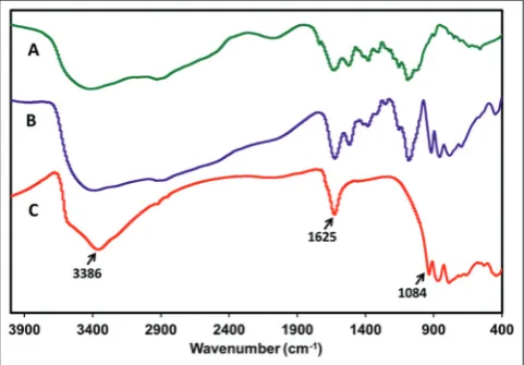
(Fig. 1) describes the FT-IR spectrum of bare CoW11CpTi, native
CSYC100 and CoW11CpTi -CSYC100 nanocomposites. FT-IR spectrum of CSYC100 is represented in Fig. 1a, which signifies all the characteristic peaks of chitosan at 3420/cm appeared, and this can be credited to the -NH2 and -OH groups stretching vibration and a peak at 1620/cm for
the amide similar to as characterized earlier [12-14]. FT-IR spectrum of bare CoW11CpTi denoted in Fig. 1c portrays characteristic peak sharp
absorption at 1620/cm. This feature is typical of the C-C stretching for ɳ5-C
5 H5 ligand attached to Ti11 which exhibit the presence of
cyclopentadienyl ligand.
The spectrum of CoW11CpTi-CSYC100 nanocomposite (Fig. 1b) shows
all the representative peaks existing in bare CoW11CpTi representing
successful complex development. Furthermore, the peak at 1060/cm corresponds to the bridge oxygen (C–O–C) stretching bands [12-14].
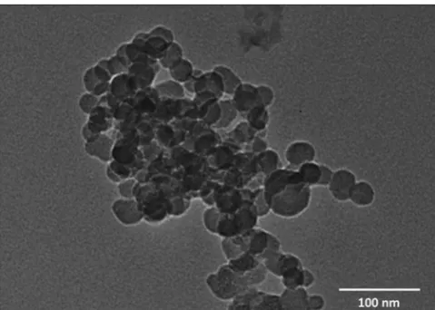
Fig. 2 represents the TEM images of CoW11CpTi -CSYC100
nanocomposite. It is a clear evident from the images that approximately all composites obtained by this technique were (1) monodispersed (2) possessed spherical shape morphology and (3) had sizes ranging from 35 to 80 nm in the diameter.
DLS analysis portrayed hydrodynamic size of the nanocomposites (Fig. 3). DLS analysis unveiled that 98% or more particles possess
Fig. 1: Comparative Fourier transformed infrared spectrum of (a) ChitosanYC-100 (CSYC100), (b) CoW11CpTi-CSYC100
hydrodynamic diameter size <100 nm (Number % distribution). CoW11CpTi-CSYC100 nanocomposite, size distribution of particles
ranges from 42.82 to 72.82nm, and 35.9% (highest) particles are of 50.75 d nm size.
Thermograms of CSYC100, CoW11CpTi-CSYC100 nanocomposite and
bare CoW11CpTi, shown in Fig. 4 describes that the thermal strength
of the nanocomposite is better when compared to the bare CSYC100. This may be credited due to the formation of a complex by CSYC100 with thermally stable CoW11CpTi having lesser degradation as those
are heavy metal salts. From the thermogram of CSYC100 in Fig. 4, the degradation observed up to 100°C can also be due to the loss in moisture and then polymer remains stable till 228°C as there is minimal change in the weight loss from 100 to 228°C. Afterward, an exponential degradation of was observed up to 380°C which may be due to degradation of CSYC100 polymeric structure.
In case of nanocomposite, thermogram showed the degradation starting at 180°C, further with a slow degradation till 320°C. The percentage weight residual at 900°C for the nanocomposite was more when related to CSYC100. Furthermore, the sharpness of the curve was reduced for nanocomposite, representing its debilitated degradation in comparison to that of bare CSYC100. CoW11CpTi exhibited a small
degradation due to the moisture loss at around 100°C. Further, there was no degradation observed up to 900°C which represents ∼90% of the particles did not undergo any degradation and stayed stable. Basically, the thermal stability of CSYC100 is significantly improved when developing composites with the thermally stable CoW11CpTi.
To study the release of CoW11CpTi from nanocomposites, lysozyme
was dissolved in PBS at pH 7.4, and this solution was used as release medium. Lysozyme was used as it enzymatically degradation of the glycosidic bonds in CSYC100 polymer. Lysozyme concentration was
adjusted to mimic the in vivo system. Here, lysozyme weakens the electrostatic interaction between CoW11CpTi and CSYC100 which
would cause its controlled release from composites. It was seen that there was a slow and steady release of CoW11CpTi for 11 hrs, with the
98% of collective release at the end (Fig. 5).
Results of MTT assay were performed to study the toxicity of bare CoW11CpTi and hybrid CoW11CpTi -CSYC100 nanocomposites on C2C12
and A549 cell line is represented in (Fig. 6). Concentrations of bare CoW11CpTi and CoW11CpTi-CSYC100 were varied from 0.019 mM to
5mM range. At very high concentration of 5 mM, CoW11CpTi and hybrid
CoW11CpTi-CSYC100 nanocomposite were toxic to the C2C12 and
A549 cells. However, there was significant variation in the percent (%) viability of C2C12 cells and A549 cells at their lower concentrations. Calculated IC50 values for bare CoW11CpTi and hybrid CoW11
CpTi-CSYC100 nanocomposite on C2C12 cells were 4.8 mM and 5.3 mM; similarly, IC50 values for these compounds on A549 were 0.9 and
4.9 mM, respectively. This show Hybrid POM was less toxic at higher concentrations as compared to bare POM. Furthermore, cancerous cells A459 can easily be inhibited by CoW11CpTi-CSYC100 nanocomposite at
very low concentrations where normal C2C12 cells remain viable and unaffected.
DISCUSSION
Initially, the research portrayed in this manuscript deals with the synthesis of CoW11CpTi. This desired POM was synthesized using the
Fig. 2: Transmission electron microscopy images of CpTi-ChitosanYC-100 nanocomposite
Fig. 3: Particle size distribution (number %) using dynamic light scattering analysis of CoW11CpTi-ChitosanYC-100 nanocomposite
Fig. 4: Comparative thermo gravimetric analysis of ChitosanYC-100 (CSYC100), CoW11CpTi-CSYC100 nanocomposite,
bare CoW11CpTi
Fig. 5: Release of CoW11CpTifrom CoW11CpTi-ChitosanYC-100
standard protocol reported by Wang et al. [8]. Successful synthesis of CoW11CpTi was confirmed by comparing the FT-IR spectra of the same POM originally synthesized by Wang et al. [8].
Once the desired POM (CoW11CpTi) was synthesized, its nanocomposite was synthesized. This is the first report for the synthesis of nanocomposites using P2W17Ni. To categorize any composite particles
as “nano” requires strict implementation on the limitation of the particle size which is to be <100 nm. Hence, the first important characterization was to study the size of the CoW11CpTi-CSYC100 formed. With the aid of particle size analysis by DLS and by TEM imaging, it was noted that the average size of CoW11CpTi-CSYC-100 composites was 50 nm. Thus, with this data, CoW11CpTi-CSYC100 composites can be classified as “nano” in nature [15]. For successful nanocomposite formation to have occurred, they were characterized for the linking of POM with CSYC100
using FT-IR spectral analysis where FT-IR spectrum signifies all the characteristic peaks of chitosan at 3420/cm appeared, and this can be credited to the -NH2 and -OH groups stretching vibration and a peak at 1620/cm for the amide similar to as characterized by Anitha et al. [1]; this proves that CSYC100 forms linkage with POM.
Thermograms of CSYC100, CoW11CpTi-CSYC100 nanocomposite,
and bare CoW11CpTi describes that the thermal strength of the
nanocomposite is better when compared to the bare gelator. This may be credited due to the formation of a complex by CSYC100 with thermally stable CoW11CpTi having lesser degradation as those are heavy metal salts similar results are also reported by Geisberger et al. [3].
Release of CoW11CpTi from CoW11CpTi-CSYC100 nanocomposite was
analyzed to understand the profile of POM release from nanocomposite, here it was observed that the release of POM occurred till 11 hrs under simulated physiological conditions. In other words, for 98% of POM to escape from the matrix of nanocomposites requires 11 hrs. This is greatly significant for drug development as POMs at high dosages are lethal of the cells of the body, so its sustained release is inevitable [8]. This can be seen when the cytotoxicity of bare CoW11CpTi and CoW11CpTi -CSYC100
nanocomposite was tested in vitro on normal myoblast cell lines C2C12 and A549 lung cancer cell line using MTT assay. Results on normal myoblast cell lines C2C12 are essentially important because it mimics the behavior of normal healthy cells of the body. It was observed that CoW11CpTi-CSYC100 nanocomposite was comparatively less toxic than
bare CoW11CpTi; this can be due to the phenomena of sustain release
of CoW11CpTi from the CSYC100 nanocomposites [6,8,10,13]. Despite
such positive results, extensive research is still needed to be done on model organisms to make highly specific and finely tuned POM baring nanocomposites as a futuristic medicine.
CONCLUSIONS
Under this study, cytotoxicity of an anticancer POM, CoW11CpTi was
reduced by preparing their nanocomposite using CSYC100. To start with, bare CoW11CpTi was prepared and their purity was analyzed using
FT-IR. This POM was then used to prepare nanocomposites namely CoW11CpTi-CSYC100 which was characterized using DLS, FT-IR, TEM,
and TGA analysis. The release profile of CoW11CpTi from CoW11
CpTi-CSYC100 nanocomposite was recorded at physiological pH conditions which showed CoW11CpTi was released from CSYC100 matrix at slow
rate, and thus, we concluded that the release was sustained. Finally, the cytotoxicity assays of CoW11CpTi-CSYC100 nanocomposite were
performed on normal myoblast cell lines C2C12 and A549 lung cancer cell line. CoW11CpTi -CSYC100 nanocomposite at the concentrations of
1.25 mM and lower did not exhibit toxic effect on C2C12 cells as 95% total C2C12 cell mass remained viable. While in case of A549 cells highest 5 mM concentration of bare CoW11CpTi is toxic to the cancer
cells and after encapsulation cell viability increases from 10% to 55%. Cytotoxicity assays show that CoW11CpTi in complex with CSYC100
had reduced toxic effect on C2C12 mouse myoblast cells. Thus, this study has designated the feasibility of consuming CoW11CpTi-CSYC100
chitosan nanocomposite for lesser toxic biomedicinal applications.
ACKNOWLEDGMENTS
Authors VP and SJ acknowledge Department of Science and Technology (DST), Ministry of Science and Technology, Government of India (GoI) for financial assistance through DST Fast-Track Scheme No. SR/FT/CS-133/2011.
REFERENCES
1. Anitha A, Rani VD, Krishna R, Sreeja V, Selvamurugan N, Nair SV,
et al. Synthesis, characterization, cytotoxicity and antibacterial studies of chitosan, O-carboxymethyl and N, O-carboxymethyl chitosan nanoparticles. Carbohydr Polym 2009;78(4):672-7.
2. Dolbecq A, Dumas E, Mayer CR, Mialane P. Hybrid organic − Inorganic
polyoxometalate compounds: From structural diversity to applications. Chem Rev 2010;110(10):6009-48.
Fig. 6: In vitro cytotoxicity investigation of CoW11CpTi and CoW11CpTi-ChitosanYC-100 nano-composite on (a) A549 (adenocarcinomic human alveolar basal epithelial
cells) and (b) C2C12 (mouse myoblast cell line) by 24 hrs [3-(4,5-dimethylthiazol-2-yl)-2,5-diphenyltetrazolium bromide]
assay. ns(p>0.05) non-significant as compared to control, *(p
between 0.05 and 0.01) signinificant at 5%, **(p between 0.01 and 0.001) significant at 1% as compared to control, ***(p<0.001)
significant at 0.1% as compared to control b
3. Geisberger G, Paulus S, Carraro M, Bonchio M, Patzke GR. Synthesis, characterisation and cytotoxicity of polyoxometalate/carboxymethyl chitosan nanocomposites. Chem Eur J 2011;17(16):4619-25.
4. Hasenknopf B. Polyoxometalates: Introduction to a class of inorganic
compounds and their biomedical applications. Front Biosci 2005;10(1-3):275-84.
5. Knoth WH, Domaille PJ, Farlee RD. Anions of the type (RMOH2)3W18P2O689- and [H
2OCo]3W18P2O6812-. A reinvestigation of
“B,β-W9PO349-”. Organometallics 1985;4(1):62-8.
6. Kumar MN, Muzzarelli RA, Muzzarelli C, Sashiwa H, Domb AJ. Chitosan chemistry and pharmaceutical perspectives. Chem Rev 2004;104(12):6017-84.
7. Liu Y, Peterson DA, Kimura H, Schubert D. Mechanism of cellular 3-(4,5-dimethylthiazol-2-yl)-2,5-diphenyltetrazolium bromide (MTT) reduction. J Neurochem 1997;69(2):581-93.
8. Wang X, Liu J, Li J, Yang Y, Liu J, Li B, et al. Synthesis and antitumor activity of cyclopentadienyltitanium substituted polyoxotungstate [CoW11O39(CpTi)]7−(Cp= η 5-C 5 H 5). J Inorg Biochem
2003;94(3):279-84.
9. Rajitha P, Gopinath D, Biswas R, Sabitha M, Jayakumar R. Chitosan
nanoparticles in drug therapy of infectious and inflammatory diseases. Expert Opin Drug Deliv 2016;13:1177-94.
10. Prasanth N, Dilip C, Sanal D, Lis A, Saraswathi R. Evaluation of
in-vitro cytotoxic and antioxidant activities of Ipomoea batatas. Int J
Pharm Pharm Sci 2010;2(3):91-2.
11. Vijayabaskaran M, Venkateswaramurthy N, Arif Pasha MD, Babu G, Sivakumar P, Perumal P, Jayakar B. In vitro cytotoxic effect of ethanolic extract of Pseudarthria viscida Linn. Int J Pharm Pharm Sci
2010;2(3):93-4.
12. Rhule JT, Hill CL, Judd DA, Schinazi RF. Polyoxometalates in medicine. Chem Rev 1998;98(1):327-58.
13. Singh R, Lillard JW Jr. Nanoparticle-based targeted drug delivery. Exp Mol Pathol 2009;86(3):215-23.
14. Menon D, Thomas RT, Narayanan S, Maya S, Jayakumar R, Hussain F,
et al. A novel chitosan/polyoxometalate nano-complex for anti-cancer applications. Carbohydr Polym 2011;84(3):887-93.
![Fig. 6: In vitroand 0.001) significant at 1% as compared to control, ***(p<0.001) [3-(4,5-dimethylthiazol-2-yl)-2,5-diphenyltetrazolium bromide] between 0.05 and 0.01) signinificant at 5%, **(p between 0.01 assay](https://thumb-us.123doks.com/thumbv2/123dok_us/999945.1123733/4.595.44.289.69.504/vitroand-significant-compared-control-dimethylthiazol-diphenyltetrazolium-bromide-signinificant.webp)
