MEFENAMIC ACID-NICOTINAMIDE CO-CRYSTAL SYNTHESIZED BY USING MELT
CRYSTALLIZATION METHOD AND ITS SOLUBILITY STUDY
DWI UTAMI
1,2*, ILMA NUGRAHANI
1, SLAMET IBRAHIM
11Department of Pharmacochemistry, School of Pharmacy, Institute Technology Bandung, Lab Tech VII ITB, Ganesha 10, Bandung 40132,
Indonesia. 2Department of Pharmacochemistry, Faculty of Pharmacy, Ahmad Dahlan University, Jln. Prof. Dr. Soepomo, Yogyakarta.
Email: [email protected]
Received: 25 October 2016, Revised and Accepted: 10 February 2017
ABSTRACT
Objective: The focus of this study is to develop the formation mefenamic acid (MFA) - nicotinamide (NCT) co-crystal by using melt crystallization method and investigate its solubility.
Methods: Co-crystal was prepared by using melt crystallization method of MFA-NCT (1:2) at (220±3°C). The initial co-crystal formation was performed by powder X-ray diffraction (PXRD). The thin-layer chromatography (TLC) method was done to confirm the chemical stability of MFA and NCT due to synthesized process. The melt crystallization of MFA-NCT (1:2) was characterized by differential scanning calorimetry (DSC)/thermal gravimetric, infrared spectrophotometry, and scanning electron microscopy. The solubility of the melt crystallization of MFA-NCT (1:2) was evaluated by incubating the samples in water at 25°C and shaken for 24 hrs. The solubility of MFA was measured by ultraviolet-visible spectrophotometer.
Result: Characterization of a co-crystal MFA-NCT (1:2) including PXRD, Fourier transform infrared, DSC, and SEM have indicated the formation of new solid crystal phase that differs from MFA, NCT, and its physical mixture. The chromatogram of the TLC study exhibited two spot that corresponds to MFA and NCT. The solubility of the melt crystallization of MFA-NCT (1:2) was 57.97% higher than MFA solubility.
Conclusion: These results suggest that MFA-NCT co-crystal can be synthesized by using melt crystallization method without decomposition of its component and provides an opportunity for the development of MFA solid form.
Keywords: Co-crystal, Mefenamic acid, Nicotinamide, Melt crystallization, Solubility.
INTRODUCTION
Pharmaceutical co-crystals are defined as multiple component crystals in which at least one component is molecular and a solid at room temperature (the co-former) and forms a supramolecular synthon with a molecular or ionic active pharmaceutical ingredients (APIs) [1]. The new crystalline form may exhibit distinctive physicochemical properties and could in turn affect the dissolution, manufacturing, physical stability, permeability, and oral bioavailability of an API [2]. There are numerous co-crystals that have an improvement in their physiochemical properties as advantages in manufacturing such as carbamazepine-cinnamic acid co-crystal (dissolution properties); apovincamine-terephthalic acid co-crystal (stability); and paracetamol-dipicolinic acid co-crystal (tabletability) [3-5].
Co-crystal can be defined as crystalline complexes of two or more neutral molecular constituents bond together in the crystal lattice through noncovalent interaction, primarily hydrogen bond [6]. In crystal engineering, crystals can be viewed as supramolecules consisting of superatoms bound together with intramolecular interactions instead of covalent bonds. Supramolecular synthons are defined as structural units within supermolecules which can be formed and/or assembled by known or conceivable synthetic operations involving intramolecular interaction [7]. The key in designing co-crystals is choosing a synthon, which is likely to form a crystallization process.
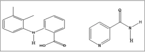
One of such potential APIs was identified to be mefenamic acid (MFA), an anthranilic acid and non-steroidal anti-inflammatory drugs with anti-inflammatory, antipyretic, and analgesic activities. MFA is a Biopharmaceutics Classification System class II drug with low solubility and high permeability [8]. MFA has the chemical name
2-((2,3-dimethylphenyl)amino)benzoic acid with molecular weight 241.28 g/mol. MFA has 2 hydrogen donor and 3 hydrogen bond acceptor based as computed properties [9]. Structure of MFA is shown in Fig. 1. In the term co-crystal design, this property is useful to construct the hydrogen bond with co-former as the heterosynthon supramolecular interaction.
Chemically, MFA has a carboxylic acid functional group and amine functional group. In the supramolecular architecture of co-crystal design, MFA may exhibit a carboxylic acid-amide heterosynthon or carboxylic acid dimer homosynthon. In the Cambridge Structural Database, a carboxylic acid-amide is more favorable than carboxylic acid dimer homosynthon [10,11]. Nicotinamide (NCT) is one of the well-known co-crystal formers with carboxylic acid. The amide synthon in the chemical structure of NCT is a functional group that is responsible for co-crystallization forming of APIs such as carbamazepine, indomethacin, and artesunate [12-14]. The chemical structure of NCT is shown in Fig. 1.
© 2017 The Authors. Published by Innovare Academic Sciences Pvt Ltd. This is an open access article under the CC BY license (http://creativecommons. org/licenses/by/4. 0/) DOI: http://dx.doi.org/10.22159/ajpcr.2017.v10i5.15863
Research Article
There were several co-formers that have been used to form supramolecular heterosython with MFA, such as NCT, isonicotinamide, nicotinic acid, urea, oxalic acid, 4-aminobenzoic acid, 2-aminobenzoic acid, pyridoxine, isonicotinic acid hydrazide, 4,4’-bipyridyl, 2,2’-bipyridyl, and 2-aminopyride [15]. The successful MFA co-crystal formation was performed by 4,4’-bipyridyl by solvent evaporation
method and NCT by neat grinding method [16]. In the pharmaceutical
solid form development, NCT is more suitable to be used as co-former than 4,4’-bipyridyl. Li et al. showed the neurotoxicity of 4,4’-bipyridyl compound [17]. To the best of our knowledge, only one reference involving the successful co-crystal of MFA with NCT has been reported until now, which is obtained by neat grinding method. The co-crystal of MFA-NCT by neat grinding method could only be prepared as polycrystalline powders, and the structure of its co-crystal was determined from powder diffraction data.
Therefore, the studies of MFAc-crystal with various co-formers especially NCT are still challenged to be explored. Although the MFA-NCT co-crystal have been reported before by neat grinding, but there were several limitations of neat grinding as co-crystal synthesis method. The disadvantages were that grinding methods often need special equipment to keep the grinding frequency, and the resulting product often contains amorphous impurities [18]. On the other hand, as reported by Ulrich [19], the melt crystallization method has a positive aspect in high selectivity to obtain the more pure product. In this study, we reported the melt crystallization method as the alternative method in MFA-NCT co-crystal synthesis and also its solubility study. Physicochemical characterization was performed by powder X-ray diffraction (PXRD), Fourier transform infrared spectroscopy (FTIR), thermal analysis differential scanning calorimetry/thermal gravimetric analysis (DSC/TGA), and scanning electron microscope (SEM) observation. The chemical stability of starting component was also investigated by thin-layer chromatography (TLC) analysis to ensure that both MFA and NCT did not possess chemical decomposition during the heating process. The solubility was done by incubating the samples in water for 24 hrs. These co-crystals are expected to be more soluble in water than parent acids, due to the possibility of complex formation in solution and the higher solubility of NCT.
MATERIALS AND METHODS
Materials
MFA (2-((2,3-dimethylphenyl)amino) benzoic acid) (99.0% Pyridam), NCT (>99.5%, E-Merck) used without further purification.
Method
Co-crystal preparation
Physical mixing
Physical mixtures were obtained by gentle mixing of MFA and NCT in a 1:2 molar ratio in a glass mortar with a glass pestle for 1 minute.
Melt crystallization
The physical mixtures (2 g) of MFA and NCT (1:2 molar ratio) were heated in a porcelain dish on a paraffin oil bath maintained at 200°C. Molten mass was incubated in a vessel containing 25 ml water at 90°C using water bath. The obtained product was dried at room temperature overnight and further to be analyzed for its characterization.
Characterization of co-crystals
The pure drug, co-former, and the product obtained from melt crystallization were subjected to PXRD, TLC, FTIR, DSC/TGA, and SEM analyzed.
PXRD
The PXRD analysis was performed in Philips PW 1710 BASED and
illuminated with Cu-Kα radiation (λ=1.5418) at a tube voltage of 40 kV and a tube current of 35 mA. The samples were analyzed over a 2θ
range of 5-45° with increase of 0.019° at a rate of 7/minutes.
TLC analysis
TLC analysis of melt crystallization of MFA-NCT (1:2) was done for the identification of the parent compound decomposition as the result of melt crystallization process. The chloroform: Methanol (9:1) was designated as solvent system and the silica gel GF 254 nm as stationary
phase. The spot detection was recorded under ultraviolet (UV) 254 nm.
FTIR spectroscopy
The spectra of drug, co-former, physical mixtures, and the melt crystallization product were recorded in Infrared Spectrophotometer (Jasco-4200 type A) and KBr beam splitter. Potassium bromide pellet method was employed, and background spectrum was collected. The range was set from 450 to 4000 cm−1 with a 4 cm−1 resolution.
DSC
Thermal analysis of drug, co-former, physical mixtures and the melt crystallization product were recorded on a DSC (Linseis sta pt 1600 and Q-20 Thermal Analysis). Thermograph was recorded under a constant nitrogen gas flow of 50 mL/minutes. The DSC apparatus was calibrated with regard to temperature and enthalpy using indium as a standard. The heating was set to 10 K/minutes in a range from 30 to 250°C.
SEM
The surface and shape characteristic of pure MFA, NCT, and melt crystallization of MFA-NCT (1:2) were studied by SEM (JEOL JSM 6510). Powder samples were mounted onto aluminum stub using double-sided adhesive tape and sputter coated with a thin layer of gold at 5.6 Torr vacuum before examination. The samples were scanned by an
electron beam of acceleration potential of 10 kV.
Solubility study
An excess amount of MFA, physical mixture of MFA-NCT (1:2), and melt crystallization of MFA-NCT (1:2) were suspended in 10 ml of distilled water in capped glass vials, and the slurries were agitated in an orbital shaker (150 rpm/minutes) at room temperature for 24 hrs. After 24 hrs, the suspension was filtered through a paper filter, and the amount of drug solubilized was analyzed spectrophotometrically
(UV-1800, Shimadzu, Japan) at 330 nm. The solubility experiments
were conducted in triplicate.
RESULT PXRD analysis
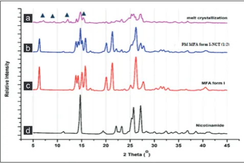
The result of PXRD analysis of MFA, NCT, its physical mixture, and melt crystallization product is shown in Fig. 2. The diffractogram of MFA
showed characteristic peak at 2θ value of 6.4°; 21.35°; and 26.21°, and NCT exhibited diffractogram peak 2θ at 14.7°; 22.1°; 23.3°; 25.7°;
and 27.2°. The physical mixture showed diffractogram combination of both starting components. The diffractogram of melt crystallization of
Fig. 2: The X-ray powder diffraction patterns of mefenamic acid (MFA) form I (a), nicotinamide (NCT) (b), physical mixture of MFA-NCT (c), and the melt crystallization of MFA-NCT (1:2) (d)
MFA-NCT showed new peaks at 2θ value of 7.2°; 9.3°; 12.1°; and 15.4°
(Fig. 2).
TLC analysis
The result of TLC analysis of the melt crystallization of MFA-NCT (1:2) exhibited two spot that corresponds to the spots of MFA and NCT pure compound (Fig. 3). The MFA eluated in Rf: 0.85, whether NCT at Rf: 0.68. Except the two spots above, there was not observed another spot of melt crystallization of MFA-NCT product.
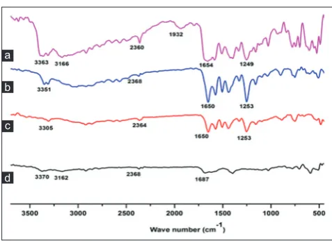
FTIR spectroscopy analysis
The FTIR spectrum of MFA showed characteristic peak with high intensity at 1650 cm−1, presenting C=O functional group (Fig. 4),
3305 cm−1 as vibration of N-H functional group and carboxylic acid at
1253 cm−1. The NCT spectrum exhibited the N-H stretching at 3370 cm−1
and 3162 cm−1 and C=O functional group at 1681 cm−1. The spectrum
of physical mixture showed characteristic peaks of MFA and NCT. The melt crystallization of MFA-NCT (1:2) exhibited spectrum at 1654 cm−1,
1249 cm−1, 3166 cm−1, 3363 cm−1, and two new bands at 2360 cm−1 and
1932 cm−1. In the fingerprint region (1500-500 cm−1), the result was
presented the same spectrum pattern.
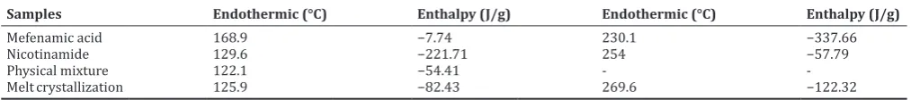
Thermal analysis
The thermal behavior of each sample (MFA, NCT, its physical mixture, and melt crystallization) was recorded by DSC-TGA. The MFA showed two endothermic peaks at 168.9°C and 230.1°C as MFA form I. NCT only showed an endothermic peak at 129.9°C as melting point. The physical
mixture of MFA-NCT (1:2) showed an endothermic peak at 122.1°C. The DSC parameters of MFA, NCT, its physical mixture, and the melt crystallization of MFA-NCT (1:2) are presented in Fig. 5 and Table 1.
SEM
SEM analysis was performed for the pure MFA, co-former NCT, and the melt crystallization of MFA-NCT (1:2). The pure MFA exhibited cube shape with rough surface; the co-former NCT showed cube shape with long size and smooth surface. The melt crystallization of MFA-NCT (1:2) presented a cube shape with smooth surface and showed reducing of particle size (Fig. 6).
Solubility study
The solubility test was done with the standard pure MFA, the physical mixture of MFA-NCT (1:2), and the melt crystallization of MFA-NCT (1:2) by dissolving the samples in the distilled water equipped with an orbital shaker at room temperature and was done in triplicate. The results from the solubility test were then measured with a
spectrophotometer UV-visible absorbance maximum of 332 nm. The
results are shown in Table 2.
DISCUSSION
The new crystalline phase of interaction experiment between two components can be identified by PXRD method. If the new crystalline phases are formed from the interaction between the two components [20], it will show clearly from X-ray diffractogram. The different diffractogram pattern showed differences in the character of the crystal lattice. By comparing the diffractogram pattern of its starting components, its physical mixture, and the product of co-crystallization experiment, the new solid phase formation can be adjusted [21,22].
Fig. 3: Thin-layer chromatography profile of mefenamic acid (MFA), nicotinamide (NCT), physical mixture of MFA-NCT (1:2),
and the melt crystallization of MFA-NCT (1:2)
Fig. 5: The differential scanning calorimetry profiles of mefenamic acid (MFA) form I, nicotinamide (NCT) and the melt
crystallization of MFA-NCT (1:2) (a) and thermal gravimetric analysis result of the melt crystallization of MFA-NCT (1:2) (b)
b a
Fig. 6: The scanning electron microscope of mefenamic acid (MFA) (a), nicotinamide (NCT) (b), and melt crystallization of
MFA-NCT(1:2) (c)
c
b a
Fig. 4: The Fourier transform infrared pattern of nicotinamide (NCT) (a), mefenamic acid (MFA) form I (b), physical mixture
of NCT (1:2) (c), and the melt crystallization of MFA-NCT(1:2) (d)
This result was relevant to MFA-NCT co-crystal diffractogram reported by Fabian et al., 2011, that have been synthesized by the neat grinding method. Based on PXRD analysis, the melt crystallization of MFA-NCT (1:2) exhibited a co-crystal formation.
Furthermore, the co-crystal formation of melt crystallization of MFA-NCT (1:2) confirmed by several analytical methods such as FTIR spectrophotometry, thermal analysis (DSC-TGA), SEM, and also the TLC method for the possibilities of chemical decomposition of starting component as heating process consequences.
In the pharmaceutical co-crystal term, the interaction between API and co-former was only exhibiting the hydrogen bond or noncovalent bond that can be separated as a parent compound during the dissolution process [23]. The new compound forming was not being expected in co-crystallization process. In some case, the melt crystallization method triggered the chemical decomposition and resulted in the new compound that differs with parent compound. Nugrahani et al. reported the chemical decomposition of MFA with oxalic acid during the melting process [24].
As shown in Fig. 3, the TLC analysis of the melt crystallization product resulted two spot that corresponds to MFA and NCT. This result was indicated that MFA and NCT did not possess chemical decomposition or other chemical reaction during heating process. This finding was also supported by FTIR spectrum of the melt crystallization product as shown in the fingerprint region. The importance of the fingerprint region is that each different compound produces a different pattern of troughs in this part of the spectrum [25]. As mentioned in the FTIR result, in the fingerprint region, all samples presented the same spectrum pattern.
Despite that, the FTIR analysis was also used as co-crystal analytical method for co-crystal formation due to the formation of the hydrogen bond between API and former. The hydrogen bond of API and co-former is identified as the result of synthon-synthon interaction. The possibilities of the hydrogen bond formation in the melt crystallization of MFA-NCT (1:2) can be identified by FTIR and compared to the starting components and their physical mixture.
In the melt crystallization of MFA-NCT (1:2), there were occurred
three major wavenumber shifting of FTIR spectrum. First, the ν(C=O)
stretching band of MFA (1650 cm−1) and NCT (1681 cm−1) were fused
and appeared in the co-crystal spectrum at 1654 cm−1. The fused and
shifting of C=O was due to the possibilities of hydrogen bond formation between the C=O of MFA and the NH2 of NCT. Second, there was shifting from N-H vibration of NCT to lower frequency from 3370 cm−1 to
3363 cm−1 and from 3162 cm−1 to 3166 cm−1. And the third, there was
also the new band spectrum at 2360 cm−1 and 1932 cm−2. The new
band spectrum was also reported by Neurohr et al in the formation of naproxen-nicotinamide co-corystal. The new band spectrums were appeared in 2525 cm-1 and 1982 cm-1 and adjusted as O-Hcarboxylic acid Naromatic hydrogen bond [26]. According to this report, the present new band in melt crystallization of MFA-NCT (1:2) was also predicted as hydrogen bond formation of O-Hcarboxylic acid of MFA and Naromatic of NCT. Based on FTIR analysis, there was predicted the co-crystal formation of MFA and NCT as indicated by hydrogen bond occurrences.
Moreover, the thermal behavior of each sample was identified by thermogram analysis. The different melting transition from individual components suggests the formation of co-crystal [27]. As shown in Fig. 5, the new endothermic peak of melt crystallization of MFA-NCT (1:2) was presented at 125.9°C, lower than melting point of each component and higher than its physical mixture. Although the thermal pattern was similar to its physical mixture (mp: 122.1°C), the energy used was quite different as shown in Table 1 and Fig. 5a and b. The average endothermic energy of melt crystallization of MFA-NCT was shown to be higher than its physical mixture. The higher endothermic energy indicated that the melt crystallization product was more crystalline than its physical mixture. The thermogravimetric analysis showed that the melt crystallization of MFA-NCT (1:2) was stable up to 200°C before it was decomposed (Fig. 5b). A single endothermic transition of the melt crystallization of MFA: NCT (1:2) demonstrated that the new solid phase has been formed as co-crystal form, stable below its melting points and also the absence of unbound or absorbed solvent and water [28]. The presence of absorbed solvent and water in the co-crystal can influence the stability of co-crystal during manufacturing process or storage. In the other words, this co-crystal was very potentially to be developed in a solid dosage form.
As the prosperity of co-crystal formation, we evaluated the solubility of MFA-NCT co-crystal in distilled water. The solubility study was also done in pure MFA and its physical mixture. The solubility of the melt crystallization of MFA-NCT (1:2) was differed significantly to pure MFA and its physical mixture. The solubility of melt crystallization of MFA-NCT (1:2) was 57.97% higher than pure MFA. The solubility of pure MFA was lower than the melt crystallization of MFA-NCT (1:2) suggested because of the formation of hydrogen bond between MFA and NCT that increased the solubility of MFA. From the SEM analysis (Fig. 6), there was observed the smaller particle size of melt crystallization of MFA-NCT (1:2). Reducing of particle size was also contributing to the higher solubility of the melt crystallization of MFA-NCT (1:2). The other factor that influences the solubility of APIs was a decrease in energy of crystal lattice by co-crystal formation such as in paclixacel-naringenin and simvastatin-tartaric acid co-crystal formation [29,30].
Based on the result and discussion mentioned above, the co-crystal of MFA-NCT has been successfully synthesized by melt crystallization. The future development is exploring the physicochemical changes that influence in MFA manufacturing such as dissolution rate and also its stability. This finding is very useful in the development of MFA solid dosage form that has better physicochemical properties.
Table 2: The solubility of pure MFA, physical mixture of MFA-NCT (1:2), and the melt crystallization of MFA-NCT (1:2) in
distilled water
Samples Concentration (µg/mL)
1 2 3 Mean±SD
Pure MFA 6.65 6.83 6.35 6.62±0.23
Physical mixture of
MFA-NCT (1:2) 7.35 7.18 7.52 7.35±0.17
The melt crystallization of
MFA-NCT (1:2) 10.69 10.06 10.58 10.45±0.34
SD: Standard deviation, DSC: Differential scanning calorimetry,
NCT: Nicotinamide, MFA: Mefenamic acid, The data shown as mean±SD, n=3
Table 1: The DSC parameters of MFA, NCT, its physical mixture, and the melt crystallization of MFA-NCT (1:2)
Samples Endothermic (°C) Enthalpy (J/g) Endothermic (°C) Enthalpy (J/g)
Mefenamic acid 168.9 −7.74 230.1 −337.66
Nicotinamide 129.6 −221.71 254 −57.79
Physical mixture 122.1 −54.41 -
-Melt crystallization 125.9 −82.43 269.6 −122.32
CONCLUSION
This study first reports that the co-crystal of MFA-NCT (1:2) has been synthesized successfully by using melt crystallization method. The solubility evaluation of MFA-NCT co-crystal (1:2) had increased significantly compared to MFA and its physical mixture. The confirmations against co-crystal MFA-NCT (1:2) had indicated the formation of new solid crystalline phases that differ from MFA and its physical mixture.
REFERENCES
1. Qiao N, Li M, Schlindwein W, Malek N, Davies A, Trappitt G. Pharmaceutical cocrystals: An overview. Int J Pharm 2011;419(1‑2):1‑11.
2. Blagden N, de Matas M, Gavan PT, York P. Crystal engineering of active pharmaceutical ingredients to improve solubility and dissolution rates. Adv Drug Deliv Rev 2007;59(7):617‑30.
3. Shayanfar A, Zeynali KA, Jouyba A. Solubility and dissolution rate of a carbamazepine‑cinamic acid cocrystal. J Mol Liq 2013;187:171‑6. 4. Ma YH, Ge SW, Wang W, Sun BW. Studies on the synthesis, structural
characterization, Hirshfeld analysis and stability of apovincamine (API) and its co‑crystal (terephthalic acid: Apovincamine = 1:2). J Mol Struct 2005;1097:87‑97.
5. Hendrawan S, Veriansyah B, Widjojokusumo E, Soewandhi. SN, Wikarsa S, Thandrawinata RR. Simultaneous cocrystallization and micronization of paracetamol‑dipicolinic acid cocrystal by supercritical antisolvent (SAS). Int J Pharm Pharm Sci 2016;8(2):89‑98.
6. Horst JH, Deij MA, Cains PW. Discovering new cocrystals. Cryst Growth Des 2009;9(3):1531‑7.
7. Desiraju GR. Supramolecular synthon in crystal engineering‑a new organic synthesis. Angew Chem Int Ed Engl 1995;34:2311‑27. 8. Dixit M, Kini AG, Kulkarni PK. Enhancing the dissolution of
polymorphs I and II of mefenamic acid by spray drying. J Pharm Sci 2012;9(1):13‑26.
9. Mefenamic Acid, Pubchem. Available from: https://www.pubchem. ncbi.nlm.nih.gov/compound/45039685.
10. Nagia A, Desiraju GR. Supramolecular synthon and pattern recognition. Top Curr Chem 1998;198:57‑95.
11. Aakeroy CB, Salmon DJ. Building co‑crystals with molecular sense and supramolecular sensibility. CrystEngComm 2005;7:439‑48. 12. Shekh AY, Rahim SA, Hammond RB, Roberts KJ. Scalable solution co‑
crystallization: Case of carbamazepine‑nicotinamide. CrystEngComm 2009;11:501‑9.
13. Lin HL, Zhang GC, Huang YT, Lin SY. An investigation of indomethacin‑nicotinamide cocrystal formation induced by thermal stress in the solid or liquid state. J Pharm Sci 2014;103(8):2386‑95. 14. Setyawan D, Wardhana NK, Sari R. Solubility, dissolution test and
antimalarial activity of artesunate nicotinamide co‑crystal prepared by solvent evaporation and slurry methods. Asian J Pharm Clin Res 2015;8(2):164‑6.
15. Wittering KE, Agnew LR, Klapwijk AR, Robertson K, Cousen JP, Cruickshank, et al. Crystallization and physochemical property characterization of conformationally‑locked co‑crystals of fenamic acid derivates. CrystEngComm 2005;17:3610‑8.
16. Fabian L, Hamill N, Eccles KS, Moynihan R, McCausland L, Lawrence SE, et al. Cocrystal of fenamic acids with nicotinamide. Cryst Growth Des 2011;11:3522‑8.
17. Li S, Crooks PA, Wei X, de Leon J. Toxicity of dipyridyl compounds and related compounds. Crit Rev Toxicol 2004;34(5):447‑60.
18. Yan Y, Chen JM, Lu TB. Thermodynamic and preliminary pharmaceutical characterization of a melatonin‑pimelic acid cocrystal prepared by a melt crystallization method. CrystEngComm 2015;17:612‑20.
19. Ulrich J. Is melt crystallization a green technology? Cryst Growth Des 2004;4(6):879‑80.
20. Cherukuvada S, Row TN. Comprehending the formation of eutectic and cocrystals in terms of design and their structural interrelationships. Cryst Growth Des 2014;14(8):4187‑98.
21. Shanpui P, Gound NR, Khandavilli UB, Nangia A. Fast dissolving curcumin cocrystal. Cryst Growth Des 2011;11(9):4135‑45.
22. Parmar VK, Shah SA. Hydrochloride salt co‑crystals: Preparation, characterization and physicochemical studies. Pharm Dev Technol 2013;18:443‑53.
23. Shan N, Zaworotko MJ. The role of cocrystals in pharmaceutical science. Drug Discov Today 2008;13(9‑10):440‑6.
24. Nugrahani I, Ibrahim S, Puspita DD. The blue crystal of 2,3 dimethyl phenylalanine as chemical solid state interaction of mefenamic acid and oxalic acid. Math Sci J 2012;17(3):98‑104.
25. Tommasini M, Lucotti A, Alfè M, Ciajolo A, Zerbi G. Fingerprints of polycyclic aromatic hydrocarbons (PAHs) in infrared absorption spectroscopy. Spectrochim Acta A Mol Biomol Spectrosc 2016;152:134‑48.
26. Neurohr C, Revelli AL, Billot P, Marchievie M, Lecomte S, Laugier S, et al. Naproxen‑nicotinamide cocrystals produced by CO2 antisolvent. J Supercrit Fluids 2013;83:78‑85.
27. Lu EN, Rodriquez‑Hornedo N, Suryanarayanan R. A rapid thermal method for cocrystal screening. CrystEngComm 2008;10(6):665‑8. 28. Lu J, Rohani S. Synthesis and preliminary characterization of
sulfamethazine‑theophylline co‑crystal. J Pharm Sci 2010;99(9):4042‑7. 29. Muddukrishna BS, Swapnil JD, Shenoy GG, Khishnamurthy B.
Preparation, solid state characterization of paclitaxel and naringenin co‑crystals with improved slubility. Int J Appl Pharm 2016;8(4):32‑7. 30. Sopyan I, Fudholi A, Muchtaridi M, Puspitasari I. A simple effort