BRUCELLAR UVEITIS IN EASTERN INDIA
ANINDITA SEN
1, PARTHAJIT BANERJEE
1, DEVARATI DUTTA
2, MANAS KUMAR PAL
3, ATANU RAY
1, SATADAL DAS
2*
1Department of Microbiology, M G M Medical College, Kishanganj, Bihar, India. 2Brucella Research Lab, Peerless Hospital and B K Roy Research Centre, Kolkata, West Bengal, India. 3Department of Ophthalmology, Medical College, Kolkata, West Bengal, India.Email: [email protected]
Received: 16 July 2016, Revised and Accepted: 26 July 2016 ABSTRACT
Objective: Brucellosis, one of the major zoonotic diseases, still remains an uncontrolled problem, in regions of high endemicity. Ophthalmic brucellosis is not studied and overlooked in most developing countries. Considering the severe outcome of undiagnosed ophthalmic brucellosis, in this paper, we made attempts to find out whether this disease still remains a health problem in a South East Asian developing country, where the study of this disease is largely neglected.
Methods: This study was carried out over a 1-year period from January 2015 to December 2015. Blood samples were collected from clinically confirmed cases of uveitis, and they were subjected to five serological and one genus specific molecular investigations for the detection of Brucella
infection.
Results: Out of 20 uveitis cases, 4 (20%) cases confirmed as brucellar uveitis, by serological tests followed by polymerase chain reaction confirmation. After treatment of brucellosis, all the four patients were recovered uneventfully.
Conclusion:Brucella infection involving the eye is still a significant problem in South East Asian countries; hence in all uveitis cases in this reason brucellosis should be excluded by available laboratory tests.
Keywords: Brucellosis, Uveitis, Zoonotic diseases, Standard tube agglutination test, Polymerase chain reaction.
INTRODUCTION
Brucellosis is a zoonotic disease found worldwide. Although it has been eradicated in most developed countries, it still represents an important health problem in many parts of the world including Western part of Asia, Middle East, the Mediterranean, Central and South America [1-3]. In some countries such as Peru, Kuwait, and Saudi Arabia, brucellosis is endemic [3,4]. The rationale of this study is that in South East Asian countries problem of brucellosis is largely unknown, and reports of brucellar uveitis are practically missing; thus, this study has been undertaken to reveal the real scenario of brucellar uveitis in India.
METHODS
The study was carried out over a 1-year period from January 2015 to December 2015, in a tertiary care teaching hospital in Eastern India. After getting permission from Institutional Ethical Committee, 20 clinically confirmed patients with uveitis were included in this study, attaining informed consents from them. Blood samples were collected from them for serological and polymerase chain reaction (PCR) tests. Conjunctival swabs were also collected as a routine procedure to know the presence of any pathogenic microbial flora on the eye surface including Brucella. After collection, the swabs were inoculated immediately onto nutrient agar, blood agar, Sabouraud dextrose agar with chloramphenicol (SDC) slants, and in biphasic Castaneda medium. Cultures on nutrient agar and blood agar were observed after overnight incubation at 37°C to find presence of any common pathogenic bacteria; cultures on SDC were incubated at 25°C in BOD incubator and observed up to 21 days; while cultures in Castaneda media were observed for any growth of Brucella up to 21 days post-inoculation, and in subcultures on
Brucella selective agar. All isolates were identified by routine diagnostic procedures. Serum samples were kept in separate aliquots and stored at −20°C before further processing. Each sample of serum was
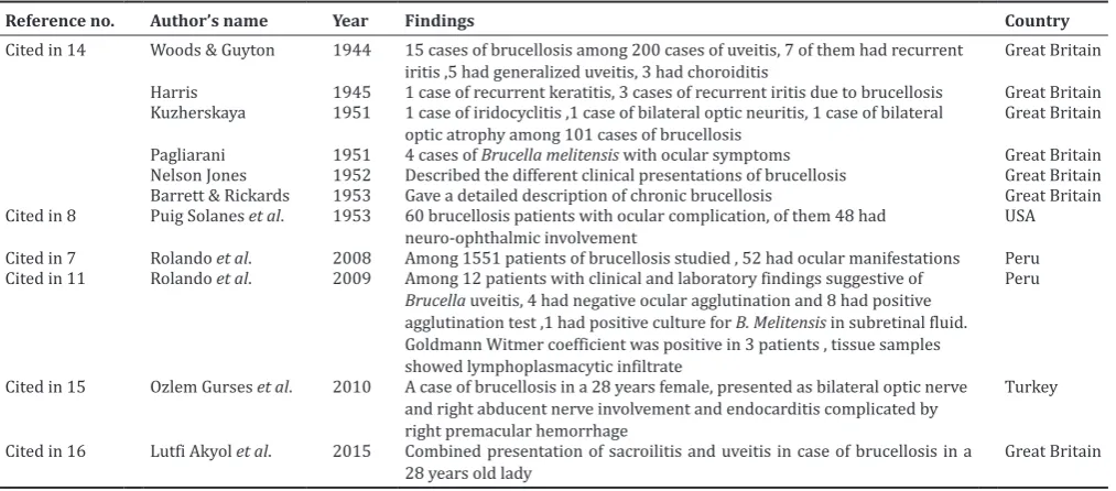
subjected to a panel of Brucella tests - Rose Bengal Plate Agglutination Test (IAHVB, Bengaluru), standard tube agglutination test (SAT; Tulip Diagnostics Pvt. Ltd.), ELISA (Immunolab GmbH, Germany) for the detection of IgM and IgG antibodies and genus-specific PCR (prime). For PCR tests, the serum samples were first subjected to DNA extraction using QIAmp DNA Mini Kit (Qiagen). The extracted DNAs were then subjected to PCR. The PCR was carried out in 50 µL reaction mixture in each PCR tube that contained 5 µL PCR buffer, 1 µL dNTP, 0.2 µL Taq Polymerase, 5 µL template DNA, 1 µL each of forward (F) and reverse (R) primers, and 36.8 µL nuclease free water. The primer sequences used were as follows [5]-BCSP-B4 (F) TGG CTC GGT TGC CAA TAT CAA; BCSP-B5 (R) CGC GCT TGC CTT TCA GGT CTG; amplicon size was 223 bp. The steps in PCR were followed as described by Baily et al. [5]. The amplified products were then subjected to agarose gel electrophoresis using a 100 bp DNA ladder and a positive and a negative control and the bands formed (Figs. 1-3) were seen in a gel doc (Biorad). Patients showing positive results with Brucella specific tests were also subjected to the following routine investigations - Mantoux test, X-ray chest posteroanterior view, toxoplasma IgM and IgG antibodies, rheumatoid arthritis factor, and treponema antibody rapid plasma reagin tests.
RESULTS
Of the 20 blood samples collected from clinically confirmed cases of uveitis, four samples (20%) were found positive for brucellosis. All other investigations in these four cases were not suggestive of any other associated disease.
Case 1 (Fig. 4)
SP, F, Hindu, 40 years, presented with 1 ½ years history that started with the sudden onset of redness and pain in the right eye, which subsided on routine medication that was subsequently followed by a recurrence of similar symptoms with bilateral involvement of both
Research Article
eyes. Associated with eye involvement, the patient also gave a history of fever that subsided on medication and was of remittent type. The patient was treated for ocular manifestations with oral and topical steroid, and other supportive treatments, which were discontinued following remission of ocular symptoms. The patient used to remain asymptomatic for 3/4 months at a time. Ophthalmological findings are given in Table 1. The patient’s serum was subjected to serological tests and showed positive Brucella SAT with a titer of 1:320 along with a positive Brucella IgM ELISA test. PCR result showed a positive band for
Brucella. The culture was negative.
Case 2 (Fig. 5)
PM, F, Hindu, 45 years, a known case of uveitis, presented with a 1-year history of recurrent bilateral redness, pain and watering from eyes and blurring of vision. The patient had 3-4 recurrences during the year. Symptoms subsided temporarily following routine treatment. On presentation, her acute inflammatory stage had subsided to some extent with previous conservative medication. The patient was from rural
background and gave history of rearing cattle at home and consumption of unpasteurized milk. The patient had two episodes of fever during this year that subsided with medications. Ophthalmological findings are given in Table 1. The patient’s serum showed a positive Brucella SAT with a titer of 1:160 and positive Brucella IgM ELISA test. PCR showed a positive band for brucellosis. Culture showed negative result.
Case 3 (Fig. 6)
HA, F, Muslim, 45 years, attended outpatient department with redness, watering, and pain of left eye 2-3 times over the last 1 year. The patient gave history of similar episodes in the left eye 4-5 times over the last 3 years. Each time, symptoms subsided with the use of medications. The patient also gave history of occasional fever that subsided with treatment after consulting physician. Ocular symptoms subsided with the use of oral and topical steroids, and topical antibiotics (moxifloxacin), topical timolol maleate, and atropine. Ophthalmological findings are given in Table 1. The patient’s serum showed a positive
Brucella SAT with a titer of 1:80 and a positive Brucella IgM ELISA test. PCR showed a positive band for brucellosis. Culture showed negative result.
Case 4 (Fig. 7)
AM, M, Muslim, 36 years, was a known case of bilateral panuveitis. He responded well to treatment (subtenon triamcenolone acetonide injection). He gave a history of recurrent attack of redness of both eyes and dimness of vision for the last 2 years and also the history of 5-6 episodes of recurrences during that period. The patient also gave a history of occasional fever of remittent type, when these symptoms started 2 years back. Ocular symptoms subsided well each time with oral and topical steroids, topical cycloplegic, topical antibiotic, and subtenon injection of triamcenolone acetonide. Ophthalmological findings are given in Table 1. The patient’s serum showed a positive
Brucella SAT with a titer of 1:80 and a positive Brucella IgM ELISA test. PCR showed a positive band for brucellosis. Culture showed negative result.
Considering the positive serological and molecular biological tests (Table 2) and excluding other causes of uveitis, these patients were diagnosed as having brucellosis. Thus in our study, we found four confirmed cases of brucellosis with the ocular presentation as chronic anterior and intermediate uveitis.
Fig 1: Gel picture represents two positive cases of uveitis with brucellosis in lanes 4 and 7. Lane 1 - Represents 100 bp DNA
ladder. Lane 2 - Positive control. Lane 3 - Negative control. Lanes 4 to 11 - Patients’ samples
Fig. 2: Gel picture represents two positive cases of uveitis with brucellosis in lanes 4 and 12. Lane 1 - Represents 100 bp DNA ladder. Lane 2 - Positive control. Lane 3 - Negative control.
Lanes 4 to 12 - Patient’ samples
Fig. 3: Gel picture three negative cases. Lane 1 - 100 bp DNA ladder, Lane 2 - Positive control, Lane 3 - Negative control,
Lanes 4 to 6: Patient samples
Fig. 4: Picture of the affected eye of case 1
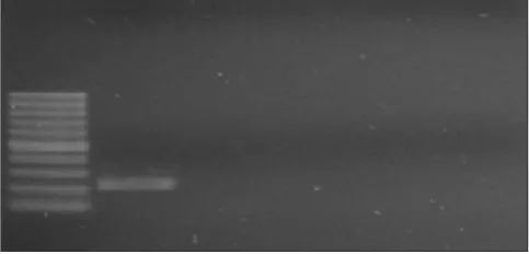
Table 1: Clinical ophthalmological findings of the four Brucella positive uveitis cases
SP, F, Hindu, 40 years PM, F, Hindu, 45 years HA, F, Muslim, 46 years AM, M, Muslim, 36 years
Right eye Left eye Right eye Left eye Right eye Left eye Right eye Left eye
V/A
(visual acuity) 6/36 6/12 6/60 Finger count 2 feet 6/9 6/60 6/60 6/60
Slit lamp examination
1. Lid Normal Normal Normal Normal Normal Normal Normal Normal
2. Conjunctiva Circumcornea l
congestion No congestion Congestion subsided Congestion subsided Normal Congested, circumcorneal congestion present
No congestion Mild congestion
3. Cornea Few KPs on the
endothelium Very old KPs on the corneal endothelium
No oedema
few old KPs Few old KPs Clear Mild oedema present, KPs seen over
Chamber Cells+2, Flair+1, Iris visible AC Normal, no cells and flare
No cell no
flair No cell no flair Normal, no cell, no flair Cells present (plenty), flair present
synechia Normal circular Pharmacol ogically mid dilated and
phakia Cataractous changes Early cataractous change
IOP Normal Normal Normal Normal 18 mmHg 30 mmHg on
the day of examination
18 mmHg 18 mmHg
Fundus
examination Retina, disc and macula visible and normal, mild Lacrimal Sac No regurgitation No
regurgitation No regurgitation Normal No abnormality detected
No abnormalit y
detected No Abnormality detected
No abnormality detected
KPs: Keratic precipitates
Fig. 6: Picture of the affected eye of case 3
Before diagnosis of brucellosis, the patients were initially given symptomatic management for uveitis, with topical steroid eye drop prednisone acetate 1% and topical antibiotic eye drop tobramycin 0.3% and mydriatic cum cycloplegic eye drop atropine 1%, (one patient needed timolol maleate eye drop so as to prevent rise of intraocular pressure, sometimes they needed oral steroid and periocular steroid injection) and responded well. Subsequently following the diagnosis of brucellosis, they were given specific treatment with oral doses of doxycycline 100 mg along with rifampicin 300 mg both twice daily continued for 6 weeks. The patients were followed up at intervals of 2-3 weeks and showed signs of improvement.
DISCUSSION
A high index of clinical suspicion coupled with appropriate diagnostic tests can detect ophthalmic brucellosis at an early stage. Brucellosis may
manifest as an acute or chronic disease. It has a wide range of clinical manifestations, which makes it diagnostically challenging. Sometimes, it is easy to identify the classical symptoms of brucellosis. However, some manifestations such as ocular brucellosis are difficult to identify. Ocular manifestations of brucellosis may be in the form of dacryoadenitis, conjunctivitis, episcleritis, keratitis, iritis, iridocyclitis, neuroretinitis, retinitis, chorioiditis, panuveitis, pars planitis, and hyalitis. The clinical symptoms and signs may include injection, blurred vision, eye pain, watering, diplopia, foreign body sensation, cotton wool lesions, exudative retinal detachment, and retinal hemorrhage [6-10]. It can only be detected if it is kept in the differential diagnosis along with other diseases. This will lead to early diagnosis and treatment and will, in turn, reduce the number of complications arising out of delayed diagnosis of the disease [6]. Lemaire in 1924, made the first diagnosis of ophthalmic brucellosis [7].
Two explanations may be there for the pathogenesis of ophthalmic brucellosis-direct invasion of Brucella and immune complex dependent pathogenesis [6,8,11]. In 2008, Rolando et al. reported about two different manifestations of brucellosis, ophthalmologic and neuro-ophthalmologic types [7]. During the long period from January 1980 to December 2005, 1551 brucellosis patients were studied by them, and 52 patients were diagnosed as having ocular brucellosis [7]. In 1953, Puig Solanes et al. identified 60 patients with ophthalmic complications of brucellosis and in them 48 patients had neuro-ophthalmic involvement [8]. In most of the studies, it has been found that uveitis is the most common ocular manifestation of brucellosis and that posterior uveitis is the most common form of uveitis [7,8,11,12]. Ocular manifestations are mainly seen during the chronic phase of the disease [7,8,13-16]. Several studies on brucellosis from Eastern India [17-21] indicated that it is prevalent Table 2: Diagnostic tests done for 20 patients
Serological tests Molecular
test Conjunctival swab culture S. No. Age
years Sex SAT titre SAT titre with 2 ME RBPT ELISA PCR N agar Blood agar Castaneda Brucella selective agar
SDC
1 50 Male 1⁄80 1⁄160 ₊ - - S. aureus S. aureus - -
-2 30 Male 1⁄40 1⁄80 - - -
-3 27 Female 1⁄20 1⁄40 - - -
-4 29 Male 1⁄80 1⁄160 ₊ - - S. pyogenes S. pyogenes - -
-5 52 Female 1⁄40 1⁄80 - - -
-6 40 Female 1⁄320 1⁄640 ₊ ₊IgM ₊ - - - -
-7 61 Male 1⁄40 1⁄80 - - -
-8 32 Female 1/80 1⁄160 - - -
-9 55 Male 1⁄20 1⁄40 - - - S. aureus S. aureus - -
-10 36 Male 1⁄80 1/160 - ₊IgM ₊ - - - -
-11 44 Female 1⁄40 1⁄80 - - -
-12 37 Female 1⁄40 1⁄80 ₊ - - - Fusarium
13 72 Male 1⁄80 1⁄160 - - -
-14 45 Female 1⁄160 1/640 - ₊IgM ₊ - - - -
-15 63 Male 1⁄8O 1⁄160 - - - S. aureus S. aureus - -
-16 39 Male 1⁄20 1⁄80 - - -
-17 57 Male 1⁄40 1⁄80 - - - A. niger
18 31 Male 1⁄20 1⁄40 - - -
-19 42 Male 1⁄40 1⁄80 - - - S. aureus S. aureus - -
-20 45 Female 1⁄80 1⁄320 - ₊IgM ₊ - - - -
-S. aureus: Staphylococcus aureus, -S. pyogenes: Streptococcus pyogenes, A. niger: Aspergillus niger. SAT: Standard tube agglutination test, RBPT: Rose bengal plate agglutination test, PCR: Polymerase chain reaction, SDC: Sabouraud dextrose agar with chloramphenicol
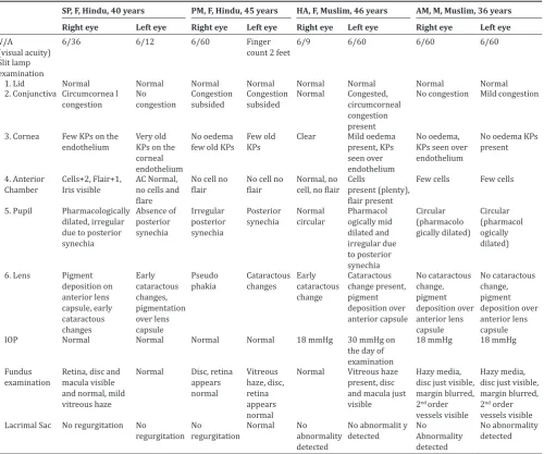
Table 3: Reported cases of Brucella uveitis
Reference no. Author’s name Year Findings Country
Cited in 14 Woods & Guyton 1944 15 cases of brucellosis among 200 cases of uveitis, 7 of them had recurrent
iritis ,5 had generalized uveitis, 3 had choroiditis Great Britain
Harris 1945 1 case of recurrent keratitis, 3 cases of recurrent iritis due to brucellosis Great Britain Kuzherskaya 1951 1 case of iridocyclitis ,1 case of bilateral optic neuritis, 1 case of bilateral
optic atrophy among 101 cases of brucellosis Great Britain
Pagliarani 1951 4 cases of Brucella melitensis with ocular symptoms Great Britain
Nelson Jones 1952 Described the different clinical presentations of brucellosis Great Britain
Barrett & Rickards 1953 Gave a detailed description of chronic brucellosis Great Britain
Cited in 8 Puig Solanes et al. 1953 60 brucellosis patients with ocular complication, of them 48 had
neuro-ophthalmic involvement USA
Cited in 7 Rolando et al. 2008 Among 1551 patients of brucellosis studied , 52 had ocular manifestations Peru Cited in 11 Rolando et al. 2009 Among 12 patients with clinical and laboratory findings suggestive of
Brucella uveitis, 4 had negative ocular agglutination and 8 had positive agglutination test ,1 had positive culture for B. Melitensis in subretinal fluid. Goldmann Witmer coefficient was positive in 3 patients , tissue samples showed lymphoplasmacytic infiltrate
Peru
Cited in 15 Ozlem Gurses et al. 2010 A case of brucellosis in a 28 years female, presented as bilateral optic nerve and right abducent nerve involvement and endocarditis complicated by right premacular hemorrhage
Turkey
Cited in 16 Lutfi Akyol et al. 2015 Combined presentation of sacroilitis and uveitis in case of brucellosis in a
in this part of our country. The previous studies on brucellar uveitis are summarized in Table 3. This study has been undertaken to diagnose the presence of brucellosis among clinically confirmed cases of uveitis. Uveitis strictly means inflammation of uveal tissue only. But clinically, there is always some associated inflammation of adjacent structures such as retina, vitreous sclera, and cornea. The infective causes of uveitis can be bacterial (Mycobacterium, Treponema, Staphylococcus, Streptococcus,
Brucella, etc.), viral, fungal, and parasitic [22]. Acute uveitis is one that persists for 6 weeks to 3 months and chronic uveitis persists for more than 3 months to years.
In our study, we found four confirmed cases of brucellosis with the ocular presentation as chronic anterior uveitis. The patients have been given the standard therapy with doxycycline and rifampicin and responded well to treatment.
CONCLUSION
To conclude, every case of systemic brucellosis should undergo routine ophthalmological evaluation and similarly all patients suffering from uveitis should be screened for brucellosis. This could reduce the possibility of blindness associated with brucellosis.
ACKNOWLEDGMENT
The authors would like to acknowledge the grant received from Government of India (Department of Biotechnology, DBT Sanction Order No.102/IFD/SAN/3141/2012-2013) for giving us the opportunity to conduct Brucella-related diagnostic tests.
REFERENCES
1. Young EJ. Brucella species. In: Mandell GL, Bennet GE, Dolin R, editors. Principles and Practice of Infectious Diseases. 5th ed. Philadelphia, PA: Churchill Livingstone; 2000. p. 2386-93.
2. Karapinar B, Yilmaz D, Vardar F, Demircioglu O, Aydinok Y. Unusual presentation of brucellosis in a child: Acute blindness. Acta Paediatr 2005;94(3):378-80.
3. Corbel MJ. Brucellosis: An overview. Emerg Infect Dis 1997;3(2):213-21.
4. Doganay M, Aygen B. Human brucellosis: An overview. Int J Infect Dis 2003;7:173-82.
5. Baily GG, Krahn JB, Drasar BS, Stoker NG. Detection of Brucella melitensis and Brucella abortus by DNA amplification. J Trop Med Hyg 1992;95(4):271-5.
6. Hatipoglu CA, Yetkin A, Ertem GT, Tulek N. Unusual clinical presentations of brucellosis. Scand J Infect Dis 2004;36(9):694-7. 7. Rolando I, Olarte L, Vilchez G, Lluncor M, Otero L, Paris M, et al.
Ocular manifestations associated with brucellosis: A 26-year experience in Peru. Clin Infect Dis 2008;46(9):1338-45.
8. Puig Solanes M, Heatley J, Arenas F, Guerrero Ibarra G. Ocular complications in brucellosis. Am J Ophthalmol 1953;36(5):675-89. 9. Rabinowitz R, Schneck M, Levy J, Lifshitz T. Bilateral multifocal
choroiditis with serous retinal detachment in a patient with Brucella infection: Case report and review of the literature. Arch Ophthalmol 2005;123(1):116-8.
10. Moutray TN, Williams MA, Best RM, McGinnity GF. Brucellosis: A forgotten cause of uveitis? Asian J Ophthalmol 2007;9:30-1. 11. Rolando I, Vilchez G, Olarte L, Lluncor M, Carrillo C, Paris M, et al.
Brucellar uveitis: Intraocular fluids and biopsy studies. Int J Infect Dis 2009;13(5):e206-11.
12. Al-Kaff AS. Ocular brucellosis. Int Ophthalmol Clin 1995;35(3):139-45. 13. Güngür K, Bekir NA, Namiduru M. Ocular complications associated
with brucellosis in an endemic area. Eur J Ophthalmol 2002;12(3):232-7. 14. Foggitt KD. Ocular disease due to brucellosis. Br J Ophthalmol
1954;38(5):273-8.
15. Sahin OG, Pelit A, Turunc T, Akova YA. Ophthalmoparesis, papillitis and premacular hemorrhage in a case with endocarditis: A rare presentation of brucellosis. Indian J Ophthalmol 2010;58(2):164-6. 16. Akyol L, Aslan K, Özgen M, Sayarlioglu M. Bilateral sacroiliitis and
uveitis comorbidity: Brucellosis? Ankylosing spondylitis? BMJ Case Rep 2015;2015.
17. Devaraj B, Kali A, Charles MV, Seetha SK. Modified biphasic media for blood culture. Asian J Pharm Clin Res 2016;9:3, 42-3.
18. Mondal I, Sanyal S, Das S. Coagulation profile in patients suffering from acute brucellosis. Asian J Phram Clin Res 2013;6(4):179-80. 19. Preman P, Sanyal S, Das S. Histological changes in mammalian uterus
in brucellosis. Asian J Biomed Pharm Sci 2013;3(22):58-61.
20. Saha S, Gupta D, Das S. Autoimmune changes in human brucellosis. Int J Biopharm 2013;4(2):131-4.
21. Srivastava R, Sanyal S, Das S. Histological changes in canine placenta during acute brucellosis. Int J Curr Microbiol Appl Sci 2013;2(8):139-43.