P61
Use of Firefly Fluorescence Imaging Technology for Robotic-assisted Partial Cystectomy and Ureteral Reconstruction
Alejandro R. Rodriguez.
Samaritan Medical Center, Watertown, NY, USA.
Background: Firefly fluorescence imaging technology has been used
dur-ing robotic assisted urological procedures, commonly for partial nephrec-tomies. In this study, we report on the use of this technology for robotic assisted partial cystectomy for bladder tumors and ureteral reconstructive procedures.
Methods: Firefly flourescence imaging technology has been used since
November 2012 at our institution. It has been used most commonly for robotic assisted partial nephrectomies. We have applied this technology during partial cystectomies and ureteral reconstructive procedures. During these procedures we either do a flexible cystoscopy (for a partial cystec-tomy) or a flexible ureteroscopy (for ureteral reconstruction). Once the lesion is identified, we point at it endourologically, and turn-on the Firefly technology on the robotic console. This technique allows us to visualize the endoscopic light in green color, and identify the exact point where the lesion is located. Patient demographics, perioperative outcomes and complications were analyzed.
Results: 2 patients were performed using this technique. In one case, the
patient (61 y.o. male, BMI of 31) had a double right ureteral stricture above the iliac vessels that had been dilated and stented multiple times. A flexible ureteroscopy was used to identify the areas of stricture. Firefly technology was used to define were the resection of the ureter was going to be performed. For this case, a robotic assisted boari flap was performed. Robotic console time was 170 min, estimated blood loss was 50 cc, hospital stay was 3 days, and JP drain time was 2 days. In the second case, the patient had a bladder tumor in the dome compatible with either a bladder adenocarcinoma versus a urachal cyst remanent. A flexible cystoscopy was performed during the robotic assisted partial cystectomy plus urachal resection. Robotic console time was 80 min, estimated blood loss was 25 cc, hospital stay was 2 days, and JP drain time was 2 days. Clear cut borders of resection were identified during both procedures. At 3 months (mean time) followup, there was no complications.
Conclusions: Firefly fluorescence imaging technology to assist in localizing
a lesion in the bladder or the ureter, proofed to be effective during both the robotic assisted partial cystectomy and the ureteral reconstruction.
P62
Robot-assisted Laparoscopic Simple Prostatectomy in patients with a Pathologic Specimen Weight >100 grams: A Multi-institutional Study of Outcomes
Vineet Agrawal¹, Claudia Berrondo¹, Anees Fazili¹, Hani Rashid¹, Guan
Wu¹, Laurence Donahue², Jean Joseph¹.
¹University of Rochester Medical Center, Rochester, NY, USA, ²Rochester General Hospital, Rochester, NY, USA.
Background: Since its first description in 2008, only 150 cases of robot
assisted simple prostatectomy (RASP) have been reported in the literature for large symptomatic benign prostatic hyperplasia. Several published series of RASP have reported small average gland weight on pathological exami-nation, where endoscopic management could have been used with equal
effectiveness, and less morbidity. We report on our experience performing RASP on patients with prostate weight of 100 grams or more.
Methods: Charts of patients undergoing RASP between 2009 and 2013
were retrospectively reviewed. Twenty-five patients with prostate weight >100 g as per the pathology report, were treated with RASP for BPH by four surgeons at two institutions. Both the extra- and transperitoneal approach were utilized to perform a retropubic (Millin’s), or suprapubic (Freyer) prostatectomy.
Results: The mean age, BMI and prostate weight on histology were 73 yrs.,
28 kg/m2 and 136 grams respectively. The median operative time was 223 mins while the EBL was 350 ccs. The average LOS was 3.6 days. 2 patients had cystolithotomy while 1 had inguinal hernia repair concurrently. 2 patients were found to have low risk prostate cancer. The Foley catheter was removed after a mean of 10 days with leak on cystogram requiring delayed removal in 1 patient. There were three and two Clavien grade 1 and 2 complications respectively. At a mean follow-up of 20 months, all patients reported a good outcome based on symptom resolution.
Conclusions: Our study has shown that robot-assisted simple prostatectomy
in very large prostates has acceptable perioperative outcomes and leads to successful resolution of patient symptoms.
P63
Renal Nephrometry Score and Predictors of Outcomes in Laparoscopic Partial Nephrectomies
Deepak K. Pruthi, Darrel E. Drachenberg, Thomas B. McGregor.
University of Manitoba, Winnipeg, MB, Canada.
Background: Nephron-sparing surgery is becoming the preferred method of
managing small renal masses. Feasibility of this approach extends beyond clinical tumor size and includes anatomical complexity of the tumor. In this study we analyze patient characteristics and anatomic tumor factors to look for variables associated with surgical complications and outcomes after laparoscopic partial nephrectomy.
Methods: We retrospectively reviewed all patients who underwent partial
nephrectomy at our academic institution between January 1, 2012 and April 30, 2014. Follow-up extended to first outpatient clinic visit 8 weeks postoperatively. All preoperative imaging was reviewed and the R.E.N.AL. Nephrometry score (radius for tumor size as maximal diameter, exophytic/ endophytic tumor properties, nearness of deepest portion of tumor to col-lecting system or sinus, anterior/posterior descriptor and location relative to polar line) was applied to each scan. Standardized grading systems were applied to data and standard statistical analysis to examine associations.
Results: Of the 102 patients who underwent partial nephrectomy seventeen
(17%) patients had 19 complications; eight were Clavien-Dindo grade 3 to 4. Two patients had laparoscopic partial nephrectomies converted intraop-eratively to radical nephrectomies; two other laparoscopic partial nephrec-tomies were converted to open partial nephrecnephrec-tomies. Fifty-one (50%) of operated patients were either obese, morbidly obese, or super obese. Nineteen (19%) of patients had pathologic benign lesions. Six (6%) patients had positive margins; Renal Nephrometry score was not correlated with positive margin status (p=0.612). In univariate analysis Charlson comor-bidity score ([[Unsupported Character - Codename ­]]>4 p=0.0055), diabetes (47% p=0.0046) were associated with complications while BMI >30 trended toward increased complications (p=0.0693). However, age
Moderated Poster Session 5: Laparoscopy/Robotics & Stones/
Endourology
(>60 p=0.461) and total R.E.N.A.L. Nephrometry score (p=0.286) were not associated with complications. A history of anticoagulation trended toward increased risk of postoperative bleeding (22%, p=0.087). Warm ischemic time did not vary significantly by Nephrometry score (low 27.8 min [SD +/- 10.0], intermediate 28.8 min [SD +/- 9.8], high 27.8 min [SD +/- 9.9]).
Conclusions: Utilization of standardized reporting may be necessary for
treatment decision making and the R.E.N.A.L. Nephrometry Score is a reproducible classification system that quantitates the salient anatomy of renal masses. However, as demonstrated in this laparoscopic series, categorizing renal masses according to nephrometry score did not signifi-cantly predict complication rates or expected WITs. Patient factors, how-ever, appear to play a larger role in the development of complications.
P64
WITHDRAWN
P65
Extraperitoneal Robot-assisted Radical Prostatectomy in the Morbidly Obese: A Propensity Score Matched Study
Vineet Agrawal, Changyong Feng, Jean Joseph.
University of Rochester Medical Center, Rochester, NY, USA.
Background: An increasing number of obese patients [body mass index
(BMI) >30 kg/m2] with localized prostate cancer are presenting as
can-didates for robot-assisted radical prostatectomy (RARP), which can be carried out using the transperitoneal (TP) or the extraperitoneal (EP) approach. It has been postulated that the EP approach may offer advan-tages in the obese. Morbidly obese (BMI >40 kg/m2) patients present as
an especially challenging surgical cohort. Only two previous studies, both utilizing the TP approach, have focused on the outcome of RARP in the morbidly obese. It has not been established if outcomes of EP-RARP in the morbidly obese differ from those of the non-morbidly obese. Herein, we sought to evaluate the perioperative and pathological outcomes associ-ated with EP-RARP in morbidly obese men.
Methods: We queried our prospectively collected database (Caisis) for
patients who underwent extraperitoneal robot assisted radical prostatec-tomy. Between July 2003 and December 2013, 1663 patients underwent EP-RARP for localized prostate cancer at our institution by a single sur-geon. 40 patients were considered morbidly obese. A propensity score-matched analysis was performed using multivariate analysis incorporating 10 co-variates to identify comparable group of patients with a BMI of >40 kg/m2 and <40 kg/m2.
Results: Table 1 shows that apart from BMI, the two groups were matched
(all p-values >0.05). Table 2 outlines the outcomes. Despite a higher total operating time and estimated blood loss in the morbidly obese (238 vs 176 mins, p <0.0001, and 235 vs 192 ccs, p=0.003 respectively), there were no differences in the ability to perform nerve-sparing or pelvic lymphad-enectomy, or the length of stay. While the morbidly obese had a higher rate of harboring more aggressive disease on final pathology (pT3 rates 27.5 vs 7.5 % respectively), there were no differences in other postopera-tive pathological parameters such as prostate weight, posipostopera-tive surgical margin status, and Gleason score sum. Moreover, there were no differ-ences in intra- or postoperative complications between the two groups.
Conclusions: EP-RARP in the morbidly obese leads to comparable
peri-operative and pathological outcomes to the non-morbidly obese.
P66
WITHDRAWN
P67
Percutaneous Nephrolithotomy in Patients with Urinary Tract Abnormalities
Philippe Violette, Marie Dion, Thomas Tailly, John Denstedt, Hassan Razvi.
Western University, London, ON, Canada.
Background: Patients with urinary tract abnormalities are at an increased
risk of stone formation. Percutaneous nephrolithotomy (PCNL) plays an important role in the management of this patient population, however outcomes are less well defined compared to patients with normal urinary tract anatomy. Our objective was to evaluate the influence of urinary tract abnormalities on intraoperative and postoperative outcomes with PCNL.
Methods: We report on a single-center prospective database of 2,284
consecutive PCNL procedures in 1,935 patients from 1990 to 2012. For the purposes of this analysis, patients were categorized by the presence or absence of a urinary tract abnormality. Multivariable analyses were used to identify independent predictors of the length of hospital stay, opera-tive time, complications and residual stones at discharge and 3 months.
Results: A urinary tract abnormality was present in 14.4% (n=330) of the
cohort. On univariable analysis patients with urinary tract abnormalities were more likely to present with urinary tract infection (28% vs. 19%, p<0.001) and less likely to present with hematuria (13% vs. 19%, p<0.02).
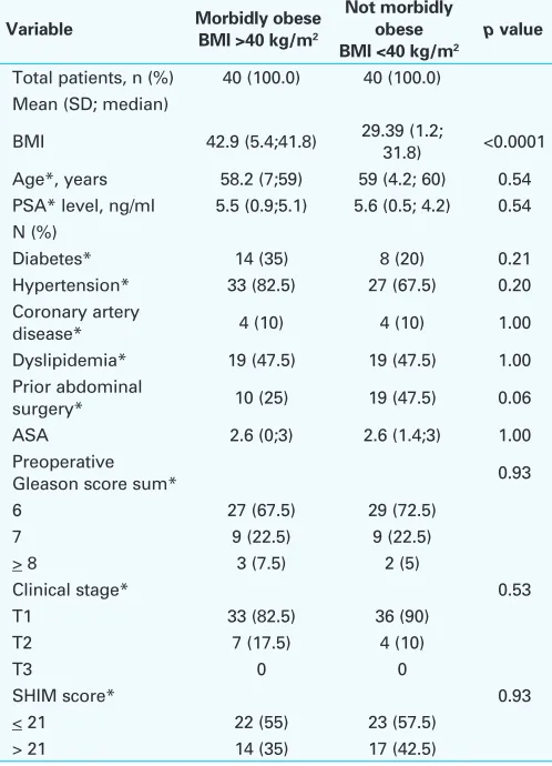
Table 1. P65. Comparison of preoperative variables for patients undergoing RARP stratified by BMI
Variable Morbidly obese
BMI 42.9 (5.4;41.8) 29.39 (1.2; 31.8) <0.0001
Age*, years 58.2 (7;59) 59 (4.2; 60) 0.54 PSA* level, ng/ml 5.5 (0.9;5.1) 5.6 (0.5; 4.2) 0.54 N (%)
Diabetes* 14 (35) 8 (20) 0.21 Hypertension* 33 (82.5) 27 (67.5) 0.20 Coronary artery
disease* 4 (10) 4 (10) 1.00
Dyslipidemia* 19 (47.5) 19 (47.5) 1.00 Prior abdominal
surgery* 10 (25) 19 (47.5) 0.06 ASA 2.6 (0;3) 2.6 (1.4;3) 1.00 Categorical data are presented as n (%)
On multivariable regression, a urinary tract abnormality was predictive of residual stone at discharge, need for a secondary procedure, but did not increase the risk of residual stone at 3 months or the development of complications. Operative time and hospital stay were only moderately prolonged.
Conclusions: Patients with urinary tract abnormalities, who undergo
PCNL, have a higher risk of residual stones at discharge and need for secondary procedures, but comparable complication rates, operative time and hospital stay.
P68
Efficacy of Tamsulosin Oxybutynin and Their Combination in the Control of Double-J Stent-related Lower Urinary Tract Symptoms
Miguel Maldonado-Avila, Rene J Jungfermann-Guzman, Leopoldo Garduño-Arteaga, Hugo A Manzanilla-García, Emmanuel Rosas-Nava, Alejandro Vela- Mollinedo, José Guzman-Esquivel, J Nestor Procuna-Hernandez, Luis Almazan-Treviño, Marcos Del Rosario-Santiago. General Hospital of México, México, City, Mexico.
Background: Indwelling double-J ureteral stents are used routinely in the
resolution of ureteral obstruction caused by different etiologies. Evaluation of urinary symptoms related to double-J stent, indicate that these affect
73-90% of patients who carry it. We conducted a prospective, random-ized study, to evaluate the efficacy of tamsulosin and oxybutinin and combination therapy in improving the urinary symptoms.
Methods: Patients who underwent ureteral stent placement after
ureteros-copy (total 51), were randomized into three groups: Group I: Tamsulosin 0.4 mg once per day( 17 patients), Group II: Oxybutinin 5 mg once per day (17 patients), Group III: Tamsulosin+oxybutynin once per day (17 patients). All the groups received the medicine for three weeks and completed a Spanish validated Ureteral Stent Symptom Questionnaire (USSQ) at day 7 and 21.
Results: Repeated measures ANOVA showed mean urinary symptom
index score was 22.3 vs. 15.5 in group three (p<0.001) at day 7 and 21 respectively. The mean work performance index was 6.6 vs 8.1 (p=0.049) favoring tamsulosin group, the mean sexual score was 0.5 vs 1.5 (p=0.03). Among additional problems the mean was 7.2 vs 6.2 (p=0.03). No sig-nificant difference was noted among pain and general health index. No side effects were reported (Table 1, Fig. 1).
Conclusions: Combination therapy with tamsulosin and oxybutynin
improved irritative symptoms and work performance as well as sexual matters. Combination therapy should be considered for patients who complained of stent related symptoms.
Table 2. P65 (continued). Comparison of preoperative variables for patients undergoing RARP stratified by BMI
Variable Morbidly obese
Wound infection 1 (2.5) 0 Bladder neck
contracture 1 (2.5) 0
Lymphocele 0 1 (2.5)
Clot retention 0 1 (2.5)
Urine leak 0 1 (2.5)
Clavien-classified complications
Grades 1-2 4 (1 0) 1 (2.5) Grades 3-4 1 (2.5) 2 (5) Categorical data are presented as n (%)
Table 2. P65. Comparison of preoperative variables for patients undergoing RARP stratified by BMI
Variable Morbidly obese
OT, min 238(70.5; 224) 176.4(46.8;
167.5) <0.0001
EBL 235 (70.7; 250) 192.4 (56.5; 160) 0.003
N (%)
Nerve sparing 0.85
None 14 (35) 16 (40)
Unilateral 11 (27.5) 9 (22.5) Bilateral 15 (37.5) 15 (37.5)
PLND 0.36
Performed 18 (45) 13 (32.5) Not performed 22 (55) 27 (67.5)
Mean (SD; median) 0.06
Prostate weight 61.2 (12.0; 61) 65.6 (8.4; 58.5)
P69
Interim Results of a Randomized Trial Comparing Narrow Versus Wide Focal Zones for Shock Wave Lithotripsy of Renal Calculi Kenneth Pace, Tarek Alzahrani, Daniela Ghiculete, R J D’A Honey.
St. Michaels Hospital, Toronto, ON, Canada.
Background: The Modulith SLX-F2 electromagnatic lithotripter (Storz
Medical) is the first lithotripter on the market with a unique design allow-ing for a dual focus of either a narrow (6x28 mm) or wide (9x50 mm) focal zones. Ex vivo data shows that disintegration capacity and renal vascular injury are independent of the focal diameter of the SW generator at the same peak pressure and disintegration power. The objective of this study is to compare single-treatment success rates of narrow and wide focal zones for the shock wave lithotripsy (SWL) of renal stones.
Methods: 118 patients with a previously untreated radio-opaque
soli-tary stone located within the kidney, measuring 5 to 15 mm in greatest diameter, were randomized to receive narrow or wide focus lithotripsy. Patients were followed with KUB x-rays and renal ultrasound at 2 and 12 weeks post lithotripsy to assess stone free status. Urinary markers indicating the degree of renal cellular damage (microalbumin and Beta-2 macroglobulin) were measured pre- and post-SWL, 24 hours post-SWL and 7 days post-treatment. Primary outcome was single-treatment success rate, defined as stone-free or adequate fragmentation (sand and asymp-tomatic fragments <=4mm) at 3 months post-treatment.
Results: 61 (51.7%) patients were randomized to narrow focus lithotripsy
versus 57 (48.3%) patients wide focus. The groups were similar in baseline characteristics including (age, gender, BMI, stone size and density and skin to stone distance). The overall success rates were not significantly different at 2 weeks post treatment (Narrow: 72.1% vs Wide: 61.4%; P=0.216) nor at 3 months (Narrow: 68.3% vs Wide: 58.9%; P=0.292). The overall complication rates was also comparable in the two groups (Narrow: 24.6% vs Wide: 17.5%; P=0.349) including similar rates of perinephric hematoma (Narrow: 3.3% vs Wide: 3.5%; P=0.945). The microalbumin-to-creatinine ratio was significantly different between the two groups (p=0.019), but that difference was gone within 24 hours after the treatment.
Conclusions: Interim results indicated that single-treatment success rate
and complications are comparable when using the narrow or wide focus of the Modulith SLX-F2. There was a difference in renal injury as measured bymicroalbumin to creatinine ratio (with lower values in the narrow focal zone group), but these differences disappeared within 24h of treatment. We are continuing to recruit patients to a pre-planned sample size of 300.
P70
Stone Burden Measurement by 2D or 3D Reconstruction Does Not Improve Prediction of Residual Stone at Discharge or 3 Months Compared to Standard Measurement With Elliptical Assumption
Philippe D. Violette, Thomas Tailly, Brandon R. Nadeau, Yige Bao, Justin
Amann, John D. Denstedt, Hassan Razvi. Western University, London, ON, Canada.
Background: Kidney stone burden has been reported as an independent
predictor of postoperative outcomes of percutaneous nephrolithotomy (PCNL). By comparing different ways of quantifying stone burden, we aimed to identify/qualify which measurement of stone burden most accu-rately predicts those outcomes.
Table 1. P68
Variable Tamsulosin Oxybutinin Tamsulosin + Oxybutinin p value
Mean Urinary symptom score Day 7 21.4 ± 4.78 22.4 ± 4.9 22.3 ± 7.2 <0.001 Day 21 21.5 ± 6.27 17.8 ± 5.5 15.5 ± 5.0
Mean pain index score Day 7 13.4 ± 2.2 11.2 ± 2.7 13.8 ± 5.0 0.207
Day 21 14.2 ± 4.3 10.9 ± 3.1 11.2 ± 5.8
Mean general health index score Day 7 11.7 ± 1.4 11.0 ± 1.4 11.5 ± 1.5 0.699 Day 21 11.6 ± 1.2 11.7 ± 1.3 11.2 ± 2.5
Mean work performance score Day 7 6.6 ± 4.0 6.3 ± 2.5 7.0 ± 2.3 0.049*
Day 21 8.1± 1.8 7.2 ± 1.9 7.7 ± 1.5
Mean sexual matters score Day 7 0.6 ± 1.1 0.5 ± 0.8 2.6 ± 3.8 0.036*
Day 21 1.2 ± 1.2 1.5 ± 1.2 2.6 ± 3.8
Mean additional problems index score
Day 7 8.05 ± 2.6 6.8 ± 1.9 7.2 ± 1.7 0.03*
Day 21 7.4 ± 3.0 6.4 ± 1.1 6.2 ± 2.8
Mean global USSQ Day 7 61.9 ± 9.1 58.47 ± 8.4 64.6± 16.8 0.932
Methods: We prospectively collected data for PCNLs performed at a
tertiary center between January 2006 and December 2013. A total of 246 patients had a preoperative CT and postoperative follow-up data at 3 months. Our primary outcome was incidence of residual stone at 3 months. Stone burden was measured three different methods for all patients on reformatted coronal CT images: 1) elliptical surface area (SA) (longest diameter x orthogonal diameter x π /4); 2) manual outline of stone surface and computer SA calculation; 3) automatic 3D volume rendering and calculation using specific CT software (AW). SA’s were described in increments 500mm². We used logistic regression and receiver operative characteristic (ROC) curve analysis and area under the curve (AUC) to evaluate the predictive value of the three measurements.
Results: Our cohort had a mean age of 55.7 years, was 42.3% female
and had an overall stone free rate at 3 months of 78.1%. The mean stone burden differed by method; 1) 644.3 ± 540.0 mm²; 2) 545.1 ± 404.2 mm²; 3) 9.33 ± 8.87 cm³ respectively. As expected, in univariate analysis, all different methods were predictive of residual stone (OR1: 1.50, CI 1.20-1.87; OR2: 1.56, CI 1.12-2.08, OR3: 1.241, CI: 1.078-1.427 respectively). Contrary to our expectation, the AUC for the three methods of measure-ment were similar (0.680, 0.665 and 0.666 respectively), demonstrating equivalent predictive value.
Conclusions: Stone burden can be used to predict incidence of residual
stone. We demonstrated that measuring the stone burden by manual outline or 3D volume assessment on reformatted NCCT images was not superior to a 2D measurement employing an elliptical assumption at predicting the incidence of residual stone.
P71
Mean, Range and Standard Deviation of Stone Density on Non-Contrast CT do not Predict Stone-Free Rates after Percutaneous Nephrolithotomy
Thomas Tailly, Philippe D. Violette, Brandon R. Nadeau, Yige Bao, Justin
Amann, John D. Denstedt, Hassan Razvi. Western University, London, ON, Canada.
Background: Stone density has been reported to influence outcomes of
both extracorporeal and endoscopic stone treatment. The standard devia-tion (SD) of Hounsfield unit (HU) measurement of the stone has been reported to influence shockwave lithotripsy outcomes. The influence of SD or range of HU on postoperative outcomes after percutaneous neph-rolithotomy (PCNL) has not been evaluated. Our objective was to identify the predictive value of stone density measurements on postoperative outcomes after PCNL.
Methods: We performed a retrospective chart review of prospectively
collected data in a single center from January 2006 to December 2013. We identified 309 PCNL treatments that had preoperative CT for density measurements and postoperative outcome data available. CT measure-ments were performed by a radiologist blinded to postoperative outcomes. Mean, SD and range of HU were measured by drawing an ellipse region of interest on the stone within the borders of the stone in bone window on the CT slice portraying the largest stone diameter in axial plane. We used Logistic regression, receiver operative characteristic (ROC) curves and area under the curve (AUC) to assess their predictive value.
Results: Our population had an overall stone free rate (SFR) at 3 months
of 78.3% and 7.8% had a second look nephroscopy. Contrary to our hypothesis, mean HU as well as SD was not found to have predictive value on SFR at 3 months. On secondary analysis, we identified mean HU by 1000 and range of HU by 100 as predictors of secondary procedure OR 2.74 (1.21, 6.20), P=0.016, and OR 1.159 (1.03, 1.30), P=0.015 respectively. Mean, SD and range of HU showed similar predictive value by ROC analysis (AUC’s 0.597, 0.601 and 0.619 respectively).
Conclusions: In contrast to previous reports, according to our data, stone
density has no predictive value on residual stone after treatment. Higher mean HU however does predict a higher chance of needing a second look nephroscopy. This preoperative assessment of stone density can be of use for surgical planning.
P72
Risk Factors for Re-admission Following Shock Wave Lithotripsy Benjamin J. Nelson¹, Anees Fazili¹, Divya Kumar², Franca Kraenzlin²,
Jason Birnbaum², Chang-Yong Feng¹, Erdal Erturk¹.
¹URMC Urology, Rochester, NY, USA, ²University of Rochester Medical School, Rochester, NY, USA.
Background: Quality improvement and pay-for-performance measures
have put an increased emphasis on reducing postoperative complications and hospital re-admissions. We therefore sought to analyze the rate of re-admission and presentation to the Emergency Department (ED) within 90 days of shock wave lithotripsy (SWL), with the hope of identifying prognostic risk factors for this adverse outcome.
Methods: We performed a retrospective review of patients that
under-went SWL therapy at our institution from 1/2011 to 5/2013 using the Storz Modulith SLX-F2 lithotripter for solitary ureteral or renal stones
≤2.0 cm. Re-admission or presentation to the ED within 90 days was our primary outcome. Secondary endpoints included stone free rates at 30 and 90 days, defined as lack of any residual fragments on follow-up KUB . Univariate and multivariate analysis were performed to identify risk factors for primary and secondary outcomes.
Results: Our study population included 307 patients with renal stones
and 270 with ureteral stones. Mean stone size was 9.2 mm. The rate of re-admission within 90 days was 11.6%. On multivariate analysis, age, BMI, ASA score, gender, history of prior nephrolithiasis, stone location, size, and presence of a ureteral stent did not affect this outcome. Patients who underwent a non-urgent SWL, however, had a lower risk of re-admission than those who had an urgent procedure performed (OR 0.2, p = 0.0005). Among patients who were re-admitted, renal colic was the most common chief complaint (67%), followed by infection (10%) and postoperative hematoma or hematuria (7.5%). Stone free rates were 57% and 78% at 30 and 90 days, respectively. The only factors that predicted stone free rates were stone size and non-urgent SWL status.
Conclusions: Our rate of re-admission 90 days following SWL was 11.6%.
Only the urgency of SWL was predictive of this outcome. Stone cen-ters should monitor their re-admission rates following SWL to establish national standards and guide decision making when considering other endourologic methods if these outcomes are considered unacceptable.
P73
Role of Tamsulosin, Tadalafil and Silodosin As The Medical Expulsive Therapy In Lower Ureteric Stone: A Randomized Trial (A Pilot Study)
Kumar Jayant¹, Santosh Kumar², Swati Agrawal¹, Mayank Mohan
Agrawal2, Shrawan Kumar Singh².
¹Sudha Hospital and Medical Research Centre, Kota, India, ²Postgraduate Institute of Medical Education and Research, Chandigarh, India.
Background: To evaluate the role of two different alpha-1 blockers and
one phosphodiesterase-5 inhibitor(PDE-5) as medical expulsive therapy for distal ureteric calculi.
Methods: Between Jan 2011 and Dec 2012, 365 patients presenting with
distal ureteric stones of size 5 to 10 mm were upon consent randomly assigned to one of three outpatient treatment arms: Tamsulosin (group A), Tadalafil (group B), Silodosin (group C). Therapy was given for a maxi-mum of four weeks. Stone expulsion rate, time to stone expulsion, anal-gesic use, number of hospital visits for pain, follow-up and endoscopic treatment and adverse effects of drugs were noted. All three groups were compared for normally distributed data by analysis of variance(ANOVA), Bonferroni or Kruskal-Wallis and Mann-Whitney U tests, as required. All the classified and categorical data were analyzed for all three groups by using the chi-square test.
Results: There was a statistically significant expulsion rate 83.3% in group
B than 64.4% , 66.7% in group A and C with lower time of stone expul-sion. (p-value=0.006, p-value=0.016). Statistically significant differences were noted in colicky episodes and analgesic requirement in group B than group A and C. There was no serious adverse event.
Conclusions: Medical expulsive therapy for the distal ureteric stones using
The result of this pilot study showed that Silodosin increases ureteric stone expulsion quite significantly along with better control of pain with significantly lesser analgesic requirement.
P74
Outpatient Bilateral Tubeless Percutaneous Nephrolithotomy: Is it Safe?
Michael Fuoco, Andrea Kokorovic, James W. L. Wilson, Darren Beiko.
Queen’s University, Kingston, ON, Canada.
Background: Bilateral tubeless percutaneous nephrolithotomy (PCNL)
has been reported to be safe and effective in select patients. Although outpatient PCNL has recently been shown to be safe and effective in a series of 50 patients, it requires further study before urologists embrace same day discharge following PCNL. The objective of this study is to report our early experience in performing bilateral PCNL on a completely outpatient basis, assessing its safety.
Methods: A review of all outpatient tubeless PCNL cases between March
2007 and May 2014 at a single Canadian centre was performed, including collection of preoperative, intraoperative and postoperative data. Strict preoperative, intraoperative and postoperative criteria were used in the selection of candidates for outpatient bilateral PCNL: no intraoperative complications including significant bleeding or collecting system perfora-tion; postoperative hemodynamic stability; adequate pain control; and reliable patient with supportive family.
Results: Forty patients underwent ambulatory PCNL during the study
period, of which 4 patients underwent bilateral ambulatory tubeless PCNL. Mean maximum stone diameter was 3.5 cm and 5 of the 8 renal units contained staghorn calculi. All 4 patients were discharged home on the same day with a mean hospital stay of 186 minutes or 3hrs 6 min. The mean narcotic requirement was 70.5 mg of oral morphine equivalents. Importantly, there were no postoperative complications, emergency room visits, hospital readmissions, ancillary procedures or deaths.
Conclusions: This small series represents the largest series of outpatient
bilateral PCNL to date. In very carefully selected patients, bilateral PCNL on a completely ambulatory basis appears safe and may be feasible. Further research on outpatient bilateral PCNL is required prior to wide-spread adoption by urologists.
P75
Practice Patterns of Extracorporeal Shockwave Lithotripsy: Variability Amongst and Between Canadian and American Urologists - Do We Need Guidelines?
Andrea G. Lantz¹, Jeff P. McKay¹, Michael Ordon², Kenneth T. Pace², John D’A Honey².
¹Dalhousie University, Halifax, NS, Canada, ²University of Toronto, Toronto, ON, Canada.
Background: Extracorporeal shockwave lithotripsy (SWL) is a widely
utilized non-invasive form of treatment for urolithiasis. Despite its wide-spread use, there are few evidence-based recommendations regarding pre-SWL patient work-up and performance of SWL. The purpose of this study is to determine practice patterns and to compare the performance of SWL in the United States and Canada to evaluate if there is variability between centers and countries.
Methods: An 18-question survey was prepared to determine
pre-pro-cedural work-up (eg: routine electrocardiograms (ECG), urine culture, discontinuation of ASA, etc) and the performance of SWL (eg: shock rate, power, stents, etc). This survey was administered in 3 phases. In Canada, SWL is a highly regionalized procedure with only 16 sites across the country. Representatives of each Canadian site were surveyed through email correspondence. The Endourology Society members were surveyed using an online survey tool, and all members of a large stone management group in the Midwest United States completed the survey. Responses across Canadian and American urologists were compared using the Chi square and Fisher’s exact test.
Results: 16 and 187 surveys were completed from Canadian and US
urologists respectively. Practice patterns varied between countries.
Specifically, routine antibiotics were more commonly given in USA (USA 78.1% vs. CAN 6.3%; p<0.001); a higher shock rate of 2Hz was more common in Canada (USA 16.2% vs. CAN 68.8%, p<0.0001); rate of discontinuing ASA for ureteral stone treatment was higher in the USA (USA 90.3% vs. CAN 50%, p<0.0002), and ureteral stents were more commonly used if treating a large stone in the USA (USA 88.8% vs. CAN 46.7%, p=0.0002). There were no significant differences between countries for use of routine pre-SWL ECG (USA 48.7% vs. CAN 43.8%, p=0.71), pre-SWL urine culture (USA 55.2% vs. CAN 56.3%, p=0.93), dose escalation (USA 87.4% vs. CAN 100%, p=0.23), discontinuation of ASA for renal stones (USA 95.7% vs. CAN 81.3%, p=0.05), and stenting for solitary kidneys (USA 66.3% vs. CAN 66.7%, p=1).
Conclusions: There are limited evidence based recommendations for the
pre-procedural work-up and performance of SWL. This study highlights the lack of standardization in the performance of SWL. Significant regional differences exist in practice patterns and performance of SWL between American and Canadian urologists.
P76
Radiation Exposure to Surgical Staff During Percutaneous Nephrolithotomy (PCNL)-A Multi-Institutional Experience Over 12 Months
Rick C. Slater¹, David Wenzler², Jeannie Su², Roger Sur², Michelle J.
Semins¹.
¹University of Pittsburgh Medical Center, Pittsburgh, PA, USA, ²UC San Diego, San Diego, CA, USA.
Background: Routine usage of fluoroscopy during endourological
pro-cedures, particularly percutaneous nephrolithotomy (PCNL), has been demonstrated to be associated with scatter radiation exposure to medical staff. Literature is sparse in reporting robust data on radiation exposure with a variety of patient demographics and procedure variables. We aim to define radiation exposure to the endourologist and their staff associated with PCNL and identify potential risk factors for higher doses.
Methods: Surgeons from 2 institutions prospectively collected data on a
per case basis for all PCNLs during a 1 year period. Patient demograph-ics and procedure variables recorded included patient age, body mass index (BMI), surgical duration, fluoroscopy time, and radiation exposure. Radiation exposure was recorded utilizing an instadoseTM dosimeter placed on the thyroid shields of the operating surgeon, resident assis-tant, scrub nurse, and anesthesia staff. Associations were assessed with Spearman’s (r) correlation coefficient.
Results: A total of 97 consecutive PCNL or second look PCNL surgeries
in adult patients by 2 surgeons were evaluated over a 12 month time span (May 2013-May 2014) at two large tertiary academic medical centers. Median patient age was 53 (range 22-87) and, median BMI was 29.76 (range 16.57-55.51). Average fluoroscopy time between the two institu-tions was 333.53 seconds/case (Median 261, SD 301.25, range 19-1809 seconds). Average dosimeter exposure for the year was 367.5mrem, 214.5mrem, 14 mrem, and12.5mrem while average dosimeter exposure per case was 7.65mrem, 4.42mrem, 0.28mrem, and 0.25mrem for oper-ating surgeon, resident assistant, circuloper-ating nurse and anesthesia staff respectfully. There was a weakly positive spearman’s rank correlation between fluoroscopy time and operating surgeon exposure (r= 0.175) and BMI and operating surgeon exposure (r = 0.171).
Conclusions: Radiation exposure to the operating surgeon during PCNL
P77
Monotherapy With a Single Session of ESWL for Kidney Stones in the Community Setting
Po Lam, Brittany Paul, Christopher Pieczonka, Bashar Omarbasha, Joel
Bass, Jeffrey Sekula, Elan Salzhauer, Richard Kronhaus, Andres Madissoo, Sasha Pavlov-Shapiro, Vladimir Mouraviev, David Albala.
Associated Medical Professionals of NY, Syracuse, NY, USA.
Background: Extracorporeal shock wave lithotripsy (ESWL) has
progres-sively acquired popularity as being the gold standard treatment for upper urinary tract urolithiasis since 1980.
Methods: A retrospective clinical study was performed on 2,316 patients
between 2010 and 2012. Main outcome measured of our study was the clearing of stones after single session of ESWL. ESWL was done on all patients under conscious sedation by 24 board-certified urologists. The Litho Tron lithotripter (Healthtronics, Atlanta, GA) was used for the treatment. Median age of the patients was 38.7 years (range: 18-95). The percent distribution of patients according to stone size was following: ≤
5mm - 15%, 5.1-10 mm- 61%, 10.1-15 mm- 15%, 15.1-20.0 mm- 7%, >20 mm-2% (Fig. 1).
Results: No significant perioperative complications were noted. All
patients tolerated the procedure well and were discharged within 2 hours after the procedure. The results demonstrated a complete destruction of the stone in 1,488 (64.3%) cases, partial stone destruction in 496 (21.3%) and treatment failure in 331 (14.3%) patients, respectively. We found a direct correlation between large stone size and failure rate. A JJ stent was inserted before the procedure in 178 patients and after procedure in 119 patients.
Conclusions: No complications following ESWL treatment were noted.
We conclude that ESWL remains efficacious and safe treatment modality for renal calculi in adults. Selection of patients is a crucial factor in treat-ment outcomes. Success rates are lower as stone size increases.
Stone Size (mm)
15.30%
60.11%
15.30%
6.56%
2.18% 0.55%
<=5.0
5.1-10.0
10.1-15.0
15.1-20.0
20.1+
Missing