ABSTRACT
MOORMAN, ERIC ALAN. Alternative Chemical Disinfection Technologies for Inactivation of Human Norovirus on Surfaces. (Under the direction of Dr. Lee-Ann Jaykus)
Human norovirus (NoV) is the most common cause of acute non-bacterial
gastroenteritis worldwide. Laboratory evidence clearly demonstrates that few antimicrobial substances are comparable to concentrated chlorine bleach solutions (1,000-5,000 ppm free available chlorine) in their efficacy for inactivation of human norovirus on solid surfaces. The purpose of the research presented in this thesis was to evaluate the efficacy of two recently developed chemical disinfectants for inactivation of human NoV on environmental surfaces.
In chapter 2, neutral electrolyzed water (NEW) was evaluated for its ability to inactivate human NoV GII.4 Sydney and cultivable surrogate Tulane virus (TuV) using two American Society for Testing and Materials (ASTM) approved antimicrobial testing
methods; suspension (ASTM 1052-11) and surface (1053-11). RT-qPCR, with and without RNase pre-treatment, was used to measure reductions in GII.4 Sydney genomic copy number and ascertain NEWs impact on the viral capsid and genome, respectively. A 1-min exposure to NEW (250 ppm free available chlorine [FAC]) reduced levels of GII.4 Sydney by 4.8 ± 0.6 log10 in suspension assays, and 0.4 ± 0.1 log10 on stainless steel surfaces. Longer exposure
supported our RT-qPCR data demonstrating that NEW mediated human NoV inactivation occurs through damage/destruction of the viral capsid. Evidence for genome degradation was provided by RT-qPCR (without RNase treatment). This study provides a proof of concept that NEW (250 ppm FAC) is capable of inactivating human NoV, but extensive contact times of 15-30 min on pre-cleaned surfaces are required for practical environmental disinfection.
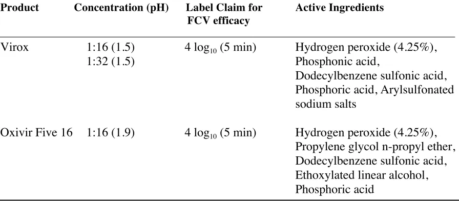
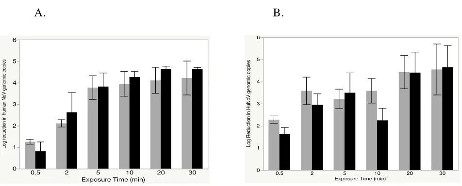
In chapter 3, accelerated hydrogen peroxide (AHP) disinfectants; Virox and Oxivir Five 16 Concentrate, were evaluated for their ability to reduce levels of human NoV GII.4 Sydney using ASTM approved antimicrobial testing methods; suspension (1052-11) and surface (1053-11). Using a 1:16 concentration (recommended by manufacturer for heavy duty cleaning and disinfection) Virox produced a 3.8 ± 0.5 and Oxivir a 3.2 ± 0.4 log10
reduction in GII.4 Sydney after a 5 minute exposure in a suspension assay. When a surface test method was used (GII.4 Sydney deposited and dried on stainless steel surfaces) Virox and Oxivir were less effective, reducing levels of GII.4 Sydney by less than 2 logs after 5 minutes. Increasing the exposure time to 30 minutes produced maximum GII.4 Sydney log10 reductions of 2.7 ± 0.2 and 2.5 ± 0.7 using Virox and Oxivir, respectively. Similar to what
we determined using neutral electrolyzed water in Chapter 2, Virox and Oxivir were significantly less effective in the presence of an additional soil load (p<0.05). This work suggests that Virox and Oxivir Five 16 are capable of inactivating human NoV, but that contact times of greater than 10 minutes are required for surface disinfection.
© Copyright 2017 Eric Alan Moorman
Alternative Chemical Disinfection Technologies for Inactivation of Human Norovirus on Surfaces
by
Eric Alan Moorman
A thesis submitted to the Graduate Faculty of North Carolina State University
in partial fulfillment of the requirements for the degree of
Master of Science
Food Science
Raleigh, North Carolina
2017
APPROVED BY:
_______________________________ _______________________________
Dr. Lee-Ann Jaykus Dr. Otto Simmons
Committee Chair
BIOGRAPHY
Eric Alan Moorman was born January 7th, 1991 in Berkeley, CA to Mark and Dawn
Moorman. Shortly after, siblings Sarah (3/28/1993) and Cole (5/20/1994) were born and the
Moorman family was officially off and running.
Eric is what you might call a ‘late bloomer’. After graduating from Portage Central
High School in Portage, MI, Eric attended Central Michigan University where he hadn’t the
slightest idea what he wanted to study. After a successful two years at CMU, Eric transferred
into Michigan State University. It was here Eric took his first Food Science course (after
advisement from his father, Mark Moorman--who happened to have a PhD from that same
Food Science program at MSU). School clicked, and Eric quickly took after his father and
developed a strong passion for food safety. Immediately after his graduation from MSU, Eric
completed a Quality Assurance internship at Campbell Soup Company in Napoleon, Ohio,
ACKNOWLEDGMENTS
First and foremost, I would like to acknowledge my major advisor, Dr. Lee-Ann
Jaykus. I am so thankful for the opportunity grow as a scientist under her guidance. It truly is
a blessing.
I would also like to thank the rest of my advisory committee, Dr. K.P. Sandeep, Dr.
Otto “Chip” Simmons, and Dr. Hal King. Each of you have been accessible, and
continuously made an effort to guide me throughout my studies. For that I am forever
grateful.
Another thank you goes to past and present members of the Jaykus lab. Dr. Blanca
Escudero-Abarca, you are an amazing person and have impacted so many students in the
Jaykus lab. Thank you for everything. Dr. Naim Montazeri, thank you sir for your
mentorship. I have never worked so well with a colleague before; it has been a pleasure.
Lastly, to Dr.’s Chip Manuel, Matthew Moore, Erin Almand, and Jon Baugher. You guys
cultivated a great dynamic in the lab, and each of you modeled what a successful graduate
student should look like. Thank you for the endless talks and advice.
Lastly, thank you to my family. Your love and support has provided me with
confidence and a strong foundation to which I can navigate the world. To my father; Dad,
you sparked my passion and your influence has made me a life-long learner. The greatest
TABLE OF CONTENTS
LIST OF TABLES ... vii
LIST OF FIGURES ... viii
CHAPTER ONE-Literature Review: Alternate Methods for Inactivation of Human Norovirus on Surfaces ...1
INTRODUCTION ...1
NON-CONVENTIONAL SURFACE INACTIVATION METHODS3 Electrolyzed Water ...3
Chlorine Dioxide ...5
Hydrogen Peroxide ...8
Copper Containing Surfaces ...9
FACTORS THAT INFLUENCE EFFICACY OF NOROVIRUS INACTIVATION MEASURES ...12
Testing Method...13
Inoculum Matrix ...13
Detection Method ...14
Use of Human Norovirus Surrogates ...17
CONCLUSION ...19
REFERENCES ...21
CHAPTER TWO-Efficacy of Neutral Electrolyzed Water for Inactivation of Human Norovirus ...30
ABSTRACT ...30
INTRODUCTION ...31
MATERIALS AND METHODS ...33
RESULTS ...41
DISCUSSION ...43
AKNOWLEDGEMENTS ...49
REFERENCES ...57
CHAPTER THREE-Efficacy of Accelerated Hydrogen Peroxide Disinfectants against Human Norovirus GII.4 Sydney as Evaluated by RNase RT-qPCR ...61
ABSTRACT ...61
INTRODUCTION ...62
MATERIALS AND METHODS ...63
RESULTS ...67
DISCUSSION ...69
AKNOWLEDGEMENTS ...73
LIST OF TABLES
Table 2.1: Impact of neutral electrolyzed water (NEW) on human NoV VLP
receptor-binding ...54
Table 3.1: Composition of accelerated hydrogen peroxide disinfectants
LIST OF FIGURES
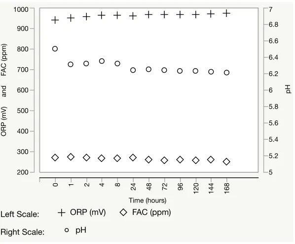
Figure 2.1: ORP, FAC, and pH stability of neutral electrolyzed water over
7 d of storage at 21°C ...48
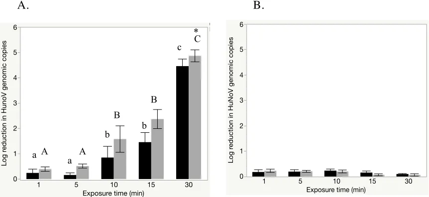
Figure 2.2: Inactivation of human NoV without (A) and with (B) additional
soil load following a one minute exposure to neutral electrolyzed
water (NEW) in suspension assay followed by RT-qPCR ...49 Figure 2.3: Inactivation of human NoV on stainless steel coupons
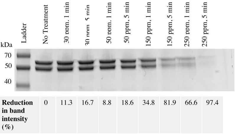
following exposure to neutral electrolyzed water ...50 Figure 2.4: SDS-PAGE analysis of HuNoV VLP major capsid protein
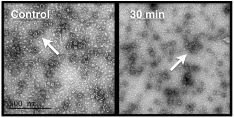
following exposure to neutral electrolyzed water ...51 Figure 2.5: Impact of neutral electrolyzed water on human NoV VLP
capsid integrity by transmission electron microscopy ...52 Figure 2.6: Inactivation of Tulane virus on stainless steel following
exposure to neutral electrolyzed water ...53 Figure 3.1: Reduction in GII.4 Sydney genomic copy number
(RNase RT-qPCR) following exposure to Virox and Oxivir
at 1:16 concentration in suspension ...73 Figure 3.2: Reduction in GII.4 Sydney genomic copy number
(RNase RT-qPCR) following exposure to Virox and Oxivir
CHAPTER 1
Literature Review: Alternative methods for inactivation of norovirus on surfaces
INTRODUCTION
Human noroviruses (NoV) are a leading food and environmental public health concern worldwide. Three of the six established genogroups are pathogenic in humans (GI, GII, and GIV), with GI and GII being the most common. Each of these genogroups
possesses multiple genotypes. Members of genogroup II, genotype 4 (GII.4) are classified as epidemic strains, with GII.4 Sydney as the current outbreak strain. The GII.4 strains
collectively were responsible for almost a fifth of all cases of global acute gastroenteritis from 2008-2014 (Ahmed et al., 2014). Human NoV are also an extremely important cause of foodborne illness, responsible for 58% of food-related illnesses, 26% of hospitalizations, and 11% of deaths in the United States annually (Scallan et al., 2011).
Human NoV symptomology includes diarrhea, vomiting, nausea, as well as abdominal cramping and fever. Diarrhea and vomiting (hallmark symptoms) result in
months (Prevost et al., 2016), and relatively unharmed by active ingredients (e.g., ethanol, quaternary ammonium compounds) in numerous conventionally used surface disinfectants and sanitizers at manufacturer recommended concentrations and contact times (Tung et al., 2013; Cromeans et al., 2014). Epidemiologic evidence continually shows that environmental transmission plays a substantial role in human NoV disease propagation, especially in institutional settings such as restaurants, hospitals, nursing homes, cruise ships, and prisons (Repp et al., 2013; Isakbaeva et al., 2005).
The major intervention strategy recommended by the U.S. Centers for Disease Control and Prevention (CDC) to inactivate human NoV on surfaces is to clean and
subsequently disinfect surfaces with 1,000-5,000 ppm free available chlorine (FAC) usually derived from dilution of household bleach (CDC, 2011). Although laboratory findings have demonstrated chlorine at these concentrations is effective against NoV (~3 log reduction in gEq after 3 min exposure to 5,000 ppm free chlorine) (Park and Sobsey, 2011), one salient caveat for the frequent use of chlorine at these concentrations is corrosion of surfaces.
other factors potentially influence antimicrobial efficacy including testing method
(suspension vs. surface); application method (e.g., aqueous vs. fog); virus suspending matrix; target virus upon which screening is based; and detection method, among others. These factors will also be discussed.
NON-CONVENTIONAL SURFACE INACTIVATION METHODS Electrolyzed water
Electrolyzed water (EW) is an aqueous chemical oxidant generated by passing a dilute salt solution (generally 0.1-1.0% sodium chloride) through an electrolytic cell in a
process known as electrolysis. Hypochlorous acid (HOCl), hypochlorite ion (OCl-) and
chlorine gas (Cl2) are the predominant oxidative species produced, the composition of which
is directly influenced by solution pH. Hypochlorous acid is a weak acid and stronger
antimicrobial relative to OCl- and Cl2, and is the most predominant species at pH values
between 2 and 7 (Dychdala et al., 1977). Electrolyzed waters aqueous chemistry suggests that
solutions with acidic or near neutral pH values (containing primarily HOCl) may achieve
similar (if not higher) antimicrobial efficacy than conventional chlorine bleach solutions
while using lower concentrations. Despite its well documented efficacy against bacterial
pathogens (Rahman et al., 2016), limited data exists on the efficacy of EW against human
NoV and its surrogates on solid non-porous surfaces. Following deposition and drying of 1%
stool specimens positive for human NoV GII.4 Sydney, or cell culture lysate containing
bacteriophage MS2 onto stainless steel and ceramic tiles, Park et al. (2007) reported a 3-log10
EW (pH 5.5-6.2) at a concentration of 188 ppm FAC. To our knowledge, this is the only
study the reported the efficacy of EW against human NoV at the time of this writing. In a
later study, Julian et al. (2014) provided additional evidence for the ability of EW to
inactivate bacteriophage MS2 on solid surfaces within 30-seconds, reporting that EW made
to deliver 500 ppm and 1,000 ppm FAC (pH 10) could reduce levels of MS2 infectivity by
1.8 and >4 log10, respectively.
When murine norovirus (MNV-1) was exposed to EW at 500 ppm FAC (pH 10) for
30-seconds on stainless steel, Julian et al. (2014) reported reductions in infectivity and gEq
were 0.1 and 0.4 log10, respectively. Similar results demonstrating moderate reductions in
MNV-1 infectivity following exposure to both acidic (pH 2.8) and neutral (pH 6.4) EW on
stainless steel surfaces were also reported by Fang et al. (2016). This group showed that
exposure to neutral EW at a concentration of 100 ppm FAC for 5 and 10 minutes was
sufficient to reduce MNV-1 infectivity by 0.9 and 1.8 log10 units, respectively. Lowering the
pH of EW from neutral (pH 6.4) to acidic (pH 2.8) produced significantly higher reductions
(p<0.05) in MNV-1 infectivity at the same concentration (100 ppm FAC) and exposure times
(5 and 10 minutes), evidenced by 1.9 and 2.9 log10 unit reductions, respectively. Although
collectively these data suggest EW is capable of modestly reducing the concentrations of
human NoV and cultivable surrogates on solid surfaces at concentrations ~200 ppm FAC and
contact times of 1-10 minutes, variability in reported efficacy does exist. Factors potentially
contributing to variation and/or limited efficacy of all products discussed will be covered in
The mechanism behind EW-mediated NoV inactivation is not well understood.
However, it is well established that exposure of dilute salt solutions (0.1-1.0% sodium
chloride) to the process of electrolysis results in the generation of pH dependent solutions
containing a mixture of predominantly chlorine species [Cl2, HOCl, and OCl-] (Rahman et
al., 2016) and potentially trace amounts of reactive oxygen species (hydroxyl radicals,
hydrogen peroxide, ozone). The balance and concentrations of these species is highly
dependent up on the device used to manufacture the EW (Jeong et al., 2009). Because of its
high oxidation reduction potential (ORP), it is reasonable to assume that EW functions
through this mechanism, primarily targeting the proteinaceous viral capsid and RNA
genome. As an example, in their study characterizing the virolysis capacity of chlorine
against feline calicivirus (FCV) and human NoV GII.4 Sydney, Nowak et al. (2011)
demonstrated that 100 ppm FAC generated in EW was sufficient to significantly reduce FCV
RNA RT-qPCR signals to <0.03% of the starting copy number. This finding led to this
authors’ assertion that chlorine mediated inactivation of NoV occurs through targeting the
viral capsid in such a way that results in exposure and subsequent degradation of viral RNA.
Chlorine Dioxide
mechanism consisting of free radical electrophilic abstraction, as opposed to oxidative substitution and/or addition carried out by free chlorine (Baribeau et al., 2002). Relative to other forms of chlorine such as sodium or calcium hypochlorite, ClO2 is less reactive with ammonia and other organic compounds, a feature that results in a lesser formation of potentially carcinogenic disinfection by-products such as chloramine and trihalomethanes (WHO, 2000; Randtke et al., 2010). Upon generation, ClO2 exists predominantly as a free radical monomer, a physical characteristic resulting in a high solubility in water and near equivalent biocidal efficacy in both aqueous and gaseous matter states (WHO, 2000).
To our knowledge, only one study was identified at the time of this writing that incorporated human strains of NoV in the assessment of ClO2 efficacy. Using a porcine gastric mucin magnetic bead binding assay followed by RT-qPCR detection, Kingsley et al. (2014) reported 0.3, 0.8, 1.5, and 2.8 log10 reductions in HuNoV GI.1 binding after treatment with 240 ppm ClO2 for 1, 10, 30 and 60 minutes, respectively.
by Lim et al. (2010) who showed ClO2 at concentrations as low as 0.3 ppm could reduce concentrations of MNV-1 infectivity by >3.5 log10 within 1-minute. This group, however used highly purified virus with significantly reduced organic load which, as discussed below, could have been responsible for the high efficacy of the product in these studies. The efficacy of ClO2 efficacy against feline calicivirus (FCV) has also been reported. In their assessments of ClO2 at concentrations of <1.0 ppm in a suspension assay format, Zoni et al. (2007) and Thurston-Enriquez et al. (2005) reported >3 log10 reductions in FCV infectivity titer after a 1-minute contact time.
The ability of ClO2 to inactivate NoV, both surrogates and human strains, while in a gaseous state has also been elucidated. Morino et al. (2009) showed that 0.05 ppm ClO2 reduced FCV by 2 log10 after a 4-hour exposure. At higher concentrations, Yeap et al. (2016) demonstrated more rapid reduction in infectivity titer, with a 3 log10 reduction in MNV-1 after 1 and 2 min exposures to 4.0 and 2.5 ppm ClO2, respectively. Transmission electron microscopy, SDS-PAGE and Western blot analyses collectively revealed that ClO2 disrupts the conformation of the MNV-1 icosahedral capsid and further degrades the capsid protein (Yeap et al., 2016). Evidence for genome degradation was also observed as signal
resulted in disruption of HAV antigenicity and destruction of the 5’ non-translated region (5’NTR) of the virus nucleic acid, respectively (Li et al., 2004).
Hydrogen Peroxide
Hydrogen Peroxide (H2O2) is another broad spectrum oxidative chemical whose use in industrial and medical settings dates back to the mid-twentieth century. Peroxide is
commonly used following nebulization into a vapor or fog (Spaulding et al., 1977), a process that has been validated for its ability to achieve >5 log10 reductions in Clostridium spp. endospores on solid inanimate surfaces (Johnston et al., 2005). The high redox potential (1.76 V) of H2O2 and subsequent high affinity for electrons is thought to contribute to its oxidative mode of action (Finnegan et al., 2010).
biological indicators containing Geobacillus stearothermophilus endospores were reduced by 3.6, 3.6, and 6.0 log10 following exposure to 500 ppm H2O2 vapor. Additional support for the ability of H2O2 to disinfect solid surfaces artificially contaminated with MNV-1 was
provided by Li et al. (2011). Using a solution containing 2.52% H2O2, they demonstrated MNV-1 titer could be reduced on stainless steel by 4 log10 units in just 10-min. Supporting evidence was provided in the surface assays of Bentley et al. (2012) and Goyal et al. (2014) who showed that ~30% H2O2 administered as a fog produced >5 log10 reductions in FCV titer within 20 min and 2 hours, respectively.
Copper-Containing Surfaces
Although methods to observe and quantitate its efficacy did not exist at the time, the exploitation of copper for its antimicrobial properties dates back to the time of ancient Egyptians (Grass et al., 2011). More recently, the incorporation of copper into high touch surfaces in health care facilities and its concomitant reduction in microbial burden has led the Environmental Protection Agency (EPA) to recognize that specific copper alloys have
antimicrobial properties (http://www.epa.gov/pesticides/factsheets/copper
Warnes and Keevil (2013) were the first to address the anti-norovirus efficacy of copper, reporting that dry surfaces containing >89% copper produced >4 log10 reductions in MNV-1 titer in just 10 minutes. A major finding from this study was the impact of surface moisture on copper efficacy, with inactivation rates 10 times faster once the inoculum had dried (Warnes and Keevil, 2013). In a follow up study, this same group suggested that surface abrasion may impact the antiviral properties of copper surfaces, as reductions in MNV-1 titer were slightly lower for surfaces receiving abrasion treatment. The group
postulated that removal of the passivating oxide later through prolonged surface use results in the attrition of antimicrobial properties (Warnes and Keevil, 2013). These findings are
aligned with Airey and Verran (2007) who observed lower efficacy of copper surfaces for the reduction of Staphylococcus aureus when the bacterial inoculum was supplemented with higher amounts of organic materials. Compared to stainless steel controls, excessive films, characterized by Airey and Verran (2007) as surface conditioning, were seen following deposition and drying on copper surfaces. This group postulated that conditioning of copper surfaces could be due to coppers high reactivity with organic material. Percent copper composition was important, with copper-brass alloys (70% copper) were more effective than nickel alloys (70% copper) (Warnes et al., 2015). Zinc is a constituent of copper-brass alloys, and zinc alloys themselves produced greater reductions in MNV-1 titer
In their study using a series of culture independent methods to assess the inactivation efficacy of copper alloy surfaces against human NoV, Manuel et al. (2015) found that surfaces containing ³ 70% copper could produce 2-3 log10 reductions in human NoV GII.4 gEq within 60 minutes of exposure. In comparison to Warnes and Keevil (2013) and Warnes et al. (2015) who utilized the cultivable surrogate MNV in their assessment of copper
surfaces and reported >4 log reductions in MNV titer in 10-min, Manuel et al. (2015) reported a slightly lower degree of efficacy, as evidenced by the need for longer contact times (60-minutes) required for a comparable degree of human NoV inactivation (2-3 log10 gEq). Nuances in experimental design that may contribute to disparate results between these two independent studies include: (i) utilization of highly purified MNV stock virus which would undoubtedly contain a lower organic load (Warnes and Keevil, 2013); Warnes et al., 2015) as compared to 20% fecal suspensions (Manuel et al., 2015); (ii) use of culture dependent methods (plaque assay) (Warnes and Keevil, 2013); (Warnes et al., 2015) vs. culture independent methods (RT-qPCR) (Manuel et al., 2015) for evaluation of disinfection efficacy; and (iii) differences in inoculum volumes between studies; [25 µl (Manuel et al., 2015) compared to 1- 20 µl (Warnes and Keevil, 2013); Warnes et al., 2015)].
These studies used RT-qPCR to determine log inactivation, however the assays targeted different regions of the viral genome; a 70 base-pair region of VPg gene within ORF1 for MNV (Warnes and Keevil, 2013), but an approximately 120 bp segment in the highly
conserved region of the ORF1-ORF2 junction for human NoV (Manuel et al., 2015). Reverse transcription and amplification efficiency during RT-qPCR is inversely proportional to fragment size. This theory, in conjunction with the differences in inherent stability of various RNA genomic regions to degradation may be factors contributing to differences in
experimental outcomes in aforementioned studies. Taken together, the data on the anti-noroviral efficacy of copper are considered strong enough for some industries to adopt its use in high touch surfaces as a means to prevent secondary transfer of NoV from contaminated environmental surfaces.
FACTORS THAT INFLUENCE EFFICACY OF NOVIRUS INACTIVATION MEASURES
In order to make conclusions about the behavior of human NoV upon exposure to select disinfecting and sanitizing compounds, one must be able to directly compare the experimental outcomes directly from one study to the next. Unfortunately, this is almost never possible when study designs are not coordinated with one another. Therefore,
Testing method
There are several ways by which antimicrobial substances are evaluated for efficacy against NoV. Testing methods reported in the literature include variations of two main categories: suspension assays (virus diluted into the antimicrobial substance); and
surface/carrier tests (antimicrobial substance added to surfaces containing wet or desiccated virus inoculum). Although suspension assay-based approaches are historically more common in the disinfection literature, their representativeness of the antimicrobial efficacy in real-world, practical applications during surface decontamination is debatable; a conclusion supported by laboratory evidence demonstrating higher antimicrobial performance in
suspension assays relative to surface tests (Park et al., 2002; Fang et al., 2016). For example, using hypochlorous acid at various concentrations, Park et al. (2007) produced a 3 log10 reduction in RT-PCR amplifiable units of human NoV and bacteriophage MS2 after just 20 seconds. When a surface testing method was used however, a contact time of 1-min was required for the same degree of hypochlorous acid efficacy. The authors postulated that desiccation of inoculum (suspending matrix) on surfaces reduced exposure of viruses to disinfectant substances during testing.
Inoculum matrix
more resistant to thermal inactivation when dispersed within shellfish tissue compared to cell culture media (Araud et al., 2016); the virus was also more resistant to chemical inactivation in the presence of increasing amounts of organic matter (Bolton et al., 2013). Compared to their more purified form after laboratory manipulation (e.g., ultracentrigation or filtration), crude viruses in fecal, environmental and/or food matrices have the potential to interact with matrix-associated particulates (e.g., protein, carbohydrates, and lipids) resulting in virus-particulate complexes. It is likely that the matrix protects the viruses from inactivation, resulting in an apparent increase in virus resistance (Rutala and Weber, 1997); Deborde and Von Gunten, 2008). Cell culture media can also be protective. MNV and FCV are commonly propagated in the presence of 10% fetal bovine serum (Fang et al., 2016; Julian et al., 2014; Warnes and Keevil, 2013), and crude virus stocks usually consist of unpurified cell culture lysate. This suspending medium has, for example, considerable chlorine demand that certainly impacts the efficacy of chemical disinfection (Nowak et al., 2011). In order to provide the most direct comparison between human NoV inactivation strategies, it is important to further characterize the impact of specific virus suspension matrices, and standardize the composition of inocula.
Detection methods
detection. Despite its high sensitivity and quantitative nature, detection only targets a small region of the viral RNA genome and is independent of physiological state of the target organisms (i.e., RNA may be detected from infectious virus, or residual RNA from
inactivated virus), a feature which may underestimate performance of inactivation measures (Cromeans et al., 2014). As a result, interpreting the public health significance of positive RT-qPCR results is nebulous. Detection of enterovirus RNA in 60/68 environmental samples using RT-PCR, but only 2/68 samples by cell culture, illustrates this discrepancy (Hot et al., 2003). A summary of limitations associated with molecular biological techniques in food virology is provided by (Richards et al., 1999).
the case (Liu et al., 2010). Limitations of this method do exist, primarily the formation of ribonucleoprotein complexes that may encapsulate, or shield RNA from the RNase enzyme, reducing the discriminatory nature of this method (Knight et al., 2013). Furthermore, virus particles may lose their functionality (i.e., receptor binding capability required for viral infectivity), but maintain their structural conformation (i.e., intact capsid) during inactivation measures. Thus, they may still be detected using RNase RT-qPCR. Incorporating ligands that are more sensitive to target conformational changes, such as host binding proteins or nucleic acid aptamers, into detection assays is a promising approach to increasing the stringency of culture independent techniques such as RT-qPCR (Moore et al., 2015; Moore et al., 2016). Viability dyes or intercalating agents such as propidium monoazide (PMA) have also been used as intended discriminatory methods for human NoV RT-qPCR detection. In their study comparing plaque assay results to PMA RT-qPCR, (Karim et al., 2015) reported that PMA was able to selectively differentiate infectious from non-infectious poliovirus following thermal and chemical inactivation procedures. Limited usefulness of this method has
al. (2008) demonstrated GII NoV could be detected with increased sensitivity (2 log10) in a variety of food samples when RT-qPCR was preceded by this ligand binding step. Porcine gastric mucin-bound magnetic beads have been incorporated into assays aimed at
characterizing the efficacy of various chemical inactivation measures against human NoV (Kingsley et al., 2014) and MNV (Li et al., 2011).
Use of human NoV surrogates
In the absence of an in vitro cultivation model for human NoV, food safety and public health virologists have used cultivable surrogate organisms in their studies. In very early days, bacteriophage MS2 within the Levivirdae family was used as a surrogate (Kniel et al., 2014). More relevant are other mammalian viruses within the Caliciviridae family that can be cultivated in cell culture. The most commonly used include: feline calicivirus (FCV), murine norovirus (MNV), and Tulane virus (TuV). FCV, although currently recognized by the U.S. Environmental Protection Agency (EPA) as the approved surrogate for use in virucidal efficacy evaluations for registered disinfectants, is transmitted by the respiratory route and relative to other surrogates displays a lower degree of environmental stability and pH resistance (Cannon et al., 2006). MNV is a member of the Norovirus genus and thus relative to FCV more genetically related to human NoV. In their study comparing the
chlorine at industrially relevant concentrations and contact times (200 and 1000 ppm chlorine for 5-min), as appears to be the case for human NoV (Cromeans et al., 2014).
Following its characterization within the Recovirus genus in 2008 (Farkas et al., 2008), TuV has become increasingly used in inactivation studies as a human NoV surrogate. Pairwise homology scores obtained from amino acid sequence alignments of several non-structural polyproteins in ORF1 of members of the Caliciviridae family indicate that TuV displays the highest amino acid similarity (20-25%) to members of the Norovirus genus. TuV is thus genetically and phylogenetically the most highly related to human NoV of all the cultivable surrogates (Farkas et al., 2008). Furthermore, TuV displays receptor binding properties similar to that of human NoV, specifically showing type A and B histoblood group antigens (HBGAs) as cellular attachment factors (Farkas et al., 2010), and perhaps sialic acid (Tan et al., 2015). In terms of relevance of the TuV surrogate, a 5-min exposure to 70% ethanol in suspension and 200 ppm chlorine on stainless steel surfaces reduced TuV
infectivity by <0.5 log10 indicating it displays resistance to chemically mediated inactivation similar to that of human NoV (Cromeans et al., 2014). This finding is further supported by Arthur and Gibson (2015) after their assessment of the physiochemical stability profile of TuV.
environmental persistence, susceptibility to chemical or physical inactivation, etc.) most relevant to the hypothesis to be tested. It is also important to note that recent advances in human NoV in vitro cultivation, i.e., viral replication in human B-cells (Jones et al., 2015) and in stem cell-derived human enteroids (Ettayebi et al., 2016), may eventually make the use of surrogates unnecessary, although these new methods need further development before that becomes a reality.
CONCLUSION
Controlling the spread of highly infectious biological agents such as human NoV will not be achieved using a ‘one size fits all’ approach, particularly in densely populated settings capable of facilitating rapid spread of the disease. Ideally, an infection control plan would encompass multiple tactics when human NoV is the suspected cause of illness. Use of household bleach at concentrations of 1,000 to 5,000 ppm chlorine (depending on surface cleanliness) is the current CDC-recommended treatment to disinfect solid, non-porous surfaces (CDC, 2011) suspected of being contaminated with human NoV. Although the use of chlorine bleach for norovirus control is empirically well defined and validated by
laboratory research, it is not without limitations, mainly it’s reduced efficacy in the presence of organic and some inorganic constituents; and corrosivity issues on sensitive surfaces such as stainless steel.
its cultivable surrogates. Results from primary literature reported over the past 10-15 years suggest that these four treatments are capable of reducing levels of NoV on solid non-porous environmental surfaces, though their efficacy is ultimately dependent upon concentration and contact time. This conclusion is supported by both data based on quantitation of viral
REFERENCES
Ahmed, S. M., A. J. Hall, A. E. Robinson, L. Verhoef, P. Premkumar, U. D. Parashar, M. Koopmans & B. A. Lopman (2014) Global prevalence of norovirus in cases of gastroenteritis: a systematic review and meta-analysis. Lancet Infect Dis, 14, 725-30. Airey, P. & J. Verran (2007) Potential use of copper as a hygienic surface; problems
associated with cumulative soiling and cleaning. J Hosp Infect, 67, 271-7.
Araud, E., E. DiCaprio, Y. Ma, F. Lou, Y. Gao, D. Kingsley, J. H. Hughes & J. Li (2016) Thermal Inactivation of Enteric Viruses and Bioaccumulation of Enteric Foodborne Viruses in Live Oysters (Crassostrea virginica). Appl Environ Microbiol, 82, 2086-99. Arthur, S. E. & K. E. Gibson (2015) Physicochemical stability profile of Tulane virus: a
human norovirus surrogate. J Appl Microbiol, 119, 868-75.
Atmar, R. L., A. R. Opekun, M. A. Gilger, M. K. Estes, S. E. Crawford, F. H. Neill & D. Y. Graham (2008) Norwalk virus shedding after experimental human infection. Emerg Infect Dis, 14, 1553-7.
Atmar, R. L., A. R. Opekun, M. A. Gilger, M. K. Estes, S. E. Crawford, F. H. Neill, S.
Ramani, H. Hill, J. Ferreira & D. Y. Graham (2014) Determination of the 50% human infectious dose for Norwalk virus. J Infect Dis, 209, 1016-22.
Baribeau, H., M. Prevost, R. Desjardins, P. Lafrance & D. Gates. 2002. Chlorite and chlorate ion variability in distribution systems. 96-105. Journal American Water Works Association.
Benarde, M. A., B. M. Israel, V. P. Olivieri & M. L. Granstrom (1965) Efficiency of chlorine dioxide as a bactericide. Appl Microbiol, 13, 776-80.
Bentley, K., B. K. Dove, S. R. Parks, J. T. Walker & A. M. Bennett (2012) Hydrogen
peroxide vapour decontamination of surfaces artificially contaminated with norovirus surrogate feline calicivirus. J Hosp Infect, 80, 116-21.
Bolton, S. L., G. Kotwal, M. A. Harrison, S. E. Law, J. A. Harrison & J. L. Cannon (2013) Sanitizer efficacy against murine norovirus, a surrogate for human norovirus, on stainless steel surfaces when using three application methods. Appl Environ Microbiol, 79, 1368-77.
Cromeans, T., G. W. Park, V. Costantini, D. Lee, Q. Wang, T. Farkas, A. Lee & J. Vinjé (2014) Comprehensive comparison of cultivable norovirus surrogates in response to different inactivation and disinfection treatments. Appl Environ Microbiol, 80, 5743-51.
Deborde, M. & U. von Gunten (2008) Reactions of chlorine with inorganic and organic compounds during water treatment-Kinetics and mechanisms: a critical review. Water Res, 42, 13-51.
Dychdala, G. R. 1977. Chlorine and Chlorine Compounds. In: Disinfection, Sterilization and Preservation. 2ed. Editor: S. S. Block. Henry Kimpton Publishers.
Escudero-Abarca, B. I., H. Rawsthorne, R. M. Goulter, S. H. Suh & L. A. Jaykus (2014) Molecular methods used to estimate thermal inactivation of a prototype human norovirus: more heat resistant than previously believed? Food Microbiol, 41, 91-5. Ettayebi, K., S. E. Crawford, K. Murakami, J. R. Broughman, U. Karandikar, V. R. Tenge, F.
H. Neill, S. E. Blutt, X. L. Zeng, L. Qu, B. Kou, A. R. Opekun, D. Burrin, D. Y. Graham, S. Ramani, R. L. Atmar & M. K. Estes (2016) Replication of human noroviruses in stem cell-derived human enteroids. Science.
Fang, J., J. L. Cannon & Y.-C. Hung. 2016. The efficacy of EO waters on inactivating norovirus and hepatitis A virus in the presence of organic matter. 13-19. Food Control.
Farkas, T., R. W. Cross, E. Hargitt, N. W. Lerche, A. L. Morrow & K. Sestak (2010) Genetic diversity and histo-blood group antigen interactions of rhesus enteric caliciviruses. J Virol, 84, 8617-25.
Farkas, T., K. Sestak, C. Wei & X. Jiang (2008) Characterization of a rhesus monkey calicivirus representing a new genus of Caliciviridae. J Virol, 82, 5408-16.
Finnegan, M., E. Linley, S. P. Denyer, G. McDonnell, C. Simons & J. Y. Maillard (2010) Mode of action of hydrogen peroxide and other oxidizing agents: differences between liquid and gas forms. J Antimicrob Chemother, 65, 2108-15.
Girard, M., K. Mattison, I. Fliss & J. Jean. 2016. Efficacy of oxidizing disinfectants at inactivating murine norovirus on ready-to-eat foods. 7-11. International Journal of Food Microbiology.
Grass, G., C. Rensing & M. Solioz (2011) Metallic copper as an antimicrobial surface. Appl Environ Microbiol, 77, 1541-7.
Holmdahl, T., M. Walder, N. Uzcátegui, I. Odenholt, P. Lanbeck, P. Medstrand & A. Widell (2016) Hydrogen Peroxide Vapor Decontamination in a Patient Room Using Feline Calicivirus and Murine Norovirus as Surrogate Markers for Human Norovirus. Infect Control Hosp Epidemiol, 37, 561-6.
Hot, D., O. Legeay, J. Jacques, C. Gantzer, Y. Caudrelier, K. Guyard, M. Lange & L. Andréoletti (2003) Detection of somatic phages, infectious enteroviruses and enterovirus genomes as indicators of human enteric viral pollution in surface water. Water Res, 37, 4703-10.
Isakbaeva, E. T., M. A. Widdowson, R. S. Beard, S. N. Bulens, J. Mullins, S. S. Monroe, J. Bresee, P. Sassano, E. H. Cramer & R. I. Glass (2005) Norovirus transmission on cruise ship. Emerg Infect Dis, 11, 154-8.
Jeong, J., C. Kim & J. Yoon (2009) The effect of electrode material on the generation of oxidants and microbial inactivation in the electrochemical disinfection processes. Water Res, 43, 895-901.
Johnston, M. D., S. Lawson & J. A. Otter (2005) Evaluation of hydrogen peroxide vapour as a method for the decontamination of surfaces contaminated with Clostridium
botulinum spores. J Microbiol Methods, 60, 403-11.
Jones, M. K., K. R. Grau, V. Costantini, A. O. Kolawole, M. de Graaf, P. Freiden, C. L. Graves, M. Koopmans, S. M. Wallet, S. A. Tibbetts, S. Schultz-Cherry, C. E. Wobus, J. Vinjé & S. M. Karst (2015) Human norovirus culture in B cells. Nat Protoc, 10, 1939-47.
Julian, T. R., J. M. Trumble & K. J. Schwab (2014) Evaluating efficacy of field-generated electrochemical oxidants on disinfection of fomites using bacteriophage MS2 and mouse norovirus MNV-1 as pathogenic virus surrogates. Food Environ Virol, 6, 145-55.
Karim, M. R., G. S. Fout, C. H. Johnson, K. M. White & S. U. Parshionikar (2015) Propidium monoazide reverse transcriptase PCR and RT-qPCR for detecting infectious enterovirus and norovirus. J Virol Methods, 219, 51-61.
Kingsley, D., E. Vincent, G. Meade, C. Watson & X. Fan. (2014) Inactivation of human norovirus using chemical sanitizers. 94-99. International Journal of Food
Kirby, A. E., A. Streby & C. L. Moe (2016) Vomiting as a Symptom and Transmission Risk in Norovirus Illness: Evidence from Human Challenge Studies. PLoS One, 11, e0143759.
Kniel, K. E. (2014) The makings of a good human norovirus surrogate. Curr Opin Virol, 4, 85-90.
Knight, A., D. Li, M. Uyttendaele & L. A. Jaykus (2013) A critical review of methods for detecting human noroviruses and predicting their infectivity. Crit Rev Microbiol, 39, 295-309.
Leifels, M., L. Jurzik, M. Wilhelm & I. A. Hamza (2015) Use of ethidium monoazide and propidium monoazide to determine viral infectivity upon inactivation by heat, UV- exposure and chlorine. Int J Hyg Environ Health, 218, 686-93.
Li, D., L. Baert, M. De Jonghe, E. Van Coillie, J. Ryckeboer, F. Devlieghere & M. Uyttendaele (2011) Inactivation of murine norovirus 1, coliphage phiX174, and Bacteroides fragilis phage B40-8 on surfaces and fresh-cut iceberg lettuce by hydrogen peroxide and UV light. Appl Environ Microbiol, 77, 1399-404.
Li, D., L. Baert, E. Van Coillie & M. Uyttendaele (2011) Critical studies on binding-based RT-PCR detection of infectious Noroviruses. J Virol Methods, 177, 153-9.
Li, J. W., Z. T. Xin, X. W. Wang, J. L. Zheng & F. H. Chao (2004) Mechanisms of
inactivation of hepatitis A virus in water by chlorine dioxide. Water Res, 38, 1514-9. Lim, M. Y., J. Kim & G. Ko. (2010) Disinfection kinetics of murine norovirus using chlorine
and chlorine dioxide. Water Research 44: 3243-3251.
Liu, P., Y. Yuen, H. M. Hsiao, L. A. Jaykus & C. Moe (2010) Effectiveness of liquid soap and hand sanitizer against Norwalk virus on contaminated hands. Appl Environ Microbiol, 76, 394-9.
Manuel, C. S., M. D. Moore & L. A. Jaykus (2015) Destruction of the Capsid and Genome of GII.4 Human Norovirus Occurs during Exposure to Metal Alloys Containing Copper. Appl Environ Microbiol, 81, 4940-6.
Moore, M. D., B. I. Escudero-Abarca, S. H. Suh & L. A. Jaykus (2015) Generation and characterization of nucleic acid aptamers targeting the capsid P domain of a human norovirus GII.4 strain. J Biotechnol, 209, 41-9.
Morino, H., T. Fukuda, T. Miura, C. Lee, T. Shibata & T. Sanekata (2009) Inactivation of feline calicivirus, a norovirus surrogate, by chlorine dioxide gas. Biocontrol Sci, 14, 147-53.
Nowak, P., J. R. Topping, K. Bellamy, V. Fotheringham, J. J. Gray, J. P. Golding, G. Wiseman & A. I. Knight (2011) Virolysis of feline calicivirus and human GII.4 norovirus following chlorine exposure under standardized light soil disinfection conditions. J Food Prot, 74, 2113-8.
Nuanualsuwan, S. & D. O. Cliver (2002) Pretreatment to avoid positive RT-PCR results with inactivated viruses. J Virol Methods, 104, 217-25.
Park, G. W., D. M. Boston, J. A. Kase, M. N. Sampson & M. D. Sobsey (2007) Evaluation of liquid- and fog-based application of Sterilox hypochlorous acid solution for surface inactivation of human norovirus. Appl Environ Microbiol, 73, 4463-8.
Park, G. W. & M. D. Sobsey (2011) Simultaneous comparison of murine norovirus, feline calicivirus, coliphage MS2, and GII.4 norovirus to evaluate the efficacy of sodium hypochlorite against human norovirus on a fecally soiled stainless steel surface. Foodborne Pathog Dis, 8, 1005-10.
Park, H., Y. C. Hung & C. Kim (2002) Effectiveness of electrolyzed water as a sanitizer for treating different surfaces. J Food Prot, 65, 1276-80.
Prevost, B., M. Goulet, F. S. Lucas, M. Joyeux, L. Moulin & S. Wurtzer (2016) Viral persistence in surface and drinking water: Suitability of PCR pre-treatment with intercalating dyes. Water Res, 91, 68-76.
Rahman, S., I. Khan & D.-H. Oh. (2016) Electrolyzed Water as a Novel Sanitizer in the Food Industry: Current Trends and Perspectives. Comprehensive Reviews in Food Science: Institute of Food Technologists 15: 471-490.
Randtke, S. (2010) Chemistry of Aqueous Chlorine. In White's Handbook of Chlorination and Alternative Disinfectants. 5th
ed. Edited by: D. Desiderio & N. Nibbering. John Wiley & Sons, Inc.
Richards, G. P. (1999) Limitations of molecular biological techniques for assessing the virological safety of foods. J Food Prot, 62, 691-7.
Rutala, W. A. & D. J. Weber (1997) Uses of inorganic hypochlorite (bleach) in health-care facilities. Clin Microbiol Rev, 10, 597-610.
Salgado, C. D., K. A. Sepkowitz, J. F. John, J. R. Cantey, H. H. Attaway, K. D. Freeman, P. A. Sharpe, H. T. Michels & M. G. Schmidt (2013) Copper surfaces reduce the rate of healthcare-acquired infections in the intensive care unit. Infect Control Hosp
Epidemiol, 34, 479-86.
Sattar, S., S. Springthorpe, O. Adegbunrin, A. Zafer & M. Busa. 2003. A disk-based quantitative carrier test method to assess the virucidal activity of chemical germicides. 3-12. J Virol Methods.
Scallan, E., R. M. Hoekstra, F. J. Angulo, R. V. Tauxe, M. A. Widdowson, S. L. Roy, J. L. Jones & P. M. Griffin (2011) Foodborne illness acquired in the United States--major pathogens. Emerg Infect Dis, 17, 7-15.
Spaulding, E., K. Cundy & F. Turner. 1977. Chemical Disinfection of Medical and Surgical Materials. In Disinfection, Sterilization, and Preservation, ed. S. S. Block, 654-683. London: Henry Kimpton Publishers.
Tan, M., C. Wei, P. Huang, Q. Fan, C. Quigley, M. Xia, H. Fang, X. Zhang, W. Zhong, J. S. Klassen & X. Jiang (2015) Tulane virus recognizes sialic acids as cellular receptors. Sci Rep, 5, 11784.
Thurston-Enriquez, J. A., C. N. Haas, J. Jacangelo & C. P. Gerba (2005) Inactivation of enteric adenovirus and feline calicivirus by chlorine dioxide. Appl Environ Microbiol, 71, 3100-5.
Tian, P., A. Engelbrektson & R. Mandrell (2008) Two-log increase in sensitivity for detection of norovirus in complex samples by concentration with porcine gastric mucin conjugated to magnetic beads. Appl Environ Microbiol, 74, 4271-6.
Tuladhar, E., P. Terpstra, M. Koopmans & E. Duizer (2012) Virucidal efficacy of hydrogen peroxide vapour disinfection. J Hosp Infect, 80, 110-5.
United States Centers for Disease Control and Prevention. 2011. Updated norovirus outbreak management and disease prevention guidelines. MMWR Recomm Rep, 60, 1-18. United States Environmental Protection Agency. 2001. Initial virucidal effectiveness test
using Feline calicivirus as surrogate for norovirus. Washington, D.C. Available at: www.epa.gov/oppad001/pdf_files/initial_virucidal_test.pdf
Vincent, M., P. Hartemann & M. Engels-Deutsch (2016) Antimicrobial applications of copper. Int J Hyg Environ Health.
Warnes, S. L. & C. W. Keevil (2013) Inactivation of norovirus on dry copper alloy surfaces. PLoS One, 8, e75017.
Warnes, S. L., E. N. Summersgill & C. W. Keevil (2015) Inactivation of murine norovirus on a range of copper alloy surfaces is accompanied by loss of capsid integrity. Appl Environ Microbiol, 81, 1085-91.
World Health Organization. (2000) Environmental health criteria 216, disinfectants and disinfection by-products. Available at: www.inchem.org/documents/ehc/ehc/ ehc216.htm#SectionNumber:8.1
Yeap, J. W., S. Kaur, F. Lou, E. DiCaprio, M. Morgan, R. Linton & J. Li (2016) Inactivation Kinetics and Mechanism of a Human Norovirus Surrogate on Stainless Steel Coupons via Chlorine Dioxide Gas. Appl Environ Microbiol, 82, 116-23.
Zoni, R., R. Zanelli, E. Riboldi, L. Bigliardi & G. Sansebastiano (2007) Investigation on virucidal activity of chlorine dioxide. experimental data on feline calicivirus, HAV and Coxsackie B5. J Prev Med Hyg, 48, 91-5.
CHAPTER 2
Efficacy of Neutral Electrolyzed Water for Inactivation of Human Norovirus
Eric Moorman, Naim Montazeri, Lee-Ann Jaykus
ABSTRACT
Human norovirus (NoV) is a leading cause of acute (epidemic and sporatic) non-bacterial gastroenteritis worldwide. The virus is stable in the environment and recent
epidemiologic evidence suggests contaminated environmental surfaces play a substantial role in accentuating the duration and severity of outbreaks. In this study, we examined the
efficacy of neutral electrolyzed water (NEW) as an alternative intervention strategy for inactivation of human NoV in suspension (ASTM 1052-11) and on stainless steel surfaces (ASTM 1053-11), both with and without an additional soil load. Impact on viral genome and capsid was assessed using real time reverse-transcription PCR (RT-qPCR) with and without RNase pre-treatment. SDS-PAGE, transmission electron microscopy (TEM), and a histo blood group antigen (HBGA) receptor-binding assay provided further evidence for capsid damage. Plaque assays using LLC-MK2 cells were also used to measure viral titer of Tulane virus (TuV) following NEW treatment. NEW (250 ppm FAC, pH 7) produced a 4.8 ± 0.6 and 0.4 ± 0.12 log10 reduction in GII.4 genome copy number after 1-min in suspension and on
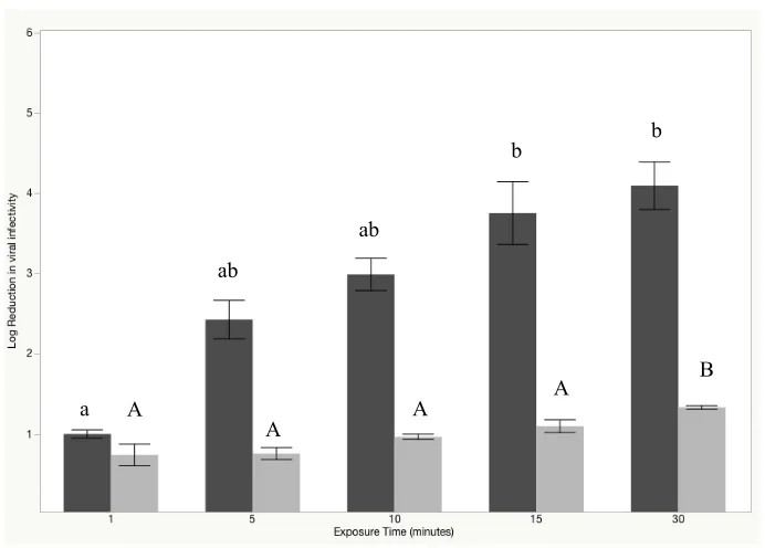
stainless steel surfaces, respectively. Increasing the contact time on surfaces to 5, 10, 15 and 30 min reduced human NoV by 0.5 ± 0.11, 1.6 ± 0.7, 2.4 ± 0.5, 5.0 ± 0.5 log10 and TuV concentrations by 2.42 ± 0.53, 2.99 ± 0.45, 3.75 ± 0.87, and 4.09 ± 0.59 log10, respectively.
of testing method and virus. The SDS-PAGE band intensity associated with the major capsid protein (VP1) of human NoV virus like particles (VLPs) was reduced by 97.1% (250 ppm FAC, 5 min) indicating capsid damage, a finding supported by increased aggregation and degradation of VLPs (TEM images) and reduced HBGA receptor binding. Results indicate that NEW shows promise as an alternate surface disinfectant when used at 250 ppm FAC on pre-cleaned surfaces.
INTRODUCTION
Human norovirus (NoV) is the leading cause of acute viral gastroenteritis (AGE) worldwide (Ahmed et al., 2014). Despite its predominance as a foodborne pathogen (Scallan et al., 2011), most illness attributed to human NoV is caused by direct contact with infected individuals and their gastrointestinal bodily fluids (feces, vomit). Both vomiting and fecal incidents no doubt lead to contamination of environmental surfaces, which can also be a source of human NoV transmission (Lopman et al., 2012; Otter et al., 2013). Previous work demonstrating high titer virus shedding by infected individuals (Teunis et al., 2015) and long-term virus persistence outside the human host both in lab-based (Lamhoujeb et al., 2009; Tung et al., 2013; Cromeans et al., 2014; Lopman et al., 2012; Kotwal and Cannon, 2014; Cook et al., 2016) and epidemiological (Isakbaeva et al., 2005; Repp et al., 2013; Cheesbrough et al., 1997) studies collectively support the phenomenon that environmental surfaces serve as reservoirs for human NoV transmission.
NoV in many different settings, for instance in healthcare and long-term care facilities, schools, and cruise ships. The current CDC recommendation for disinfecting solid surfaces presumably contaminated with NoV is 1,000-5,000 ppm free available chlorine (FAC), depending upon circumstances of the contamination event (CDC, 2011). Although effective (Tung et al., 2013), perpetual implementation of this recommendation may not be practical due to the corrosive nature of concentrated bleach solutions as well as irritancy to staff handling the solutions. Unfortunately, active ingredients within several classes of
conventionally-used surface disinfectants, e.g., quaternary ammonium compounds (QAC’s) and ethanol, do not fully inactivate human NoV at manufacturer recommended
concentrations and contact times (Tung et al., 2013; Cromeans et al., 2014; Liu et al., 2015; Girard et al., 2010). Residual infectious virus on surfaces resulting from incomplete chemical inactivation has significant public health implications, primarily by prolonging the duration and magnitude of outbreaks, further contributing to the annual U.S. healthcare costs
attributed to human NoV, estimated at $2 billion.
Electrolyzed water, also known as electrochemically activated solution, is one class of emerging disinfectants that function as a broad spectrum, aqueous chemical oxidant generated by passing a dilute salt solution through an electrolytic cell in a process known as electrolysis. Electrolyzed water has been evaluated for several food safety industrial
applications including use as a fresh produce wash (Pangloli et al., 2009), as well as a cleaning and sanitation agent in dairy manufacturing clean-in-place (CIP) systems (Wang et al., 2012). The microbiolocidal efficacy of electrolyzed water is attributed to three
[HOCl], hypochlorite ion [OCl
-]), and oxidation-reduction potential (ORP). HOCl is a more effective biocide relative to its dissociated form (OCl
-), which is the active ingredient of bleach. Manipulating the pH-dependent aqueous chemistry of electrolyzed water to a near neutral pH ensures that the HOCl- molecule predominates (Dychdala et al., 1977). Neutral
electrolyzed water (NEW) has been shown to be effective at reducing or eliminating bacterial pathogens (Kim et al., 2000; Park et al., 2002) and cultivable human NoV surrogates, e.g., murine norovirus (Park et al., 2007; Fang et al., 2016). However, its ability to inactivate human NoV has not been established.
The purpose of this study was to examine the efficacy of NEW for inactivation of human NoV. In the absence of a readily available infectivity assay for human NoV, and in order to amass a body of evidence supporting or refuting the efficacy of NEW, we used a multi-pronged approach in this study. Specifically, we characterized the effect of NEW on human NoV viral genome and capsid integrity using a combination of RT-qPCR, sodium dodecyl-sulfate polyacrylamide gel electrophoresis (SDS-PAGE), transmission electron microscopy (TEM), and a receptor-binding assay. Additional studies were done to evaluate the performance of the product using the most current cultivable surrogate, Tulane virus (TuV), under the same experimental conditions.
MATERIALS AND METHODS
Human NoV and Virus-Like Particles (VLPs)
Stool specimens obtained from confirmed human NoV GII.4 Sydney outbreaks were
These were diluted to 20% (w/v) by suspension in 1X PBS (pH 7.2), clarified by
centrifugation at 3,100 x g for 2 min, and used as stock virus in experiments. Fecal
suspension drying experiments (dry weight/wet weight) indicated stocks contained 2.5-3.0%
solids. Working fecal suspensions with increased organic load were prepared by adding 160
µl of ASTM standardized soil load [i.e., a solution consisting of bovine serum albumin (5%),
yeast extract (7%), and bovine mucin (20%)], to 340 µl clarified fecal suspension as
previously described (ASTM, 2011). The virus titer of stock fecal suspensions was ~7.0 log10
RT-qPCR amplifiable units (RT-qPCRU)/ml. GII.4 Grimsby and GII.4 New Orleans
virus-like particles (VLPs) at respective concentrations of 1.35 and 1.1 µg/µl were provided
courtesy of R. Atmar (Baylor College of Medicine, Houston, TX). Fecal suspensions and
VLPs were stored at -80 and -20°C until use, respectively.
Tulane virus (TuV) stock preparation and enumeration
Tulane virus was propagated in LLC-MK2 cells cultured in M199 media (Corning,
Manassas, VA) supplemented with 10% fetal bovine serum (FBS; Thermo Scientific,
Waltham, MA) and 1% Pen/Strep antibiotic (Thermo Scientific) (Farkas et al., 2008). For
virus stock preparation, 90% confluent monolayers were infected with TuV and incubated at
37°C in 5% CO2 for 48 h, or until overt cytopathic effects were seen. Viruses were harvested
by three consecutive rounds of freeze-thaw followed by centrifugation at 3,000 x g for 15
min, after which virus-laden supernatant was collected and stored at -80°C until use. Plaque
(Farkas et al., 2008). Stock virus titer, as determined by plaque assay on MK2 cells, was ~7.0
log10 PFU/ml.
Neutral Electrolyzed Water (NEW) generation
NEW was generated using a Mini-UL-75a device (Clarentis Technologies, Palm
Beach Gardens, FL)according to manufacturer’s instructions. Each batch was tested for
concentration of FAC using the idodometric titration method (Hach Co, Loveland, CO). The
pH and oxidation-reduction potential (ORP) of each NEW batch was measured using a dual
scale pH/ORP meter (Orion Versa Star Pro, Fischer Scientific, Pittsburg, PA). To reach the
desired FAC concentration for testing purposes, the stock NEW solution was diluted with DI
water and used within 30 min of generation for all experiments. To determine the pH, ORP,
and FAC stability of NEW in freshly generated solutions, 1 liter was stored in a sealed
container in the dark at room temperature and the pH, ORP, and FAC concentration was
measured periodically over 7 days.
Disinfection protocols
Preliminary suspension assays were performed in accordance with ASTM method
E1052-11. Briefly, 25 µl human NoV fecal suspension (with or without added soil load) or
TV cell culture lysate was suspended into 225 µl NEW at concentrations of 50, 150, and 250
ppm FAC (pH 7.0) for 1 min. Disinfection reactions were terminated by diluting the reaction
mixture 1:10 in 10% D/E neutralization broth (Sigma-Aldrich, St. Louis, MO) and storing on
added to 225 µl PBS) and neutralization control (25 µl fecal suspension added to 225 µl
neutralized disinfectant [250 ppm FAC NEW diluted 1:10 in 10% D/E broth]) was also
performed with each experiment.
Surface (carrier) tests were performed in accordance with ASTM method E1053-11,
with minor modifications (i.e., lower sample volumes, stainless steel carriers instead of glass,
and vortexing as a means to remove virus film instead of scraping). Non-adhesive stainless
steel tape (Newell Rubbermaid, Atlanta, GA) was cut into 1.0 x 0.5 inch coupons and
sterilized by soaking in acetone for 2-min followed by autoclaving prior to use. Coupons
were placed in plastic disposable petri dishes, inoculated with 20 µl human NoV fecal stock
(with or without additional soil load) or TuV cell culture lysate, and allowed to dry 2 h under
ambient conditions. One hundred and eighty microliters NEW (250 ppm FAC, pH 7.0) was
pipetted onto coupons which were held for 1, 5, 10, 15, and 30 min contact times, after which
the coupon and its entire liquid volume were carefully transferred to a 15-ml conical tube
containing 1.8 ml 10% D/E broth for neutralization. Virus elution from coupons was
achieved by vortexing for 30 sec. Positive (180 µl PBS) and neutralization controls (180 µl
neutralized disinfectant [250 ppm FAC NEW diluted 1:10 in 10% D/E broth]) were
performed with each experiment.
RNA Extraction and RT-qPCR
Because molecular amplification methods cannot be relied upon to definitively
discern infectivity status from target nucleic acid, human NoV samples both before and after
(Nuanualsuwan and Cliver, 2002). RNase degrades unencapsulated viral RNA, the
amplification of which may lead to an overestimation of the amount of infectious virus and
subsequent under-estimation of disinfectant efficacy. It was thus imputed in this study that
sample pre-treatment with RNase prior to RT-qPCR analysis provides an indirect
measurement of capsid dissociation as only RNA from structurally intact capsids will be
detected. Alternatively, reductions in RNA obtained from RT-qPCR analysis without RNase
pre-treatment provided insight into genome integrity after treatment (Knight et al., 2013). For
RNase pre-treatment, 2 µl RNase One (Promega, Madison, WI) and 22 µl reaction buffer
was added to 200 µl of post-neutralization sample eluate and incubated at 37°C for 15 min.
Samples were placed on ice for 5 min to abolish RNase enzyme activity prior to RNA
extraction, which was done using an automated NucliSENS easyMag system (bioMerieux,
St. Louis, MO) in accordance with manufacturer instructions. The resulting RNA pellet was
resuspended in 25 µl proprietary buffer and stored at -80°C until analysis by RT-qPCR.
The conserved region at the ORF1-ORF2 junction of the human NoV GII genome
was targeted in the amplifications. Primers JJV2F (5’
CAAGAGTCAATGTTTAGGTGGATGAG-3’) and COG2R
(5’-TCGACGCCATCTTCATTCACA-3’) and probe RING2P
(5’FAM[6-carboxyfluorescein]-TGGGAGGGCGATCGCAATCT-BHQ-3’) were used as previously described (Jothikumar
et al., 2005). RT-qPCR was performed using the SuperScript III Platinum One-Step
Quantitative RT-PCR system (Invitrogen, Carlsbad, CA) and a CFX96 Touch Real Time
PCR Detection System (Bio-Rad, Hercules, CA). A 25 µl reaction mixture containing 7.55 µl
0.5 µl SuperScript III reverse transcriptase/Platinum Taq DNA polymerase, 0.25 µl RNasin
Plus RNase inhibitor, and 2.5 µl RNA template, was subject to the following amplification
conditions: (i) reverse transcription for 15 min at 50°C; (ii) activation of Hot-Start DNA
polymerase for 2 min at 95°C; (iii) and 40 cycles of 15 sec at 95°C and 30 sec at 55°C.
For purposes of standard curve generation, the in vitro MEGAshortscript High Yield
Transcription kit (Ambion, Austin, TX) was used to generate RNA transcripts at a
concentration of 12 log10 RNA copies/µl (Escudero et al., 2012). Absolute quantification was
performed by comparing RT-qPCR derived cycle threshold (Ct) values of 10-fold serially
diluted RNA transcripts to experimental samples. Log10 inactivation was calculated by
subtracting log-transformed genomic copy number of treatment samples from a
corresponding neutralization control sample. Log-transformed virus titer was reported as
mean ± standard error.
Receptor-binding assay
A histo-blood group antigen (HBGA) receptor-binding assay was performed using
methods adapted from a previous study (Manuel et al., 2015). Virus-like-particle affinity for
HBGAs was quantitated following a 30 sec treatment with NEW (5, 10, 15, 20 ppm FAC).
Briefly, medium binding 96-well polystyrene plates (Costar 3591; Thermo Fisher Scientific,
Waltham, MA) were coated with 100 µl of human NoV GII.4 New Orleans VLPs (diluted
from stock solutions to a concentration of 2 µg /ml) and incubated with gentle shaking at 4°C
skim milk in PBS supplemented with 0.05% (v/v) Tween 20 (PBST) for 2 h at room
temperature with gentle shaking. Wells were washed with 200 µl PBST and treated with 100
µl of the appropriate concentration of NEW for 30 sec, after which 100 µl of D/E
neutralization broth (10% v/v) was added to halt oxidation reactions. After washing 3 times
with D/E broth, 100 µl biotinylated blood type A HBGA (10 µg/ml; catalog number 01-032;
Clyco Tech, Gaithersburg, MD) diluted in 0.25% skim milk-PBST was added to each well
and allowed to incubate for 1 h with gentle shaking. Wells were washed three times with
PBST and subsequently incubated for 15 min with 100 µl of 1:5000 diluted
streptavidin-horseradish peroxidase conjugate (Invitrogen, Carlsbad, CA). Development was achieved
using 100 µl/well TMB substrate solution (KPL, Gaithersburg, MD). Absorbance was read at
450 nm using a Tecan Infinity M200 Pro plate reader (Tecan, Morrisville, NC). Positive and
negative controls included neutralized disinfectant and DEPC treated water without VLPs,
respectively. Reduction in binding affinity was calculated by normalizing treatment
absorbance values to the positive control.
SDS-PAGE
SDS-PAGE experiments were performed using human NoV VLPs in accordance with
ASTM method 1053-11 with modified volumes. A 2 µl volume of GII.4 Grimsby VLPs (1.1
µg/µl) was added to stainless steel coupons and allowed to dry for 30 min. VLPs were then
exposed to 5 µl NEW (30, 50, 150 and 250 ppm FAC) for 1 and 5 min. Reactions were
then mixed 1:1 with 2x Laemmli buffer (Bio-Rad, Hercules, CA) and heated for 5 min at
95°C. Electrophoresis was performed using Mini Protean TGX gels (Bio-Rad) loaded with
20 µl VLP samples and 10 µl Spectra broad-range protein ladder (Thermo Scientific). Gels
received a 30 min Coomassie blue stain (Bulldog Bio, Portsmouth, NH) and were subject to
imaging using Image Studio Software (LI-COR Biosciences, Lincoln, NE) to provide a
quantitative analysis of band intensity.
Transmission electron microscopy (TEM)
Transmission electron microscopy experiments were performed on human NoV GII.4
Grimsby VLPs following suspension in NEW (150 ppm FAC, 10 min). After neutralization,
the disinfectant-treated VLP solutions were placed on carbon substrate grids (Ladd Research,
Williston, VT) and negatively stained using 2% uranyl acetate. A JEOL 1210 transmission
electron microscope (JEOL-USA, Inc., Peabody, MA) located at the Center for Electron
Microscopy (North Carolina State University, Raleigh, NC) was set at 80 kV and used to
visualize the VLPs.
Data analysis
All quantitative experiments were replicated in triplicate. Statistical comparisons
between treatments were made with Tukey honestly significant difference (HSD) analysis
using JMP®, version Pro 12 (SAS Institute Inc., Cary, NC). A P-value of < 0.05 was
RESULTS
NEW Stability
Free available chlorine, ORP, and pH of NEW at time zero was 270 ppm, 939.7 mV,
and 6.50, respectively. NEW properties remained relatively stable over a 7-day period
retaining respective FAC, ORP and pH values of 250 ppm, 974.7, and 6.21 (Figure 1).
Efficacy of NEW based on RT-qPCR analysis
Neutral electrolyzed water was screened for antinoroviral efficacy using two
ASTM-approved methods, with minor modification: suspension assay (E1052-11), and surface test
(E1053-11), both with and without added soil load. Human NoV inactivation following a 1
min exposure to 50, 150, and 250 ppm FAC in suspension is shown in Figure 2. There were
no statistically significant differences at any one concentration between samples processed
with or without RNase pre-treatment. Statistically significant differences (p<0.05) were
observed when comparing the three NEW concentrations to each other, as well as when
comparing samples with and without added soil load treated at any given NEW
concentration.
Stainless steel coupons were used to evaluate the anti-norvial efficacy of NEW on
non-porous surfaces. Because of the superior efficacy of the 250 ppm FAC treatment by
suspension, this concentration was chosen for the surface studies, with contact times ranging
from 1-30 min (Figure 3). Based on an assay lower limit of detection of 1.4 log10 genomic
copies, NEW (250 ppm FAC) produced a 1.6 ± 0.7, 2.4 ± 0.5, and 5.0 ± 0.5 log10 reduction in
experiments were done without additional soil load. Soil load significantly reduced NEW efficacy (p<0.05) producing <0.3 log10 reduction in genome copy number following
exposure, regardless of contact time.
Evidence of viral capsid degradation upon exposure to NEW
SDS-PAGE analysis was performed to provide supportive evidence of human NoV
capsid degradation following exposure to NEW. As shown in Figure 4, untreated human
NoV VLPs produced two protein bands corresponding to the VP1 and cleaved VP1 (cVP1)
proteins, with respective molecular masses of approximately 50 kDa, and 60 kDa. Exposure
to lower concentrations of NEW resulted in minimal reduction in protein band intensities
(reductions of <12%, 9%, and 35% following treatment with 30, 50, and 150 ppm FAC for 1
min, respectively). Treatment at higher concentration (250 ppm FAC) and longer contact
time (5 min) produced greater reduction in band intensity (97%). TEM images showed loss
of capsid structural integrity and increased VLP aggregation following NEW treatment (150
ppm FAC, 10 min), providing further support for capsid degradation upon exposure to this
disinfectant (Figure 5).
Impact of NEW on human NoV receptor binding
Receptor-binding assays were used as an indirect measurement of virus infectivity.
The ability of human NoV VLPs to bind HBGA receptors was quantitated following
treatment with NEW for 30 sec. As seen in Table 1, binding affinity, reported as absorbance
NEW, with near complete signal abolishment following exposure to 15 and 20 ppm FAC
concentrations.
Impact of NEW on Tulane virus surrogate infectivity
When solid stainless steel coupons were inoculated with ~7.0 log10 TV cell culture
lysate, dried, and treated with 250 ppm FAC, infectivity reductions of 3.0 ± 0.5, 3.8 ± 1.0,
and 4.1 ± 0.7 were observed after 10, 15 and 30 min, respectively (Figure 6). Differences in
TuV reduction were non-significant between 10, 15, and 30 min exposure times (p<0.05). In
the presence of an additional 5% (v/v) organic load, NEW (250 ppm FAC) was less effective
against TV, providing infectivity reductions of 0.7 ± 0.1, 1.0 ± 0.2, and 1.2 ± 0.1 after 10, 15,
and 30 min, respectively. These data were statistically significantly lower than those
observed for assays without added organic load (P<0.05).
DISCUSSION
Human NoV is one of the most prevalent pathogens worldwide, and evidence
continues to mount that contaminated environmental surfaces enhance its transmission and
intensify the duration and magnitude of outbreaks (Kotwal and Cannon, 2014; Lopman et al.,
2012). Unfortunately, many commonly used surface disinfectants e.g., ethanol and
quaternary ammonium compounds (QACs), do not completely inactivate human NoV at
manufacturer recommended concentrations and contact times (Tung et al., 2013; Cromeans
et al., 2014; Liu et al., 2015; Girard et al., 2010). Although chlorine bleach at 1,000-5,000
this is an unrealistically high concentration for routine use in commercial and institutional
settings. There is a need for more effective surface disinfectants having enhanced efficacy
against human NoV but with reduced corrosivity