R E S E A R C H
Open Access
Prognostic and predictive value of Phospho-p44/
42 and pAKT in HER2-positive locally
advanced breast cancer patients treated with
anthracycline-based neoadjuvant chemotherapy
Liang Huang
1,2†, Tianwen Chen
3†, Canming Chen
1,2*, Sheng Chen
1,2, Yin Liu
1,2, Jiong Wu
1,2and Zhiming Shao
1,2Abstract
Background:To evaluate the predictive and prognostic value of various molecular factors associated with the Ras/MAPK and PI3K/Akt signaling pathways in HER2-positive locally advanced breast cancer patients treated with anthracycline-based neoadjuvant chemotherapy (NAC).
Methods:A total of 113 patients were recruited in this retrospective study. Core needle biopsies and excision samples were assessed through immunohistochemistry for various biomarkers, including IGF-1R, Phospho-p44/42, Ki67, pAKT, PTEN, p27, and cyclinD1. The changes in these biomarkers after NAC and their predictive and prognostic values were investigated.
Results:Significant decreases in Ki67, Phospho-p44/42, and pAKT expression were observed after treatment (30.7% vs. 18.1%, 36.4% vs. 18.9%, and 35.1% vs. 16.4%, respectively). The decreases in Phospho-p44/42, pAKT, and Ki67 expression were strongly associated with the response to anthracycline treatment (P= 0.027,P= 0.031, and P= 0.008, respectively). In a multivariate survival analysis, Phospho-p44/42 expression after neoadjuvant chemotherapy and lymph node status were significant independent prognostic factors of both relapse-free survival and overall survival. Conclusions:Reductions in Ki-67, Phospho-p44/42, and pAKT expression are related to the clinical response to
anthracycline-based NAC in HER2-positive breast cancer patients. High pAKT expression prior to NAC had a better clinical response. Phospho-p44/42 expression and lymph node status after NAC could be useful for determining relapse-free survival and overall survival.
Keywords:Breast cancer, HER2/neu, Neoadjuvant chemotherapy, Overall survival, pAKT, Prognostic factors, Phospho-p44/42, Relapse-free survival
Background
Human epidermal growth factor receptor 2 (HER2) is a tyrosine kinase receptor; up to 25% of women with early breast cancer are HER2 positive. HER2 is associ-ated with a more aggressive biological behavior, a higher likelihood of recurrence after initial treatment,
and poorer prognosis [1]. Three publications have sug-gested that HER2 positivity is associated with a relative benefit from anthracycline-containing chemotherapy compared with non-anthracycline-containing regimens, which is in agreement with the results of two meta-analyses [2-6]. Since trastuzumab was approved for use in HER2-positive breast cancer patients, the prognosis of breast cancer patients has improved. When used as a single agent, overall response rates ranging from 15% to 30% have been reported [7].
Neoadjuvant chemotherapy (NAC) has been used in locally advanced breast cancer to convert previously unresectable cancer into operable cancer. More recently,
* Correspondence:fdhlyx@gmail.com †Equal contributors
1Department of Breast Surgery, Fudan University Shanghai Cancer Center/ Cancer Institute, 399 Ling-Ling Road, 200032 Shanghai, People’s Republic of China
2
Department of Oncology, Shanghai Medical College, Fudan University, Shanghai, People’s Republic of China
Full list of author information is available at the end of the article
it has been widely administered in primarily operable breast cancer to reduce tumor volume and allow conser-vative surgery. The complete pathological response rate for patients with HER2-positive tumors is nearly 23%, but the rate has been shown to increase to 40% with trastuzumab [8]. The additional use of anthracyclines in combination with trastuzumab is thought to explain the higher complete pathological response rates observed in the GeParQuinto trial [9].
HER2 overexpression may lead to increased receptor homodimerization and heterodimerization, which causes intrinsic receptor tyrosine kinase activity and induces phosphorylation of the intracellular domain. The growth factor receptors utilize several signaling pathways, in-cluding the Ras/MAPK pathway, which is important for mitogenic stimulation. They also activate the PI3K/Akt cascade, which has been shown to be important for cell survival and inhibiting apoptosis. The PI3K pathway is downstream of HER2 and is activated to catalyze the phosphorylation of inositol lipids to produce PIP3 from PIP2. PIP3 recruits protein kinases and activates the pro-tein kinase B/AKT pathway. AKT phosphorylation can inhibit cell cycle arrest. The mitogen-activated protein kinase (MAPK) signaling pathway is known to be acti-vated in breast cancer. Extracellular signal-related kinase (ERK), a member of the MAPK pathway, promotes cell proliferation, angiogenesis, cell differentiation, and cell survival.
Insulin-like growth factor receptor-1 (IGFR-1) is a transmembrane heterotetrameric protein. It is the dom-inant receptor for the IGF family of molecules, and it promotes the oncogenic transformation, growth and survival of cancer cells. IGF-I/II ligand binding induces intracellular tyrosine kinase activity and triggers a cas-cade of reactions involving signal transduction pathways,
including the Ras, Raf, MAPK, and PI3K–AKT
path-ways. In breast cancer, IGFR-1 expression and activation have been linked to disease progression and poor prog-nosis [10,11].
To improve the efficacy of treatment in HER2-positive breast cancer patients, it is critical to study the correl-ation between HER2 signaling pathway activity and the efficacy of adjuvant treatment. Therefore, we initiated a retrospective study to collect serial samples of HER2-positive breast cancer for molecular analyses in patients undergoing anthracycline-based neoadjuvant chemotherapy.
Methods
Ethics statement
The retrospective study was approved by the Ethics Committee of Shanghai Cancer Center. Written in-formed consent was obtained from each patient involved in the study.
Patients and clinical samples
From May 2002 to September 2007, 113 patients with HER2-overexpressing (defined as either 3+ or 2+ with
confirmed c-erbB2 gene amplification by fluorescencein
situ hybridization) stage II to III breast cancer were
retrospectively recruited [12]. Core needle biopsy was performed for every patient to confirm the diagnosis of invasive cancer. A complete history, including patient characteristics, clinical and imaging examinations, and pathologic assessments of the morphologic and biologic features of the cancers, was collected. Patients with metastatic diseases, inflammatory breast cancer or male breast cancer were not included in this study. All patients were treated with CEF (cyclophosphamide
600 mg/m2, epirubicin 80 mg/m2 and fluorouracil
500 mg/m2, q3w) or NE (vinorelbine 25 mg/m2on days
1 and 8 and epirubicin 60 mg/m2on day 1, q3w).
Fol-lowing completion of neoadjuvant treatment, all patients underwent breast surgery. For adjuvant chemotherapy, 74.3% of cases had an anthracycline-based regimen, and 10.6% of cases had a paclitaxel regimen; two patients re-ceived trastuzumab treatment for 1 year. Other standard therapies, including radiation therapy and endocrine therapy, were administered at the discretion of the treat-ing clinician followtreat-ing NCCN guidelines. All patients were followed-up every 3 months for the first year and then every 6 months until death.
Assessment of the response to neoadjuvant chemotherapy
All surgical specimens were submitted for pathological evaluation. A complete pathological response was de-fined as no residual invasive carcinoma in the breast or lymph nodes. The clinical stage and size of the primary tumor measured by MRI or ultrasonography were re-corded before treatment. The primary tumor was mea-sured as the product of its greatest diameter. The clinical response was evaluated at each cycle of chemo-therapy and prior to definitive surgery on day 21 of the last cycle of chemotherapy as the product of the primary tumor diameters and the axillary clinical status and clas-sified as a complete response, partial response, stable disease, or progressive disease according to the solid tumors criteria (RECIST 1.1).
Immunochemistry
Table 1 Clinical characteristics of HER2-positive breast cancer patients and the univariate analysis of predictive biomarkers of the response to anthracyclines
Factors Number of patients (%)
Clinical response Pathological response
Stable disease + progressive disease
Partial response + complete response
P Complete pathological response
Incomplete pathological response
P
Age
45 years 34 (30%) 10 24 0.975 3 31 0.450
≥45 years 79 (70%) 23 56 11 68
Menopausal status
Postmenopausal 69 (61%) 20 49 0.949 7 62 0.364
Premenopausal 44 (39%) 13 31 7 37
Regimen
CEF 55 (49%) 15 40 0.660 9 46 0.212
NE 58 (51%) 18 40 5 53
Tumor size
≤5 cm 46 (41%) 21 25 0.001 4 42 0.323
>5 cm 67 (59%) 12 55 10 57
Lymph node status
0 43 (38%) - - -
-1 to 3 31 (27%) - - -
-4 to 9 25 (22%) - - -
-≥10 14 (13%) - - -
-Pre-estrogen receptor
Negative 78 (69%) 21 57 0.426 10 68 0.835
Positive 35 (31%) 12 23 4 31
Pre-progesterone receptor
Negative 83 (73%) 24 59 0.911 9 74 0.518
Positive 30 (27%) 9 21 5 25
Pre-pMAPK
Negative 34 (30%) 13 21 0.166 8 26 0.855
Positive 79 (70%) 20 59 6 73
Pre-pAKT
Negative 30 (27%) 14 16 0.014 4 26 0.855
Positive 83 (73%) 19 64 10 73
Pre-PTEN
Negative 40 (35%) 13 27 0.568 5 35 0.979
Positive 73 (65%) 20 53 9 64
Pre-P27
Negative 35 (31%) 15 20 0.432 5 40 0.737
Positive 68 (69%) 18 50 9 59
Pre-IGF-1R
Negative 47 (42%) 12 35 0.469 5 42 0.634
Positive 66 (58%) 21 45 9 57
Pre-cyclinD1
antibody for 60 minutes at room temperature, the appropriate secondary antibody (Dako), labeled strepta-vidin-horseradish-peroxidase (Dako), DAB chromogen, and 0.2% osmium tetroxide (Sigma Chemicals, St Louis, MO), followed by counterstaining with light hema-toxylin. Appropriate positive controls for each antibody and negative controls using species-matched immuno-globulin to replace the primary antibody were run with each batch. Positive tumor cells were quantified by evaluating at least 1,000 cells and expressed as percent-ages. Samples were evaluated by two trained pathologists (Dr. Zou, Dr. Li) who were blinded to the patient back-ground and clinical outcome. If the difference between the two results was more than 10%, a third pathologist (Dr. Zhou) was consulted.
The cut-off for estrogen receptor (clone 1D5, Dako) and progesterone receptor (clone PgR 636, Dako) posi-tivity was 1% of tumor cells with positive nuclear stain-ing. For Phospho-p44/42 (clone 20G11, Cell Signaling) and pAKT (clone 736E11, Cell Signaling), the cut-off for positive expression was 20% of cells with nuclear and cytoplasmic expression [13]. The cut-off for Ki67 (clone MIB-1, Dako) positivity was 14% of tumor cells with positive nuclear staining. The cut-off for other markers was 10%, including nuclear staining for P27 (clone SX53G8, Dako) and cyclinD1 (clone EP12, Dako), mem-branous and cytoplasmic staining for IGF-1R (clone 3027, Cell Signaling), and cytoplasmic and nuclear staining for PTEN (clone 6H2.1, Dako) [14-16]. The re-duction between the pre-NAC average percentage and
post-NAC average percentage was defined as the reduc-tion cut-off for Ki67, pAKT, and Phospho-p44/42 (13%, 19%, and 18%, respectively).
Statistical analysis
Descriptive statistics were calculated to summarize pa-tient characteristics, tumor size, and the biomarker levels in the core needle biopsies and surgical tumor samples. Biomarker expression levels in pre- and post-chemotherapy tumor samples were compared using a pairedttest. The initial biomarker levels were compared between responders and non-responders using the chi-square test. Fisher’s exact test was performed when ne-cessary. A simultaneous analysis of the biomarkers that were significantly predictive of tumor response in the univariate analysis was performed using a multivariate logistic regression.
Survival results were last updated in September 2012. Relapse-free survival was defined as the elapsed time between the date of first diagnosis and the date of first relapse. Overall survival was calculated from the date of diagnosis to the date of death or last follow-up. Patients without events or death were censored at the last follow-up. Survival curves were established according to the Kaplan-Meier method. The log-rank test was used for univariate comparison of survival endpoints. A Cox regression was used to assess the relative influence of prognostic factors on relapse-free survival and overall survival. All tests were considered significant at a
two-Table 1 Clinical characteristics of HER2-positive breast cancer patients and the univariate analysis of predictive biomarkers of the response to anthracyclines(Continued)
Positive 71 (63%) 19 52 10 61
Pre-Ki67
Negative 43 (38%) 15 28 0.298 2 41 0.076
Positive 70 (62%) 18 52 12 58
IGF-1R, insulin-like growth factor 1 receptor; pAKT, phosphorylated AkT; pMAPK, phosphorylated mitogen-activated protein kinase.
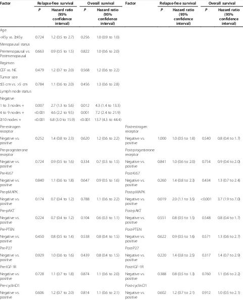
Table 2 Univariate analysis of relapse-free survival and overall survival
Factor Relapse-free survival Overall survival Factor Relapse-free survival Overall survival
P Hazard ratio (95% confidence
interval)
P Hazard ratio (95% confidence
interval)
P Hazard ratio (95% confidence
interval)
P Hazard ratio (95% confidence
interval)
Age
<45y vs.≥45y 0.724 1.2 (0.5 to 2.7) 0.256 1.0 (0.9 to 1.0)
Menopausal status
Premenopausal vs. Postmenopausal
0.663 0.9 (0.5 to 1.5) 0.822 1.0 (0.6 to 2.0)
Regimen
CEF vs. NE 0.479 1.2 (0.7 to 2.0) 0.568 1.2 (0.6 to 2.2)
Tumor size
≤5 cm vs. >5 cm 0.784 1.1 (0.6 to 2.0) 0.456 1.3 (0.6 to 2.8) Lymph node status
Negative
1 to 3 nodes + 0.007 2.7 (1.3 to 5.6) 0.012 4.3 (1.4 to 13.3)
4 to 9 nodes + <0.001 4.6 (2.2 to 9.5) 0.001 7.2 (2.4 to 21.9) ≥10 nodes + <0.001 6.8 (3.0 to 15.9) <0.001 13.7 (4.3 to 44.4) Pre-estrogen
receptor
Post-estrogen receptor
Negative vs. positive
0.252 1.4 (0.8 to 2.3) 0.620 1.2 (0.6 to 2.2) Negative vs. positive
1.000 1.0 (0.5 to 1.8) 0.540 0.8 (0.4 to 1.7)
Pre-progesterone receptor
Post-progesterone receptor
Negative vs. positive
0.724 0.9 (0.5 to 1.6) 0.334 0.7 (0.3 to 1.5) Negative vs. positive
0.841 1.0 (0.6 to 2.0) 0.754 0.9 (0.4 to 2.0)
Pre-Ki67 Post-Ki67
Negative vs. positive
0.840 1.1 (0.6 to 1.8) 0.647 0.9 (0.5 to 1.6) Negative vs. positive
0.260 1.4 (0.8 to 2.3) 0.434 1.3 (0.7 to 2.4)
Pre-pMAPK Post-pMAPK
Negative vs. positive
0.174 0.7 (0.4 to 1.2) 0.788 1.1 (0.6 to 2.2) Negative vs. positive
0.019 2.0 (1.1 to 3.5) <0.001 3.7 (1.9 to 7.0)
Pre-pAKT Post-pAKT
Negative vs. positive
0.224 0.7 (0.4 to 1.2) 0.104 0.6 (0.3 to 1.1) Negative vs. positive
0.551 0.8 (0.5 to 1.5) 0.548 0.8 (0.4 to 1.7)
Pre-PTEN Post-PTEN
Negative vs. positive
0.450 0.8 (0.5 to 1.4) 0.538 0.8 (0.4 to 1.5) Negative vs. positive
0.622 0.9 (0.5 to 1.6) 0.571 1.3 (0.6 to 2.7)
Pre-P27 Post-P27
Negative vs. positive
0.929 1.0 (0.6 to 1.6) 0.439 0.8 (0.4 to 1.5) Negative vs. positive
0.220 1.4 (0.8 to 2.5) 0.317 1.4 (0.7 to 2.9)
Pre-IGF-1R Post-IGF-1R
Negative vs. positive
0.728 1.1 (0.7 to 1.8) 0.874 1.1 (0.6 to 2.0) Negative vs. positive
0.388 0.8 (0.5 to 1.3) 0.760 1.1 (0.6 to 2.2)
Pre-cyclinD1 Post-cyclinD1
Negative vs. positive
0.606 1.2 (0.7 to 2.0) 0.814 1.1 (0.6 to 2.1) Negative vs. positive
0.602 1.2 (0.7 to 2.1) 0.912 1.0 (0.5 to 2.1)
sided P< 0.05. All analyses were performed using SPSS 17.0 (SPSS, Chicago, IL).
Results
Clinical characteristics and responses to NAC
A total of 113 HER2-positive breast cancer patients were recruited in this retrospective study. The average age of patients at diagnosis was 49 (range 26 to 78) years; 69 patients were premenopausal at presentation. There were 67 patients with a baseline tumor size greater than 5 cm. The CEF regimen was given to 55 patients, and the other patients received the NE regimen. The mean number of NAC cycles was 3.52 (range 1 to 8), and the objective response (complete response + partial response) and non-response rates (progressive disease + stable dis-ease) were 70.8% and 29.2%, respectively. Eight patients underwent breast-conserving surgery. The clinical charac-teristics of these patients are shown in Table 1.
Changes in biomarker expression after NAC
The expressions of various biomarkers (in Figure 1) before and after NAC were compared in 99 patients who did not achieve complete pathological response after NAC. A paired t test analysis found no significant changes in p27, PTEN, IGF-1R, or cyclinD1 expression (15.7% vs. 16.5%, 17.0% vs. 18.3%, 49.0% vs. 51.0%, and 18.2% vs. 16.9%, respectively). A significant decrease in Ki67, Phospho-p44/42, and pAKT expression was ob-served after treatment (30.7% vs. 18.1%, 36.4% vs. 18.9%, and 35.1% vs. 16.4%, respectively). We defined positive-staining tumor cells decreasing by more than the cut-off
value as positive decrease in biomarker expression. The details are shown in Table 2. Ki67, Phospho-p44/42, and pAKT expression were all significantly decreased after anthracycline-based neoadjuvant chemotherapy, as shown in Figure 2.
Predictors and response to NAC
Biomarkers and clinical characteristics were examined to investigate their value in predicting the NAC response (Table 2). Univariate analysis demonstrated that tumor size and pre-pAKT were predictive factors of the
response to anthracyclines (P= 0.001 and P= 0.014,
respectively). Multivariate analysis demonstrated that primary tumor size and pAKT expression remained in-dependent predictive factors of the clinical response to
anthracycline-based NAC (P= 0.012 and P= 0.031,
re-spectively). However, no biomarker could predict a pathologic complete response. We also found that the clinical response was coincident with decreased bio-marker expression, including Ki67, pAKT, and Phospho-p44/42 (P= 0.001,P= 0.002, andP= 0.007, respectively), as shown in Figure 3.
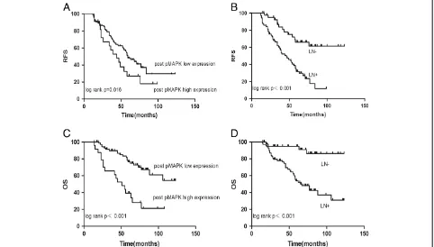
Prognostic markers
The median follow-up time was 60 months (ranging from 14 to 123 months). The overall 5-year relapse-free survival was 50.4%, and the overall survival was 72.6%. A univariate analysis (Table 2) demonstrated that the num-ber of positive lymph nodes and post-Phospho-p44/42 expression were prognostic factors for relapse-free sur-vival. In the multivariate analysis, the number of positive
Figure 2Ki67, Phospho-p44/42 and pAKT expression were significantly decreased after anthracycline treatment. (A)Ki67, (B)Phospho-p44/42,(C)pAKT.
lymph nodes (hazard ratio, 2.0; 95% confidence interval, 1.6 to 2.6; P< 0.001) and post-Phospho-p44/42 expres-sion (hazard ratio, 2.3; 95% confidence interval 1.3 to 4.1; P< 0.001) remained significantly independent prog-nostic factors. In the univariate analysis (Table 2), lymph node status (P< 0.001) and post-Phospho-p44/42 expres-sion (P= 0.019) showed clear associations with overall survival. In the multivariate analysis, lymph node status (hazard ratio, 2.3; 95% confidence interval, 1.7 to 3.3; P< 0.001) and post-Phospho-p44/42 expression (hazard ratio, 4.3; 95% confidence interval, 2.2 to 8.4; P< 0.001) were also significant predictors of overall survival. Repre-sentative survival curves are shown in Figure 4.
Discussion
To our knowledge, this is the largest analysis of PI3K-Akt and MAPK pathway activation in HER2-overexpressing breast cancer patients who received an anthracycline-based neoadjuvant chemotherapy regimen without trastuzumab.
Owing to its aggressive nature and poor prognosis, a number of preclinical and clinical studies have focused on the HER2-positive subtype. The use of anthracyclines in neoadjuvant treatments for HER2-positive breast can-cer in addition to trastuzumab is still controversial [17].
HER2 signaling activates pathways (PI3K/Akt and Ras/MAPK) regulating cell cycle progression and cell
proliferation. In HER2-overexpressing MBC group,
Gori (et al. found that Phospho-p44/42 and pAKT were
not associated with the clinical outcome, although low Phospho-p44/42 expression showed a trend for associ-ation with a longer overall survival [18]. In our study, the pre-pAKT and pre-Phospho-p44/42 expression rates were high, and this finding has been confirmed by other recent studies [19-21]. The results indicate that the Ras/MAPK and PI3K/Akt pathways are universally active in locally advanced breast cancer with HER2 over-expression. However, the PI3K/Akt and Ras/MAPK pathways have been related to resistance to doxorubicin and paclitaxel in breast cancer cells [22,23]. The level of pre-pAKT expression has been shown to have a signifi-cant correlation with the objective response rates to anthracycline treatment. It is possible that Akt isoforms have a distinct impact on the cellular resistance to a given drug and, in fact, Akt activity does not confer equal resistance to different chemotherapeutic agents. For example, the overexpression of constitutively active Akt isoforms in HeLa cells has been shown to induce isoform-specific sensitivity to doxorubicin [24]. The role of pAKT in the neoadjuvant setting is still controversial, owing to limited investigations, even in large clinical tri-als. However, decreases in Phospho-p44/42 and pAKT expression are related to the response to anthracyclines. Higher levels of active MAPK may have aggressive
biological behavior, which has been found to be associ-ated with lymph node metastasis [25]. In the TNBC subgroup, high ERK protein expression levels and shorter survival times have been observed [26]. After anthracycline-based adjuvant treatment, a higher score was significantly associated with poorer survival follow-ing relapse compared to a lower expression score among patients with MAPK overexpression [21].
Furthermore, few studies have presented precise values for pAKT and Phospho-p44/42 in neoadjuvant chemo-therapy. In our study, low Phospho-p44/42 expression after neoadjuvant chemotherapy was a strong prognostic factor for these patients. Patients with high post-Phospho-p44/42 expression had a higher recurrence rate (up to 68%) in the first 5 years, while patients with low post-Phospho-p44/42 expression had a lower recurrence rate (45%), and the survival difference between the two groups was highly significant. It is possible that de-creased pAKT and Phospho-p44/42 expression promotes tumor apoptosis and inhibits tumor proliferation, result-ing in a survival benefit for HER2-positive breast cancer patients treated with anthracyclines. It is also possible
that topoisomerase IIα expression is regulated by Ras
pathways and tumor proliferation status. The activation of the Ras/Raf/MAPK pathway has been shown to be involved in the induction of MRP-1 activity and
topoisomerase IIα downregulation, which are the main
mechanisms of anthracycline resistance [27].
Higher levels of the proliferation marker Ki67 are as-sociated with poorer survival in breast cancer patients, but we found no prognostic value for pre-NAC Ki67, post-NAC Ki67, or the Ki67 fold change. Other studies have reached controversial conclusions, and it is there-fore difficult to choose reasonable predictive and prog-nostic factors among pre-NAC Ki67, post-NAC Ki67, and Ki67 reduction [14,28-30].
This study has limitations common to all retro-spective analyses, and it lacked a control group, such as patients treated with trastuzumab-containing NAC. However, based on results of big clinical trials, such as HERA, BCIRG 006, NCCTG N9831, and NSABP B-31, trastuzumab was approved for adjuvant chemo-therapy after 2005. In our study, 71% patients were treated before 2005 when most patients did not re-ceive trastuzumab in developing countries. Addition-ally, the number of patients was small. However, the scientific and clinical community must establish and evaluate these biomarkers and standardize cut-off
levels. Although a measurement of topoisomerase II-α
amplification was not within the scope of this study, a possible explanation for our findings may be that other confounding molecular factors are involved in the mechanism of the anthracycline response in HER2-positive patients.
Conclusions
In HER2-positive breast cancer patients treated with anthracyclines, the expression of pAKT, Phospho-p44/ 42, and Ki67 decreased significantly after treatment. Furthermore, patients with high pAKT expression before NAC had higher objective response rates to anthracy-clines. The results of our study demonstrate that Phospho-p44/42 expression after neoadjuvant chemo-therapy is a strong predictor of outcome. It will be necessary and valuable to further evaluate potential therapeutic targets of the PI3K and MEK signaling path-ways in HER2-positive breast cancer patients.
Abbreviations
ERK:Extracellular signal-related kinase; HER2: Human epidermal growth factor Receptor 2; IGF-1R: Insulin-like growth factor 1 receptor; MAPK: Mitogen-activated protein kinase; NAC: Neoadjuvant chemotherapy; pAKT: Phospho-protein kinase B; Phospho-p44/42: Phospho-mitogen-activated Phospho-protein kinase; pMAPK: Phosphorylated mitogen-activated protein kinase.
Competing interests
All authors declare that they have no potential conflict of interest.
Authors’contributions
LH and T-WC have made substantial contributions to the conception and design of the study, and the acquisition of data. SC and YL analyzed and interpreted the data. JW and Z-MS revised the manuscript critically for important intellectual content. C-MC gave final approval of the version to be published. All authors read and approved the final manuscript.
Acknowledgements
This research is supported by Multidiscipline Comprehensive Treatment Cooperation Group Foundation of Fudan University Cancer Hospital, Shanghai, China (DXK200801), the Key Clinical Program of the Ministry of Health (2010 to 2012). The funders had no role in study design, data collection and analysis, decision to publish, or preparation of the manuscript.
Author details
1Department of Breast Surgery, Fudan University Shanghai Cancer Center/ Cancer Institute, 399 Ling-Ling Road, 200032 Shanghai, People’s Republic of China.2Department of Oncology, Shanghai Medical College, Fudan University, Shanghai, People’s Republic of China.3Department of Thyroid and Breast Surgery, Affiliated Nanshan Hospital of Guangdong Medical College, Shenzhen, People’s Republic of China.
Received: 23 April 2013 Accepted: 9 November 2013 Published: 30 November 2013
References
1. Slamon DJ, Clark GM, Wong SG, Levin WJ, Ullrich A, McGuire WL:Human breast cancer: correlation of relapse and survival with amplification of the HER-2/neu oncogene.Science1987,235:177–182.
2. De Placido S, Perrone F, Carlomagno C, Morabito A, Pagliarulo C, Lauria R, Marinelli A, De Laurentiis M, Varriale E, Petrella G, Gallo C, Bianco AR:CMF vs alternating CMF/EV in the adjuvant treatment of operable breast cancer. A single centre randomised clinical trial (Naples GUN-3 study).
Br J Cancer1995,71:1283–1287.
3. Pritchard KI, Shepherd LE, O’Malley FP, Andrulis IL, Tu D, Bramwell VH, Levine MN:HER2 and responsiveness of breast cancer to adjuvant chemotherapy.N Engl J Med2006,354:2103–2111.
4. Paik S, Bryant J, Park C, Fisher B, Tan-Chiu E, Hyams D, Fisher ER, Lippman ME, Wickerham DL:Wolmark N: erbB-2 and response to doxorubicin in patients with axillary lymph node-positive, hormone receptor-negative breast cancer.J Natl Cancer Inst1998,90:1361–1370.
5. Dhesy-Thind B, Pritchard KI, Messersmith H, O’Malley F, Elavathil L, Trudeau M:
6. Gennari A, Sormani MP, Pronzato P, Puntoni M, Colozza M, Pfeffer U, Bruzzi P:
HER2 status and efficacy of adjuvant anthracyclines in early breast cancer: a pooled analysis of randomized trials.J Natl Cancer Inst2008,
100:14–20.
7. Montemurro F, Aglietta M:Incorporating trastuzumab into the neoadju-vant treatment of HER2-overexpressing breast cancer.Clin Breast Cancer 2005,6:77–80.
8. von Minckwitz G, Untch M, Blohmer JU, Costa SD, Eidtmann H, Fasching PA, Gerber B, Eiermann W, Hilfrich J, Huober J, Jackisch C, Kaufmann M, Konecny GE, Denkert C, Nekljudova V, Mehta K, Loibl S:Definition and impact of pathologic complete response on prognosis after neoadjuvant chemotherapy in various intrinsic breast cancer subtypes.J Clin Oncol 2012,30:1796–1804.
9. Untch M, Loibl S, Bischoff J, Eidtmann H, Kaufmann M, Blohmer JU, Hilfrich J, Strumberg D, Fasching PA, Kreienberg R, Tesch H, Hanusch C, Gerber B, Rezai M, Jackisch C, Huober J, Kühn T, Nekljudova V, von Minckwitz G, German Breast Group (GBG), Arbeitsgemeinschaft Gynäkologische Onkologie-Breast (AGO-B) Study group:Lapatinib versus trastuzumab in combination with neoadjuvant anthracycline-taxane-based
chemotherapy (GeparQuinto, GBG 44): a randomised phase 3 trial.
Lancet Oncol2012,13:135–144.
10. Creighton CJ, Casa A, Lazard Z, Huang S, Tsimelzon A, Hilsenbeck SG, Osborne CK, Lee AV:Insulin-like growth factor-I activates gene transcrip-tion programs strongly associated with poor breast cancer prognosis.
J Clin Oncol2008,26:4078–4085.
11. Turner BC, Haffty BG, Narayanan L, Yuan J, Havre PA, Gumbs AA, Kaplan L, Burgaud JL, Carter D, Baserga R, Glazer PM:Insulin-like growth factor-I receptor overexpression mediates cellular radioresistance and local breast cancer recurrence after lumpectomy and radiation.Cancer Res 1997,57:3079–3083.
12. Wolff AC, Hammond ME, Schwartz JN, Hagerty KL, Allred DC, Cote RJ, Dowsett M, Fitzgibbons PL, Hanna WM, Langer A, McShane LM, Paik S, Pegram MD, Perez EA, Press MF, Rhodes A, Sturgeon C, Taube SE, Tubbs R, Vance GH, van de Vijver M, Wheeler TM, Hayes DF, American Society of Clinical Oncology/College of American Pathologists:American Society of Clinical Oncology/College of American Pathologists guideline
recommendations for human epidermal growth factor receptor 2 testing in breast cancer.J Clin Oncol2007,25:118–145.
13. Antonelli M, Massimino M, Morra I, Garre ML, Gardiman MP, Buttarelli FR, Arcella A, Giangaspero F:Expression of pERK and pAKT in pediatric high grade astrocytomas: correlation with YKL40 and prognostic significance.
Neuropathology2012,32:133–138.
14. Chen S, Chen CM, Yu KD, Yang WT, Shao ZM:A prognostic model to predict outcome of patients failing to achieve pathological complete response after anthracycline-containing neoadjuvant chemotherapy for breast cancer.J Surg Oncol2012,105:577–585.
15. Taunk NK, Goyal S, Moran MS, Yang Q, Parikh R, Haffty BG:Prognostic significance of IGF-1R expression in patients treated with breast-conserving surgery and radiation therapy.Radiother Oncol2010,96:204–208.
16. Sangale Z, Prass C, Carlson A, Tikishvili E, Degrado J, Lanchbury J, Stone S:
A robust immunohistochemical assay for detecting PTEN expression in human tumors.Appl Immunohistochem Mol Morphol2011,19:173–183. 17. Von Minckwitz G, Loibl S, Untch M:What is the current standard of care
for anti-HER2 neoadjuvant therapy in breast cancer?Oncology2012,
26:20–26.
18. Gori S, Sidoni A, Colozza M, Ferri I, Mameli MG, Fenocchio D, Stocchi L, Foglietta J, Ludovini V, Minenza E, De Angelis V, Crinò L:EGFR, pMAPK, pAkt and PTEN status by immunohistochemistry: correlation with clinical outcome in HER2-positive metastatic breast cancer patients treated with trastuzumab.Ann Oncol2009,20:648–654.
19. Andre F, Nahta R, Conforti R, Boulet T, Aziz M, Yuan LX, Meslin F, Spielmann M, Tomasic G, Pusztai L, Hortobagyi GN, Michiels S, Delaloge S, Esteva FJ:
Expression patterns and predictive value of phosphorylated AKT in early-stage breast cancer.Ann Oncol2008,19:315–320.
20. Gallardo A, Lerma E, Escuin D, Tibau A, Muñoz J, Ojeda B, Barnadas A, Adrover E, Sánchez-Tejada L, Giner D, Ortiz-Martínez F, Peiró G:Increased signalling of EGFR and IGF1R, and deregulation of PTEN/PI3K/Akt path-way are related with trastuzumab resistance in HER2 breast carcinomas.
Br J Cancer2012,106:1367–1373.
21. Derin D, Eralp Y, Ozluk Y, Yavuz E, Guney N, Saip P, Igci A, Ozmen V, Kücücük S, Aslay I, Aydiner A, Topuz E:Lower level of MAPK expression is associated with anthracycline resistance and decreased survival in patients with hormone receptor negative breast cancer.Cancer Invest 2008,26:671–679.
22. Liedtke C, Cardone L, Tordai A, Yan K, Gomez HL, Figureoa LJ, Hubbard RE, Valero V, Souchon EA, Symmans WF, Hortobagyi GN, Bardelli A, Pusztai L:
PIK3CA-activating mutations and chemotherapy sensitivity in stage II-III breast cancer.Breast Cancer Res2008,10:R27.
23. Stemke-Hale K, Gonzalez-Angulo AM, Lluch A, Neve RM, Kuo WL, Davies M, Carey M, Hu Z, Guan Y, Sahin A, Symmans WF, Pusztai L, Nolden LK, Horlings H, Berns K, Hung MC, van de Vijver MJ, Valero V, Gray JW, Bernards R, Mills GB, Hennessy BT:An integrative genomic and proteomic analysis of PIK3CA, PTEN, and AKT mutations in breast cancer.Cancer Res2008,
68:6084–6091.
24. Gagnon V, Van Themsche C, Turner S, Leblanc V, Asselin E:Akt and XIAP regulate the sensitivity of human uterine cancer cells to cisplatin, doxorubicin and taxol.Apoptosis2008,13:259–271.
25. Adeyinka A, Nui Y, Cherlet T, Snell L, Watson PH, Murphy LC:Activated mitogen-activated protein kinase expression during human breast tumorigenesis and breast cancer progression.Clin Cancer Res2002,
8:1747–1753.
26. Bartholomeusz C, Gonzalez-Angulo AM, Liu P, Hayashi N, Lluch A, Ferrer-Lozano J, Hortobagyi GN:High ERK protein expression levels correlate with shorter survival in triple-negative breast cancer patients.
Oncologist2012,17:766–774.
27. McCubrey JA, Steelman LS, Abrams SL, Lee JT, Chang F, Bertrand FE, Navolanic PM, Terrian DM, Franklin RA, D’Assoro AB, Salisbury JL, Mazzarino MC, Stivala F, Libra M:Roles of the RAF/MEK/ERK and PI3K/PTEN/AKT pathways in malignant transformation and drug resistance.Adv Enzyme Regul2006,46:249–279.
28. Jones RL, Salter J, A’Hern R, Nerurkar A, Parton M, Reis-Filho JS, Smith IE, Dowsett M:The prognostic significance of Ki67 before and after neoadju-vant chemotherapy in breast cancer.Breast Cancer Res Treat2009,116:53–68. 29. Tanei T, Shimomura A, Shimazu K, Nakayama T, Kim SJ, Iwamoto T, Tamaki
Y, Noguchi S:Prognostic significance of Ki67 index after neoadjuvant chemotherapy in breast cancer.Eur J Surg Oncol2011,37:155–161. 30. Fasching PA, Heusinger K, Haeberle L, Niklos M, Hein A, Bayer CM, Rauh C,
Schulz-Wendtland R, Bani MR, Schrauder M, Kahmann L, Lux MP, Strehl JD, Hartmann A, Dimmler A, Beckmann MW, Wachter DL:Ki67, chemotherapy response, and prognosis in breast cancer patients receiving neoadjuvant treatment.BMC Cancer2011,11:486.
doi:10.1186/1477-7819-11-307
Cite this article as:Huanget al.:Prognostic and predictive value of Phospho-p44/42 and pAKT in HER2-positive locally advanced breast cancer patients treated with anthracycline-based neoadjuvant chemotherapy.World Journal of Surgical Oncology201311:307.
Submit your next manuscript to BioMed Central and take full advantage of:
• Convenient online submission
• Thorough peer review
• No space constraints or color figure charges
• Immediate publication on acceptance
• Inclusion in PubMed, CAS, Scopus and Google Scholar
• Research which is freely available for redistribution