DOI: 10.1534/genetics.106.063966
Antagonizing Scalloped With a Novel Vestigial Construct Reveals an
Important Role for Scalloped in
Drosophila melanogaster
Leg, Eye
and Optic Lobe Development
Ankush Garg, Ajay Srivastava, Monica M. Davis, Sandra L. O’Keefe,
Leola Chow and John B. Bell
1Department of Biological Sciences, University of Alberta, Edmonton, Alberta T6G 2E9, Canada
Manuscript received July 25, 2006 Accepted for publication November 1, 2006
ABSTRACT
Scalloped (SD), a TEA/ATTS-domain-containing protein, is required for the proper development of
Drosophila melanogaster. Despite being expressed in a variety of tissues, most of the work on SD has been restricted to understanding its role and function in patterning the adult wing. To gain a better under-standing of its role in development, we generated sd47M flip-in mitotic clones. The mitotic clones had developmental defects in the leg and eye. Further, by removing the VG domains involved in activation, we created a reagent (VGDACT) that disrupts the ability of SD to form a functional transcription factor complex and produced similar phenotypes to the flip-in mitotic clones. The VGDACT construct also disrupted adult CNS development. Expression of the VGDACT construct in the wing alters the cellular localization of VG and produces a mutant phenotype, indicating that the construct is able to antagonize the normal function of the SD/VG complex. Expression of the protein:protein interaction portion of SD is also able to elicit similar phenotypes, suggesting that SD interacts with other cofactors in the leg, eye, and adult CNS. Furthermore, antagonizing SD in larval tissues results in cell death, indicating that SD may also have a role in cell survival.
T
HROUGHOUT the course of development a single transcription factor may often be used to control the patterning of different tissues. For example, in Dro-sophila melanogaster, the paired domain protein Eyeless is required for the proper development of the adult compound eye (Halderet al.1995; Shenget al.1997)as well as of the adult central nervous system (Callaerts
et al.2001), while the Drosophila GATA factor Pannier is required for sensory organ development (Ramain
et al.1993; Heitzleret al.1996), cardioblast
differen-tiation (Gajewskiet al.1999, 2001), and dorsal closure
(Herranzand Morata 2001). The Drosophila TEA/
ATTS domain (TEAD) protein, Scalloped (SD), is a tran-scription factor that is expressed in several different tissues throughout development. The existance of lethal alleles ofsd(Campbellet al.1991, 1992) implies that this
gene is vital for proper development.
The TEAD is a highly conserved DNA-binding domain that recognizes the M-CAT motif (59-TCATTCCT-39) (Hwang et al. 1993; Stewart et al. 1994).
TEAD-containing proteins generally bind directly to tissue-specific transcriptional intermediary factors (TIFs) to properly function as a specific transcription factor (TF) complex (Halder et al. 1998; Simmonds et al. 1998;
Vaudinet al.1999; Jianget al.2000; Maedaet al.2002;
Chenet al.2004a,b; Mahoneyet al.2005). Since
TEAD-containing proteins, such as SD, often lack an activation domain (Hwang et al. 1993; Vaudinet al. 1999), the
associated TIFs may provide this function (Halder
et al. 1998; Simmonds et al. 1998; Vaudinet al. 1999;
Maedaet al. 2002; Chen et al.2004a,b). Alternatively,
the activation domains of TEAD-containing proteins may associate with TIFs that can act as coactivators or corepressors. To date, two TIF-interacting domains have been identified for TEAD proteins: the Vestigial inter-acting domain (VID) (Halder et al. 1998; Simmonds
et al.1998) and the C-terminal YAP/TAZ-transactivating domain (TD) (Vassilevet al.2001; Fossdalet al.2004).
GST pull-down experiments with the human homolog transcription enhancer factor-1 (TEF-1) loosely posi-tion the VID and the TD between amino acids 221 and 329 (Vassilevet al.2001) and 224 and 426 (Vassilev
et al. 2001), respectively. The VID interacts with the Vestigial protein (VG) (Halderet al.1998; Simmonds
et al.1998) and with Vestigial-like proteins (VGL) (Vaudin
et al.1999; Halderand Carroll2001; Mielcareket al.
2002; Chen et al.2004a,b) in flies and mammals,
res-pectively. To date, the TD has been shown to be func-tionally important only in the human TEAD proteins and to interact with the yes-associating protein 65 (YAP65) (Vassilev et al. 2001) and with the Yap65 homolog
TAZ (Mahoneyet al.2005).
The gene scalloped (sd) is the only TEAD-encoding gene in the Drosophila genome. Enhancer trap studies
1Corresponding author:Department of Biological Sciences, University of Alberta, Edmonton, AB T6G 2E9, Canada. E-mail: [email protected]
reveal thatsdis first expressed at stage 14 in the periph-eral nervous system (PNS), the antennomaxillary com-plex, and the supraesophageal ganglion. By stage 16,sd
expression is expanded to the anterior sense organs and the sense organs of the gnathal regions (Campbellet al.
1992). SD is also thought to be expressed in embryonic cardiac cells (Bidetet al.2003). In third instar larvae,sd
is present in the optic lobes and in a few discrete cells of the cerebral hemisphere and the ventral nerve cord. In the wing imaginal disc,sdis expressed in the wing blade, scutellum, and the mesopleura, while in the eye imagi-nal disc, staining is restricted to cells behind the mor-phogenetic furrow (Campbellet al.1992).
Most of our understanding of SD function is derived from work done with the wing imaginal disc, where SD interacts with the TIF, VG (Halder et al. 1998;
Simmonds et al. 1998). VG does not contain a
DNA-binding motif, but contains a SD-interacting domain (SID) and two regions important for activation of down-stream genes, located at the N- and C-terminal ends of the protein (Vaudinet al. 1999; MacKay et al. 2003).
These three regions are required for the proper develop-ment of the wing blade (MacKayet al.2003). Binding of
SD to VG is necessary for the formation of a functional transcription complex that is able to specifically activate the expression of downstream wing genes (Halderet al.
1998; Simmondset al.1998). Ectopic expression of VG
in cells expressing SD but not VG causes activation of downstream wing genes and directs the developmental fate of that tissue into a wing (Kimet al.1996; Halder
et al.1998). SD is also required to localize VG to the nucleus (Halder et al. 1998; Simmonds et al. 1998;
Srivastava et al. 2002). In vitro experiments have
shown that the binding of VG to SD can alter the DNA motif that SD recognizes. These experiments have also shown that the interaction of a truncated form of VG, containing only the SID, with SD is able to disrupt the complex from binding to wing-specific DNA-binding motifs. Proper recognition of the wing-specific DNA-binding motifs by SD requires the SID and at least one of the VG domains involved in activation (Halder
and Carroll2001). This is consistent with the findings
that the protein encoded by a fusion of thesdTEAD domain with a full-lengthvggene can rescuesdandvg
wing mutations (Srivastavaet al.2002). In the absence
of VG, SD is still able to bind to DNA, but recognizes a different motif (Halderand Carroll2001).
The function of SD in nonwing tissues is poorly un-derstood. Several lethal alleles ofsdhave been isolated (Campbellet al.1991). The lethal alleles ofsdfall into
two classes: embryonic and pupal lethal (Campbellet al.
1991; Srivastavaet al. 2004). Since wings are not
es-sential to the survival of laboratory stocks, SD likely plays a vital role in nonwing tissues. To gain a better un-derstanding of the role that SD plays in development, we generated mitotic clones using an embryonic lethal allele of SD. Loss of SD caused blistering in the wings
(Liu et al. 2000), alterations in eye bristle shape and
patterning, and truncation of the legs. We further as-sessed the role of SD by antagonizing its function with a novel allele of VG. By expressing a truncated form of VG, VGDACT (Figure 1), that is able to bind to SD but not activate transcription, we were able to recapitulate all the phenotypes observed with the flip-in clones as well as cause defects in the optic lobe and promote fu-sions between ommatidia. Despite the VGDACT pro-tein’s ability to antagonize SD in larval tissues, it did not affect the development of the embryonic PNS, central nervous system (CNS), or cardiac cells. To test whether the phenotypes seen in the affected tissues are due to the inability of SD to form a functional TF, we also ex-pressed a truncated form of SD, SDD200 (Figure 1), that is unable to bind DNA but retains the TIF-interacting domains (Chowet al.2004). The SDD200 protein should
be able to bind to any TIFs that normally interact with the C-terminal 244 amino acids of the SD protein. Expression of the SDD200 protein induces phenotypes similar to those caused by the flip-in clones and the VGDACT protein, indicating that SD likely binds to currently unknown TIFs in the eye, leg, and optic lobe. Through these studies we show that the level of SD is important in these tissues and that the SD/TF complex is involved in promoting cell survival in the leg imaginal disc.
MATERIALS AND METHODS
Drosophila stocks:All crosses were done at 25°. The UASsd, UASsdD200, UASsdD88-123, and UASsdD88-159 constructs are described in Chow et al. (2004). The ptcGal4 and vgGal4
strains were a gift from S. Carroll, thesd47MFRT 18A was a gift from K. Irvine, andw1
,ry1
2[p-Myc] FRT18A; Sb FLP/Tb was Figure1.—Schematic of the VG and SD constructs used
a gift from S. Hughes. The 24BGal4, 109(2)80Gal4, 167YGal4, 179YGal4, C147Gal4, c698aGal4, ActinGal4,atoGal4, ChaGal4, CQ2Gal4, DllGal4, eyGal4, GMRGal4, Pan-R7Gal4, PdfGal4, RN2Gal4, sd72b, and sevEPGal4 stocks were obtained from the Bloomington Stock Center. Thesd72ballele is a deficiency stock containing a deletion the spans the entiresdgene.
Flip-in clones: The sd47M allele is a 157-bp deletion (Srivastavaet al.2004) and homozygous individuals exhibit
an embryonic lethal phenotype (Campbellet al.1991). The
sd47Mallele was recombined onto a w, P(ry, neoFRT)18A chro-mosome (Liuet al.2000). Mitotic clones of this allele were
generated by flipase-mediated mitotic recombination (Golic
and Lindquist1989; Xuand Rubin1993) by repeated daily
heat shocking at 39°for 45-min intervals.
Construction of VGDACT: Using a polymerase chain reaction (PCR) protocol, the nucleotides that encode amino acids 171–335 of VG were amplified with the following primers: 59-TCG AGG CCT CAC ACA CAC ACG CAT ACG-39
and 59-GGG CTC GAG TTA GTG CAC GTA ATT GCT GTT-39. The PCR products were digested with XhoI and StuI and cloned into a p131 vector (Abu-Shaaret al.1999).
PCR conditions:ATaq:Pfu(20:1) mix was used in standard PCR conditions. The template was allowed to initially denature at 94°for 5 min. DNA was amplified with 30 cycles at 94°for 30 sec (denaturing) followed by 55°for 30 sec (annealing) and 68°for 90 sec (extension).
Micro-injections: Micro-injections were performed as de-scribed in Rubinand Spradling(1982) using the VGDACT
construct in pUAST and a helperD2-3 plasmid provided by S. Campbell. Four independent transgenic lines were isolated and tested. All the results shown make use of the VGDACT-22 line.
Scanning electron microscopy: Adult flies were fixed in a 2% glutaraldehyde, 13PBS solution for 1 hr at room tem-perature. Samples were washed twice in 13PBS before being dehydrated in increasing concentrations of ethanol. Ethanol was removed by bathing the samples in increasing concen-trations of hexamethyldisilazane:ethanol solutions. Samples were dried and gold coated before visualization by scanning electron microscopy (SEM).
Silver staining: Adult flies were fixed overnight in a 10% formalin solution, dehydrated, and embedded in paraffin. Sam-ples were sectioned at 15mm and mounted on charged glass slides, after which the paraffin was removed and the samples were rehydrated. Silver staining of samples was performed as described in Naoumenkoand Feigin(1967).
Acridine–orange staining: Imaginal discs were dissected in 13PBS and incubated in a 1.6310 6msolution of acridine– orange solution for 10 min. Discs were rinsed and mounted in a 13PBS solution.
RESULTS
SD47M mitotic clones affect the development of
bristles and legs:To gain a better understanding of the
role thatsdplays in development, we generated flip-in mitotic clones using an embryonic lethal allele of sd,
sd47M. The sd47M allele contains a 157-bp deletion
span-ning intron 8 and exon 9 of the gene (Srivastavaet al.
2004). Because the deletion spans a splice site, it pre-sumably disrupts the VID and the C-terminal region of the protein. The induction ofsd47M
mitotic clones causes defects in the wing (Liuet al.2000), eye bristles, and the
leg. Similar to the wing, clones in the eyes were very small. Now,ryclones were visible in the eye. In wild-type
eyes, bristles are found at alternating vertices of each ommatidium (Figure 2, A and B). The effect of clonal induction on bristles varied. SEM analysis revealed that a few of the bristles were either smaller (Figure 2C) or mispatterned (Figure 2D). Wild-type legs are made up of three basic parts: the femur, the tibia, and the tarsus (Figure 2E). The distal end of the fly leg contains a metatarsal claw (Figure 2E, arrow and inset). Inducing
sd47Mclones leads to truncations in the leg (Figure 2F;
arrow and inset emphasize the absence of the metatarsal claw).
VGDACT is able to antagonize SD function in the
wing imaginal disc: In the wing, induction of mitotic
clones causes only blistering, whereas homozygous viable mutant alleles of sdcause a loss of wing tissue.
Figure2.—Mitotic clones ofsd47Mcause defects in the eye
Because the clones are surrounded by a population of wild-type cells, it is possible that the loss ofsdin the eye and leg is partially masked. To create a more robust phenotype, we created a reagent, VGDACT, which is able to bind to SD but is not able to activate trans-cription. The VGDACT construct was generated by re-moving the two activation domains, ACT1 (amino acids 1–65) and ACT2 (amino acids 356–453) (Halderand
Carroll2001; MacKayet al.2003), fromvg(Figure 1),
leaving the entire scalloped interacting domain intact. Thus, the expectation is that the VGDACT protein will cause a dominant-negative phenotype. To test if the VGDACT construct can cause a dominant-negative effect, we first expressed it in the wing. Using the
vgGAL4 driver, we expressed the VGDACT transgene along the dorsal–ventral axis of the wing disc. This ec-topic expression causes a loss of wing bristles and wing tissue (compare Figure 3B to Figure 3A). The extent of the phenotype varies considerably from almost a com-plete loss of the wing (not shown) to small patches of missing bristles in the anterior margin and loss of wing tissue along the posterior margin (Figure 3B). To test whether the observed effects are due to the VGDACT protein antagonizing the endogenous SD protein, we attempted to rescue the dominant-negative phenotype by coexpressing length SD. Coexpression of full-length SD and VGDACTrescued the bristle and the wing margin phenotypes (Figure 3C).
VGDACT is not able to antagonize SD function in
the embryo:Since the VGDACT protein is able to cause
a dominant-negative phenotype in the wing, it may be able to have a similar effect in other tissues where SD is expressed. To test the ability of VGDACT to antagonize endogenous SD in other cells, we expressed the trans-gene in the embryo using an actin GAL4 driver and a muscle-specific 24BGAL4 driver (Kidd et al. 1999).
Overexpression of SD using the 24BGAL4 driver has been shown to cause embryonic lethality and to have an effect on the development of cardiac cells (Bidetet al.
2003). Furthermore, we found that tinman-expressing cells are mispatterned in asdnull background (data not shown). Neither embryonic lethality nor tinman mis-patterning is seen when the VGDACT or SD deletion transgenes are expressed under the control of either driver. However, larval lethality is seen when the trans-genes are expressed using the actin GAL4 driver.
VGDACT is able to antagonize SD function in tissues
outside the wing disc: Driving the VGDACT transgene
with the widely expressedptcGAL4 driver causes more robust defects in the fly (Figure 4) than those seen with the mitotic clones. Antagonizing SD in the eye causes the eyes to protrude (compare Figure 4B to Figure 4A). We also observe more severe defects that affect bristle patterning, number, and morphology. In VGDACT/1;
ptcGAL4/1flies, bristles can occur at adjacent vertices (Figure 4C; the ommatidium marked with an asterisk has bristles at four vertices) or they can be absent (Figure 4D, arrow). Duplications of bristles are also visible (Figure 4C, arrowhead) and in some of the more severe cases as many as three extra bristles at a single vertex are seen (not shown). Expression of the VGDACT protein also affects bristle size (Figure 4C; note the size differences between the bristles identified by the arrows). Interestingly, bristle defects are more frequent along the tip of the eye protrusion. In some eyes, defects in the ommatidia are also present. Changes in omma-tidia vary from increases in size (data not shown) to fusions between adjacent ommatidia (Figure 4D; com-pare fused ommatidia marked by a single asterisk to wild-type ommatidia marked by a double asterisk).
Four structures make up the optic lobe: lobulla, lob-ulla plate, medlob-ulla, and lamina (Figure 4E) (Armstrong
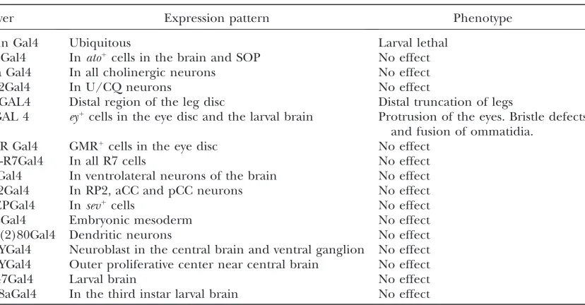
et al. 1995). In wild-type flies, the lamina makes di-rect contact with the basal membrane of ommatidia (Figure 4E). Silver staining of horizontal sections from VGDACT/1; ptcGAL4 flies reveals an ectopic cluster of cells between the lamina and the basal membrane (Figure 4F, arrowhead) that correlates with the location of the protrusion (Figure 4F, arrow). All of the compo-nents of the optic lobe are present and appear normal in these flies. To try to identify the cells affected in the eye and brain, we expressed our construct with a variety of drivers (see Table 1). Only the eyeless GAL4 driver was able to cause optic lobe and eye phenotypes. None of the other eye- or brain-specific drivers had any effect.
Truncation can occur in all three leg segments when the VGDACT transgene is under the control of the
DllGAL4 driver (Figure 4G; arrow and inset emphasize the absence of the metatarsal claw). The degree of leg truncation varies from loss of only a few of the metatar-sals (Figure 4G, arrow) to the loss of all the metatarmetatar-sals, tarsus, and tibia (not shown). Sharp bends in the femur
Figure3.—VGDACT interacts with SD and
dis-rupts SD function. (A) A wild-type wing. (B) Wing bristles and wing tissue are lost in avgGAL4/1;
UAS VGDACT/1 fly. (C) Rescue of the
or tibia are also sometimes seen (Figure 4G, arrow-head). In addition to causing truncations as seen in the mitotic clones, the VGDACT protein is also able to cause duplications of the leg. However, only the VGDACT/1;
ptcGAL4 flies show duplications of the leg (Figure 4H, arrowhead) and these occur only in the T2 and T3 legs.
SD likely binds to other cofactors:To help determine
if the VGDACT protein hinders the binding of impor-tant cofactors to SD, we expressed a truncated form ofsd
(SDD200) that lacks the coding capacity for the first 200 amino acids of SD (Figure 1). Although the SDD200 protein is missing the TEA/ATTS DNA-binding do-main, it should still be able to bind to TIFs and compete with endogenous SD for binding to them. Thus, the expectation is that if SD binds to and interacts with
other cofactors via a domain located in the terminal 244 amino acids of the protein, the SDD200 transgene should induce a phenotype similar to those seen in the VGDACT/1;ptcGAL4 flies. If, however, SD does not interact with cofactors, or interacts with cofactors via a domain located within the first 200 amino acids of the protein, the SDD200 transgene should have no effect. Expression of the SDD200 protein with the ptcGAL4 driver causes phenotypes similar to those observed with the VGDACT construct (Figure 5). In the head, the SDD200 protein causes an eye protrusion (data not shown), alters bristle patterning (Figure 5A, arrow) and morphology (Figure 5B, arrow), causes duplication of bristles (Figure 5A, arrowhead), induces the fusion of ommatidia (Figure 5C, asterisk), and produces an ectopic cluster of cells between the lamina and omma-tidia (Figure 5D). SDD200 is also able to cause duplica-tions in the legs (Figure 5E) and a loss of wing bristles and tissue (Figure 5F).
SD likely functions as a TF in tissues outside the wing
disc: To determine if the sole function of SD in larval
tissues is to transport proteins into the nucleus, we overexpressed SDD88-123 and SDD88-159 (Figure 1) using the ptcGAL4 driver. The protein encoded by the SDD88-123 transgene contains a deletion in the first half of the TEA/ATTS DNA-binding domain that does not compromise the nuclear localization signal (NLS), while the deletion in the SDD88-159 removes the entire TEA/ATTS DNA-binding domain and the majority of the NLS. If the sole function of SD is to transport proteins into the nucleus, expression of the SDD88-159 but not the SDD88-123 transgene should be able to induce phenotypes similar to those observed by express-ing VGDACT and SDD200. Expression of either the SDD88-123 or the SDD88-159 proteins is able to induce similar eye, brain, wing, and leg phenotypes (data not
Figure 4.—Antagonizing SD in tissues outside the wing.
shown), indicating that the role of SD in these tissues is not limited to transporting proteins into the nucleus.
Levels of SD are important in tissues outside the wing disc:To assess whether the relative levels of SD are as important for the proper development of the eye, brain, and leg as they are in the wing (Halderet al.
1998; Simmonds et al. 1998), we overexpressed the
full-length SD transgene (Figure 1) using theptcGAL4 driver. Unfortunately, this causes pupal lethality. Re-moval of pharate adults from the pupal cases reveals phenotypes similar to those seen in the presence of the VGDACT and the SDD200 proteins. Protrusion of the eyes (Figure 6A, arrowhead), truncations of the legs (Figure 6B, arrowheads), and an ectopic cluster of cells below the basal membrane of the ommatidia (Figure 6C) are all seen. In most of the pupae, the head of the fly
is found in the abdomen (Figure 6D). Silver staining of the horizontal sections in these flies shows that the lamina and regions of the optic lobe are missing (Figure 6, C and E). The brain is also physically separated from the optic lobe (Figure 6E). However, the development of the ommatidia remains intact (Figure 6F).
SD is required for cell survival in the leg disc: One
function of the SD/VG complex in the wing is to pro-mote cell survival (Liu et al. 2000; Delanoue et al.
2004). To ascertain if SD also plays a role in promoting cell survival in other larval tissues, we looked for the presence of increased cell death in our transgenic flies using acridine–orange staining. In the wild-type wing disc, SD expression is restricted to the wing pouch and the periphery of the notum. Very little to no cell death is visible in the wing (Figure 7A) and leg (Figure 7B)
TABLE 1
Eye and neural drivers used to express VGDACT and SDD200
Driver Expression pattern Phenotype
Actin Gal4 Ubiquitous Larval lethal
AtoGal4 Inato1cells in the brain and SOP No effect
Cha Gal4 In all cholinergic neurons No effect
CQ2Gal4 In U/CQ neurons No effect
DllGAL4 Distal region of the leg disc Distal truncation of legs
eyGAL 4 ey1cells in the eye disc and the larval brain Protrusion of the eyes. Bristle defects and fusion of ommatidia.
GMR Gal4 GMR1
cells in the eye disc No effect
Pan-R7Gal4 In all R7 cells No effect
PdfGal4 In ventrolateral neurons of the brain No effect
RN2Gal4 In RP2, aCC and pCC neurons No effect
sevEPGal4 Insev1cells No effect
24BGal4 Embryonic mesoderm No effect
109(2)80Gal4 Dendritic neurons No effect
167YGal4 Neuroblast in the central brain and ventral ganglion No effect
179YGal4 Outer proliferative center near central brain No effect
C147Gal4 Larval brain No effect
c698aGal4 In the third instar larval brain No effect
Figure5.—The SDD200 protein can also
antag-onize SD function.ptcGAL4/1; UAS SDD200/1
flies (32000) have mutant phenotypes similar to
imaginal discs of the wild-type fly. Staining ofptcGAL4; UAS SDD200 (Figure 7C, arrow) and ptcGAL4; UAS VGDACT (data not shown) imaginal discs shows only increased acridine–orange staining along the anterior/ posterior (A/P) boundary of the wing pouch. No in-crease in staining is seen along the A/P boundary of the notum (Figure 7C, arrowhead). Increased staining is also seen in the leg imaginal disc (Figure 7D). In addition to seeing an increase in staining in the leg discs, the leg discs may also be duplicated (Figure 7D). No notable change in acridine staining is visible in the larval optic lobes or in the eye-antennal imaginal disc (data not shown).
DISCUSSION
The generation of sd47M mitotic clones resulted in a
mild phenotype in the eye and leg and had no effect in the optic lobe. Considering that induction of sd47M
mitotic clones also caused a weak blistering phenotype in the wing (Liuet al.2000), it is not surprising that they
had a minor effect in tissues outside the wing. The weak phenotype is due to the inability of the clones to survive. No w, ryclones were seen in the eye, despite the fact that numerous blisters are present in the wing. However, the induction of clones is still able to cause a bristle phenotype, indicating that SD may be required for the survival of some of the eye cells.
To create a stronger phenotype, we attempted to antagonize the SD protein in the developing embryo and larva. This was accomplished by ectopically express-ing a truncated form of VG, VGDACT, which can still bind to SD but cannot activate transcription of down-stream genes. Thus, we hoped that the interaction between the VGDACT protein and SD would create an inert complex and consequently affect overall SD function. To test if the VGDACT protein can antagonize SD function, we expressed it in the wing. Similar tosd
Figure 7.—SD promotes cell
survival in the leg imaginal disc. Acridine–orange staining in a (A) wild-type wing imaginal disc, (B) wild-type leg imaginal disc, (C)ptcGAL4; UAS SDD200 wing imaginal disc, and (D)ptcGAL4; UAS SDD200 leg imaginal disc. Very little cell death is visible in the wild-type wing and leg imagi-nal discs. Ectopic expression of SDD200 induces cell death only in the (C) wing pouch (arrow) and not in the notum (arrowhead). Increased cell death is also visible in the (D) leg imaginal disc.
Figure6.—SD levels are important for proper
development. Overexpression of wild-type SD in
mutant flies, the VGDACT protein in the wing is able to induce wing-pouch-specific cell death (Figure 7C) (Liu et al. 2000) and to promote the loss of bristles
and wing tissue (Figure 3B) (Campbell et al. 1992;
Simmondset al.1998). These observations indicate that
the VGDACT protein likely binds to SD and disrupts its function in the wing.
While ectopic expression of full-length SD using either the actin or the 24BGal4 drivers causes embryonic lethality, the VGDACT and the SD deletion proteins are unable to affect embryonic development. Several possibilities may explain why the VGDACT protein is unable to disrupt SD function in the embryo: (1) proteins with which SD interacts in the embryo may utilize a domain that the VGDACT protein is unable to affect or may bind to a region outside of the SDD200 protein, (2) the TIFs with which SD interacts in the em-bryo have a higher binding affinity than the VGDACT protein, or (3) SD may have a TIF-independent role in the embryo. The inability of our construct to inhibit SD function in the embryo is consistent with the ob-servation that lethal alleles of sd carrying a point mutation in either the VID or the putative TD do not cause embryonic lethality (Srivastavaet al.2004), but
do cause pupal lethality. Insight into the regions of SD required for embryonic development may come from the fact that the SD human homolog TEF-1 is able rescue the embryonic lethal allelesdETX81
(Deshpande
et al.1997). These observations, taken together, indicate that the mechanism by which SD functions in the embryo is likely different from that in larval tissues.
The VGDACT protein, however, is able to antagonize SD function in larval tissues. Ectopic expression of the protein is able to disrupt leg, eye, and brain develop-ment. In the leg, expression of the VGDACT transgene is able to induce duplications and truncations (Figure 4, G and H). Several lines of evidence indicate that the phenotypes seen in the leg may be related to an induction of cell death, rather than to a defect in pattern formation. Previous reports have shown that high levels of apoptosis induced by ultraviolet (UV) light in the leg are able to induce splitting and duplication of the T2 and T3 but not T1 appendage (Arking1974). Similar to the UV experiments,
dupli-cations caused by the VGDACT protein in the leg disc are seen only in the second and third appendages. The lack of duplications seen in the T1 segment supports the idea that the phenomenon is related to cell death. Furthermore, dissections of VGDACT/1; ptcGAL4/1
larvae show splitting of the leg imaginal disc and an increased level of cell death (Figure 7D). Finally, ex-pression of the construct at the distal end of the leg with theDllGAL4 driver induces truncations at the tip of the appendage (Figure 4G).
Antagonizing the SD protein in the eye affects proper development of the bristles and ommatidia. Defects in bristles include changes in morphology and patterning.
Previous reports have shown that SD is able to activate genes involved in sensory organ development (Halder
et al.1998; Srivastavaand Bell2003) and expression
of thesdTEATVG fusion construct in the eye is able to
alter bristle morphology (Srivastavaet al.2002). Thus,
it is likely that SD has a role in patterning eye bristles. Another possible explanation for the bristle defects may be related to the cone cell phenotype. Pigment cell identity in the developing eye is based on cues provided by precursor cone cells (Caganand Ready1989a). The
secondary and tertiary pigments can be sacrificed to form extra bristle cells (Cagan and Ready 1989b).
Thus, it possible that the bristle phenotype may be due to defects in cone cell patterning. The ommatidial defects are seen on the surface of the fly eye and not in the horizontal sections. The inability to see these effects in the horizontal sections may be because ommatidial fusions occur at a low frequency in the eye (typically four to five fused ommatidia are seen in each eye) or because this inability is caused by cone-cell-patterning defects. Expression of the VGDACT protein is also able to cause a protrusion in the adult eye (Figure 4B). Horizontal sec-tions show that the protrusion is due to an unidentified ectopic layer of cells (Figure 4F). These cells are located between the lamina and the ommatidia (Figure 4F, arrowhead). Overexpression of SD induces similar, but more severe, phenotypes than when the VGDACT or SDD200 proteins are expressed (Figure 6). In flies where SD is overexpressed, components of the optic lobe are lost (Figure 6, C and E). Thus, it is possible that the ectopic layer of cells originates from the optic lobe.
To determine if SD forms a TF complex in larval tissues, we expressed a truncated form of SD, SDD200 (Figure 1). The DNA-binding domain of SD and the NLS are deleted in the SDD200 transgene. However, the SDD200 protein should still bind to and potentially sequester SD-specific TIFs. Expression of the SDD200 protein (Figure 5) is able to induce phenotypes similar to those seen with the VGDACT protein, suggesting that SD probably binds to cofactors in the eye, leg, and brain. In larval tissues, SD could interact with a putative TIF via the VID or perhaps even the TD. The existence of a TD domain in SD is supported by the observation that a mutation in the C-terminal end of the gene causes pu-pal lethality, but does not inhibit wing development (Srivastavaet al.2004). However, whether or not this
association forms a TF complex is not clear. In the wing, SD is required to transport VG into the nucleus (Halderet al.1998; Simmondset al.1998; Srivastava
In other species, whenever a TEAD protein interacts with a TIF it forms a TF complex. In mammals, the TEAD protein (TEF-1) is known to interact with a variety of different TIFs, such as VGL-1 and VGL-3 in the placenta (Halderet al.1998; Maedaet al.2002), VGL-2
in skeletal muscles (Maeda et al.2002), VGL-4 in the
heart (Chenet al.2004b), YAP65 (Vassilevet al.2001),
and p160 (Belandiaand Parker2000). In each case,
the interaction between TEF-1 and its relevant TIF results in a TF complex. Thus, it is likely that in larval tissues the association between SD and putative TIFs also results in the formation of a TF complex (SD/TIF). The identity of putative TIFs with which SD may interact is currently unknown. Homology searches reveal three potential novel interacting partners for SD. The VGL-4 homolog in Drosophila is an unidentified gene, CG10741. To date there have been no studies done with CG10741. The p130 homolog, taiman, is widely ex-pressed in the embryo and follicle cells in the larva (Bai et al. 2000). The YAP65 homolog, yorkie (yki), is
another putative SD-interacting partner. Yki is known to control cell survival and cell proliferation (Huanget al.
2005). Yeast two-hybrid analysis shows with high confi-dence that SD and Yki interact with each other (Giot
et al. 2003). Similar to VG, Yki contains an activation domain (Yagiet al.1999); thus it is possible that Yki and
SD interact with each other to form a functional TF complex. Presumably, this interaction occurs through the yet to be identifiedsdTD, as in humans YAP65 is known to interact with the TD of TEF-1. If Yki and SD form a complex, then it is possible that the Yki/SD complex may be the entity that is antagonized in the leg, eye, and/or brain.
One interesting property of SD is that its amount is important for proper wing development (Simmonds
et al.1998). A change in the ratio of SD to VG affects the development of the wing. Overexpressing SD causes phenotypes in leg, eye, and brain similar to those ob-served when the SD/TIF complex is antagonized. Why the levels of SD are important for its proper function remains unclear. In the wing, the SDVG complex re-quires two SD molecules and two VG molecules (Halder
and Carroll2001). SD can form homo-dimers (Halder
et al.1998) and thus the overexpression of SD may pro-mote the formation of SD dimers as opposed to form-ing a tetrameric TF complex with its TIF. Another possibility may be that free SD may act as a repressor (personal communication with S. Veeraraghavan) and
that overexpressing SD may lead to suppression of genes that are required to promote cell survival in the brain, eye, and leg.
When SD function is disrupted in the leg and the eye, a common phenotype observed is the loss of tissue. Bristles are lost in the eye (Figure 4D) and truncation can be seen in the legs (Figure 4G). Furthermore, the induction of clones in eye did not give rise to anyw,ry
clones. In the wing, SD has a crucial role in promoting
cell survival (Liuet al.2000; Delanoueet al.2004) and
this role may also be important in the eye and the leg. Acridine–orange staining of leg discs where SD is antag-onized shows an increased amount of cell death (Figure 7D), suggesting that the complex is required to promote cell survival. Whether this is a direct relationship is currently unclear. The increased cell death may be be-cause the cells in these tissues fail to properly differen-tiate, and consequently are unable to interpret the proper cues to survive. No increase in acridine–orange staining was observed in the eye disc (not shown). There is a high level of background acridine–orange staining (data not shown) and missing bristles occur only in a small percentage of the ommatidia, but this does not exclude the possibility that the SD/TIF complex is re-quired to promote cell survival in these tissues.
Determining the role that SD has in promoting the fate of eye cells, leg cells, or optic lobe cells is difficult without knowing the identity of the possible TIFs with which it associates. Thus, the exact role for such a complex in these tissues must still be determined. However, we were able to gain some insight into the function of SD in these tissues by generating mitotic clones and expressing a truncated form of VG, VGDACT. The VGDACT protein is able to bind to SD and likely prevents TIFs from interacting with SD protein:protein interaction domains that include any of the amino acids between positions 220 and 344 (Vaudinet al.1999). In the wing, the SD/VG complex
is required to determine wing fate (Halderet al.1998)
and to promote cell survival (Liuet al.2000; Delanoue
et al. 2004). We have shown that the mechanism by which SD functions in the eye, leg, and brain is similar to that of SD in the wing imaginal disc. SD likely binds to tissue-specific TIFs to form a TF and this interaction occurs by a domain located in the C-terminal portion of the protein. The TF complex is also sensitive to the levels of SD present in the system and antagonizing SD function induces cell death. Thus, the SD/TIF com-plexes in larval tissues may be involved in promoting optic lobe, bristle, ommatidia, and leg fate or be involved in regulating cell proliferation and/or pro-moting cell survival. Studies aimed at identifying the TIFs or at determining the exact protein:protein in-teraction domains that these TIFs recognize may be able to identify the role of SD in these tissues.
We thank Ross Hodgetts and Andrew Simmonds for their help with the manuscript; Rakesh Bhatnagar and Jack Scott for their assistance with the confocal and scanning electron microscopes; and Randy Mandryk for his help with preparation and staining the fly horizontal sections. This work was funded by the National Science and Engineer-ing Research Council (Canada) and the Alberta Ingenuity Fund.
LITERATURE CITED
Abu-Shaar, M., H. D. Ryooand R. S. Mann, 1999 Control of the
Arking, R., 1974 Temperature-sensitive cell-lethal muntants of
Dro-sophila: isolation and characterization. Genetics30:519–537. Armstrong, J. D., K. Kaiser, A. Muller, K. F. Fischbach, N.
Merchantet al., 1995 Flybrain, an on-line atlas and database
of the Drosophila nervous system. Neuron15:17–20.
Bai, J., Y. Ueharaand D. J. Montell, 2000 Regulation of invasive
cell behavior by taiman, a Drosophila protein related to AIB1, a steroid receptor coactivator amplified in breast cancer. Cell
103:1047–1058.
Belandia, B., and M. G. Parker, 2000 Functional interaction
be-tween the p160 coactivator proteins and the transcriptional en-hancer factor family of transcription factors. J. Biol. Chem.
275:30801–30805.
Bidet, Y., T. Jagla, J. P. Da Ponte, B. Dastugue and K. Jagla,
2003 Modifiers of muscle and heart cell fate specification iden-tified by gain-of-function screen in Drosophila. Mech. Dev.120:
991–1007.
Cagan, R. L., and D. F. Ready, 1989a The emergence of order in
the Drosophila pupal retina. Dev. Biol.136:346–362.
Cagan, R. L., and D. F. Ready, 1989b Notch is required for
succes-sive cell decisions in the developing Drosophila retina. Genes Dev.3:1099–1112.
Callaerts, P., S. Leng, J. Clements, C. Benassayag, D. Cribbset al.,
2001 Drosophila Pax-6/eyeless is essential for normal adult brain structure and function. J. Neurobiol.46:73–88.
Campbell, S. D., A. Duttaroy, A. L. Katzen and A. Chovnick,
1991 Cloning and characterization of the scalloped region of Drosophila melanogaster.Genetics127:367–380.
Campbell, S., M. Inamdar, V. Rodrigues, V. Raghavan, M.
Palazzoloet al., 1992 The scalloped gene encodes a novel,
evolutionarily conserved transcription factor required for sen-sory organ differentiation in Drosophila. Genes Dev.6:367–379. Chen, H. H., T. Maeda, S. J. Mullett and A. F. Stewart,
2004a Transcription cofactor Vgl-2 is required for skeletal mus-cle differentiation. Genesis39:273–279.
Chen, H. H., S. J. Mullettand A. F. Stewart, 2004b Vgl-4, a novel
member of the vestigial-like family of transcription cofactors, reg-ulates alpha1-adrenergic activation of gene expression in cardiac myocytes. J. Biol. Chem.279:30800–30806.
Chow, L., J. Berube, A. Fromontand J. B. Bell, 2004 Ability of
scalloped deletion constructs to rescue sd mutant wing pheno-types inDrosophila melanogaster. Genome47:849–859.
Delanoue, R., K. Legent, N. Godefroy, D. Flagiello, A. Dutriaux
et al., 2004 The Drosophila wing differentiation factor vestigial-scalloped is required for cell proliferation and cell survival at the dorso-ventral boundary of the wing imaginal disc. Cell Death Differ.11:110–122.
Deshpande, N., A. Chopra, A. Rangarajan, L. S. Shashidhara, V.
Rodrigues et al., 1997 The human transcription enhancer
factor-1, TEF-1, can substitute for Drosophila scalloped during wingblade development. J. Biol. Chem.272:10664–10668. Fossdal, R., F. Jonasson, G. T. Kristjansdottir, A. Kong, H.
Stefanssonet al., 2004 A novel TEAD1 mutation is the causative
allele in Sveinsson’s chorioretinal atrophy (helicoid peripapillary chorioretinal degeneration). Hum. Mol. Genet.13:975–981. Gajewski, K., N. Fossett, J. D. Molkentin and R. A. Schulz,
1999 The zinc finger proteins Pannier and GATA4 function as cardiogenic factors in Drosophila. Development126:5679–5688. Gajewski, K., Q. Zhang, C. Y. Choi, N. Fossett, A. Dang et al.,
2001 Pannier is a transcriptional target and partner of tinman during Drosophila cardiogenesis. Dev. Biol.233:425–436. Giot, L., J. S. Bader, C. Brouwer, A. Chaudhuri, B. Kuanget al.,
2003 A protein interaction map of Drosophila melanogaster. Science302:1727–1736.
Golic, K. G., and S. Lindquist, 1989 The FLP recombinase of yeast
catalyzes site-specific recombination in the Drosophila genome. Cell59:499–509.
Halder, G., and S. B. Carroll, 2001 Binding of the Vestigial
co-factor switches the DNA-target selectivity of the Scalloped selec-tor protein. Development128:3295–3305.
Halder, G., P. Callaerts and W. J. Gehring, 1995 Induction of
ectopic eyes by targeted expression of the eyeless gene in Dro-sophila. Science267:1788–1792.
Halder, G., P. Polaczyk, M. E. Kraus, A. Hudson, J. Kimet al.,
1998 The Vestigial and Scalloped proteins act together to
directly regulate wing-specific gene expression in Drosophila. Genes Dev.12:3900–3909.
Heitzler, P., M. Haenlin, P. Ramain, M. Callejaand P. Simpson,
1996 A genetic analysis ofpannier, a gene necessary for viability of dorsal tissues and bristle positioning in Drosophila. Genetics
143:1271–1286.
Herranz, H., and G. Morata, 2001 The functions of pannier
dur-ing Drosophila embryogenesis. Development128:4837–4846. Huang, J., S. Wu, J. Barrera, K. Matthewsand D. Pan, 2005 The
Hippo signaling pathway coordinately regulates cell proliferation and apoptosis by inactivating Yorkie, the Drosophila homolog of YAP. Cell122:421–434.
Hwang, J. J., P. Chambonand I. Davidson, 1993 Characterization of
the transcription activation function and the DNA binding domain of transcriptional enhancer factor-1. EMBO J.12:2337–2348. Jiang, S. W., M. A. Trujillo, M. Sakagashira, R. A. Wilkeand N. L.
Eberhardt, 2000 Novel human TEF-1 isoforms exhibit altered
DNA binding and functional properties. Biochemistry39:3505– 3513.
Kidd, T., K. S. Blandand C. S. Goodman, 1999 Slit is the midline
repellent for the robo receptor in Drosophila. Cell96:785–794. Kim, J., A. Sebring, J. J. Esch, M. E. Kraus, K. Vorwerk et al.,
1996 Integration of positional signals and regulation of wing formation and identity by Drosophila vestigial gene. Nature
382:133–138.
Liu, X., M. Grammontand K. D. Irvine, 2000 Roles for scalloped
and vestigial in regulating cell affinity and interactions between the wing blade and the wing hinge. Dev. Biol.228:287–303. MacKay, J. O., K. H. Soanes, A. Srivastava, A. Simmonds, W. J.
Brooket al., 2003 An in vivo analysis of the vestigial gene in
Dro-sophila melanogasterdefines the domains required for Vg function. Genetics163:1365–1373.
Maeda, T., D. L. Chapmanand A. F. Stewart, 2002 Mammalian
vestigial-like 2, a cofactor of TEF-1 and MEF2 transcription fac-tors that promotes skeletal muscle differentiation. J. Biol. Chem.
277:48889–48898.
Mahoney, W. M., Jr., J. H. Hong, M. B. Yaffeand I. K. Farrance,
2005 The transcriptional co-activator TAZ interacts differen-tially with transcriptional enhancer factor-1 (TEF-1) family mem-bers. Biochem. J.388:217–225.
Mielcarek, M., S. Gunther, M. Krugerand T. Braun, 2002 VITO-1,
a novel vestigial related protein is predominantly expressed in the skeletal muscle lineage. Gene Expr. Patterns2:305–310. Naoumenko, J., and I. Feigin, 1967 A stable silver solution for axon
staining in paraffin sections. J. Neuropathol. Exp. Neurol.26:
669–673.
Ramain, P., P. Heitzler, M. Haenlinand P. Simpson, 1993 Pannier,
a negative regulator of achaete and scute in Drosophila, encodes a zinc-finger protein with homology to the vertebrate transcrip-tion factor gata-1. Development119:1277–1291.
Rubin, G. M., and A. C. Spradling, 1982 Genetic transformation of
Drosophila with transposable element vectors. Science218:348– 353.
Sheng, G. J., E. Thouvenot, D. Schmucker, D. S. Wilsonand C.
Desplan, 1997 Direct regulation of rhodopsin 1 by Pax-6/
eyeless in Drosophila: evidence for a conserved function in pho-toreceptors. Genes Dev.11:1122–1131.
Simmonds, A. J., X. Liu, K. H. Soanes, H. M. Krause, K. D. Irvine
et al., 1998 Molecular interactions between Vestigial and Scal-loped promote wing formation in Drosophila. Genes Dev.12:
3815–3820.
Srivastava, A., and J. B. Bell, 2003 Further developmental roles
of the Vestigial/Scalloped transcription complex during wing development in Drosophila melanogaster. Mech Dev 120: 587– 596.
Srivastava, A., J. O. MacKay and J. B. Bell, 2002 A Vestigial:
Scalloped TEA domain chimera rescues the wing phenotype of a scalloped mutation in Drosophila melanogaster. Genesis
33:40–47.
Srivastava, A., A. J. Simmonds, A. Garg, L. Fossheim, S. D.
Campbellet al., 2004 Molecular and functional analysis of
scal-lopedrecessive lethal alleles inDrosophila melanogaster.Genetics
166:1833–1843.
Stewart, A. F., S. B. Larkin, I. K. Farrance, J. H. Mar, D. E. Hall
elements from muscle-specific promoters and differentially acti-vate transcription. J. Biol. Chem.269:3147–3150.
Vassilev, A., K. J. Kaneko, H. J. Shu, Y. M. Zhao and M. L.
DePamphilis, 2001 TEAD/TEF transcription factors utilize the
activation domain of YAP65, a Src/Yes-associated protein local-ized in the cytoplasm. Genes Dev.15:1229–1241.
Vaudin, P., R. Delanoue, I. Davidson, J. Silber and A. Zider,
1999 TONDU (TDU), a novel human protein related to the product of vestigial (vg) gene ofDrosophila melanogasterinteracts
with vertebrate TEF factors and substitutes for Vg function in wing formation. Development126:4807–4816.
Xu, T., and G. M. Rubin, 1993 Analysis of genetic mosaics in
develop-ing and adult Drosophila tissues. Development117:1223–1237. Yagi, R., L. F. Chen, K. Shigesada, Y. Murakamiand Y. Ito, 1999 A
WW domain-containing yes-associated protein (YAP) is a novel transcriptional co-activator. EMBO J.18:2551–2562.