Jun Xu,* Bhylahalli P. Srinivas,* Shang Yew Tay,* Alicia Mak,
†Xianwen Yu,*
Serene G. P. Lee,
†Henry Yang,
‡Kunde R. Govindarajan,
†Bernard Leong,
†Guillaume Bourque,
†Sinnakarupan Mathavan
†,1,2and Sudipto Roy*
,§,2*Institute of Molecular and Cell Biology, Proteos, 61 Biopolis Drive, Singapore 138673,†Genome Institute of Singapore, Genome, 60 Biopolis Street, Singapore 138672,‡Bioinformatics Institute, Matrix, 30 Biopolis Street, Singapore 138671 and
§Department of Biological Sciences, National University of Singapore, Singapore 117543
Manuscript received May 31, 2006 Accepted for publication July 18, 2006
ABSTRACT
Hedgehog proteins play critical roles in organizing the embryonic development of animals, largely through modulation of target gene expression. Little is currently known, however, about the kinds and numbers of genes whose expression is controlled, directly or indirectly, by Hedgehog activity. Using tech-niques to globally repress or activate Hedgehog signaling in zebrafish embryos followed by microarray-based expression profiling, we have discovered a cohort of genes whose expression responds significantly to loss or gain of Hedgehog function. We have confirmed the Hedgehog responsiveness of a rep-resentative set of these genes with whole-mountin situhybridization as well as real time PCR. In addition, we show that the consensus Gli-binding motif is enriched within the putative regulatory elements of a sizeable proportion of genes that showed positive regulation in our assay, indicating that their expression is directly induced by Hedgehog. Finally, we provide evidence that the Hedgehog-dependent spatially restricted transcription of one such gene, nkx2.9, is indeed mediated by Gli1 through a single Gli recognition site located within an evolutionarily conserved enhancer fragment. Taken together, this study represents the first comprehensive survey of target genes regulated by the Hedgehog pathway during vertebrate development. Our data also demonstrate for the first time the functionality of the Gli-binding motif in the control of Hedgehog signaling-induced gene expression in the zebrafish embryo.
D
URING animal development, cells communicate with each other to coordinate their proliferation and differentiation and to ensure that the right kind of tissues are assembled spatially and temporally within the embryo. Surprisingly, only a handful of intercellular signaling molecules have so far been identified and shown to participate in this process, their reiterative use in different cellular situations governing the genera-tion of cellular diversity. The Hedgehog (Hh) family of lipid-modified secreted proteins are one such group of intercellular signals that have profound effects on the regulation of embryonic development of all animals (Ingham and McMahon 2001; Hooper and Scott2005). Although the signaling pathway was first dis-covered in Drosophila, where it primarily acts to pat-tern the cuticle of the larva and the appendages of the adult fly, in vertebrates, a multitude of developmental processes in the embryo and the adult organism have now been shown to be regulated by Hh activity. These include effects on cell proliferation and cell survival as well as cell fate determination. Consistent with all of
these influences of Hh on normal development and physiology, loss of Hh signaling in humans has been linked to a number of congenital abnormalities like holoprosencephaly, whereas excessive activity of the pathway appears to be the etiological factor in the ini-tiation and growth of some of the most common forms of cancers (Altabaet al.2002; McMahonet al.2003).
Much of our understanding of the molecular details of the Hh pathway has come from genetic and bio-chemical experiments with components required for transduction of the signal in Drosophila. From all of these investigations it is evident that nuclear access of the transcriptional activator form of the zinc finger protein, Cubitus interruptus (Ci), and its activation of target gene transcription marks the culmination of events in the Hh transduction cascade (Ingham and
McMahon 2001; McMahon et al. 2003; Hooper and
Scott2005). In vertebrates, three distinct Ci homologs,
the Gli proteins, have subsumed the function of Ci in the regulation of target gene expression. Because Dro-sophila Ci and the Gli3, and possibly Gli2, proteins of vertebrates undergo phosphorylation and proteolytic processing in the absence of Hh to yield truncated transcriptional repressor forms, optimal induction of target gene expression is critically determined by the
1Corresponding author: Genome Institute of Singapore, Genome, 60
Biopolis St., Singapore 138672. E-mail: [email protected]
2These authors contributed equally to this work.
ratio of the activator (Gliact)vs. the repressor variants (Glirep) of these proteins within the nucleus. In addi-tion, since Hh signaling can influence a wide diversity of developmental processes, it is clear that the kinds of genes that are activated in each of these circumstances varies with the cellular context and developmental time, and is likely to require cell-type-specific cofactors. For example, the gene encoding the Hh receptor protein Patched (Ptc) is a conserved and a direct target of Gliact in all cells that respond to Hh activity, irrespective of their lineage (Ingham and McMahon2001; Hooper
and Scott 2005). By contrast, in situations where Hh
instigates cell proliferation, as among the precursors of the cerebellar granule cells within the mammalian brain, Gli proteins induce the expression of cyclin genes for stimulation of the cell cycle (Wechsler-Reyaand
Scott2001; Royand Ingham2002). In the developing
spinal cord, on the other hand, a concentration gradient of Hh is translated into graded Gli activity, which in turn activates the expression of a number of transcriptional regulators in a neuronal progenitor cell-type-specific manner ( Jacob and Briscoe 2003). Thus, overall
changes in gene expression in response to Hh can be accounted for by the summation of those whose expression is directly induced through the loss of Glirep and/or the activity of Gliact, together with others, whose expression is modulated (through activation or repres-sion) by secondary and tertiary transcription factors acting downstream of Gli. Although it is clear that the endpoint in the Hh signal transduction cascade is the regulation of a diversity of target gene transcription leading to specific cellular responses, our knowledge on the number and the kinds of genes that are directly or indirectly regulated by Hh is quite inadequate.
We have been using genetic and cell biological analysis in the zebrafish to study the mechanism of Hh signaling in vertebrates and to understand how Hh specifies individual cell fates within specific lineages (Royet al.2001a,b; Wolffet al.2003, 2004; Baxendale
et al.2004; Tayet al.2005). As in amniotes, the primary
source of Hh ligands within the early zebrafish embryo is the axial midline cells that compose the developing notochord and the floor plate (Krauss et al. 1993;
Ekkeret al.1995; Currieand Ingham1996). Hh activity
that emanates from these restricted sources directs the specification of unique cell identities within the neural tube (Lewisand Eisen 2003). Hh also regulates
spec-ification of cell patterns within the myotome of verte-brate embryos, and in the zebrafish, it directs the induction of the slow-twitch muscle fiber progenitor fate (Blagdenet al.1997; Duet al.1997; LEWIS et al.
1999b; ROY et al. 2001b; Baxendale et al. 2004).
Mutational inactivation of key components within the signaling pathway such as Smoothened (Smo), the trans-membrane protein that is activated when Hh binds Ptc and functions to transmit the signal intracellularly, completely inhibits development of ventral neuronal
fates in the spinal cord and the acquisition of the slow-twitch muscle fiber identity in the myotome (Barresi
et al. 2000; Varga et al. 2001). Conversely, ectopic
activation of the Hh pathway by misexpression of Hh or a dominant negative (dn) version of protein kinase A (PKA), which antagonizes endogenous PKA-mediated phosphorylation and subsequent proteolysis of Gli into Glirep, results in the development of supernumerary ventral neural cell types at the expense of those in the dorsal neural tube (Krausset al.1993; Concordetet al.
1996; Hammerschmidt et al. 1996). Within the
myo-tome, excessive numbers of slow-twitch fibers are speci-fied, with a concomitant loss of the fast-twitch muscles—a cell type that represents the default fate of the muscle progenitors (Blagdenet al.1997; Duet al. 1997; Roy
et al.2001b; Baxendaleet al.2004).
Microarrays have been demonstrated to be a powerful technological platform for genomewide expression profiling of gene activity (Schena et al. 1998). The
under conditions of aberrant signaling that instigates a variety of disease states in humans.
MATERIALS AND METHODS
Zebrafish strains:Wild-type and mutant strains of zebrafish were maintained under standard conditions of fish husbandry. The strain carrying a mutation in the zebrafish gli1 gene, detourts269, has been described previously.
Embryo samples and RNA extraction: Zebrafish embryos were collected immediately after fertilization, maintained at 28.5°, and staged by developmental time (hours post-fertilization, hpf) and using morphological criteria (Kimmelet al.1995).
For RNA extraction, embryos (wild-type and experimental samples) were harvested at 24 hpf and stored at80°. Total RNA was extracted from the frozen embryos using Trizol reagent (Gibco-BRL, Grand Island, NY), purified using Qiagen columns, and its quality was evaluated using gel electrophoresis. Reference RNA was prepared from stage-matched wild-type embryos. Sufficient amounts of reference RNA required for the entire project were prepared at one time and stored as aliquots at80°.
Cyclopamine treatment, DNA and mRNA injection,in situ
hybridization, and antibody labeling: Cyclopamine samples were purchased from Toronto Research Chemicals. Fertilized eggs were soaked in medium containing 100mmcyclopamine
and grown to desired stages essentially as described previously (Barresi et al.2001; Wolffet al.2003). In vitrosynthesized
capped mRNA encoding dnPKA, Gli1 (approximately 0.1mg/ ml), or the plasmid containing thenkx2.9-gfpreporter trans-gene (approximately 25 ng/ml) was injected at the single cell stage and the embryos were allowed to develop until 12 or 24 hpf before harvesting. Whole-embryoin situhybridization and antibody labeling with anti-GFP antibodies (from Abcam) were performed according to standard protocols.
Microarray analysis: Zebrafish microarrays (Compugen, San Jose, CA) containing 16,416 oligonucleotide probes of se-lected genes were used in this study. Details of array compo-sition, putative annotation of the genes in the array, and other information are given elsewhere (Mathavanet al.2005). The
oligonucleotide probes were spotted onto poly-l-lysine coated
microscope slides using a custom-built DNA microarrayer. Printed arrays were post-processed following the standard procedure described for cDNA arrays (Eisen and Brown
1999). Sample and reference RNA were reverse transcribed in the presence of Cy3–dUTP and Cy5–dUTP (Amersham), respectively, to fluorescently label the target cDNAs. The arrays were hybridized following the strategies described by Eisenand Brown(1999) with minor modifications. For each
sample, a minimum of four hybridizations were performed. The signal intensities of Cy5 and Cy3 dyes in each spot and the local background were measured using the GenePix 4000B microarray scanner (Axon Instruments, Foster City, CA) to calculate the net intensity of each spot for analysis.
Normalization of the two channels (sample and control) was performed for each slide using the intensity-based log ratio median method (Yanget al.2003). To select genes that
are differentially expressed in cyclopamine treatment and dnPKA mRNA injection experiments, a two-step filtering process was utilized. First, only those genes that were differ-entially expressed in cyclopamine treatment or dnPKA mRNA
used as the fudge constant; the criticalPvalue was set at 0.01, and the critical median fold change was set at 2. Data sets extracted using above statistical analysis were clustered and visualized (Cluster and Tree View; Eisenet al.1998). Putative
annotations of the differentially expressed genes were ob-tained from the zebrafish chip annotation database (http:// giscompute.gis.a-star.edu.sg/govind/zebrafish/version2/). Procedures for annotation retrievals and use of the database have been explained earlier (Mathavanet al.2005).
Real-time PCR: For transcript quantification by real-time PCR, the SYBR Green I (Roche Applied Science) RNA am-plification kit was used on the LightCycler according to the manufacturer’s instructions. The primers used for each of the genes are as follows:
AW777717: F- 59TCGCGTGTTATTCTCCAAAG 39, R- 59AAG ACTATGCGCAACACAGG 39;
AW777561: F-59 TCATTCCACTGGTTTGCTCT 39, R-59TTG GAATGACCGAGTGGTTA 39;
AW342624: F-59TCCAATGCAGGTTTCACATT 39, R-59ACAA CAATTGGACCCCTGTT 39;
BG985673: F-59AGTCGCAGATCCAGGAGTTT 39, R-59CTT CTCTCCGAACATGCTGA 39;
AJ317957: F-59ACGTTCTTGAACCCCTTCAC 39, R-59GCCG CACTCAACAATGATAC 39;
AJ311846: F-59ACCATTCTTTGGCAAGGTTC 39, R-59TACG GCTGTTCATCTTCTGC 39;
BI866326: F-59CAACTGGAACGGAATGATTG 39, R-59ATGG AGGTCAACAGGTAGGC 39;
BI533161: F-59ATGATTTGGTTTCTGCCACA 39, R-59TTGC CTAAAACCCTCAGCTT 39.
Prior to quantification, the optimal concentrations of tem-plate, primers, and magnesium were determined. Serially diluted plasmid DNA samples were used to construct a stan-dard curve to quantify the test samples as well as the efficiency of amplification.
Construction of the nkx2.9-gfp transgene: A 1.75-kb frag-ment of genomic DNA immediately upstream of the nkx2.9 translation initiation site was amplified using the following pair of primers
Forward: 59-CACGCAGATCTGCTCTTGGGTGTTTGAGA GC-39
Reverse: 59 -CAGCTGTCGACTACACTGATGTCGCGGTGTT-39and cloned into the pEGFP1 plasmid (Clontech).
Microscopy and image processing: Stained embryos were dissected from their yolk and mounted in 70% glycerol. All imaging was done using a Zeiss Axioplan 2 compound microscope equipped with a Nikon digital camera and image capture software. The images were subsequently arranged into montages using Adobe Photoshop 6.
for this analysis were obtained from the zebrafish UniGene database (Unigene build 89, http://www.ncbi.nlm.nih.gov/ entrez/query.fcgi?db¼unigene). When available, the desig-nated genomic sequences were extracted from the zebrafish Zv5 assembly, using the University of California, Santa Cruz (UCSC) genome browser (http://genome.ucsc.edu). Intra-genic sequences were extracted only for genes with full cds. Position weight matrices were constructed from the positive instances of the Gli_m1 motif found in sequences associated with upregulated genes and zebrafish unigene clusters using an expectation maximization algorithm. Sequence logos were generated using the standard program WebLogo (v. 2.8) (Crookset al.2004).
RESULTS
Strategy to globally repress or activate the Hh pathway in the zebrafish embryo: We reasoned that a differential analysis of RNA expression profiles from embryos with complete absencevs. those with ectopic levels of Hh activity is likely to uncover genes that are specifically regulated by Hh signaling during develop-ment. To achieve this, we either treated batches of zebra-fish embryos with cyclopamine, an antagonist of Smo, or injected them with mRNA-encoding dnPKA to hyper-activate the pathway. Using the expression of the zebra-fish homolog of theptcgene,ptc1, as a sensitive readout of Hh signaling levels as well as the expression patterns of several cell-type restricted markers as our assay, we and others have provided substantial evidence in earlier studies that cyclopamine is a specific inhibitor of Hh signaling, whereas loss of PKA activity results in the spe-cific and global activation of the pathway in the zebra-fish embryo (Concordetet al.1996; Hammerschmidt
et al. 1996; BARRESI et al. 2001; Wolff et al. 2003).
Although zebrafishhhgenes show dynamic expression in a variety of organizing centers throughout embryonic development (Krauss et al. 1993; Ekker et al. 1995;
Currie and Ingham 1996), for the purpose of this
screen, we selected a representative developmental stage—24 hpf—by which time the majority of the cell fates that depend upon Hh signaling from midline tissues become specified.
The relative mRNA abundance levels of embryos treated with cyclopamine or injected with dnPKA RNA were measured from genomewide expression profiling using zebrafish microarrays containing probes for more than 16,000 genes (Mathavanet al.2005). The profiles
of transcripts of a selected cohort of differentially ex-pressed genes were grouped into two different clusters (Figure 1). A class of genes that did not significantly change their expression levels in the above treatments was also identified; these genes may not be involved directly or indirectly in the Hh pathway and were not considered for further investigation. For the rest of the analysis, we focused on genes that were differentially expressed due to loss or gain of Hh function. To reliably identify genes whose expression is critically dependent on Hh activity from the set of about 16,000 probes on
the arrays, we have primarily focused on those whose transcript levels showed more than twofold change (increase or decrease) between the cyclopamine and dnPKA-treated embryos (Pvalue of 0.01; seematerials and methods).
Genes whose expression showed substantial positive regulation by Hh signaling: The genes that were sig-nificantly (median log2ratio.1) upregulated in dnPKA mRNA-injected embryos (101 genes) compared to their transcript levels in the cyclopamine-treated group are presented in cluster I (Figure 1A; supplemental Figure 1 at http://www.genetics.org/supplemental/). A critical analysis of the expression profiles of the genes within this cluster, however, showed different degrees of mod-ulations in the suppression or induction of transcript accumulation as a result of loss or gain of Hh function, respectively, with two readily apparent patterns.
1. Some genes showed a dramatic reduction of expres-sion in cyclopamine-treated embryos and a strong upregulation in those injected with dnPKA mRNA (Figure 1A and Figure 2A, B; supplemental Figure 2 at http://www.genetics.org/supplemental/).
2. For others, the expression did not change apprecia-bly on loss of Hh signaling, but the levels of their transcripts increased substantially on ectopic Hh pathway activity (Figure 1A and Figure 2C(i), D(i); supplemental Figure 2 at http://www.genetics.org/ supplemental/).
On an average, the genes changed their levels of ex-pression from a minimum of 2-fold to a maximum of about 13-fold (median log2 ratio 1.0–3.7), indicating that transcript abundance of some of the genes dramat-ically increased in dnPKA mRNA-injected embryos (Figure 1B).
Putative annotation of the genes in cluster I re-vealed that 26 of them have full annotation, while the rest are zebrafish gene collection (zgc) clones or ESTs (supplemental Figure 1 at http://www.genetics.org/ supplemental/). Of the annotated clones, a number of them are known to be involved in the Hh signaling pathway from previous investigations, while the rest of the annotated genes and ESTs have so far not been linked, directly or indirectly, to Hh signaling. A classical example of an annotated gene involved in the pathway is ptc1, which is known to be upregulated in response to Hh in all cellular contexts that have been examined so far (Inghamand McMahon2001; Hooperand Scott
2005). Expression profiles obtained in this study show thatptc1is upregulated by about 10-fold in the dnPKA mRNA-injected embryos, compared to its expression in those treated with cyclopamine (Figure 1B). Besides ptc1, genes that have been known to be positively reg-ulated by the Hh pathway in different developmental contexts that figured prominently in this cluster include
cyclinD1(ccnd1) (Kenneyand Rowitch2000; Duman
Figure 1.—Expression
profiles of significantly upregulated and down-regulated genes in dnPKA mRNA-injected embryos compared to their expres-sion in those treated with cyclopamine (minimum twofold change; Pvalue .
and2.2b(Barthand Wilson1995; Schaferet al.2005),
foxa (Odenthal et al. 2000), foxa2 (Norton et al.
2005), and netrin1a (net1a) (Lauderdale et al.1998)
among others. On the basis of the above results, we can conclude that the remainder of the genes in this cluster, which display expression profiles similar to that of the known Hh-regulated genes, can be expected to play a role in Hh-dependent developmental processes. For example, the EST clones with GenBank accession num-bers AW777717 (UniGene id: Dr.2317; zebrafish Uni-Gene build 85) and AI641630 (UniUni-Gene Id: Dr.27431; zebrafish UniGene build 85), increased their expression by about 13-fold in dnPKA mRNA-injected embryos compared to their levels in the cyclopamine-treated group (Figure 1B). The levels and profiles of transcript abundance of the above genes in response to loss or gain of Hh function are almost identical to the expression level ofptc1, implying that they are likely to be targets whose expression is critically dependent on Hh.
Genes that exhibited significant negative regulation by the Hh pathway:Cluster II represents a collection of genes whose transcript abundance profile is somewhat diametrically opposite to the pattern observed in clus-ter I (Figure 1C). Following dnPKA mRNA injection, 114 genes displayed reduced amounts of transcript ac-cumulation compared to their levels in cyclopamine-treated embryos (Figure 1C; supplemental Figure 3 at
http://www.genetics.org/supplemental/). The differen-tial expression of the genes between these groups ranged from two- to eightfold (Figure1D). For the vast major-ity of the genes, the levels were significantly down-regulated in the dnPKA-injected embryos, while their expression was not that strongly influenced by cyclop-amine treatment (Figure 2C(ii), D(ii); supplemental Fig-ure 2 at http://www.genetics.org/supplemental/). Only a handful of genes showed a slight increase on cy-clopamine treatment while their transcript levels ei-ther did or did not decrease substantially in the dnPKA mRNA-injected batch (Figure 2, E and F; supplemental Figure 2 at http://www.genetics.org/supplemental/). Of the total of 114 genes in cluster II, only 23 had full annotation with gene ontology (GO) terms; several of them appear to be related to the differentiation of the fast-twitch muscle cell type like fast muscle-specific myosin heavy chains (myhz1 andmyhz2), fast myosin light
chain (mylz3), and parvalbumin (pvalb)). Other genes
have been previously linked to the development of eye structures (crystallins,pax6b, andretinal homeobox 1,rx1) and a number of them represent zinc finger proteins (zic1,zic2b,zic6) (see Figure 1C and supplemental Figure 3 at http://www.genetics.org/supplemental/). As men-tioned previously, functional studies from our group and those of others have shown that midline-derived Hh activity promotes the specification of slow-twitch muscle
Figure 2.—Three different patterns of gene
fibers in the somite, and ectopic Hh signaling can convert the entire myotome to the slow-twitch fate at the expense of fast fibers (Blagdenet al.1997; Duet al.
1997; Roy et al. 2001b; Baxendale et al. 2004). On
similar lines, cell fates along the proximo-distal axis of the developing eye are also regulated by Hh signaling, and high levels of Hh activity can suppress the specifi-cation of distal eye cell types(like the lens) at the ex-pense of proximal fates (Ekkeret al.1995; MacDonald
et al.1995). Thus, detection of the downregulated genes in our microarray analysis is commensurate with their expression in tissues and cell types that are normally antagonized by Hh signaling.
Developmental expression patterns of selected novel target genes recapitulates their Hh signaling-dependent profiles on the microarrays:To corroborate our micro-array data, we have subjected a number of selected clones to whole-mountin situhybridization on wild-type embryos at two distinct developmental stages, 12 hpf and 24 hpf, and compared their expression patterns with that observed in embryos treated with cyclopamine or injected with dnPKA mRNA.
a. Genes that showed positive regulation in the micro-array analysis (genes are referred to by their Gen-Bank accession numbers)
AW777717: The expression of this gene is restricted exclusively to the developing central nervous system (CNS) in both of the developmental stages examined (Figure 3). Within the CNS, expression is observed in the ventral neuronal cells whose
specification is dependent on Hh signals from the midline (Figure 3, A, D, G, and J). Consistent with this, the expression of this gene is completely lost in embryos exposed to cyclopamine (Figure 3, B, E, and H), while dnPKA mRNA injections resulted in its dramatic misexpression within the neural tube (Figure 3, C, F, I, and K). Consideration of sequence homology as well as expression profile indicates that this gene encodes the zebrafish ortholog of the mammalian homeodomain containing tran-cription factor Nkx2.9 (supplemental Figure 4A at http://www.genetics.org/supplemental/; Pabst
et al. 1998). Previous studies have identified the zebrafish homologs of the nkx2.2 (nkx2.2a and nkx2.2b) (Barthand Wilson1995; Schaferet al.
2005) and thenkx6genes (Cheesmanet al.2004),
which, like nkx2.9 described here, are definitive markers of ventral cell types in the neural tube and are induced by high levels of Hh activity. Our discovery of a zebrafish nkx2.9 gene ortholog further reinforces the view that the combinatorial activity ofnkxfamily members in the patterning of the neural tube is well conserved in vertebrate embryos.
AI416034: This gene is expressed predominantly in the adaxial cells, the precursors of the slow-twitch muscle fibers, in 12-hpf embryos (Figure 4A). This expression appears to be transient, since only rem-nants of the pattern are visible in the slow muscles in the tail end of embryos at 24 hpf (Figure 4, D and G). Because Hh activity is essential for the
Figure3.—In situhybridization analysis of AW777717. (A–C) Expression in the developing CNS (arrows) of a 12-hpf wild-type
specification of the slow muscle cells, embryos with loss of Hh signaling exhibited little or no expression of this gene (Figure 4, B, E, and H), while those with ectopic Hh signaling resulting from overexpression of dnPKA showed upregu-lation of expression within the myotome (Figure 4, C, F, and I). Using 59and 39RACE PCR we am-plified and assembled the full-length sequence for this gene that shows complete sequence identity to the zgc clone zgc:103659. BLAST searches re-vealed that this gene encodes a protein that shows the most significant homology to mammalian fib-ulins, extracellular matrix-associated proteins that contain epidermal growth factor (EGF)-like Ca21
ion-binding domains (supplemental Figure 4B at http://www.genetics.org/supplemental/).
AF281003: At 12 hpf, this gene is expressed in a zone of cells around the developing tail bud of wild-type embryos (data not shown). By 24 hpf, expression is observed prominently in all slow-twitch muscle cells (Figure 5A, D). In addition, this gene is expressed in the developing lens at this stage (data not shown). Consistent with its expression in slow muscle fibers, we did not observe any expression of the gene at 24 hpf in the myotome of embryos treated with cyclopamine (Figure 5B). On the contrary, in dnPKA mRNA-injected embryos there was ubiquitous expression of the gene throughout the myotome (Figure 5, C and E). We were able to identify a full-length clone of this gene in the zgc database, zgc:86932. Sequence analysis of the
encoded protein indicates that it has strong ho-mology to troponin C (supplemental Figure 4C at http://www.genetics.org/supplemental/). Tropo-nins bind Ca21ions and are components of muscle
sarcomere; this particular protein, therefore, is likely to represent the slow-twitch muscle-specific variant of troponin C.
BM102082: In 12-hpf wild-type embryos, expression of this gene figures most prominently in the dorsal portion of the forebrain primordium and in the ventral region of the developing neural tube (Figure 6A). Subsequently, at 24 hpf, high levels of transcription can be observed all along the ventral region of the brain and the spinal cord, but in a much broader area than nkx2.9 (Figure 6, D and G). Embryos treated with cyclopamine showed a consistent loss of expression in the ven-tral neural tube at 12 hpf (Figure 6B). Marked loss of transcription is also apparent in the brain at 24 hpf, concomitant with a reduction in the overall levels of expression along the ventral spinal cord at this stage (Figure 6, E and H). In response to ectopic Hh signaling, there was a clear increase in the levels and an expansion in the domain of expression of the gene to the dorsal extent of the brain and spinal cord at 12 and 24 hpf (Figure 6, C, F, and I). This gene encodes a zebrafish homolog of the mammalian Heparan sulfate 6-o
-endosulfatase, Sulfatase FP2 (supplemental Figure 4D at http://www.genetics.org/supplemental/), that is identical to a full-length clone, AY332607,
Figure4.—Expression pattern of AI416034. (A–C) Expression in the slow-twitch muscle precursors, the adaxial cells (arrows) of
available in the sequence database. In the quail embryo, the expression of a similar gene,sulfatase FP1 (also called qsulf1), is regulated by Shh signaling in the myotome (Dhoot et al. 2001).
Although, expression of sulfatase FP2 has been
examined in the developing CNS of the mam-malian embryo (Nagamine et al. 2005), our
analysis presents the first explicit connection of the regulation of expression of the gene by Hh activity.
Figure 5.—Expression pattern of AF281003.
(A–C) Expression in the slow-twitch muscle fibers of a 24-hpf wild-type embryo (A), after cyclop-amine treatment (B), and on dnPKA injection (C). (D, E). Transverse sections showing expres-sion in a 24-hpf wild-type embryo (D) and an em-bryo injected with dnPKA (E). The slow-twitch muscle fiber layer expressing the gene is indi-cated with arrows in D. A–C represent lateral views with anterior to the left and dorsal to the top; D and E are frontal views of transverse sections.
Figure6.—Expression pattern of BM102082. (A–C) Expression in the ventral neural tube (short arrows) of a 12-hpf wild-type
AI965251: We observed a rather ubiquitous expres-sion of this gene in embryos at 12 hpf (data not shown). In 24-hpf embryos, however, a clear spatial pattern is apparent with high levels of expression all along the ventral region of the brain and spinal cord (Figure 7, A and D), a pattern quite similar to that of the sulfatase gene described above. In cyclopamine-treated embryos, there was a distinct reduction in the levels of expression in the spi-nal cord, and more prominently in the brain (Fig-ure 7, B and E), whereas in those injected with dnPKA mRNA, strong upregulation of expression occurred dorsally in the brain and the spinal cord (Figure 7, C and F). The predicted protein en-coded by the longest EST sequence available for this gene in the zebrafish unigene cluster exhibits significant homology to mammalian GREB1 (Gene regulated in breast cancer 1; Ghoshet al.2000), a
novel protein that is induced in response to estro-gen in breast cancer cells (supplemental Figure 4E at http://www.genetics.org/supplemental/). b. Genes that showed downregulation in the
micro-array analysis
AI722462: The expression of AI722462 localizes preferentially to the dorsal cells of the CNS of wild-type embryos at 12 as well as 24 hpf (Figure 8, A, D, and G and data not shown). This expression is distinctly expanded ventrally in embryos that lack Hh activity (Figure 8, B, E, and H). Con-versely, in line with the ventralization of the neu-ral tube on ectopic activation of Hh signaling, we found complete absence of expression of this gene from the dorsal neural cells in embryos overexpressing dnPKA (Figure 8, C and F). The
protein encoded by this gene is the zebrafish ho-molog of Scube2, one member of a family of ver-tebrate extracellular matrix associated proteins that contain a signal peptide, CUB domain and EGF repeats (Yanget al.2002). Recent functional
studies with zebrafish embryos have suggested that Scube2 functions as a positive modulator of the Hh pathway (Kawakami et al. 2005; Woods
and Talbot 2005; Hollway et al. 2006; our
unpublished observations). The molecular mech-anism of its action remains enigmatic, although it has been postulated that the protein functions by inhibiting the antagonizing effects of BMP-like molecules that are secreted from the dorsal neural tube, on Hh signaling (Kawakamiet al.2005). In
light of this, our present analysis has uncovered an interesting negative regulation that exists between Hh activity and the expression of the scube2gene in the dorsal neural tube.
BI866326: The expression of this gene, like that of scube2, is confined to the dorsal neural tube in wild-type embryos at 24 hpf (Figure 9, A and D). Unlike scube2, however, we could not detect any specific pattern of expression at 12 hpf (data not shown). In line with the similarity in the domain of expression of this gene and scube2, we found that its expression expanded on cyclopamine treatment (Figure 9, B and E) and is completely repressed in embryos that were injected with dnPKA mRNA (Figure 9C). Database searches have revealed that this gene encodes a protein that is similar to glutamate ionotropic receptors of mammals (supplemental Figure 4F at http:// www.genetics.org/supplemental/), which possibly
Figure 7.—Expression pattern of AI965251.
explains the rather late onset of its expression in the differentiating dorsal neurons of the spinal cord.
Analysis of real-time transcript accumulation of rep-resentative up- and downregulated genes further corrob-orates the Hh-dependent control of their expression: In addition toin situ hybridizations, we have also per-formed real time PCR for eight clones that exhibited Hh-dependent expression profiles on the microarrays: four genes that are upregulated and another four that are
downregulated by Hh signaling. Two of the clones that were tested byin situhybridization were also subjected to real-time PCR and they displayed identical patterns of expression in both of these analyses (supplemental Figure 5 at http://www.genetics.org/supplemental/). Although the actual values obtained in the PCRvs.the array hybridization studies were not identical owing to variations in the sensitivity of the two methods, however, the overall patterns of expression were again highly compatible in both of these experiments (supplemen-tal Figure 5 at http://www.genetics.org/supplemen(supplemen-tal/).
Figure 9.—Expression pattern of BI866326.
(A–C) Pattern of expression in the spinal cord (arrow) of a 24-hpf wild-type embryo (A), an em-bryo treated with cyclopamine (B), and after dnPKA mRNA injection (C). (D, E) Transverse sections showing expression in the spinal cord (arrow) of a 24-hpf wild-type embryo (D) and af-ter cyclopamine treatment (E). A–C represent lat-eral views with anterior to the left and dorsal to the top; D and E are frontal views of transverse sections.
Figure8.—In situhybridization analysis of AI772462. (A–C) Expression pattern in the brain (arrows) of a 24-hpf wild-type
Thus, the real-time PCR data provide an additional line of validation of the results obtained from the microarrays.
Gli protein-binding motif exists within the regulatory elements of zebrafish genes that are evolutionarily conserved direct Hh targets: To understand how Hh activity controls the genes that showed differential expression in our screen, especially those that exhibited an upregulation of their expression, we have to de-termine whether they are directly activated by the Gli proteins or are induced by secondary transcriptional events downstream. The first line of investigation in this direction will involve the identification of the consensus Gli-binding motif within the regulatory sequences of these genes. In humans, the direct Hh targetPTCH1, is controlled by the consensus Gli-binding motif GAC CACCCA (hereafter referred to as Gli_0) in its pro-moter (Kinzler and Vogelstein 1990; Agren et al.
2004), and all of the three mammalian Gli proteins, Gli1, Gli2, and Gli3, are capable of binding to this consensus motif (Agrenet al.2004). In addition, there
are several instances, such as for mammalianGli1and Myf5, in which Gli-dependent regulation of expression of Hh target genes is mediated through binding sites that are variations from the consensus (Daiet al.1999;
Gustafssonet al.2002; Ikram et al.2004). The
zebra-fish Gli1 and Gli2 proteins can induce reporter gene expression through such a variant Gli-binding motif,
GAACACCCA (hereafter any variant Gli-binding mo-tif with one mutation is referred to as Gli_m1), lo-cated within the 39enhancer region of the mouseFoxa2 (HNF3b) gene (Sasaki et al. 1997; Karlstrom et al.
2003), suggestive of a highly stringent level of conser-vation in Gli protein–DNA interaction. However, the presence of the Gli_0 or Gli_m1 sites within the genome of the zebrafish, even with respect to genes likeptc1and
patched2 (ptc2), that are thought to be evolutionarily
conserved direct targets of Hh signaling, has not been analyzed to date. Using a bioinformatics approach, we were able to identify conserved enhancer elements with the perfect Gli-binding motif in the upstream sequences of zebrafish ptc1andptc2implicating the existence of common mechanisms in the regulation of Hh target gene expression across vertebrate species (Figure 10).
Gli-binding sites are enriched in the regulatory ele-ments of genes that showed positive induction in re-sponse to Hh: Prompted by the discovery of the Gli_0 motif in the upstream regulatory region of the zebra-fish homologs of the evolutionarily conserved Hh target genes, we decided to look for the distribution of Gli_0 and/or the Gli_m1 motifs in the genes that displayed positive or negative regulation on the microarrays in response to Hh signaling. For the purpose of this search, we restricted our analysis to 5 kb of upstream and downstream (intragenic) sequences relative to the translation start site (since transcription start sites have
Figure10.—Sequence alignments of the
ped to specific genomic locations. The Gli_0 motif was observed in 12 of these genes (8 sites in the 59
upstream region, 2 in the intragenic, and 3 in the 39
downstream region) (Figure 11A and supplemental Fig-ure 6 at http://www.genetics.org/supplemental/). In ad-dition toptc1(CK681296) andptc2(AJ007742), another well-established Hh target in this list is the hedgehog
interacting protein gene (hhip; BM859918, DT078309;
Chuangand McMahon1999). Others, such as thenkx
family members (nkx2.2a, BC076402;nkx2.2b, BC091555;
nkx2.9, BC091676) andfoxa(AF052247) have been
rec-ognized as targets of Hh signaling; however, it has so long been unclear whether they are directly induced by the Gli proteins. This study shows that these genes do con-tain the Gli_0 motif within their regulatory sequences, a finding that substantiates the view that their expression is indeed directly induced by Hh activity (Figure 10; Fig-ure 11A; supplemental FigFig-ure 6 at http://www.genetics. org/supplemental/). The remainder of the genes with the Gli_0 motif are sulfatase fp2 (AY332608), foxd1 (BC075922),cellular retinoic acid binding protein 2(crabp2, BC091960), a slow muscle-specific myosin essential light
chain(BC062288), and a novel gene (CK678864) that
shows homology to a new class of cysteine- and tyrosine-rich proteins from humans. Since we were able to detect the Gli_0 motif in only 10% of the upregulated genes, we scanned for its variant, the Gli_m1 site, and detected the motif in 31 additional genes that includedistal-less homeobox 2a(dlx2a), cyclinD1(cnnd1), foxa1,foxd5, and foxa2. While the majority of these have not been as-sociated with Hh signaling previously, the expression of some like thedlx2a,ccnd1, andfoxa2has been doc-umented to be controlled by the Hh pathway (Sasaki
et al. 1997; Kenney and Rowitch 2000; Rallu et al.
2002; Duman-Scheelet al.2002; Nortonet al.2005). In
fact, the Gli_m1 site in the 39 end of zebrafish foxa2 is identical to the Gli1-binding site present in the 39
enhancer of thefoxa2gene of mammals. We found that a number of genes had more than one Gli_m1 motif, thus taking the total number of Gli-binding sites present among the 82 genes analyzed to 119 (50 in the 59 re-gion, 18 in intragenic, and 51 in the 39region) (Figure 11A; supplemental Figure 6 at http://www.genetics.org/ supplemental/).
Using all of the Gli_m1 sequences observed in the upregulated genes, we constructed a position weight matrix in the form of sequence logos using the expectation-maximization algorithm to identify the Gli_m1 consensus sequence associated with upregu-lated genes (Figure 11C). It appears that of the nine bases in the Gli_0 consensus, the first and last base were not mutated in the upregulated genes while the other
bases showed more variations, a finding that fully concurs with the efficacy of binding of mammalian Gli1, 2, and 3 to all possible base substitutions within the consensus motif (Hallikaset al.2006).
Figure11.—The distribution of the Gli_0 and Gli_m1
A similar analysis for the downregulated genes re-vealed that only two genes, fast muscle-specific myhz2 (BC071279) andeomesodermin (eomes, AF287007), have the Gli_0 motif in their 59 upstream regions. On the whole, 38 of the 114 downregulated genes have the Gli_0 or the Gli_m1 motifs in the 59upstream (48 sites), in the intragenic (18 sites), and in the 39downstream regions (44 sites) (Figure 11B and supplemental Figure 7 at http://www.genetics.org/supplemental/). As indi-cated for the upregulated genes, we also found that some of the downregulated genes have more than one Gli_m1 motif in their sequence. A position weight matrix analysis for this motif in the 59 sequences of 26,164 zebrafish unigenes (only those genes for which sufficient upstream sequence information are available have been considered) is presented in Figure 11D. It is noteworthy, that unlike the upregulated genes, the identified Gli_m1 motifs in this instance exhibited mutations in the flanking bases (Figure 11D). Overall, the Gli_m1 motif is marginally enriched (approximately 3%) in the upregulated genes compared to its dis-tribution in the unigene clusters. This could indicate a possible functional relevance of the motif in the upregulated genes.
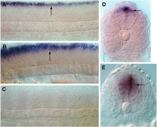
nkx2.9is a direct target of Gli1 activity in the ventral neural tube of the zebrafish embryo: The final
veri-fication of the biological significance of the Gli-binding sites that we have been able to identify using bioinfor-matics will require the functional validation of their ability to direct the spatio-temporal transcription of the relevant genes in a Gli-dependent manner during the course of embryogenesis. As a starting point for this kind of analysis, we investigated whether the expression
of nkx2.9, a gene whose transcription showed
maxi-mal positive responsiveness to Hh signaling in our mi-croarray assay, is directly regulated by the Gli proteins through the Gli_0 motif in its enhancer. In contrast to mammals, where Gli2 and Gli3 are the primary media-tors of Hh signaling in the neural tube, in the zebrafish embryo, the Gli1 protein appears to have assumed a more significant role. Mice that are homozygous for loss-of-function alleles of Gli1 are viable and are not associated with any developmental abnormalities (Park
et al.2000); however,gli1 mutant zebrafish are embry-onic lethal and exhibit striking defects in the specifica-tion of Hh-dependent cell fates in the ventral neural tube (Karlstromet al.2003). In embryos homozygous
for mutations in the detour(dtr) gene, which encodes zebrafish Gli1, there was a dramatic reduction innkx2.9 expression at all levels along the antero-posterior axis of the developing neural tube (Figure 12, A, E, and F). On the contrary, injection of syntheticgli1mRNA into
Figure12.—nkx2.9expression in the ventral neural tube is directly regulated by Gli1 activity. (A) A 12-hpfgli1mutant embryo,
enhancer sequence encompassing the Gli_0 motif fused upstream of thegfpreporter gene into newly fertilized zebrafish eggs. A previous analysis in the mouse embryo has indicated that the homologous enhancer fragment of the mouse Nkx2.9 gene is sufficient for directing reporter gene expression in the ventral neural tube (Santagati et al. 2003). In line with this mammalian
study, we found that the injected zebrafish embryos displayed faithful expression of GFP in cells along the ventral region of the brain and spinal cord, recapit-ulating the endogenous expression pattern of nkx2.9 (Figure 12, G and I; 69 of 86 injected embryos showed this pattern). By contrast, injection of the same con-struct harboring a deletion of the Gli-binding site com-pletely abolished GFP expression in the ventral neural tube (Figure 12, H and J; none of the 71 injected embryos showed GFP expression in thenkx2.9domain). All of these findings not only provide incontrovertible evidence that the Hh-dependent induction of nkx2.9 expression is directly mediated by the activity of the Gli1 protein, but also validate, for the first time in the zebrafish, the functionality of the Gli_0 motif in Hh-dependent target gene regulation.
DISCUSSION
Genetic studies, in Drosophila as well as vertebrates, have shown that the zinc finger containing Gli family of proteins is the primary mediator of Hh-dependent transcriptional regulation of target genes in responding cells (Inghamand McMahon2001; Hooperand Scott
2005). It is now largely accepted that Hh modulates target gene transcription by inhibiting the formation of the proteolytically processed transcriptional repressor form of Gli, Glirep, and by promoting the accumulation of the transcriptional activator variant, Gliact, in the nucleus (Ingham and McMahon 2001; Hooper and
Scott2005). Therefore, genes that are
transcription-ally activated due to the loss of Glirepand/or the gen-eration of Gliactcan be classified as direct targets of the Hh pathway. If a direct target gene encodes a transcrip-tional regulator it can, in turn, activate or repress many other downstream target genes. These downstream genes are indirect or secondary targets of Hh signaling. In our microarray-based screen for Hh pathway targets, the expression pattern of several genes mimicked that of ptc1 with strong decline in levels of expression in cyclopamine-treated embryos and prominent levels of ectopic induction in embryos overexpressing dnPKA, implicating that Hh signaling plays a central role in positively regulating their expression during develop-ment. Other genes were not affected as dramatically on
within which the genes are normally expressed. Con-sequently, loss of Hh signaling does not dramatically af-fect their overall levels of transcript accumulation, but ectopic Hh activity can result in ectopic induction of their expression. Among the genes that were strongly repressed in response to Hh, only a handful seemed to show some degree of ectopic induction in cyclopamine-treated embryos, while the vast majority did not exhibit any significant degree of upregulation on the loss of Hh activity. The expression of none of these genes is likely to be regulated through direct repression since Hh signal-ing does not induce the formation of Glirep. For ex-ample, ventralization of the neural tube in embryos with ectopic Hh signaling occurs through the ectopic dorsal activation of the ventrally expressed genes like thenkx family members. The Nkx proteins, in turn, repress genes likepax6anddbx2, which are normally expressed in the dorsal region within the neural tube ( Jacob
and Briscoe2003). This paradigm is likely to be true
in many other instances where alternative fates are adopted by cells in response to Hh activity. The ex-pression of genes that showed negative regulation in our assay could be inhibited by Hh through such indirect repressive effects.
Genomewide computational search for enhancer modules containing the Gli-binding motif represents a strategy that is complementary to our functional screen for the identification of Hh target genes (for example, see Hallikaset al.2006). While the former is restricted
exclusively to the discovery of the direct targets, a functional approach such as the one described here has the merit of sampling, in an unbiased way, for all kinds of target genes—both direct and indirect. This additional benefit is significant when one considers that for many biological outcomes of Hh signaling, the central determinant of a specific phenotype is the activity of an indirect target gene. To cite an example, expression of Tbx1, which has a critical role in cranio-facial development in mammals, is indirectly regulated by Hh signaling. The primary targets for Hh in this context are genes encoding the Fox proteins, which in turn, directly activateTbx1transcription through a Fox protein-binding motif in the enhancer of theTbx1gene (Yamagishiet al.2003). Nevertheless, throughin silico
and Basler 1999; Muller and Basler 2000). Cirep
switches the expression ofdppoff in the absence of Hh signaling, while the loss of Cirepand generation of Ciact that result from Hh pathway activation induce high levels of dpp expression in the same cells. In the ptc promoter, however, the Gli motif is targeted exclusively by Ciact (Methot and Basler 1999; Muller and Basler 2000), indicating that differential access of
Glirepand Gliactto the Gli-binding site imposes another dimension in the regulation of Hh signaling-dependent gene expression. The Gli sites that we have identified in the positively regulated genes from our screen can be envisaged to function through similar mechanisms— their biological relevance, however, will have to await functional characterization through procedures exem-plified by our analysis of thenkx2.9enhancer. We also recognize that the limited amount of sequence infor-mation that has been examined for the presence of these motifs must have resulted in the underestimation of the precise numbers of direct target genes. It is worth noting though, that for the majority of the well-established direct target genes, we were able to identify the Gli-binding motif(s) within the 5 kb of flanking sequences in the proximity of the translation start and stop sites.
Strikingly, many genes among those that showed downregulation in response to Hh also contain the Gli_0 or the Gli_m1 motifs within their flanking and/or intragenic sequences. Although the functionality of these binding sites must again be confirmed before we can attribute any biological importance, assuming that some of these are indeed targeted by Gli, we can envisage at least two possible reasons for their presence in this class of genes. First, the Gli recognition sites in many of these genes could be exclusively recognized by the repressor, and not the activator forms of the Gli proteins. In Drosophila, thehhgene itself is a target of Ci activity, but the Gli-binding site inhhappears to be accessible only to Cirep.hhtranscription is induced when Ci activity, both repressor and activator, is completely abolished, but the gene is never activated even in cells that are exposed to the highest levels of Hh (Methot
and Basler 1999; Muller and Basler 2000). Thus,
even small amounts of Cirepare sufficient to prevent hh transcription, and the preponderance of Ciact in these maximally responding cells cannot overcome this repression and promotehh induction. Such a mecha-nism of control could be operative for several of the downregulated genes that have the Gli-binding motif. Second, Hh affects morphogenesis through several temporally distinct inductive interactions, sometimes within the same cell or tissue type, during the course of development (McMahonet al.2003). We have shown in
a previous report that the timing of exposure of the zebrafish somitic muscle precursors to Hh determines the kinds of fates that they will ultimately adopt (Wolff
et al. 2003). In the early embryo, Hh promotes
slow-twitch muscle specification in myoblasts in proximity to the midline, at the expense of the fast-twitch fate. However, later in embryogenesis, Hh activity in fast muscle precursors is necessary for the generation of diversity within the fast lineage itself, as exemplified by the Hh-dependent expression of the engrailed (eng) genes in a subset of fast fibers. Thus, Hh can antagonize the development of one cell type at a particular time and have a completely different effect on the same lineage at a later point in development. Given this scenario, genes that are downregulated in response to Hh in one particular cellular context or developmental time could be positively regulated in a different tissue or even within the same cell type, but at a different time point in embryogenesis. Our screen was confined to the as-sessment of transcriptional changes ensuing from the global loss or activation of Hh signaling in early em-bryogenesis. Taking this limitation into account, it will be of interest to perform similar screens in the future with regulated administration of cyclopamine at specific developmental stages, as well as the use of conditional transgenes to temporally or spatially activate the path-way (Wolffet al.2003).
The novel target genes of the Hh pathway that have been identified from this study should be of consider-able significance in helping us build a more compre-hensive picture of how an Hh-induced network of gene activity translates into the regulation of cell fates during development. Determination of the expression pat-terns, effects of loss of function, and analyses of the regulatory elements of these genes will clarify their tissue-specific requirements and provide insights into whether their expression is influenced directly or in-directly by Hh activity. Given the remarkable conserva-tion in the molecular details of vertebrate development, it can be expected that homologs of many of the genes that our assay has recognized as Hh pathway targets in the zebrafish will also be involved in mediating Hh-dependent responses within the developing mam-malian embryo. Moreover, several studies have now underscored a dramatic and direct association of aber-rations in the Hh pathway, with a wide spectrum of congenital abnormalities and malignancies in humans (Altaba et al. 2002). In spite of this connection, we
currently have strikingly little appreciation of the cel-lular and molecular basis for these Hh signaling-related anomalies. It is conceivable that misregulated expres-sion of the human homologs of some of the target genes that we have discovered in the zebrafish could be the critical determinants of certain Hh signaling associated pathologies.
in cancer: tumors, embryos and stem cells. Nat. Rev. Cancer.2:
361–372.
Barresi, M. J., H. L. Stickneyand S. H. Devoto, 2000 The zebra-fish slow-muscle-omitted gene product is required for Hedgehog signal transduction and the development of slow muscle identity. Development127:2189–2199.
Barresi, M. J., J. A. D’angelo, L. P. Hernandezand S. H. Devoto, 2001 Distinct mechanisms regulate slow-muscle development. Curr. Biol.11: 1432–1438.
Barth, K. A., and S. W. Wilson, 1995 Expression of zebrafish nk2.2 is influenced by sonic hedgehog/vertebrate hedgehog-1 and de-marcates a zone of neuronal differentiation in the embryonic forebrain. Development121:1755–1768.
Baxendale, S., C. Davison, C. Muxworthy, C. Wolff, P. W. Ingham
et al., 2004 The B-cell maturation factor Blimp-1 specifies verte-brate slow-twitch muscle fiber identity in response to Hedgehog signaling. Nat. Genet.36:88–93.
Blagden, C. S., P. D. Currie, P. W. Inghamand S. M. Hughes, 1997 Notochord induction of zebrafish slow muscle mediated by Sonic hedgehog. Genes. Dev.11:2163–2175.
Cheesman, S. E., M. J. Layden, T. VonOhlen, C. Q. Doeand J. S. Eisen, 2004 Zebrafish and fly Nkx6 proteins have similar CNS expression patterns and regulate motoneuron formation. Development131:5221–5232.
Chuang, P. T., and A. P. McMahon, 1999 Vertebrate Hedgehog sig-naling modulated by induction of a Hedgehog-binding protein. Nature397:617–621.
Concordet, J. P., K. E. Lewis, J. W. Moore, L. V. Goodrich, R. L. Johnsonet al., 1996 Spatial regulation of a zebrafish patched homologue reflects the roles of sonic hedgehog and protein kinase A in neural tube and somite patterning. Development
122:2835–2846.
Crooks, G. E., G. Hon, J. M. Chandonia and S. E. Brenner, 2004 WebLogo: a sequence logo generator. Genome Res.14:
1188–1190.
Currie, P. D., and P. W. Ingham, 1996 Induction of a specific mus-cle cell type by a hedgehog-like protein in zebrafish. Nature382:
452–455.
Dai, P., H. Akimaru, Y. Tanaka, T. Maekawa, M. Nakafukuet al., 1999 Sonic Hedgehog-induced activation of the Gli1 promoter is mediated by GLI3. J. Biol. Chem.274:8143–8152.
Dhoot, G. K., M. K. Gustafsson, X. Ai, W. Sun, D. M. Standiford
et al., 2001 Regulation of Wnt signaling and embryo patterning by an extracellular sulfatase. Science293:1663–1666.
Du, S. J., S. H. Devoto, M. Westerfield and R. T. Moon, 1997 Positive and negative regulation of muscle cell identity by members of the hedgehog and TGF-beta gene families. J. Cell. Biol.139:145–156.
Duman-Scheel, M., L. Weng, S. Xinand W. Du, 2002 Hedgehog regulates cell growth and proliferation by inducing cyclin D and cyclin E. Nature417:299–304.
Eisen, M. B., and P. O. Brown, 1999 DNA arrays for analysis of gene expression. Methods Enzymol.303:179–205.
Eisen, M. B., P. T. Spellman, P. O. Brown and D. Botstein, 1998 Cluster analysis and display of genome-wide expression patterns. Proc. Natl. Acad. Sci. USA95:14863–14868.
Ekker, S. C., A. R. Ungar, P. Greenstein, D. P. VonKessler, J. A. Porteret al., 1995 Patterning activities of vertebrate hedgehog proteins in the developing eye and brain. Curr. Biol.5:944– 955.
Ghosh, M. G., D. A. Thompsonand R. J. Weigel, 2000 PDZK1 and GREB1 are estrogen-regulated genes expressed in hormone-responsive breast cancer. Cancer Res.60:6367–6375.
Gustafsson, M. K., H. Pan, D. F. Pinney, Y. Liu, A. Lewandowski
et al., 2002 Myf5 is a direct target of long-range Shh signaling and Gli regulation for muscle specification. Genes. Dev. 16:
114–126.
Hallikas, O., K. Palin, N. Sinjushina, R. Rautiainen, J. Partanen
et al., 2006 Genome-wide prediction of mammalian enhancers
fish embryo. Dev. Biol.294:104–118.
Hooper, J. E., and M. P. Scott, 2005 Communicating with Hedge-hogs. Nat. Rev. Mol. Cell. Biol.6:306–317.
Ikram, M. S., G. W. Neill, G. Regl, T. Eichberger, A. M. Frischauf
et al., 2004 GLI2 is expressed in normal human epidermis and BCC and induces GLI1 expression by binding to its promoter. J. Invest. Dermatol.122:1503–1509.
Ingham, P. W., and A. P. Mcmahon, 2001 Hedgehog signaling in an-imal development: paradigms and principles. Genes. Dev. 15:
3059–3087.
Jacob, J., and J. Briscoe, 2003 Gli proteins and the control of spinal-cord patterning. EMBO Rep.4:761–765.
Karlstrom, R. O., O. V. Tyurina, A. Kawakami, N. Nishioka, W. S. Talbotet al., 2003 Genetic analysis of zebrafish gli1 and gli2 reveals divergent requirements for gli genes in vertebrate devel-opment. Development130:1549–1564.
Kawakami, A., Y. Nojima, A. Toyoda, M. Takahoko, M. Satohet al., 2005 The zebrafish-secreted matrix protein you/scube2 is im-plicated in long-range regulation of hedgehog signaling. Curr. Biol.15:480–488.
Kenney, A. M., and D. H. Rowitch, 2000 Sonic hedgehog pro-motes G(1) cyclin expression and sustained cell cycle progres-sion in mammalian neuronal precursors. Mol. Cell. Biol. 20:
9055–9067.
Kimmel, C. B., W. W. Ballard, S. R. Kimmel, B. Ullmannand T. F. Schilling, 1995 Stages of embryonic development of the ze-brafish. Dev. Dyn.203:253–310.
Kinzler, K. W., and B. Vogelstein, 1990 The GLI gene encodes a nuclear protein which binds specific sequences in the human genome. Mol. Cell. Biol.10:634–642.
Krauss, S., J. P. Concordetand P. W. Ingham, 1993 A functionally conserved homolog of the Drosophila segment polarity gene hh is expressed in tissues with polarizing activity in zebrafish em-bryos. Cell75:1431–1444.
Lauderdale, J. D., S. K. Pasquali, R. Fazel, F. J. VanEeden, H. E. Schauerteet al., 1998 Regulation of netrin-1a expression by hedgehog proteins. Mol. Cell. Neurosci.11:194–205.
Lewis, K. E., and J. S. Eisen, 2003 From cells to circuits: devel-opment of the zebrafish spinal cord. Prog. Neurobiol.69:419– 449.
Lewis, K. E., J. P. Concordet and P. W. Ingham, 1999a Char-acterization of a second patched gene in the zebrafish Danio rerio and the differential response of patched genes to Hedge-hog signaling. Dev. Biol.208:14–29.
Lewis, K. E., P. D. Currie, S. Roy, H. Schauerte, P. Haffteret al., 1999b Control of muscle cell-type specification in the zebrafish embryo by Hedgehog signaling. Dev. Biol.216:469–480. MacDonald, R., K. A. Barth, Q. Xu, N. Holder, I. Mikkolaet al.,
1995 Midline signaling is required for Pax gene regulation and patterning of the eyes. Development121:3267–3278.
Mathavan, S., S. G. Lee, A. Mak, L. D. Miller, K. R. Murthyet al., 2005 Transcriptome analysis of zebrafish embryogenesis using microarrays. PLoS. Genet.1:260–276.
McMahon, A. P., P. W. Inghamand C. J. Tabin, 2003 Developmental roles and clinical significance of hedgehog signaling. Curr. Top. Dev. Biol.53:1–114.
Methot, N., and K. Basler, 1999 Hedgehog controls limb devel-opment by regulating the activities of distinct transcriptional activator and repressor forms of Cubitus interruptus. Cell96:
819–831.
Muller, B., and K. Basler, 2000 The repressor and activator forms of Cubitus interruptus control Hedgehog target genes through common generic gli-binding sites. Development 127: 2999– 3007.
Norton, W. H., M. Mangoli, Z. Lele, H. M. Pogoda, B. Diamond
et al., 2005 Monorail/Foxa2 regulates floorplate differentia-tion and specificadifferentia-tion of oligodendrocytes, serotonergic raphe neu-rones and cranial motoneuneu-rones. Development132:645–658. Odenthal, J., F. J. Van Eeden, P. Haffter, P. W. Ingham and
C. Nusslein-Volhard, 2000 Two distinct cell populations in the floor plate of the zebrafish are induced by different pathways. Dev. Biol.219:350–363.
Pabst, O., H. Herbrandand H. H. Arnold, 1998 Nkx2–9 is a novel homeobox transcription factor which demarcates ventral do-mains in the developing mouse CNS. Mech. Dev.73:85–93. Park, H. L., C. Bai, K. A. Platt, M. P. Matise, A. Beeghlyet al.,
2000 Mouse Gli1 mutants are viable but have defects in SHH signaling in combination with a Gli2 mutation. Development
127:1593–1605.
Rallu, M., R. Machold, N. Gaiano, J. G. Corbin, A. P. McMahon
et al., 2002 Dorsoventral patterning is established in the telen-cephalon of mutants lacking both Gli3 and Hedgehog signaling. Development129:4963–4974.
Roy, S., and P. W. Ingham, 2002 Hedgehogs tryst with the cell cycle. J. Cell. Sci.115:4393–4397.
Roy, S., T. Qiao, C. Wolffand P. W. Ingham, 2001a Hedgehog signaling pathway is essential for pancreas specification in the zebrafish embryo. Curr. Biol.11:1358–1363.
Roy, S., C. Wolffand P. W. Ingham, 2001b The u-boot mutation identifies a Hedgehog-regulated myogenic switch for fiber-type diversification in the zebrafish embryo. Genes. Dev. 15:1563– 1576.
Santagati, F., K. Abe, V. Schmidt, T. Schmitt-John, M. Suzukiet al., 2003 Identification of cis-regulatory elements in the mouse
Pax9/Nkx2–9genomic region: implication for evolutionary con-served synteny. Genetics165:235–242.
Sasaki, H., C. Hui, M. Nakafukuand H. Kondoh, 1997 A binding site for Gli proteins is essential for HNF-3beta floor plate en-hancer activity in transgenics and can respond to Shh in vitro. Development124:1313–1322.
Schafer, M., D. Kinzel, C. Neuner, M. Schartl, J. N. Volffet al., 2005 Hedgehog and retinoid signaling confines nkx2.2b ex-pression to the lateral floor plate of the zebrafish trunk. Mech. Dev.122:43–56.
Schena, M., R. A. Heller, T. P. Theriault, K. Konrad, E. Lachenmeier
et al., 1998 Microarrays: biotechnology’s discovery platform for functional genomics. Trends Biotechnol.16:301–306.
Tay, S. Y., P. W. Inghamand S. Roy, 2005 A homologue of the Dro-sophila kinesin-like protein Costal2 regulates Hedgehog signal transduction in the vertebrate embryo. Development132:625– 634.
Tusher, V. G., R. Tibshiraniand G. Chu, 2001 Significance analysis of microarrays applied to the ionizing radiation response. Proc. Natl. Acad. Sci. USA98:5116–5121.
Varga, Z. M., A. Amores, K. E. Lewis, Y. L. Yan, J. H. Postlethwait
et al., 2001 Zebrafish smoothened functions in ventral neural tube specification and axon tract formation. Development128:
3497–3509.
Wechsler-Reya, R., and M. P. Scott, 2001 The developmental bi-ology of brain tumors. Annu. Rev. Neurosci.24:385–428. Wolff, C., S. Royand P. W. Ingham, 2003 Multiple muscle cell
iden-tities induced by distinct levels and timing of hedgehog activity in the zebrafish embryo. Curr. Biol.13:1169–1181.
Wolff, C., S. Roy, K. E. Lewis, H. Schauerte, G. Joerg-Rauchet al., 2004 iguana encodes a novel zinc-finger protein with coiled-coil domains essential for Hedgehog signal transduction in the zebrafish embryo. Genes Dev.18:1565–1576.
Woods, I. G., and W. S. Talbot, 2005 The you gene encodes an EGF-CUB protein essential for Hedgehog signaling in zebrafish. PLoS. Biol.3:e66.
Yamagishi, H., J. Maeda, T. Hu, J. McAnally, S. J. Conwayet al., 2003 Tbx1 is regulated by tissue-specific forkhead proteins through a common Sonic hedgehog-responsive enhancer. Genes Dev.17:269–281.
Yang, H., H. Haddad, C. Tomas, K. Alsakerand E. T. Papoutsakis, 2003 A segmental nearest neighbor normalization and gene identification method gives superior results for DNA-array anal-ysis. Proc. Natl. Acad. Sci. USA100:1122–1127.
Yang, R. B., C. K. Ng, S. M. Wasserman, S. D. Colman, S. Shenoy
et al., 2002 Identification of a novel family of cell-surface pro-teins expressed in human vascular endothelium. J. Biol. Chem.
277:46364–46373.