RNA Interference in the Pathogenic Fungus
Cryptococcus neoformans
Hong Liu,* Tricia R. Cottrell,* Lynda M. Pierini,
†William E. Goldman* and Tamara L. Doering*
,1*Department of Molecular Microbiology, Washington University School of Medicine, Saint Louis, Missouri 63110 and†Department of Biochemistry, Weill Medical College of Cornell University, New York, New York 10021
Manuscript received July 11, 2001 Accepted for publication November 13, 2001
ABSTRACT
Cryptococcus neoformansis a pathogenic fungus responsible for serious disease in immunocompromised individuals. This organism has recently been developed as an experimental system, with initiation of a ge-nome project among other molecular advances. However, investigations of Cryptococcus are hampered by the technical difficulty of specific gene replacements. RNA interference, a process in which the presence of double-stranded RNA homologous to a gene of interest results in specific degradation of the correspond-ing message, may help solve this problem. We have shown that expression of double-stranded RNA cor-responding to portions of the cryptococcalCAP59 andADE2genes results in reduced mRNA levels for those genes, with phenotypic consequences similar to that of gene disruption. The two genes could also be subjected to simultaneous interference through expression of chimeric double-stranded RNA. Specific mod-ulation of protein expression through introduction of double-stranded RNA thus operates inC. neoformans, which is the first demonstration of this technique in a fungal organism. Use of RNA interference in Cryptococcus should allow manipulation of mRNA levels for functional analysis of genes of interest and enable efficient exploration of genes discovered by genome sequencing.
T
HE challenge of using eukaryotic genome sequence 1998) and its high frequency of nonhomologous recom-bination. Despite these obstacles several dozen targeted information to focus research efforts on productiveareas is shared by those who investigate organisms with gene replacements have now been accomplished, mainly by the groups of J. Perfect (Duke University Medical genomes of all magnitudes and complexities. One
fun-gal genome, that ofCryptococcus neoformans(24 Mb), has Center), J. Heitman (Duke University Medical Center), and J. Kwon-Chung (National Institutes of Health), been currently sequenced to approximately seven times
shotgun coverage by the Stanford Genome Technology which have tremendous value for functional analysis of cryptococcal genes and virulence studies in animal Center (http://www-sequence.stanford.edu) and The
models. To exploit the forthcoming genome sequence Institute for Genomic Research (http://www.tigr.org).
efficiently, however, it will be necessary to develop more
C. neoformans is an encapsulated fungal pathogen
re-rapid methods to test the function of gene products, sponsible for severe disease in immunocompromised
instead of relying on individual gene disruptions. individuals. It usually grows as a haploid yeast that
repro-One recently developed method for downregulating duces by budding, but under certain environmental
gene function that may facilitate studies inC. neoformans
conditions it may also undergo a sexual cycle resulting
is double-stranded RNA interference (RNAi). In this in the production of basidiospores (Kwon-Chung1975).
process double-stranded RNA (dsRNA) induces the spe-In recent years Cryptococcus has been developed as an
cific destruction of mRNA to which it is homologous experimental system, with advances in molecular
tech-(Fireet al. 1998; reviewed in Sharp 1999;Bass 2000; niques enabling detailed investigation of virulence
fac-Carthew 2001; Hammond et al. 2001). The dsRNA
tors and interactions with the mammalian host. Notable
“trigger” is thought to be cleaved into shorter fragments among these advances have been the establishment of
(21–25 nucleotides; Zamore et al. 2000), which then transformation systems suitable for episomal expression
guide specific degradation of the corresponding mRNA, and chromosomal integration (Edman and
Kwon-catalyzed by a protein or protein complex with nuclease Chung1990;Toffalettiet al.1993) and the initiation
activity (Baulcombe2001). Before cleavage the trigger of genome sequencing efforts (Heitman et al. 2000).
can be as short as 26 nucleotides and has no specific More difficult has been the development of methods
requirements as far as sequence motif or base composi-for efficient gene replacement inC. neoformans, which
tion, but must be double stranded (Parrishet al.2000). has been hampered by both the propensity of
cryptococ-Interference can persist for many rounds of cell division cus to modify exogenous DNA (Kwon-Chung et al.
and growth and may even be passed through the germ-line (Fireet al.1998). Specific inhibition of gene expres-sion by RNAi has been demonstrated in a range of
1Corresponding author: Campus Box 8230, 660 S. Euclid Ave., St.
Louis, MO 63110. E-mail: [email protected] organisms, from the initial report onCaenorhabditis
Texas Health Science Center). (This plasmid contains AmpR
ans (Fire et al.1998) to works on trypanosomes (Ngo
andC. neoformans URA5markers, as well as theGUSreporter
et al. 1998), Drosophila (Kennerdell and Carthew
gene flanked by theC. neoformans GAL7promoter and termina-1998), early mouse embryos (Svobodaet al.2000;Wi- tor; digestion with NdeI and BglII releases theGUS coding anny and Zernicka-Goetz 2000), and mammalian sequence.) The ligation product was isolated fromEscherichia
coli, digested with BglII, and ligated to the BglII restricted cells in culture (Elbashiret al.2001). RNAi has proved
product B. Products were tested by restriction digestion to a valuable tool for probing functions of individual gene
select one in which theCAP59repeats were in opposite orienta-products (e.g.,KennerdellandCarthew1998;Bastin
tions, and this was digested with NdeI and ClaI to remove
et al. 2000) and offers a powerful approach for func- theGAL7promoter. To form pCAP59i (Figure 1) theGAL7 tional genomic studies (KuwabaraandCoulson2000; promoter was replaced with a cryptococcal ACT promoter Kim2001). In the best-studied and most tractable sys- amplified with primers 7 and 8 from plasmid GMC200 (from Dr. Gary Cox, Duke University Medical Center). An identical tem,C. elegans, over a third of all genes have now been
cloning scheme was used to construct pADE2i, except that assessed for the phenotypic effects of RNAi. We
hypothe-primers 1*–6* (Table 1) were used in place of hypothe-primers 1–6 sized that this technique could be useful in C. neo- above. All PCR reactions were done using Taq polymerase formans, both to circumvent the difficulties of gene re- under standard conditions and PCR products were purified placement for functional studies of individual gene before use in subsequent reactions. Both plasmids were checked
by DNA sequencing. products and to initiate larger-scale investigations of
To incorporate cryptococcal telomere sequences, the 1.4-kb genes discovered through sequencing efforts. In this
NotI fragment of pTEL-HYG (from Dr. Gary Cox, Duke Univer-article we show thatC. neoformansgene expression can
sity) was inserted into the uniqueNotI site of pCAP59i, forming be specifically inhibited by RNA interference. pCAP59i-tel. pCAP59/ADE2i was generated by adding dupli-cates of the same portion ofADE2used in pADE2i to pCAP59i, in the positions and orientations shown in Figure 1 (cloning
MATERIALS AND METHODS details available on request). For the promoterless version of
pCAP59i the plasmid was digested withNdeI andClaI to re-Strains and cell growth:C. neoformanswas grown with
contin-move the actin promoter, the sticky ends were filled in with uous shaking at 30⬚ in YPD medium [1% (w/v) Bacto yeast
T4 DNA polymerase, and the plasmid was religated. extract; 2% (w/v) peptone, 2% dextrose] or minimal medium
JEC43 cells were transformed by electroporation as de-lacking uracil (Ausubelet al.2001). Low adenine plates
con-scribed (WickesandEdman1994) using 5g of linearized tained yeast nitrogen base supplemented with (per liter) 20 g
DNA per 3 ⫻ 108 cells with selection of transformants on glucose; 24 mg uracil; 40 mg each arginine, histidine,
isoleu-minimal plates lacking uracil. pCAP59i-tel was linearized with cine, leucine, lysine, methionine, and tryrosine; 60 mg
phenyl-SceI so that the telomeres formed the ends of the DNA used alanine and tryptophan; 120 mg homoserine; 180 mg valine;
for transformation; all other plasmids were linearized with and 10 mg adenine. For experiments using 5-fluoroorotic acid
NotI. Transformants were streaked to master plates of the same (5-FOA), plates contained the same medium with adenine
medium and stored at 4⬚. raised to 40 mg/liter and the addition of 1 g/liter 5-FOA.
RNA preparation and reverse transcription-PCR:C. neoformans
Wild-type serotype D strain B4500 andcap59 strain TYCC33
cells were washed with pyrocarbonic acid diethyl ester-treated (ChangandKwon-Chung1994) were from Dr. June
Kwon-water, suspended in Trizol Reagent (GIBCO BRL, Gaithers-Chung (National Institutes of Health), andura5strain JEC43
burg, MD), and broken in a MiniBeadbeater-8 (Biospec Prod-(Wickes et al. 1997) was from Dr. Joseph Heitman (Duke
ucts, Bartlesville, OK) in the presence of 0.5-mm glass beads University Medical Center). JEC43 cells transformed with a
(3 ⫻ 1-min bursts, 4⬚). Cell lysis was typically 60–80% for control plasmid alone (CIP-GUST.Cla.Kpn; see below) are
encapsulated strains and 80–100% for acapsular strains. Total designated “control.” JEC43 cells transformed with a plasmid
RNA was precipitated using standard methods (Ausubel et
designed to interfere with CAP59expression (described
be-al.2001) with a yield of 1.5–3.0 mg/500 OD600units of cells. low) were termed CAP59i, with numbers following this
desig-For cDNA synthesis, 100 g of total RNA was treated with nation to indicate individual transformants (e.g., CAP59i-1).
amplification grade DNase I (GIBCO BRL), ethanol precipi-A similar convention was used for naming constructs designed
tated, and used in the Superscript preamplification system for to interfere withADE2or with bothCAP59andADE2.
first strand cDNA synthesis using Oligo(dT) primer (GIBCO Constructs and transformation:Constructs for RNA
inter-BRL) according to manufacturer’s instructions. ference were designed with inverted repeats ofⵑ500 bp of
For reverse transcription (RT)-PCR experiments,ⵑ2g of coding sequence of the gene of interest separated by a spacer
cDNA was amplified in a 100-l reaction using Taq polymerase segment of green fluorescent protein (GFP) sequence. For
and appropriate primers (see Table 2 and text). The amplifi-construction of pCAP59i (Figure 1, top), a portion of the
cation program was (i) 94⬚for 2 min; (ii) 26–35 cycles of 94⬚ coding sequence of CAP59 was PCR amplified from B4500
for 1 min, 55⬚ for 1 min, 68⬚ for 1 min; and (iii) 68⬚ for genomic DNA using primers 1 and 2 (Table 1) to add anNdeI
7 min. Ten-microliter samples were removed every 3 cycles restriction site at the 5⬘end and a portion ofGFPsequence
immediately after the 68⬚ incubation, beginning as early as at the 3⬘end (product A). Product A was further amplified
the eleventh cycle. PCR products were resolved on 1.5% aga-using primers 3 and 4 to incorporateBglII restriction sites at
rose gels run in TAE buffer and stained (45 min, RT) with each end (product B). A segment ofGFPwas amplified (from
SYBR Green I nucleic acid gel stain (Molecular Probes, Eu-an enhEu-anced version designed forC. elegansfrom Dr. Andrew
gene, OR) in TAE buffer. DNA-associated fluorescence was Fire, Carnegie Institute of Washington) using primers 5 and
visualized using a FluorImager SI (Molecular Dynamics, Sun-6 to incorporate the 3⬘end of theCAP59sequence in product
nyvale, CA), and data were analyzed using Scanalytics IPLab A at the 5⬘ end and a BglII restriction site at the 3⬘ end
gel and Microsoft Excel software with subtraction of back-(product C). PCR products A and C were then used as the
ground fluorescent signal. template for a PCR reaction with primers 1 and 6 to amplify
Cell imaging:For immunofluorescence, cells were grown a 750-bp fragment (product D); this was then digested with
in YPD medium, washed twice in PBS, and treated with
anticap-NdeI andBglII and ligated into the similarly restricted plasmid
TABLE 1
Primers used in plasmid construction
Primer Sequence (5⬘to 3⬘) Description
1 GGAATTCCATATGGAATTCCATGCTCCCCTCCATCGAGC NdeI site,CAP59nt 1–19 sense (underlined)
2 GAAAAGTTCTTCTCCTTTACTCATCGGTGGTTGAACAGACCT GFPnt 24–1 antisense (underlined),CAP59nt 520–502 antisense
3 GGAGATCTGCCGGTGGTTGAACAGACCTC BglII site,CAP59nt 520–500 antisense (underlined)
4 GAAGATCTTCATGCTCCCCTCCATCGAG BglII site,CAP59nt 1–20 sense (underlined)
5 AGGTCTGTTCAACCACCGATGAGTAAAGGAGAAGAACTTTTC The antiparallel sequence of primer 2
6 GGAGATCTCATAACAGAAAGTAGTGACAA BglII site,GFPnt 230–250 antisense (underlined)
7 CCATCGATGGCTGCGAGGATGTG Clasite, actin promoter nt 1–14 sense (underlined)
8 GGAATTCCATATGGTTGGGCGAG NdeI site, actin promoter nt 834–825 antisense (underlined)
1* GGAATTCCATATGGAATTCCATGGCCCCCAGAAAGACTG NdeI site,ADE2nt 1–19 sense (underlined)
2* GTTCTTCTCCTTTACTCATTGCGGATTTAAGAGGAGAG GFPnt 24–1 antisense (underlined),ADE2nt 520–502
antisense
3* GAAGATCTTCATGCGGATTTAAGAGGAGAG BglII site,ADE2nt 521–502 antisense (underlined)
4* GAAGATCTTCATGGCCCCCAGAAAGACTG BglII site,ADE2nt 1–19 sense (underlined)
5* CTCTCCTCTTAAATCCGCAATGAGTAAAGGAGAAGAAC The antiparallel sequence of primer 2*
6* GAAGATCTTCTGCGGATTTAAGAGGAGAGTT BglII site,GFPnt 250–230 antisense (underlined)
Restriction sites are in boldface type and sequences homologous to genes of interest are designated by the nucleotide (nt) position in the corresponding cDNA coding sequence (1 being the A of the initiating ATG) and whether they correspond to the sense or antisense strand. For theACTpromotor the nt numbers correspond to those in GenBank entry AF156670. Primers specific to pADE2i are designated with an asterisk (seematerials and methods).
Albert Einstein College of Medicine) that was tagged with Cy3 in whichCAP59is disrupted are acapsular and avirulent (as inPieriniandDoering2001). Differential interference (ChangandKwon-Chung1994). The second gene tested contrast (DIC) and fluorescence images were acquired simul- wasADE2, which encodes phosphoribosylaminoimida-taneously with a Zeiss ( Jena, Germany) LSM510 laser scanning
zole carboxylase; disruption of this gene yields pink confocal microscope. All samples were imaged with identical
colonies due to accumulation of adenine biosynthetic acquisition settings to allow direct quantitative comparison.
Average fluorescence intensities were based on measurements intermediates.
of at least 25 cells per condition. Postacquisition image analysis Several approaches have been used to express double-was performed with MetaMorph imaging software (Universal stranded RNA for interference testing. It can be synthe-Imaging Corporation, West Chester, PA), and images were
sizedin vitroorin vivoeither as independent RNA mole-prepared for publication using Adobe Photoshop (Adobe
Sys-cules corresponding to a sequence and its complement tems Inc., San Jose, CA).
or as a single polynucleotide containing duplicate se-quences in opposite orientation separated by a spacer.
RESULTS In the former situation the sense and antisense strands,
which may be formed by convergent promoters tran-To test RNAi inC. neoformanswe chose two genes that
scribing the same DNA sequence, anneal to form the would produce clear phenotypes if the technique was
required dsRNA. In the latter case the single RNA forms successful. The first of these isCAP59, which encodes
a hairpin with a double-stranded stem. dsRNA may be a product required for synthesis of the polysaccharide
delivered by injection (Fireet al.1998;Guru2000), elec-capsule in cryptococcus. The elec-capsule is absolutely
re-troporation, soaking, or feeding (the last two inC. elegans; quired for virulence of this fungal pathogen, and cells
TABLE 2
Primers used for RT-PCR
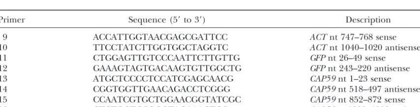
Primer Sequence (5⬘to 3⬘) Description
9 ACCATTGGTAACGAGCGATTCC ACTnt 747–768 sense
10 TTCCTATCTTGGTGGCTAGGTC ACTnt 1040–1020 antisense
11 CTGGAGTTGTCCCAATTCTTGTTG GFPnt 26–49 sense
12 GAAAGTAGTGACAAGTGTTGGCTG GFPnt 243–220 antisense
13 ATGCTCCCCTCCATCGAGCAACG CAP59nt 1–23 sense
14 CGGTGGTTGAACAGACCTCGGG CAP59nt 518–497 antisense
15 CCAATCGTGCTGGAACGGTATCGC CAP59nt 852–872 sense
16 CTCTGGTCCGGGTACAAACTCC CAP59nt 1229–1208 antisense
Figure1.—Plasmid design. pCAP59i was used to interfere withCAP59expression, and pCAP59/ADE2i was used for tan-dem interference with bothCAP59andADE2.
Tabaraet al.1998;TimmonsandFire1998), or it may be expressed within the cell of interest (Ngoet al.1998; KennerdellandCarthew2000;Wanget al.2000). For initial attempts to interfere with CAP59 expression in cryptococcus, we designed a plasmid with convergent promoters flanking a segment of the gene. These studies were not successful (not shown), so we designed another
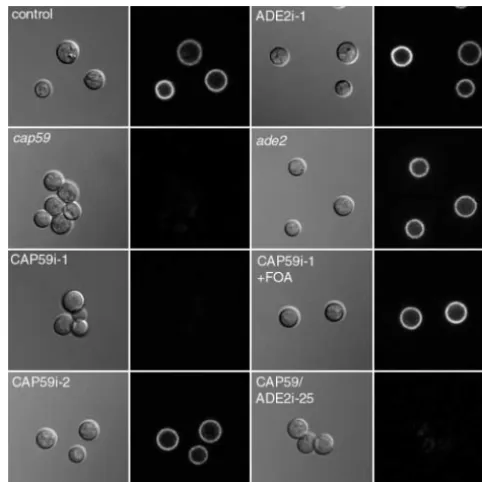
construct employing a hairpin scheme with duplicate se- Figure 2.—Light microscopy of CAP59 interference and control strains. The indicated strains (see text) were labeled quences of ⵑ500 bp ofCAP59in opposite orientation
with Cy3-conjugated monoclonal antibody 2H1 and then ex-separated by a 250-bp spacer ofGFPsequence (pCAP59i,
amined by confocal microscopy. DIC (left image of each pair) Figure 1). Constitutive expression was driven by a
crypto-and fluorescence (right image of each pair) images are shown. coccal actin promoter, and the chimeric sequence was CAP59i-1, like cap59, demonstrates clumping and a lack of followed by aGAL7 terminator. capsule, which is reversed after growth on 5-FOA (Cap59i-1⫹ FOA). An acapsular phenotype is also seen with double
inter-C. neoformanscells of serotype D were transformed by
ference cells (CAP59/ADE2i). Capsule is not affected by inhi-electroporation with the linearized interference
con-bition or mutation ofADE2alone (ADE2i-1 andade2, respec-struct (pCAP59i) or a control plasmid. Transformants
tively). Bar, 10m. were restreaked to a master plate and replica plated
to medium for capsule induction. Ten percent of the
pCAP59i transformants and 0% of the control trans- case of parental and control cells. CAP59i-1 andcap59
cells exhibited no halo, but CAP59i-2 showed an inter-formants demonstrated a clear mutant phenotype, with
dull colonies due to the absence of polysaccharide cap- mediate phenotype with a mixture of encapsulated and a few acapsular cells.
sule (Table 3). Cells from one dull and one shiny colony
transformed with pCAP59i (CAP59i-1 and CAP59i-2, re- To test the presence of capsule with a more sensitive method, we treated cells with a monoclonal antibody spectively) were chosen for closer examination.
By light microscopy (Figure 2, first column) CAP59i-1 to cryptococcal capsule, which we conjugated to Cy3 (Pierini andDoering 2001), and examined them by cells were noted to clump together, similar to strains in
which CAP59has been disrupted (cap59). In contrast, confocal microscopy (Figure 2, second column). Cells transformed with a control plasmid showed brightly fluo-parental cells (not shown) or those transformed with a
control plasmid rarely clump. Examination of cells in rescent surface staining, as was the case with CAP59i-2 cells. In contrast, and similar tocap59mutants, CAP59i-1 the presence of india ink (not shown), which is excluded
from the capsule, showed a characteristic halo in the cells showed fluorescence⬍1% of control (Figure 2),
TABLE 3
Efficiency of generation of mutant phenotypes
Control pCAP59i pCAP59-noP pCAP59i-tel pADE2i pCAP59/ADE2i
Dull ⬍1 10 ⬍1 27 ⬍1 24.5
Pink ⬍1 ⬍1 ⬍1 ⬍1 7 25.5
even when grown in capsule-inducing medium (not shown). Quantitation showed no difference in fluores-cence intensity between CAP59i-1 andcap59cells (P⬍
0.001), and electron microscopy confirmed the ab-sence of capsule on CAP59i-1 andcap59cells (not shown). These results suggest that a fraction of transformants (e.g., CAP59i-1) show complete interference with nor-mal gene expression, while others (e.g., CAP59i-2) do not (seediscussion).
To confirm that the interference phenotype depended on exogenous DNA, CAP59i-1 cells were streaked on 5-FOA plates to select against maintenance of theURA5 -marked interference construct. Colonies that grew un-der these conditions regained a shiny appearance and displayed wild-type capsule staining by immunofluores-cence (Figure 2), demonstrating association of the acap-sular phenotype with presence of the interfering plas-mid. Southern blotting and PCR experiments showed that the endogenousCAP59gene was intact in CAP59i-1 and confirmed that the interfering construct had not been integrated and was lost after growth on 5-FOA (not shown). To assess dependence of interference on
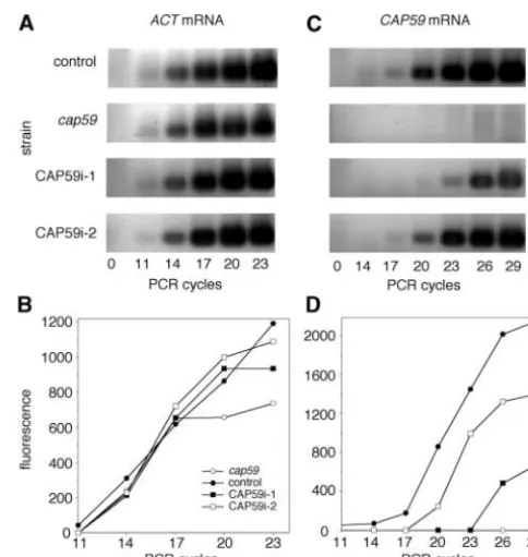
tran-Figure3.—RT-PCR analysis ofCAP59expression. RT-PCR scription, pCAP59i was modified by removing the
pro-analysis of the indicated strains was performed as described motor sequence; this plasmid (pCAP59i-noP) yielded
in the text, using amounts of cDNA normalized to actin mRNA no phenotypically altered transformants (Table 3).
expression. The results of RT-PCR reactions using primers 9 If the observed acapsular phenotype of CAP59i-1 re- and 10 for detection ofACTmRNA are shown in A and quantita-sulted from RNA interference, the levels ofCAP59RNA tion of the corresponding PCR product is plotted in B in arbitrary fluorescence units. RT-PCR reactions using primers in those cells should be reduced compared to controls.
15 and 16 to detectCAP59mRNA are shown and plotted in To test this we used a quantitative RT-PCR method.
C and D. Control cells, solid circles; cap59, open circles; cDNA prepared from cells to be tested was used as a
sub-CAP59i-1, solid squares; and CAP59i-2, open squares. strate for PCR reactions that were sampled every three
cycles (see materials and methods), and products
were quantitated using SYBR green dye (sensitivity 60 ment of the cryptococcalADE2gene (pADE2i, see ma-terials and methods). Similar to results with pCAP59i, pg dsDNA). Fluorescence was plotted vs. number of
cycles, and the starting amounts of cDNA from each 7% of cells transformed with pADE2i showed an inter-ference phenotype (Table 3), producing pink colonies strain were adjusted such that the exponential phase of
PCR product accumulation for a constitutively expressed (e.g., ADE2i-1 in Figure 4). RT-PCR experiments con-firmed significant reduction inADE2mRNA in the cells control gene (ACT, amplified with primers 9 and 10 in
Table 2) overlapped closely for all strains (Figure 3, A (not shown). The phenotype was plasmid dependent, as growth of ADE2i-1 on 5-FOA to select against mainte-and B). These normalized amounts of cDNA were then
used in experiments to quantitate the presence of spe- nance of pADE2i yielded colonies of the wild-type cream color, which regained adenine prototrophy (Figure 5). cific RNA species in each strain. PCR product
accumula-tion was plotted as above, and comparisons between Inhibition of expression ofADE2, as ofCAP59, was spe-cific, with no alterations of growth or morphology detected several points in the exponentially increasing portions
of the curves were used to calculate relative amounts to suggest that dsRNA initiated nonspecific protein inhi-bition (as observed in some mammalian systems; Wil-of product. As expected,GFP-specific sequences were
detected only in CAP59i-1 and CAP59i-2 cells, and se- liams 1999). The successful inhibition of this second gene product indicates that dsRNAi offers a general quences fromCAP59that were also present in pCAP59i
were detected in all cells exceptcap59(not shown). PCR technique for gene suppression inC. neoformans. In studies with both pCAP59i and pADE2i we noted was also performed with primers homologous toCAP59
sequences that were absent from pCAP59i, to specifically that some transformants remained phenotypically wild type, as exemplified by CAP59i-2 (Figure 2) and ADE2i-2 assess amounts of cellular mRNA (Figure 3, C and D).
In these reactions CAP59i-1 cells showed⬍23% as much (Figure 4). Other transformants demonstrated interme-diate phenotypes, such as reduced fluorescence with
CAP59-specific mRNA as control cells. cap59 cells, as
expected, had noCAP59-specific mRNA. cell clumpiness for CAP59i or less intense pink color for pADE2i (not shown). We hypothesized that these To examine the effect of dsRNAi on another gene
Figure5.—5-FOA reversal ofADE2interference. ADE2i-1 cells and cells transformed with a control plasmid were grown on medium without (⫺) or with (⫹) 5-FOA. Growth in the pres-ence of 5-FOA led to reversal of the ADE2i-1 mutant phenotype.
Figure4.—Interference withADE2. Cells were streaked on YPD plates and incubated for 3 days at 30⬚to demonstrate the
pink color resulting from the accumulation of adenine biosyn- again suggesting some variation in efficiency of inhibi-thetic intermediates. Two ADE2i strains are shown, one that tion (seediscussion). Even with this variation, however, shows the mutant phenotype (ADE2i-1) and one that does ⬎
80% of pink colonies were also acapsular, showing that not (ADE2i-2), along withade2cells and cells transformed with
this approach could effectively indicate cells in which a control plasmid for comparison. The mutant phenotype is also
exhibited by a double interference transformant (CAP59/ interference was operating. ADE2i-25), but is not induced by inhibition or mutation of
CAP59alone (CAP59i-1,cap59).
DISCUSSION
We have explored the utility of double-stranded RNA transforming plasmid, possibly resulting from variable
modification of the exogenous DNA expression (Edman interference as a tool for specific inhibition of gene ex-pression inC. neoformans. Our goal was to find a method 1992;Varmaet al.1992;Kwon-Chunget al.1998). To test
this idea we quantitated the CAP59 mRNA in CAP59i-2 to efficiently assess the phenotypic consequences of pre-venting production of specific gene products, both to cells (Figure 3) and found it to be 72% as abundant as
control, which is severalfold higher than the phenotypi- circumvent the difficulty of gene disruption in this or-ganism and to exploit the rapidly emerging genome cally mutant CAP59i-1 cells. We also examined the DNA
from CAP59i-1 and CAP59i-2 cells by Southern blotting sequence of this fungus for studies of its biology and pathogenicity. We have shown that double-stranded RNA and noted that while the plasmids in both had been
modified and enlarged, probably by a combination of interference occurs in C. neoformansby demonstrating specific inhibition of expression of genes involved in degradation and telomere addition (Kwon-Chunget al.
1998), the former had been extended to a greater extent capsule synthesis and adenine biosynthesis, both individ-ually and together. To date, there are no published re-(not shown). Intermediate phenotypes are discussed
further below. ports of dsRNAi in a yeast or fungus. These observations
should be of great practical value to investigations of im-To stabilize the plasmid and reduce modifications,
which might alter expression of the interference con- portant pathogens likeC. neoformansthat are less amenable to genetic manipulation than are model systems. struct, we incorporated cryptococcal telomere sequences
into pCAP59i. In transformations with this plasmid Studies of RNAi-resistant mutants inC. eleganshave iden-tified several genes believed to encode proteins involved (pCAP59i-tel) we found a higher fraction of
phenotypi-cally mutant cells (Table 3), and we observed reduced in interference (Tabaraet al.1999). Interestingly, several of these have homologs known to participate in other post-modification of the input plasmid (not shown).
Because not all cells transformed with interfering plas- transcriptional gene silencing (PTGS) processes, such as the quelling phenomenon inNeurospora crassa(whereby mids demonstrate mutant phenotypes, investigation of
novel genes could be slowed, as it would require initial endogenous genes are silenced by introduction of a transgenic copy) or the PTGS observed in plants, sug-assessment of mRNA levels in a number of transformants
before phenotypes could be examined with confidence gesting shared mechanisms (Catalanottoet al.2000; Fagardet al.2000;Maine2000;Vaucheretet al.2001). that interference was occurring. One way to mark
trans-formants that exhibit interference would be to simulta- We examined the available C. neoformans genome se-quence for homology to the Arabidopsis AGO1 gene, neously interfere with the gene of interest and with a
marker gene, which would provide an easily tracked phe- one member of a gene family conserved across the three systems (Fagardet al.2000). The longest available con-notype. To test this approach we constructed an
interfer-ence plasmid incorporating portions of bothCAP59and tiguous stretch of cryptococcal sequence was 33% identi-cal and 50% positive, as scored in an NCBI BLAST search
ADE2(Figure 1, pCAP59/ADE2i). Transformants were
both dull and pink, indicating simultaneous inhibition with the BLOSUM62 matrix (Altschul et al. 1997), when compared to the region of Ago1 from amino acids of expression of both genes (Table 3). Some colonies did
(corre-sponding to amino acids 772–986 of Ago1) was 46% interest and then further analysis of those transformants exhibiting appropriate levels of interference. To sim-identical and 59% positive. Notably, the latter group of
identical residues included 45 of the 61 residues that plify this process we propose use ofADE2, or another readily observed marker, in tandem interference con-are identical in Ago1 and its homologs in Neurospora
(QDE-2) andC. elegans(RDE-1). The cryptococcal ge- structs with the gene of interest. For example, trans-formants that are pink, indicating thatADE2expression nome project also has generated sequences encoding
polypeptides with significant homology to theC. elegans is inhibited and interference is operating, could be eas-ily selected for further study of the second gene. Tan-Ego-1, which encodes a putative RNA-dependent RNA
polymerase with homologs in Arabidopsis thaliana and dem interference may also be useful for cases where a double mutant is desired for study, as in Cryptococcus
N. crassa, and to theN. crassaQDE-3, a putative helicase.
this will certainly be more rapid than either sequential The presence in the cryptococcal database of an
Ago-gene disruptions or independent disruptions and ge-1-like sequence and homologs of other proteins
impli-netic crossing. It may also be possible to interfere with cated in RNAi is consistent with this pathogen utilizing
more than two genes simultaneously. In our experiments RNAi or related processes in post-transcriptional gene
we have used relatively large portions of the genes to be silencing.
inhibited (500 bp), but the literature suggests that much For the two genes we tested, 7–10% of transformants
smaller segments are effective. This indicates that plas-demonstrated a complete mutant phenotype, although
mid size will not be limiting and it will be possible to others with less mRNA inhibition displayed
intermedi-test multi-interfering constructs. It may also be possible ate or wild-type phenotypes (Figures 2 and 4 and data
to interfere with several related genes simultaneously if not shown). This is consistent with observations in
Trypa-they share specific sequences that may be incorporated
nosoma brucei, where individual transformants
demon-into the interfering plasmid; this may allow coordinate strate variable efficiency of interference (E. Ullu,
per-inhibition of a family of genes (Fireet al.1998). sonal communication). Our results suggest that for the
Double-stranded RNA interference holds great prom-observation of some phenotypes (e.g., complete lack of
ise for studies of cryptococcus and other fungi. In par-capsule detected by antibody binding) mRNA levels
ticular, it should provide a way to exploit the rapidly must drop below a threshold, which may be different
accumulating genome sequence ofC. neoformans with-for each protein depending on its mechanism of action
out requiring disruption of each gene of potential inter-and regulation. This would explain why in experiments
est. Because only a short segment of the gene sequence with simultaneous interference of two genes we did
ob-is needed for interference, open reading frame identi-tain colonies where only one mutant phenotype was
fication will suffice for this method and complete anno-expressed and also why in almost all of these cases the
tation will not be required. This will allow screening of same single phenotype was observed. It is not clear why
sequences of interest and subsequent choice of genes a range of interference occurs in cryptococcus, but it
for further study. Other advantages of this method in-may be that the interference plasmid is variably
modi-clude the rapidity of functional disruption, the potential fied by the cell before stabilization by addition of
telo-for regulated specific inhibition of gene expression, and meres (Edman1992;Varmaet al.1992;Kwon-Chunget
the feasibility of simultaneous inhibition of several genes
al.1998). This could result in plasmids that are variably
or a gene family. Together these features indicate that expressed and thus have different degrees of efficacy
the specific inhibition of gene expression in cryptococ-in mediatcryptococ-ing mRNA degradation. Addition of telomeric
cus using RNA interference will be a valuable tool in sequences to the interfering plasmid did increase the
an organism where gene disruption is a time-consuming yield of mutant phenotypes and decrease the degree of
and labor-intensive process. plasmid modification after transformation.
We appreciate the efforts of Erin Brosnahan, Scott Handley, and
For some purposes the availability of cells with
differ-Jacquelyn Engle in helping to develop the quantitative RT-PCR method
ent degrees of RNA interference may have advantages
and of Wandy Beatty for her examination of cells by electron
micro-over complete disruptions, as intermediate phenotypes
scopy. We are grateful for helpful discussions with Elisabetta Ullu,
might be available for investigation. For example, if Mark Johnston, members of the Doering lab, and colleagues at the complete inhibition of a gene were lethal, this technique Molecular Mycology Course at the Marine Biological Laboratories. We thank June Kwon-Chung and Joe Heitman for strains, Brian Wickes
might still yield cells for study. Similarly, regulated
ex-and Andy Fire for DNA, ex-and Arturo Casadevall for antibody. T.R.C.’s
pression of interfering constructs might permit
manipu-work on this project was supported by an American Society for
Microbi-lation of levels of inhibition. This could be useful for ology Undergraduate Research Fellowship and Howard Hughes Medi-investigation of nonessential genes and also for poten- cal Institute Summer Research funds to Washington University. L.M.P. tially determining whether a gene is essential, at present was supported by an Atorvastatin Research Award from Pfizer/Parke Davis. W.E.G. was supported by the National Institutes of Health
a difficult question to address directly in cryptococcus.
(AI25584). T.L.D. and work in her laboratory were supported by the
One disadvantage of the spectrum of interference
ob-National Institutes of Health (AI49173), ob-National Science Foundation
served is that to investigate novel genes would require (POWRE 0096115), a Burroughs Wellcome Fund Junior Investigator two phases of study: initial investigation of a series of Award in Molecular Pathogenic Mycology, the Andrew and Virginia Craig Faculty Research Fund, and a Neose Corporation GRANT award.
Kwon-Chung, K. J., 1975 A new genus,Filobasidiella, the perfect
LITERATURE CITED
state ofCryptococcus neoformans.Mycologia68:821–833.
Kwon-Chung, K. J., W. E. Goldman, B. KleinandP. J. Staniszlo,
Altschul, S. F., T. L. Madden, A. A. Scha¨ffer, J. Zhang, Z. Zhang
et al., 1997 Gapped BLAST and PSI-BLAST: a new generation 1998 Fate of transforming DNA in pathogenic fungi. Med. My-col.36(Suppl. 1): 38–44.
of protein database search programs. Nucleic Acids Res. 25:
3389–3402. Maine, E. M., 2000 A conserved mechanism for post-transcriptional gene silencing? Genome Biol.1:1018.1011–1018.1014.
Ausubel, F. M., R. Brent, R. E. Kingston, D. D. Moore, J. G. Seidman
et al.(Editors), 2001 Current Protocols in Molecular Biology.John Ngo, H., C. Tschudi, K. GullandE. Ullu, 1998 Double stranded RNA induces mRNA degradation in Trypanosoma brucei.Proc. Wiley & Sons, New York.
Bass, B. L., 2000 Double-stranded RNA as a template for gene silenc- Natl. Acad. Sci. USA95:15502–15507.
Parrish, S., J. Fleenor, X. S. C. MelloandA. Fire, 2000 Functional
ing. Cell101:235–238.
Bastin, P., K. Ellis, L. KohlandK. Gull, 2000 Flagellum ontogeny anatomy of a dsRNA trigger: differential requirement for the two
trigger strands in RNA interference. Mol. Cell6:1077–1087. in trypanosomes studied via an inherited and regulated RNA
interference system. J. Cell Sci.113:3321–3328. Pierini, L. M., andT. L. Doering, 2001 Spatial and temporal se-quence of capsule construction inCryptococcus neoformans.Mol.
Baulcombe, D., 2001 Diced defense. Nature409:295–296.
Carthew, R. W., 2001 Gene silencing by double-stranded RNA. Microbiol.41:105–115.
Sharp, P. A., 1999 RNAi and double-strand RNA. Genes Dev.13:
Curr. Opin. Cell Biol.13:244–248.
Catalanotto, C., G. Azzalin, G. Macino andC. Cogoni, 2000 139–141.
Svoboda, P., P. Stein, H. HayashiandR. M. Schultz, 2000
Selec-Gene silencing in worms and fungi. Nature404:245.
Chang, Y. C., andK. J. Kwon-Chung, 1994 Complementation of a tive reduction of dormant maternal mRNAs in mouse oocytes by
RNA interference. Development127:4147–4156. capsule-deficient mutation ofCryptococcus neoformansrestores its
virulence. Mol. Cell. Biol.14:4912–4919. Tabara, H., A. GrishokandC. Mello, 1998 Soaking in the genome sequence. Science282:430–431.
Edman, J. C., 1992 Isolation of telomerelike sequences from
Crypto-Tabara, H., M. Sarkissian, W. G. Kelly, J. Fleenor, A. Grishoket
coccus neformansand their use in high-efficiency transformation.
al., 1999 The rde-1gene, RNA interference, and transposon Mol. Cell. Biol.12:2777–2783.
silencing inC. elegans.Cell99:123–132.
Edman, J. C., andJ. K. Kwon-Chung, 1990 Isolation of theURA5
Timmons, A., andA. Fire, 1998 Specific interference by ingested
gene fromCryptococcus neoformansvar.neoformansand its use as a
dsRNA. Nature395:854. selective marker for transformation. Mol. Cell. Biol.10:4538–4544.
Toffaletti, D. L., T. H. Rude, S. A. Johnston, D. T. Durackand
Elbashir, S., J. Harborth, W. Lendeckel, A. Yalcin, K. Weberet
J. R. Perfect, 1993 Gene transfer inCryptococcus neoformansby
al., 2001 Duplexes of 21-nucleotide RNAs mediate interference
use of biolistic delivery of DNA. J. Bacteriol.175:1405–1411. in cultured mammalian cells. Nature411:494–498.
Varma, A., J. C. EdmanandK. J. Kwon-Chung, 1992 Molecular and
Fagard, M., S. Boutet, J.-B. Morel, C. BelliniandH. Vaucheret,
genetic analysis ofURA5transformants ofCryptococcus neoformans.
2000 AGO1, QDE-2, and RDE-1 are related proteins required
Infect. Immun.60:1101–1108. for post-transcriptional gene silencing in plants, quelling in fungi,
Vaucheret, H., C. BeclinandM. Fagard, 2001 Post-transcriptional
and RNA interference in animals. Proc. Natl. Acad. Sci. USA97:
gene silencing in plants. J. Cell Sci.114:3083–3091. 11650–11654.
Wang, Z., J. C. Morris, M. E. DrewandP. T. Englund, 2000
Inhibi-Fire, A., S. Xu, M. K. Montgomery, S. A. Kostas, S. E. Driveret
tion ofTrypanosoma bruceigene expression by RNA interference
al., 1998 Potent and specific genetic interference by
double-using an integratable vector with opposing T7 promoters. J. Biol. stranded RNA inCaenorhabditis elegans.Nature391:806–811.
Chem.275:40174–40179.
Guru, T., 2000 A silence that speaks volumes. Nature404:804–808.
Wianny, F., andM. Zernicka-Goetz, 2000 Specific interference
Hammond, S. M., A. A. CaudyandG. J. Hannon, 2001
Post-tran-with gene function by double-stranded RNA in early mouse de-scriptional gene silencing by double stranded RNA. Nat. Rev.2:
velopment. Nat. Cell Biol.2:70–75. 110–119.
Wickes, B. L., andJ. C. Edman, 1994 Development of a
transforma-Heitman, J., J. K. Lodge, A. CasadevallandJ. R. Perfect, 2000
tion system forCryptococcus neoformans, pp. 309–313 inMolecular Cryptococcus neoformansgenome sequencing project.
Mycopatho-Biology of Pathogenic Fungi: A Laboratory Manual, edited by B.
logia148:1–7. MarescaandG. C. Kobayashi. Telos Press, New York.
Kennerdell, J. R., andR. W. Carthew, 1998 Use of dsRNA-medi- Wickes, B. L., U. EdmanandJ. C. Edman, 1997 TheCryptococcus
ated genetic interference to demonstate thatfrizzledandfrizzled neoformans STE12alpha gene: a putativeSaccharomyces cerevisiae 2act in the wingless pathway. Cell95:1017–1026. STE12homologue that is mating type specific. Mol. Microbiol.
Kennerdell, J. R., andR. W. Carthew, 2000 Heritable gene silenc- 26:950–960.
ing inDrosophilausing double-stranded RNA. Nat. Biotechnol. Williams, B. R., 1999 PKR; a sentinel kinase for cellular stress.
18:896–898. Oncogene18:6112–6120.
Kim, S. K., 2001 Functional genomics: the worm scores a knockout. Zamore, P. D., T. Tuschl, P. A. SharpandD. P. Bartel, 2000 RNAi:
Curr. Biol.11:R85–R87. double-stranded RNA directs the ATP-dependent cleavage of
Kuwabara, E., andA. Coulson, 2000 RNAi—Prospects for a gen- mRNA at 21–23 nucleotide intervals. Cell101:23–33.
eral technique for determining gene function. Parasitol. Today