Scholarship@Western
Scholarship@Western
Electronic Thesis and Dissertation Repository
1-26-2017 12:00 AM
Discovery of Novel Diagnostic Biomarkers on Prostate Tumor
Discovery of Novel Diagnostic Biomarkers on Prostate Tumor
Microparticles for Discriminating Between Low and High Risk
Microparticles for Discriminating Between Low and High Risk
Prostate Cancer and Improving Prostate Cancer Screening
Prostate Cancer and Improving Prostate Cancer Screening
Sabine Brett
The University of Western Ontario
Supervisor Dr. Hon Leong
The University of Western Ontario
Graduate Program in Microbiology and Immunology
A thesis submitted in partial fulfillment of the requirements for the degree in Master of Science © Sabine Brett 2017
Follow this and additional works at: https://ir.lib.uwo.ca/etd
Part of the Cancer Biology Commons, and the Diagnosis Commons
Recommended Citation Recommended Citation
Brett, Sabine, "Discovery of Novel Diagnostic Biomarkers on Prostate Tumor Microparticles for
Discriminating Between Low and High Risk Prostate Cancer and Improving Prostate Cancer Screening" (2017). Electronic Thesis and Dissertation Repository. 4547.
https://ir.lib.uwo.ca/etd/4547
This Dissertation/Thesis is brought to you for free and open access by Scholarship@Western. It has been accepted for inclusion in Electronic Thesis and Dissertation Repository by an authorized administrator of
There are few protein-based biomarkers to accurately distinguish between patients with low
risk prostate cancer from those with high risk disease in a non-invasive manner. Prostate
specific antigen (PSA) is used for clinical follow-up of prostate cancer; however, it is not
effective as a screening tool. As a result, many men with non-life threatening disease having
to undergo unnecessary and painful biopsies. Therefore, there is a dire need for minimally
invasive platforms for monitoring patients with clinically significant prostate cancer. Prostate
cell microparticles (PCMPs) released by prostate epithelial cells into plasma are a potential
source of biomarkers specific for prostate cancer. I undertook a translational prostate cancer
research project to detect biomarkers expressed in PCMPs isolated from patient plasmas
representing low and high grade prostate cancer, with the goal to differentiate patients. These
novel biomarkers will offer a non−invasive means to differentiate between these two disease
states.
Keywords
Prostate Cancer, Gleason Score, Biomarkers, Microparticles, STEAP1, Prostate Specific
Antigen, Prostate Specific Membrane Antigen, Nanoscale Flow Cytometry, Atomic Force
ii
Co-Authorship Statement
Chapter 1, sections 1.1 through 1.8 were adapted from the published review:
S. I. Brett, Y. Kim, C. N. Biggs, J. L. Chin, and H. S. Leong, Prostate Cancer and Prostatic
iii
Acknowledgments
So many amazing people have made the past two years an enjoyable experience. I have
learned so much from each and one of you, and I feel so blessed to finish my degree with so
many new friends.
Firstly, I must thank my supervisor, Dr. Hon Leong, for taking me as his first graduate
student. Without his constant support, expertise and advice, I could have never developed
this project into the thesis it is today. I cannot thank him enough for giving me the
opportunity to learn and grow as a scientist-in-training.
To my committee members, Dr. Joseph Mymryk and Dr. Gregory Dekaban, thank you for all
the challenging questions, for your input throughout the course of my project.
A very special thank-you to all the past members of the Leong lab, especially Coleen Biggs
for being my first mentor in the arts of microparticle purification. Also, to our post-docs Dr.
Karla Chinnery-Williams, Dr. Thamara Dayarathna, and Dr. Patrick Telmer; thank you for
letting me plug my brain into yours, and learn as much as I could from you. To our
technicians, Dr. Johanna Garzon, Yaroslav Fedyshyn, thank you for all your hard work; your
daily efforts are the reason our lab operates like a well-oiled machine.
I am also immensely grateful for all the volunteers and students in the Leong Lab; thank you
for all the laughs and the good times. Good luck to all of you in your future endeavours.
Lastly, I am extremely lucky to have made lifetime friends in the lab, like Dr. Sohrab Ali,
Yohan Kim, and Rachel Kim; you became my family during our time together in the lab, and
I will be always grateful to count on such incredible people in my life.
I would also like to thank Charles Guo and DongXing from Dr. Jun Yang’s laboratory in the
Department of Mechanical and Materials Engineering at Western University, for their
collaboration and expertise in atomic force microscopy.
A special thanks to the staff of the Biological Mass Spectrometry Laboratory at the
University of Western Ontario, namely to Paula Pittock for her help with conducting mass
iv the mass spec result back to me in a timely manner.
I want to express sincere gratitude to my parents, my brother, and my boyfriend for their
encouragement, patience, support, and love. Thank you for believing in me and pushing me
to persevere, even when I doubted myself; I love you all very much. Last but never least, I
would like to extend my gratitude to my aunt Dr. Adina Brett-Morris; as the only two
scientists in the family, you always are an incredible source of knowledge and advice.
Sincerely,
v
Table of Contents
Abstract ... i
Co-Authorship Statement... ii
Acknowledgments... iii
Table of Contents ... v
List of Tables ... ix
List of Figures ... x
List of Appendices ... xi
List of Abbreviations ... xii
Chapter 1 ... 1
« Introduction» ... 1
Extracellular Vesicles ... 1
Intercellular mode of communication ... 3
Non-tumor derived EVs of physiological importance ... 6
1.3.1 Apoptotic bodies ... 6
1.3.2 Platelet MPs ... 7
1.3.3 Endothelial MPs ... 7
1.3.4 Leukocyte MPs ... 8
Limitation of previous methods of EV characterization and quantification ... 9
A nanoscale based approach to EV purification and evaluation... 11
Current state of prostate cancer diagnosis ... 15
Prostate MPs ... 16
1.7.1 Prostasomes... 16
1.7.2 PCa cell fragments ... 17
vi
Chapter 2 ... 21
Materials and Methods ... 21
Patient plasma ... 21
Antibodies and isotype controls ... 21
Buffers and reagents ... 21
Confirmation of the sizing resolution of the apogee A50-micro nanoscale flow cytometer... 24
Immunoaffinity isolation of prostate cancer cell fragments (PCCFs) from patient plasma with PSMA ... 24
Tandem immunoaffinity isolation of PCCFs with Protein G agarose beads ... 24
EV isolation from patient plasma using exosome isolation kits ... 25
2.7.1 ExoQuick-TC™ ... 25
2.7.2 ExoSpin™ ... 26
2.7.3 Total Exosome™ ... 26
Enumerating the PSMA positive populations of PCCFs ... 26
Nanoscale flow cytometric detection of dual positive PCCF populations ... 27
Atomic force microscopy ... 27
Western blotting ... 28
Mass spectrometry and proteomic analysis of PSMA isolated samples ... 28
In-solution digestion ... 29
2.13.1 List of solution ... 29
2.13.2 Sample preparation and disulfide reduction ... 29
2.13.1 Sulfhydryl alkylation ... 30
2.13.2 Stopping alkylation ... 30
2.13.3 Trypsin digest... 30
vii
Chapter 3 ... 32
« Results» ... 32
The A50-micro nanoscale flow cytometer analyzes events within the submicron size range, and detects PSMA positive extracellular vesicles ... 32
Immunoaffinity isolation using PSMA antibodies enriches extracellular vesicles from prostate cancer patient plasma ... 35
Atomic force microscopy (AFM) evaluation of controls shows size distribution of soluble proteins ... 38
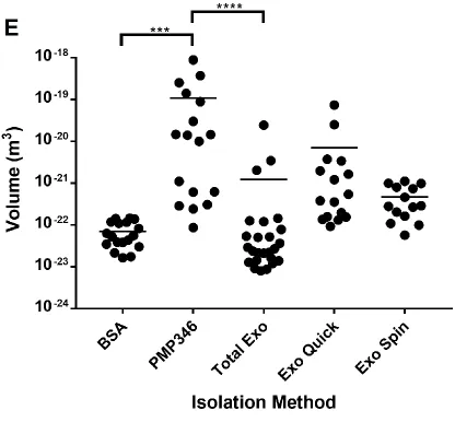
Multimodal characterization of PCCFs isolated using different techniques reveals that the immunoaffinity method is the most efficient at eliminating background proteins ... 41
Atomic force microscopy resolves three dimensional surface characteristics of isolated PCCFs ... 45
Mass spectrometry analysis of PSMA-isolated PCCFs identifies an abundance of albumin and protein peptides from tissues other than prostate ... 48
Nanoscale flow cytometry detects STEAP1 positive events in prostate cancer patient plasma as well as dual positive PSMA-STEAP1 PCCF events ... 49
Tandem immunoaffinity isolation of PCCFs significantly reduces non-target MP populations, while maintaining PSMA+STEAP1 dual events ... 52
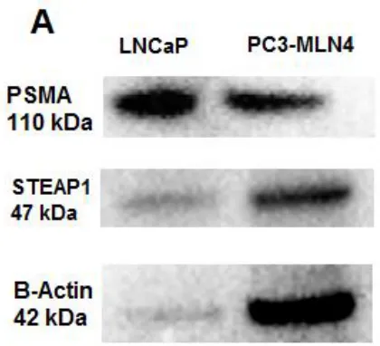
Western blot detection of prostate proteins in prostate cell lysate and PSMA immuno-purified PCCFs, but not in tandem isolated PCCF samples ... 55
Mass spectrometry results of tandem isolated samples reveal abundance of plasma and cytoskeletal proteins. ... 57
Chapter 4 ... 59
Discussion ... 59
Extracellular vesicles such as prostate cancer cell fragments as a fluid biopsy for prostate cancer ... 59
Immunoaffinity isolation of PCCFs ... 60
Multi-modal nano-characterization of purified extracellular vesicles from
viii
isolated samples ... 62
4.3.2 Atomic force microscopy for visual characterization and validation of the isolated PCCFs ... 62
Mass spectrometry ... 63
Significance... 64
Future directions and conclusions ... 65
References ... 68
Appendices ... 78
ix
List of Tables
Table 2: Summary of antibodies and isotype controls used in this study ... 22
Table 3: Summary of buffers used in this study. ... 23
x
List of Figures
Figure 1.Schematic representation of the biogenesis of different extracellular vesicles. ... 2
Figure 2. Summary of purification and evaluation strategy for extracellular vesicles (EVs)
from biological samples. ... 13
Figure 3. Nanoscale flow cytometry analysis of sizing beads and PCa patient plasma measure
events within the submicron range. ... 34
Figure 4. Working model of biotinylated-PSMA technique enriches PCCFs from patient
plasma and are quantified using nanoscale flow cytometry. ... 37
Figure 5. Atomic force microscopy images of bovine serum albumin and platelet-poor
plasma reveal size and distribution of proteins at varying concentrations. ... 40
Figure 6. Atomic force microscopy and nanoscale flow cytometry reveal differences in
particle size and distribution in PCCF samples obtained using different isolation methods. . 44
Figure 7. Atomic force microscopy resolves three-dimensional structures of PCCF isolated
from patient plasma and reveals small peaks in PCCF surface. ... 47
Figure 8. Nanoscale flow cytometry reveals the incidence of STEAP1 positive PCCF events
in PCa patient plasma samples, and also detects dual PSMA-STEAP1 positive events in PCa
patient plasma samples. ... 51
Figure 9. Nanoscale flow cytometry analysis of tandem immunoaffinity isolated PCCFs from
patient plasma show the enrichment of STEAP1 positive as well as dual PSMA-STEAP1
positive events. ... 54
Figure 10. PSMA and STEAP1 protein expression in LNCaP and PC-3M-LN4 prostate
cancer cell lines and PCCFs isolated with biotinylated-PSMA method. ... 56
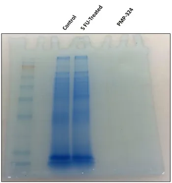
Figure 11. SDS-PAGE gel show the difference in protein band identification between loading
xi
List of Appendices
Appendix A: List of proteins identified in PCCF isolated using biotinylated PSMA
immunoaffinity method from plasma of patients with Gleason score 6. ... 78
Appendix B: List of proteins identified in PCCF isolated using tandem immunoaffinity
method from plasma of patients with Gleason score 8. ... 90
Appendix C: List of proteins identified in PCCF isolated using tandem immunoaffinity
method from plasma of patients with Gleason score 6. ... 100
Appendix D: List of proteins identified in PCCF isolated using tandem immunoaffinity
xii
List of Abbreviations
PSA Prostate specific antigen
PCa Prostate cancer
PSMA Prostate specific membrane antigen
STEAP1/2 Sixth transmembrane epithelial antigen of
the prostate 1/2
EV Extracellular vesicles
MPs Microparticles
APB Apoptotic bodies
miRNAs microRNAs
PCCFs Prostate cancer cell fragments
CTC Circulating tumor cells
GS Gleason score
NFC Nanoscale flow cytometry
AFM Atomic force microscopy
MVBs Multivesicular bodies
EM Electron microscopy
BPH Benign prostatic hyperplasia
PSCA Prostate stem cell antigen
xiii
SEM Scanning electron microscopy
Chapter 1
« Introduction»
Extracellular Vesicles
Extracellular vesicles (EVs) are a family of heterogeneous, cell-derived fragments or
vesicles, which can be generated by cell membrane shedding or storage vesicle
exocytosis. EV generation typically occurs following biological processes such as cell
activation and modes of cell death such as necrosis and apoptosis (1). Initially perceived
as cellular by-products or ‘dust’ of insignificant biological importance, recent research
has shed light on the role of EVs as mediators of intercellular communication, blood
coagulation and disease progression. Major sources or contributors of EVs in the blood
are platelets, leukocytes and endothelial cells (2). Secretory glands comprised of
epithelial cells also are a major source of EVs (3), but their contribution to the EV pool in
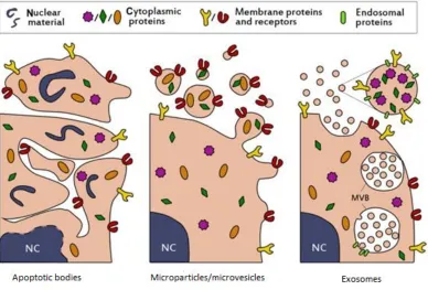
blood is unclear. Several types of EVs are described throughout the literature (Fig.1);
they are categorized according to their size, contents and mechanism by which they are
released (4). For example, exosomes (30–100 nm) are EVs of cytoplasmic origin,
released or exocytosed into the extracellular environment upon fusion of multivesicular
bodies (MVBs) with the plasma membrane (5). Microparticles (MPs; Fig.1, middle
panel), also known as microvesicles, are larger than exosomes measuring 100–1000 nm,
and are the primary result of membrane blebs released from the surface of cells (1).
Lastly, apoptotic bodies (APBs; Fig. 1, left panel) are larger cell derived vesicles,
measuring up to 4000 nm, and are eponymously generated during cellular apoptosis (6).
Given their origin and release from cells, EVs are commonly endowed with a portion of
membrane proteins, and in some APBs, genetic remnants of the parent cell (4). It is now
generally accepted that EVs, such as platelet MPs, play a significant role in modulating
normal physiological processes, such as coagulation (7) via expression of multifunctional
cellular signaling proteins such as tissue factor (8). However, despite observations of
elevated EVs in cancer patient plasmas (9) and other diseases (10), it has still not been
reservoir of biomarkers for disease detection and the role they play, if any, in disease
progression.
Figure 1.Schematic representation of the biogenesis of different extracellular vesicles.
The most common populations of extracellular vesicles found in biological fluids (saliva,
plasma, semen, etc.) include apoptotic bodies (APBs, left panel),
microparticles/microvesicles (middle panel), and exosomes (right panel). As an outcome
of their biogenesis during cell apoptosis, APBs package a variety of cellular contents
including DNA, RNA, and signaling molecules. During the process of cell membrane
blebbing, membrane and cytosolic proteins are selectively packaged into
microparticles/microvesicles (middle panel), resulting in the enrichment of specific
proteins from the parent cell. Lastly, exosomes contain proteins that are primarily
incorporated during formation of multivesicular bodies, such as tetraspanins CD9 and
Intercellular mode of communication
Heralded as an auxiliary means of signaling across vast cellular distances, the ability of
EVs to transport oncogenic factors and regulatory RNA in a vesicle format has been a
topic of intense debate that requires a rollback in perspective. First, classic examples of
cell-to-cell communication are hormone-based paracrine signaling circuits. As a specific
example, testosterone secreted by the testicles or adrenal glands into the circulation
reaches the prostate to sustain gland viability. Testosterone, primarily in a soluble form,
is an essential growth factor for prostate epithelium and only requires nanogram
quantities to elicit a physiological impact. Upstream of this, an additional paracrine
signaling circuit that relies on a brain–gland axis of communication represents another
complex and sensitive means of cell-to-cell communication that occurs between the
pituitary and testicles, in which luteinizing hormone is released into the circulation to
induce testosterone production by Leydig cells.
In contrast to those classic examples, recent key studies have revealed processes by
which EVs are able to interact with their target microenvironment, delivering various
cargo types and facilitating cell-to-cell communication. As an example, leukocyte EVs
are able to modulate endothelial cell activation by delivering pro-inflammatory agonists
onto endothelial cells, resulting in the release of endothelium-derived cytokines and
surface expression of ICAM-1, which is normally agonist-induced (12). Within an
oncology context, glioblastoma cells have been shown to release EVs that express
oncogenic factors such as EGFRvIII on their surface, impacting adjacent cells through
vesicle–cell interactions (13). This represents the first description of ‘oncosomes’,
wherein EVs that express transforming factors such as EGFRvIII are released from
parental tumor cells via membrane blebs and merge with the plasma membranes of
adjacent glioma cells lacking EGFRvIII receptor, resulting in the activation of
transforming signaling pathways and alteration of EGFRvIII-regulated gene expression
(13). Similarly, Peinado et al. (14) investigated the transfer of MET oncogene from
tumor-derived exosomes to bone marrow progenitor cells, wherein exchange of MET
oncogene induced the formation of a pro-angiogenic bone microenvironment and a
oncogenic factors can accelerate oncogenesis or metastasis, the potency of these
tumor-derived EVs falls far behind that of classic paracrine signaling mechanisms
(pituitary-luteinizing hormone-leydig cells) because of the submilligram quantities needed to elicit
a measurable effect in vivo. Hence, these observations may be overly optimistic given the
field’s lack of knowledge regarding the half-life of tumor-derived EVs and the propensity
of the immune system to also release counteracting measures potentially in the form of
exosomes.
Reports of nucleic acid transport via EVs have been the driving force for heightened
awareness in EV research across many different disciplines. For example, Valadi et
al.(15) recently demonstrated that RNA resident within exosomes derived from mouse
mast cells was transferred to human mast cells, resulting in ectopic expression of mouse
proteins in recipient cells (15). This advancement suggests that EVs are envoys between
cells, able to deliver mRNA that can impact protein production in recipient cells of a
measurable magnitude, akin to a hormone operating at the genetic level. Similarly,
exosomes from colorectal cancer cells determined to be enriched with 15 mRNAs
associated with M-phase processes of the cell cycle were delivered to healthy endothelial
cells in vitro, subsequently stimulating the proliferation of recipient endothelial cells (16).
Although the efficiency of this communication delivery system remains unclear, these
findings suggest that EVs from malignant cells can facilitate the delivery of RNAs that
encode factors responsible for cell proliferation. While it is unlikely that entire mRNA
coding regions are transported via EVs, much smaller miRNA present within EVs are
much more likely to be transferred (15). Studies of miRNA residing within EVs have
dominated the field because of their regulatory nature and robustness against degradation.
miRNAs are a family of small, non-coding RNAs (17–22 nucleotides in length) that
regulate gene expression by degrading target mRNAs and nullifying translation of those
target mRNAs (17). miRNAs can directly contribute to tumorigenesis through
modulation of oncogenic or tumor suppressor pathways by targeting mRNAs of
Given their impact and contributions to tumorigenesis, miRNA can also be used as
biomarkers to identify patients with aggressive or life-threatening tumors in a
non-invasive manner. As one example, Taylor and Gercel-Taylor (19) isolated exosomal
miRNA from serum samples of patients with benign ovarian disease, patients with
adenocarcinoma of the ovary and healthy volunteers to profile stage-specific
miRNA-based biomarkers. The amount of total miRNA was significantly elevated in
adenocarcinoma patients compared with patients with benign growth, and with minimal
exosomal miRNA detected in healthy controls. The diversity or levels of most miRNAs
was not significantly different between patients with early vs late-stage ovarian cancers,
but expression profiles of exosomal and tumor cell-derived miRNAs were similar (19). In
parallel, biomarker development for lung cancer has also resulted in a panel of miRNAs
(20) validated as biomarkers for diagnosis and prognosis for this disease. As described
previously in ovarian cancer patients, the total amount of exosomal miRNA was also
elevated in patients with lung adenocarcinoma and low or undetectable in control
samples. Most importantly, no significant differences were found when comparing
miRNAs derived from circulating exosomes and miRNAs derived from lung tumors,
indicating that exosomal miRNAs reflect the genomic identity of the tumor and can be
used as a potential blood-based marker for lung adenocarcinoma. Hence, EVs derived
from malignant cells may act as a system of miRNA transport to distant cells and used as
a novel biomarker platform for cancer progression.
EVs generated by breast cancer cells have also been implicated in de novo miRNA
processing and biogenesis due to the presence of Dicer, AGO2 and TRBP proteins within
purified EVs (21). Overlooked in this same study is the contribution of EVs generated by
other sources (endothelial, leukocyte and so on) that are present in the bloodstream with
unknown miRNA content. If in fact miRNA biogenesis is exclusive to breast or breast
cancer cells, this mechanism is unclear and conveniently specific to these sites and
ultimately may not be applicable to prostate cells because these proteins are not present in
prostasomes generated by the prostate (22). Studies like these continue to fall short in
determining whether EV preparations are free of any plasma proteins, which could
dominate currently published reports, lacking sufficient attention to the depletion of
plasma proteins that continue to be present in purified samples. Although the use of
atomic force microscopy is key to these evaluations (23), these reports still fail to
examine the EV preparations at the nanoscale resolution to quantitate soluble protein
contamination, marking these findings as suspect until further validations are performed.
Non-tumor derived EVs of physiological importance
1.3.1
Apoptotic bodies
Damaged, senescent and/or infected cells are often destined to undergo programmed cell
death or apoptosis (Note to Sabine: some infections are lytic and cause necrosis, have to
waffle a bit here or its an overstatement). This process is followed by degradation to
maintain tissue homeostasis and a normal physiologic milieu (24). Although mechanisms
of bleb formation are unclear during apoptosis, cells break apart and form membrane
blebs called APBs, which can contain nucleic acids such as miRNA, mRNA or genomic
DNA (25). Most importantly, APBs also display phosphatidylserine on their surface, an
‘eat-me’ signal for engulfment by phagocytes (for example, macrophages, dendritic cells)
and some fibroblast cells in an immune silent manner (26,27). These APBs play key roles
in adaptive immune responses in which self vs non-self antigens are processed for
subsequent development of T- or B-cell-mediated immune responses, depending on the
ongoing background of immune-based ‘danger signals’ in the body.
In cancer research, APBs have been shown to function as carriers for horizontal transfer
of oncogenic DNA. In one such study, APBs transported oncogenic H-rasV12 and
c-myc, to nearby normal mouse embryo fibroblast cells with a p53 knockout background
(28), resulting in tumor-like growth and progression in vitro. Furthermore, phagocytosis
of tumor APBs mediated by immature dendritic cells can induce immune tolerance by
cross-presentation and activation of regulatory T cells (29), revealing a potential
1.3.2
Platelet MPs
The discovery of EVs occurred in parallel with the initial studies of blood coagulation,
when researchers observed platelet-like activity in otherwise platelet-free serum samples
(30). However, they were not formally described until the late 1960s, when Peter Wolf
(31) used the term ‘platelet dust’ to describe small, membrane coated fragments he
observed from activated platelets. Wolf considered these vesicles as by-products of
platelet activation during storage, and concluded that coagulation activity in platelet-free
samples was due to the action of ‘platelet dust’. The term ‘platelet dust’ was later replaced with ‘microparticles’ (32) and ‘exosomes’ (33), in which platelet MPs are
membrane-derived and exosomes being the exocytosed storage granules (alphagranules,
dense granules) of platelets. EVs, either MPs or exosomes, are secreted by platelets (34),
endothelial cells (32) and leukocytes (33), and these types of cell fragments are relatively
abundant in different bodily fluids (2). Through quantification of all circulating MPs in
vivo, it is now understood that platelet EVs are the most abundant types of EVs, when
compared with MPs from other circulating cells (10). Platelet EVs functionally contribute
to coagulation and thrombosis because they are enriched in membrane receptors for key
coagulation factors and prothrombotic proteins. For example, MPs derived from activated
platelets express a high density of prothrombotic proteins on their surface, such as
adhesive receptor P-selectins (8), plasminogen activator inhibitor-1 (PAI-1) and
vitronectin (VN) (35), making thrombi resistant to fibrinolysis. In terms of cancer
research, platelet EVs (MPs and/or exosomes) have been shown to promote tumor cell
invasion in vitro by induction of MMP-2 synthesis in a prostate cell line (Clone-1) (36),
but the impact of platelet MPs on several other prostate cell lines in vitro and in vivo is
still unclear.
1.3.3
Endothelial MPs
Endothelial MPs are membrane-derived particles released upon apoptosis or necrosis,
whereas endothelial EVs are likely exocytosed storage granules of endothelial cells, such
as Weibel–Palade bodies, and are released upon activation by cytokine agents such as
endothelial cells were incubated with tumor necrosis factor-α and anti-tumor necrosis
factor antibody, it was found that tumor necrosis factor-α elevated endothelial EV
formations by a maximum of 2.5-fold within a 24-hour period (38). Recent studies have
also demonstrated that neoplastic cells induce the release of endothelial EVs, revealing
their potential as a novel biomarker for the detection of cancer and disease progression.
For lung cancer, levels of circulating endothelial EVs were found to be significantly
higher in lung cancer patients than in healthy control subjects suggesting that endothelial
EVs may be involved in endothelial cell proliferation as occurs in angiogenesis (39),
underscoring their pro-angiogenic effect in cancer progression. A subsequent study by
this group examined the potential use of endothelial EVs for predicting 1- year mortality
in patients with end-stage non-small cell lung cancer (34). In accordance with previous
findings, the results of this study revealed that circulating levels of endothelial EVs were
significantly higher in patients with 1-year mortality than in patients within the 1-year
and above mortality category, demonstrating the potential of endothelial EVs as
biomarkers for lung cancer prognosis (34). Yet again, these findings will require further
research to validate endothelial EVs as a prognostic biomarker.
1.3.4
Leukocyte MPs
Leukocyte EVs are released by almost all immune cells when activated by inflammatory
stimuli (40), further activating receptors on other leukocytes, resulting in the secretion of
inflammatory and chemotactic cytokines. In detail, in in vitro co-cultures of leukocyte
EVs with human umbilical vein endothelial cells, leukocyte EVs acted as inflammatory
agonists on endothelial cells which resulted in the release of cytokines interleukin-6 and
interleukin-8, and upregulation of leukocyte-endothelial cell adhesion molecules such as
ICAM-1 (13). These findings suggest that circulating leukocyte EVs can activate a stress
signaling pathway in endothelial cells, leading to an increase in coagulant and
pro-inflammatory activities.
Circulating leukocyte EVs have also been proposed as determinants for cardiovascular
risk factors in asymptomatic subjects. Chironi et al. (37) examined the carotid, abdominal
asymptomatic subjects without previous cardiovascular diseases. Levels of leukocyte
EVs were higher in subjects who carried atherosclerotic plaques in two or three sites
compared with those without plaque at any sites. Therefore, the measurement of
leukocyte EVs has demonstrated biomarker potential in cardiovascular disease in
asymptomatic patients, thus offering encouraging signs of their application in other
disease contexts but none as of yet with oncology although clearly applicable given the
recent advances of immunotherapy for PCa treatment.
Limitation of previous methods of EV characterization and
quantification
Several techniques have been described for the isolation and identification of EVs from
different bodily fluid samples. Minimizing the amount of non-target EVs and other
contaminants is a crucial step towards obtaining a homogenous mixture of EVs.
Scientists have largely relied upon serial centrifugation and ultracentrifugation steps at
increasing speeds and time intervals to isolate EVs from cells, proteins and large cellular
debris (41). However, this method does not guarantee elimination of all non-target
fragments from samples, resulting in enrichment, as opposed to purification of the desired
EV population. This was presented by Mrvar-Brečko et al. (42), who reported that after
several centrifugation steps, samples predominantly contained populations of unwanted
cells mixed with EVs. This method is also time consuming, as it requires repeated
centrifugation steps lasting hours. Immunoaffinity approaches that utilize paramagnetic
beads conjugated with an antibody specific for antigens expressed on the surface of a
target EV are rapid and more target-MP/exosome specific. Beads are mixed with the
sample containing target cell fragments and passed through a column based magnet
separation system (43). The vast majority of non-bead bound cells, non-target MPs and
plasma proteins will pass through the column, whereas antigen-positive EVs bound to
paramagnetic beads become indirectly immobilized to the column via magnets (44). For
purification of prostate-derived EVs, magnetic beads can be conjugated with anti-CD9 or
anti-prostate-specific membrane antigen antibody, and incubated with peripheral blood or
customizable, in which the amount of ‘bait’ antibody used can be varied and the number
of times the beads + EVs are washed can vary from two to eight times to fully eliminate
plasma proteins and non-target EVs. Unfortunately, the major limitation of this method is
the lack of bait antibodies available for target EVs and the cost of presently available
ones.
Electron microscopy (EM) is a powerful method to visually characterize cell-derived
vesicles, and scanning EM (SEM) is specifically used to visualize the morphology and
relative size of platelet-derived EVs (31,42). In most cases, SEM can be used to visually
differentiate EVs from erythrocytes and other circulating cells (42). SEM allows certain
analyses that are not possible by other techniques such as determining whether objects
are vesicular or proteinaceous by their ultrastructure; however, it does not quantify the
concentration of vesicles isolated from a sample in a high-throughput manner. SEM also
has significant drawbacks related to preparation of sample(s), with purified EVs
inconsistently being immobilized onto the substrate (silica wafers or mica) for SEM
imaging (32). Atomic force microscopy is another valuable tool that enables the
determination of ultrastructure for all entities (EVs, plasma proteins) at an atomic
resolution (23). Most recently, this technique was used to understand the ultrastructure of
purified platelet MPs (23) and is the only instrument that offers information regarding
protein contamination in purified EV fraction owing to its atomic level of resolution.
To enumerate EVs in any given sample, flow cytometry (FC)techniques would be best
for quantitation of EV subpopulations given the multi-parametric nature of the technique.
Hamilton and colleagues (45) first described the use of FC for detection of cell-derived
vesicles released by human umbilical vein endothelial cells. The reliability of FC to
characterize EVs has substantially improved despite previous ill-informed claims
regarding the inability of optics to accurately acquire scatter of events smaller than 1
micron (μm). The emergence of nanoscale FC has made high-throughput,
multi-parametric analysis of all events between 110 and 880 nm possible (46) regardless of the
incident wavelength of light used. Using the nanoscale flow cytometer Apogee
100 nm increments (47), revealing the potential of this instrumentation to become first in
class for analysis of MPs in complex biological mixtures (48).
A nanoscale based approach to EV purification and
evaluation
In Figure 2, we present an idealized approach to purifying EVs, such as MPs,
microvesicles and exosomes. The sequence of techniques proposed is important because
they will allow the experimenter to evaluate their preparations at a nanoscale resolution
while analyzing each EV as a single discrete event. First, either plasma, serum or urine
can be used as the starting material and submitted to isolation or purification with the
three main techniques. Technique selection for purification is dependent on the
resources, instrumentation and amount of starting material available to the experimenter.
We recommend immunoaffinity based approaches to generate ultra-pure preparations of
antigen specific EVs, such as prostasomes or prostate cell fragments.
Immunoaffinity-based approaches also enable the experimenter to ‘wash’ their sample repeatedly prior to
elution, to maximally reduce the presence of non-target EVs and plasma or urine
proteins. The first evaluation step should focus on the enrichment ratio of target EVs vs
non-target EVs. In the case of PCCFs, nanoscale FC is recommended because all EV
events in the sample will be evaluated and the percentage of events that bind the prostate
biomarker can be used to infer enrichment. With exosomes, dynamic light scattering
instruments must be used because nanoscale FC cannot analyze events smaller than 100
nm in diameter. Although this is the ideal instrument for that purpose, it does not inform
the user of the target vs non-target EV ratio unless single fluorescence channel dynamic
light scattering instruments are used. If these instruments are not available, then ELISA
followed by sequential western immunoblotting is recommended. Finally, if an EV
preparation is maximally enriched for target EVs, then the next step is to determine the
extent of plasma protein ‘contamination’ in the sample. This is significant because of the
potential for soluble RNA/DNA and miRNA–protein complexes to be present outside
and alongside the EVs in the preparation. The definitive instrument to determine the
force microscopy eliminates all washing and processing of the sample and can be
performed ‘dry’, wherein the solvent is dried off, leaving behind only EVs, ions and
proteins. Owing to its atomic resolution, all events can be volumetrically analyzed, with
events smaller than 100 nm in diameter quantitated and compared with much larger
structures such as MPs. Alternatively, if there are suspicious ultra-structures present in
the sample, SEM can be performed to determine whether the structure is vesicular in
structure, or a protein aggregate. By following this scheme, an experimenter can readily
purify EVs with the full knowledge of the contribution of non-target EVs and
Figure 2. Summary of purification and evaluation strategy for extracellular vesicles (EVs) from biological samples.
This scheme can be used to isolate and evaluate EVs from plasma, sera or seminal fluid.
Various techniques can be used to isolate EVs based on size, immunoreactivity to
antibodies, or samples can be sent out for purification by third party vendors. Once
purified, the experimenter may wish to consider the proportion of target EVs to
non-target EVs using western blot, nanoscale flow cytometry, dynamic light scattering or
techniques to determine the extent of contamination from non-EV proteins can be
performed using either atomic force microscopy or scanning electron microscopy which
Current state of prostate cancer diagnosis
Prostate cancer is the most commonly diagnosed visceral cancer among Canadian men.
In 2016 it accounted for 21% of all newly diagnosed cancers, and for ~ 10 % of all
cancer-related deaths among men in Canada (72). This translates to 65 Canadian men
being diagnosed and another 11 dying from PCa every day. The prostate specific antigen
(PSA) testing continues to be heavily relied upon as a monitoring and prognostication
tool; however, it is produced and secreted by both normal prostate epithelium and PCa
into the circulation (4). For this reason, PSA-based screening is discouraged for screening
of PCa because of its low specificity, which means that a high number of PCa cases are
of a low-risk phenotype forcing men to undergo painful and repeated biopsies to ensure
the tumor has not upstaged (73). Most of the time, PSA acts as a "red flag" that causes
considerable anxiety for a patient until the definitive prostate biopsy is taken and
examined by a pathologist (3). Prostate biopsy is the gold standard for diagnosis, as it
provides very important histological information regarding the 5 different patterns of
acinar arrangement and glandular characteristics for grading the tumor with the Gleason
Score (GS) system. Gleason grade 1 represents the most well-differentiated lesion,
whereas Gleason grade 5 represents the most poorly differentiated lesion, and hence a
highly aggressive phenotype of PCa. The most predominant lesion in the specimen also
known as the primary pattern or first number of the GS, and the second most common
pattern in the specimen (the secondary pattern) becomes the second number of the score.
Thus, the Gleason score is the sum of two grades. Only Gleason grade 3, 4, and 5 are
considered histologically and clinically relevant, therefore only a GS of 6 and higher is
considered to be PCa. However, only GS ≤ 7 is regarded as clinically significant prostate
cancer, whereas GS 6 prostate cancer is considered low-risk (73). The recommendation
options for most patients with Gleason Score 6 PCa is active surveillance, which requires
regular PSA testing, physical examination and periodic biopsy to determine if the cancer
has "upstaged" or progressed (74). However, repeated biopsy also submits patients to
potential complications such as hematuria, rectal bleeding, and urinary tract infection
with rare cases leading to mortality (75). Also, PSA levels do not correlate with the
to accurately identify patients with aggressive forms of PCa from those with low-risk
disease.
Prostate MPs
The first studies on prostate EVs in 1977 by Ronquist and Hedström (49) described
vesicles generated within prostate epithelial cells and released via exocytosis into seminal
fluid. These EVs were subsequently termed ‘prostasomes’ (50). In seminal studies
comparing prostasomes from both benign and malignant prostate cells, no significant
differences were reported regarding synthesis and release of these prostate-derived EVs
(51). Although there are few reports describing prostate cell MPs in healthy individuals,
the presence of prostasomes in prostate cancer (PCa) patient plasmas continues to be a
translational cancer research focus (51).
1.7.1
Prostasomes
These vesicles range in size from about 50–500 nm, originate from prostatic epithelial
cells and are present in seminal fluid and post prostatic massage urine (50,51). These EVs
have been shown to protect sperm within the female reproductive system, in which
cytotoxic interactions between prostasomes and natural killer cells significantly reduce
natural killer cells’ activity to prevent immune-mediated sperm destruction (52).
Prostasomes represent a novel cancer biomarker platform because of their release by
malignant prostate cells into seminal fluid and blood (53).
Tavoosidana et al. (54) suggested that the levels of prostasomes reflect disease severity,
based on the detection of prostasomes in blood samples from patients with PCa and high
Gleason score, whereas levels of prostasomes were reduced in samples from patients with
low Gleason score and benign prostatic disease or indolent PCa (54). Despite the small
sample size in this study, it demonstrated that prostasomes can be detected in patient
blood, and have the potential to distinguish aggressive PCa from low-risk or benign
1.7.2
PCa cell fragments
The potential of prostate cancer cell fragments (PCCFs) to serve as a diagnostic
biomarker platform for PCa is a topic of intense research effort because they are
independent of other serum-based biomarkers currently used for detection of PCa, such
as prostate specific membrane antigen (PSA), which is not specific for PCa (4).
Currently, there is a lack of agreement regarding the best purification strategies for
PCCFs, as well as which biomarkers should be used to characterize PCCFs. Recent
attempts to discover suitable surface markers specific for PCCFs, which relied on
proteomic analysis of isolated PCCFs present in the serum of mice grafted with human
PCa xenografts, identified putative biomarkers such as RAB5A and RAB11A (55). Other
cell-line-dependent studies reveal a higher abundance of proteins such as FASN in cell
fragments derived from PC346C and VcaP cells (4). Many of these reported biomarkers
have not been clinically validated, either in serum or plasma samples, or cross-referenced
with databases, underscoring the need to substantiate biomarkers beyond the initial
discovery phase. An approach that enumerates PCCFs based on a multi-parametric
technique may also improve sensitivity and accuracy if criteria are based on
superimposition of both prostate-specific and cancer-specific biomarkers on the same
PCCF. Clearly, PCCFs present an extracellular source of prostate-specific membrane
antigen reflecting a prostate cell origin (56,57) and should be the initial ‘capture’
biomarker for assaying other cancer-specific biomarkers. Other antigens specific to
prostatic tissue that could potentially be used are STEAP1 (58), STEAP2 (59), and PSCA
(60); however, their utility remains unclear owing to the lack of reagents such as flow
cytometry (FC)-compatible antibodies available for each of these putative
prostate-specific biomarkers.
Aside from their putative abundance in patient biofluids, PCCFs, also termed 'large
oncosomes', are also postulated to play a role in disease progression and metastasis (61).
Oncosomes, ranging from 1μm to 10μm in size, can be identified histologically in tumor
tissue sections. Additionally, they exhibit gelatin-degrading proteolytic activity by the
proteases are commonly associated with tumor cell invasion, oncosomes may serve to
concentrate proteases that assist tumor cell migration (61). Provided that oncosomes are
stable in the tumor microenvironment and in serum, they in turn could harbor clinically
valuable biomarkers to identify patients with intermediate to high-risk PCa in a
noninvasive blood-based manner.
Currently superseding PCCFs as biomarkers, are circulating tumor cells (CTC),
characterized by co-expression of EpCAM and various cytokeratins in nucleated cells
present in a 7–10 mL blood sample collected from patients. CTCs are thought to be
generated by the release or entry of tumor cells into circulation during the intravasation
step of the metastatic cascade (62). CTC enumeration via the CellSearch Instrument
(63,64) is currently the gold standard for prognostication of patients with metastatic PCa
(65). However, enumeration of CTCs is not a prognostic tool for localized PCa patients
owing to the low CTC counts even in patients undergoing salvage radiation therapy (66).
Despite the low abundance of CTCs in patient blood samples (67), several key studies
have shown that CTC enumeration can distinguish PCa patients from healthy volunteers
(68).
Emerging clinical data suggests that biomarkers derived from plasma exosomes can
similarly differentiate PCa patients exhibiting high and low Gleason scores (GS) from
those with BPH and healthy individuals. Specifically, exosome-associated Survivin is
highly expressed in plasma samples from PCa patients with Gleason score of 6 and 9,
whereas the expression of this protein is significantly lower in BPH and healthy donor
plasmas (69). However, levels of Survivin were not significantly different between the
PCa patients with different GS (6 vs 9), highlighting the need for biomarkers which are
Gleason score-specific. Other EV types such as tumor derived microparticles may offer
an equivalent, if not improved means of prognosticating PCa recurrence given the large
numbers of these submicron entities within patient plasma samples with metastatic
disease (68). Clearly, Coumans and co-workers (68) found that tumor cell MPs and tumor
cell fragments are other types of extracellular vesicle subclasses that can yield important
based testing. Various genomic tests for prognostication of early biochemical recurrence
in localized PCa patients have also prompted the notion that these biomarkers in
combination may be present in or on tumor MPs generated by the primary tumor (70).
However, transposing transcriptome-based biomarkers into a protein positive EV-based
format may be challenging given that some of these biomarkers are downregulated or
absent in the target pathology (70).
Alternative, more promising approaches may be based on the presence of microRNAs
(miRNAs) within prostate-derived EVs that are specific to each Gleason grade, or
associated with early biochemical recurrence in patients post prostatectomy or radiation
therapy. Such is the case for miRNA-34a, whose expression within EVs in patient plasma
is predictive of sensitivity to first-line treatment with Docetaxel (71). These studies are
correlative and although suggestive of a pathogenic mechanism, further investigation is
required to conclusively demonstrate compartmentalization of miR-34a within prostate
derived EVs or whether they are derived from other non-cancer sources (71).
Nonetheless, their promise as biomarkers of cancer progression is tantalizing and reflects
a world-wide intensified effort towards understanding EV biogenesis and their ability to
mediate intercellular communication during cancer progression.
Extracellular vesicles such as prostate cancer cell fragments
as a fluid biopsy for prostate cancer
Research to discover new diagnostic biomarkers that could differentiate patients with
indolent, or low-risk PCa, from those with high-risk disease has not significantly
progressed, but the need for a non-invasive test for monitoring PCa patients is of great
clinical value. PCCFs are an attractive biomarker platform for detecting PCa, as these
fragments originate from prostate epithelium or from malignant cells within the primary
tumor and are released into the blood circulation (76). Moreover, it has been previously
shown that significant quantities of PCCFs are detectable in samples from PCa patients,
Thesis hypothesis and objectives
The goal of this project is to find biomarkers on the surface of PPCFs that could
differentiate patients with high-grade PCa from those patients with low-grade disease. It
is our hypothesis that PCCFs from patients with low grade prostate cancer (GS 6) will
express different biomarkers than those found on PCCFs from patients with high grade
prostate cancer (GS 8). The following objectives will be pursued in order to reach my
goal.
1. To isolate PCCFs from patient plasmas representing low-grade prostate cancer
(GS6) and MPs from patient plasmas representing high-grade prostate cancer
(GS8) using an immunoaffinity isolation protocol developed by our lab.
2. To assess the enrichment of these isolated PCCFs samples using a nanoscale flow
cytometry and atomic force microscopy
3. To perform proteomic and bioinformatics analysis of isolated PCCFs to identify
biomarkers that differ between GS6 and GS8 patients, which could help to more
accurately diagnose patients with indolent disease from those with an aggressive
Chapter 2
Materials and Methods
Patient plasma
Prostate cancer (PCa) patient plasmas samples were attained through the Ontario Institute
for Cancer Research Tumor Bank and the University Health Network Genitourinary
BioBank (Toronto, ON) under Western University Research Ethics Board (REB)
approved Ethics Applications # 103156 and 103409. Only samples from patients with a
minimum of 3 years’ follow-up were included to avoid patients that upstaged/upgraded
during that time. Whole blood was collected into CellSave vacutainers (10mL volume,
Janssen Diagnostics Inc.). To prepare plasma from whole blood for prostate microparticle
analysis, whole blood was collected in K2-EDTA Vacutainers (BD Biosciences Inc.) and
spun at 1500 × g for 10 minutes. The plasma layer was removed, aliquoted and then
stored at -80 ˚C.
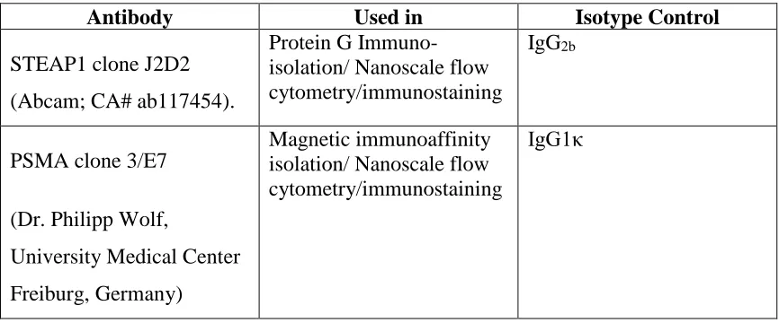
Antibodies and isotype controls
Antibodies and isotype controls used in nanoscale flow cytometry and immunoaffinity
isolation techniques have been compiled in Table 2.
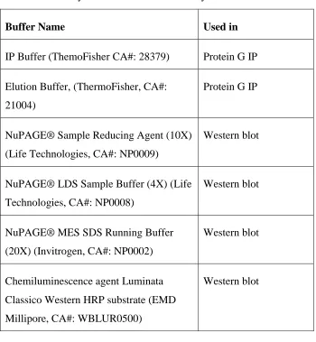
Buffers and reagents
Buffers used in the purification of proteins and experiments have been compiled in Table
Table 1: Summary of antibodies and isotype controls used in this study
Antibody Used in Isotype Control
STEAP1 clone J2D2
(Abcam; CA# ab117454).
Protein G Immuno-isolation/ Nanoscale flow cytometry/immunostaining
IgG2b
PSMA clone 3/E7
(Dr. Philipp Wolf,
University Medical Center
Freiburg, Germany)
Magnetic immunoaffinity isolation/ Nanoscale flow cytometry/immunostaining
Table 2: Summary of buffers used in this study.
Buffer Name Used in
IP Buffer (ThemoFisher CA#: 28379) Protein G IP
Elution Buffer, (ThermoFisher, CA#:
21004)
Protein G IP
NuPAGE® Sample Reducing Agent (10X)
(Life Technologies, CA#: NP0009)
Western blot
NuPAGE® LDS Sample Buffer (4X) (Life
Technologies, CA#: NP0008)
Western blot
NuPAGE® MES SDS Running Buffer
(20X) (Invitrogen, CA#: NP0002)
Western blot
Chemiluminescence agent Luminata
Classico Western HRP substrate (EMD
Millipore, CA#: WBLUR0500)
Confirmation of the sizing resolution of the apogee
A50-micro nanoscale flow cytometer
Silica microspheres (Apogee FlowSystems Inc.) of varying diameters (110 nm, 179 nm,
235 nm, 304 nm, 585 nm, 880 nm, 1300 nm) were analyzed using the A50-Micro
Nanoscale Flow Cytometer (Apogee FlowSystems Inc.). These beads were diluted
1:10000 prior to analysis on the A50-Micro Nanoscale Flow Cytometer.
Immunoaffinity isolation of prostate cancer cell fragments
(PCCFs) from patient plasma with PSMA
To isolate PCCFs from PCa patient plasma (n=10/Gleason score), the Miltenyi Biotec
MidiMACS system was used in which 100µL of plasma was incubated at 4 ºC for 30
minutes with 10 µL of biotinylated anti-PSMA antibody. Subsequently, the plasma was
diluted in 90 µL of dH2O and then incubated with 20 µL of Streptavidin microbeads
(Miltenyi Biotec; CA#130-048-102) for an additional 20 minutes at 4 ºC. The sample was
then diluted in 1 mL of dH2O, passed through a MACS- LS separation column which was
attached to a magnetic field. This step was repeated 3 times (Miltenyi Biotec;
CA#130-042-401). The column was then removed from the magnetic field and the PSMA positive
PCCFs were eluted twice with 1mL of dH2O. The eluent was then passed through another
magnetic column, and a final elution step was done with a total of 600 µL of dH2O.
Tandem immunoaffinity isolation of PCCFs with Protein G
agarose beads
The Protein G agarose beads were first to STEAP1 antibody in a microcentrifuge tube.
Briefly, 200 µL of Protein G agarose slurry were added to 10 µL of the STEAP1
antibody; the mixed was incubated overnight at 4°C. For the tandem isolation method, 50
µL of Protein G-STEAP1 agarose bead slurry was added to 600 µL of the PCCF samples
previously isolated using the biotinylated-PSMA method, and the reaction was incubated
with gentle mixing for 1 hour at room temperature. To wash the bead-PCCF immune
and subsequently centrifuged for 5 minutes at 2500×g; the supernatant was discarded.
The IP Buffer wash step was repeated a total of 3 times. To elute the PCCF’s attached to
the agarose beads, 100 µL of Elution Buffer was added to the beads and incubated for 5
minutes. The tube was centrifuged for 5 minutes at 2500 × g and the supernatant was
collected. This step was repeated a total of 2 times and the two supernatant fractions were
collected and combined. The pH of the eluate was adjusted to physiological pH by adding
~10 µL of a 1M Tris-HCl (pH 7.5-9), per 100 µL of eluate.
EV isolation from patient plasma using exosome isolation
kits
The EV fraction from PCa patient plasma were purified using the following kits:
ExoQuick-TC™ (EQ, System Biosciences Inc.; Mountain View, CA), ExoSpin™ (Cell
Guidance Systems LLC.; Carlsbad, CA), and Total Exosome (Life Technologies Inc.;
Burlington, ON), with some modification to the manufacturer’s recommendations.
2.7.1
ExoQuick-TC™
To prepare the plasma for exosome precipitation, 100 μl of the sample was centrifuged at
3000 × g for 15 minutes. The plasma samples were pre-treated with 1 μl of [500U/mL]
thrombin to make them compatible with ExoQuick exosome precipitation kit. The
mixture was incubated at room temperature for 5 minutes while mixing by gently flicking
the tube, then it was centrifuge in a standard microfuge at 10,000 x g for 5 minutes. The
supernatant was transferred to a new clean tube and treated with 25μl of ExoQuick
reagent to precipitate exosomes, and incubated for 60 minutes at 4°C. The
ExoQuick/biofluid mixture was centrifuged at 1500 × g for 30 minutes, the supernatant
was carefully aspirated, and the pellet was resuspended in 100μl of dH2O and stored at
2.7.2
ExoSpin™
100μl of plasma was centrifuged at 300 × g for 10 minutes to remove cell debris. The
supernatant was transferred to a new microcentrifuge tube and spun at 20,000 × g for 30
minutes. The supernatant was transferred to a new microcentrifuge tube and 50μl of
Buffer A was added and incubated at 4°C for 5 minutes, and then centrifuged at 20,000 x
g for 30 minutes. The supernatant was carefully removed and discarded, and the
exosome-containing pellet was resuspended in 50 μl of dH2O and stored at -20°C. The
supplied column was prepared by spinning it down at 50 x g for 30 seconds to remove
buffer from the top of the column and allowing it to enter the column bed. To wash the
column, 200µl of dH2O were added to the top, and spun down again at 50 x g for 30
seconds. The exosome-containing sample was added to the column and centrifuged at 50
x g for 60 seconds. This step was repeated once more, and the resulting eluate containing
the purified exosomes was stored at -20°C.
2.7.3
Total Exosome™
100μl of plasma was centrifuged at 2000 × g for 20 minutes at room temperature. The
supernatant containing partially clarified plasma was transferred to a new tube without
disturbing the pellet, and centrifuged at 10,000 × g for 20 minutes at room temperature.
The clarified plasma was placed in a new tube without disturbing the pellet, 50 μL of
dH2O was added and mixed thoroughly by vortexing. 30 μL of the Exosome Precipitation
Reagent (from plasma) was added to the sample, mixed by vortexing, and incubated at
room temperature for 10 minutes. The sample was centrifuged at 10,000 × g for 5
minutes at room temperature. The supernatant was removed and discarded and the
remaining exosome pellet was resuspended with 50 μL of dH2O, and stored at -20°C.
Enumerating the PSMA positive populations of PCCFs
To enumerate the populations of PCCFs in patient plasma, a phycoerythrin (PE) labelled
stain for detection of the PCCFs, 1 μL of the antibody was added to 10 μl of plasma,
incubated in the dark for 30 minutes, then diluted with 290 µL of PBS and analyzed on
the Apogee A50-Micro nanoscale flow cytometer. The negative isotype control mouse
IgG-PE was performed in parallel following the same incubation conditions. Gates for
the PCCF population were established by analyzing the isotype control first, and then
analyzing the antibody labeled samples.
Nanoscale flow cytometric detection of dual positive PCCF
populations
For detection of dual positive PCCF populations, the same protocol as section 2.8 was
followed with some modifications. In brief, 10μl of patient plasma was incubated with
1μl of anti-PSMA-PE and 2 μL of anti-STEAP1-Alexa 647 antibody at room temperature
in the dark for 30 minutes. The negative isotype controls were utilized in parallel
following the same incubation conditions. Samples were diluted with 290 µL of PBS and
analyzed on the Apogee A50-Micro nanoscale flow cytometer. Gates for each
microparticle population were established by analyzing the isotype control first,
modifying the gains for each PMT as necessary, and then analyzing the antibody labeled
samples.
Atomic force microscopy
Exosome suspensions and PCCFs were diluted in dH2O ratios of 1:10, 1:1000, and 1:
10,000. From these diluted samples, a volume of 2 µL was placed and adsorbed to a
freshly cleaved mica coverslip (Ted Pella, Inc.; Redding, California) and dried in an oven
at 60ºC for 5 minutes. Samples were analyzed with the Veeco Dimension 3100 Nanoman
AFM (Veeco Metrology, LLC; Santa Barbara, California) in tapping mode. Topographic
height and phase images were recorded at 256×256 pixels at a scan rate of 1 Hz. Image
processing was performed with Gwyddion Data Processing software, version 2.40
Western blotting
For protein extraction, isolated PCCFs were lysed in a master mix of reducing sample
buffer at a 10X concentration, and LDS sample loading buffer at a 4X concentration.
Samples were boiled for ~10 minutes at 90 ºC. Cellular proteins from LNCaP and
PC-3M-LN4 cell lysate were also extracted following the previously described steps. To
separate the PCCF and cellular proteins, 10 μg were loaded onto a NuPAGE® Novex®
4-12% Bis-Tris Gels (Invitrogen, CA#: NP0321BOX) and electrophoresed at 200V for 1
hour. Transfer of the gels to a polyvinylidene difluoride transfer membrane (Thermo
Scientific, CA#: 88518) was done at 30V for 1 hour. Blocking of the membrane was done
in 5% powdered milk in TBS-T for 1 hour at room temperature. Membranes were probed
using primary antibody STEA1 or PSMA, overnight in 4°C at a dilution ratio: 1:500, and
then with horseradish peroxidase conjugated-second antibody for 1 hour at room
temperature (Sigma Aldridge, CA#: NXA931-1ML) at a ratio of 1:2000. Protein bands
were detected by using an enhanced chemiluminescence HRP substrate, incubated for 5
minutes at room temperature, and the membrane was developed using the Bio-Rad
ChemiDoc™ MP System (Bio-Rad Laboratories Inc., Hercules, CA).
Mass spectrometry and proteomic analysis of PSMA
isolated samples
This process was contracted out to the Campus Chemical Instrument Center (CCIC) Mass
Spectrometry and Proteomics Facility at The Ohio State University (Arpad Somogyi,
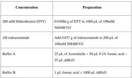
In-solution digestion
2.13.1
List of solution
Solutions prepared for in-solution digestion have been compiled in Table 4.
Table 3: Summary of solutions used for in-solution digestion.
Concentration Preparation
200 mM Dithiothreitol (DTT) 0.03086 g of DTT in 1000 µL of 100mM
NH4HCO3
1M iodoacetamide Add 0.037 g of iodoacetamide to 200 µL of
100mM NH4HCO3
Buffer A 25 µL of Acetonitrile + 50 µL 0.1% formic acid +
25 µL ddH2O
Buffer B: 1 µL formic acid + 1000 µL ddH2O
2.13.2
Sample preparation and disulfide reduction
Samples were first prepared by adding 1 uL of a 100mM NH4HCO3. In order to bring
samples to 100 μL, dH2O was added if needed. To reduce the sample, 5 μL of 200mM
2.13.1
Sulfhydryl alkylation
To alkylate the sample, 4 uL of the iodoacetamide stock was added to the sample and
vortexed, followed by brief centrifugation in a microcentrifuge to get the sample to the
bottom of the tube. The sample was incubated 1 hour at room temperature.
2.13.2
Stopping alkylation
To neutralize the remaining iodoacetamide, 20 μL of DTT stock was added to each
sample, which was then vortexed and incubated at room temperature for 1hour.
2.13.3
Trypsin digest
For trypic digestion, each sample was gently vortexed and trypsin was added at a 1:20
ratio (1mg of trypsin for every 20mg of sample). To allow complete digestion, the sample
was placed in a 37°C water bath overnight.
2.13.4
Sample clean-up
The SPE cartridges (HyperSep™ C18 columns - 50mg resin, Thermofisher CA:
#60108-390) were prepared by first washing the column 3 times with 1mL of Buffer A, then 3
times with 1 mL Buffer B, eluting the flow through into a waste beaker. Subsequently,
the samples were acidified with 0.2% formic acid and passed over the SPE cartridge
twice. The unbound components were washed off the column with 1 mL of Buffer B 3
times. The peptides were then eluted off of the column with 400 µL of Buffer A. To
reduce the volume of the samples and remove the acetonitrile, samples were concentrated
Mass spectrometry and proteomic analysis of tandem
isolated samples
This process was contracted out to the Biological Mass Spectrometry Laboratory at the
University of Western Ontario (Director: Prof. Gilles A. Lajoie;
Chapter 3
« Results»
The A50-micro nanoscale flow cytometer analyzes events
within the submicron size range, and detects PSMA positive
extracellular vesicles
The A50-Micro nanoscale flow cytometer (NFC; Apogee Flow Systems,Hertfordshire,
UK) is reported by the manufacturer to be capable of high-throughput and
multi-parametric analysis of events between 100-1000 nm, resolving various sizes of
calibration beads based on large angle light scatter (LALS) and small angle light scatter
(SALS). We ran silica beads of various diameters, 110 nm, 179 nm, 235 nm, 304 nm, 585
nm, 880 nm, 1300 nm (Fig. 3A-B), though the A50-Micro NFC. The analysis of these
beads show that the A50-Micro has the ability to analyze events within the submicron
size range, resolving discrete subpopulations when analyzed together.
The corresponding analysis of PCa patient plasma with the A50-Micro nanoscale flow
cytometer (Fig. 3C) demonstrate that when the plasma is incubated with an
anti-PSMA-PE antibody, a subpopulation of prostate cancer cell fragments (PSMA positive PCCFs)
is observed and determined to be within the 110-304 nm diameter size range (red box).
This population is distinct from other particles in the sample, which are PSMA negative
(blue box), in both size and immunofluorescence. When the isotype control mouse IgG1ƙ
-PE was incubated with PCa patient plasma, a minimal number of events were recorded
Figure 3. Nanoscale flow cytometry analysis of sizing beads and PCa patient plasma measure events within the submicron range.
The Apogee A50-Micro nanoscale flow cytometer is able to readily analyze events within
the submicron range based on the analysis of silica beads of various diameters (A and B).
When patient plasma is incubated with anti-PSMA-PE antibodies, a subpopulation of
PSMA positive PCCFs are observed within the 110-304 nm diameter size range (C).
Isotype control IgG1ƙ-PE incubated with patient plasma does not detect a significant
Immunoaffinity isolation using PSMA antibodies enriches
extracellular vesicles from prostate cancer patient plasma
The immunoaffinity method for PCCF isolation (Fig. 4A) utilizes a biotinylated PSMA
antibody and a streptavidin conjugated magnetic bead to separate PSMA positive PCCF’s
from other plasma components, such as cell debris and non-target MP’s. When patient
plasma is incubated with the biotinylated PSMA antibody and streptavidin beads and
subsequently passed through a magnetic field, the magnet attracts the PSMA positive
PCCFs, while other plasma components which are PSMA negative are not retained,
resulting in concentration and recovery of PSMA positive PCCFs.
The enrichment evaluation of immunoaffinity purified PCCFs from patient plasma shows
the relative abundance of PSMA-PE positive PCCF’s (red box) compared to the total
events of other non-target MPs (grey shaded areas) in plasma before PSMA
immunoaffinity isolation (Fig. 4B), within the first elution fraction (Fig. 4C), and within
the second elution fraction (Fig. 4D) from the same plasma sample. The population
events of non-target MP’s and other cell debris is greater in plasma samples before
immunoaffinity purification, when compared to PSMA positive PCCFs. After the first
elution, the non-target MP population is significantly reduced relative to the PSMA
positive PCCFs. After the second elution, a greater reduction of non-target MPs is
observed, and PCCF events. The relative abundance of PSMA positive PCCFs shows the
enrichment of this populations after isolation. In patient plasma (B), PSMA positive
events account for 1.1% of all events. After the last elution fraction (D), 21.5% of the