Available Online atwww.ijcsmc.com
International Journal of Computer Science and Mobile Computing
A Monthly Journal of Computer Science and Information Technology
ISSN 2320–088X
IMPACT FACTOR: 6.017IJCSMC, Vol. 7, Issue. 2, February 2018, pg.106 – 119
Using the Fourier Transform Method
to Segmentation Heart Images Using
Techniques Image Processing
N.Panah
1,
H.Masoumi
2, SH.Sharif
31,2,3
Islamic Azad University, Kazeroun, Branch Kazeroun, Iran
[email protected] mail:
-E
Abstract - In the past fifteen years, magnetic resonance imaging (MRI) has become a reference
examination for morphology, function, and injection in humans.
However, due to the characteristics of MR images of the heart and due to the variability of images among patients, the issue of the segmentation of heart cavities in MRI has not yet been resolved.
In this paper, using a MRI stage, the division carries a split in short-axis images.
Medical history problems and special categorization related to these images are provided.
For this very complicated division, previous information is required.
We then propose a basic classification for cardiopulmonary differentiation methods, as well as a special emphasis on the level of external information needed and how to limit the division.
In this paper, we perform MRI images of the heart using morphology mathematics and segmented segmentation techniques.
After examining the principles of method and analyzing the results of division, we conclude with a discussion and future directions in this field on the subject of metrological and medical discussions.
I. Introduction
The heart is a vital part of the human body's circulatory system.
Proper heart function is essential for the prevention of cardiovascular disease. Exercise, lack of exercise, stress, inappropriate diet and genetic factors all play an important role in the development and development of cardiovascular disorders.
Cardiovascular disease is now one of the first three causes of mortality and disability worldwide and is now becoming the main cause of death and disability in most countries.
Therefore, controlling and treating these diseases is an important issue.
In the past, monitoring blood pressure, blood tests to detect cholesterol and ECG were the methods used to monitor the cardiovascular health of the physician.
In recent years, with the advancement in medical science and medical engineering, imaging equipment for the diagnosis and control of diseases has come to the aid of doctors.
To the extent that the diagnosis and treatment of cardiovascular disease is largely in ways Different imaging is based on echocardiography, computer tomography (CT), corneal artery angiography, and magnetic resonance imaging (MRI).
There are several ways to diagnose heart disease, including nuclear CT scans and MRI.
Because of the high degree of MRI This method is superior to the nuclear methods, in particular The diagnosis of endocardium death is advantageous.
In addition, there is a non-invasive approach to the spatial resolution of information. Calculation of clinical parameters for the evaluation of cardiac function requires the separation of heart ventricles.
There are 2 phases in each heart cycle: the contraction phase or the accumulation of the heart and the expansion phase or the enlargement of the heart. In the contraction phase, blood is collected from all organs other than the heart.
In the contraction phase, the heart has its largest aggregate or minimal size, and in the expansion phase it is in its largest size, so the heart is a dynamic organ.
Several reasons for the failure of the proposed algorithms for segmentation and analysis of cardiac images in other articles are presented in comparison with hand-written segmentation by experts:
1- The existing methods do not answer all the segmentation problems.
2- Most of the available techniques for 3D images are not applicable.
3- Most existing methods do not consider time as an integral part.
4- It is necessary to consider proposed algorithms for a wide range of image scenarios. The difference in pictures depends on the patient's health, patient's motion, image noise and artifacts.
II. Research History
A
. The application of different methods of converting to the entire segmentation of the heart [ 1 ]
In the proposed method, the MRI images of heart originated from the National Heart Foundation of New Zealand as a database and Then, depending on the dynamics in the MRI images, cardiac separation is performed from the size of the tissues.
Using the algorithm to fill the threshold process is performed. Use the Edge Tracker canny to get edge photo information The image is then used to accelerate the Fast Fourier Transform (FFT) based on the Gaussian High Pass Filter, and then Laplace Filters are used to find areas for rapid edges.
Of course, before using Laplace to smooth the image and reduce noise, Gaussian operation (LOG) is used.
The following operations are now underway:
1- Use the Hadamard transform as a square matrix 2m*2m
2- The equivalence of Hadamard and Walsh's transformation
3- Use DCT discrete cosine transform
The final results of this study are as follows:
Fig . 1 Image enhancement results with different conversion methods (a) Split image, (b) FFT , (c) LOG (d) Hadamard 's transformation (e) DCT.
We showed the results of segmentation of the entire heart region using different conversion methods by heart films.
A Gaussian filter based method, Fast Fourier Transform (FFT), Laplace Gaussian (LOG), Discontinuous Cosine Transformation (DCT) and Hadamard 's transformation In order to analyze and overcome some of the division problems in the classification of the heart are tested.
The use of the Canny filter has proven, along with the modifiable division method, that it is a good choice to enhance the division process by using the missing portion of the missing part to be considered by the edge information.
LOG operations have produced the sharpest image, although they are prone to noise.
B
. A summary of methods for categorizing MR photographs and cardiac short-circuits [ 2 ]
The reasons for using MRI images in split methods are 1 - non-invasive, 2-have accurate information about heart morphology, 3 - tissue or circulation viability, 4 - contractile function of the heart that includes the left ventricle (LV) and right ventricle (RV) .
On the other hand, the left ventricular properties of the oxygenated pumping of the blood into the aorta, having an oval shape and surrounded by a normal muscle of the heart 6-16 mm. And the features of the right ventricle have a lower pressure for lung rejection, having a crescent complex and thickness 3 to 6 times lower than the left ventricle.
Imaging of the heart at the MRI Whole body with pieces of almost 8-10 The short axis covers, the distance between adjacent pieces is 10 to 20 mm.
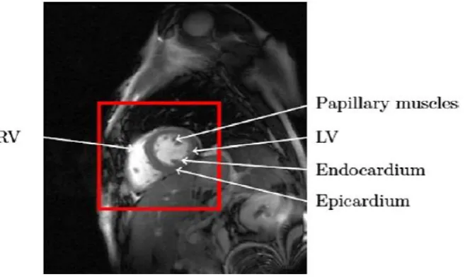
The image below shows the heart's MRI image and a full range of ROI for heart recognition.
This image remembers the following. 1 - moving the heart, 2 - obtaining images as a result of acquisition synchronization MR to ECG signal and use of movable stages MR, 3- Covering a small surface of the images by the ventricles, 4- Restrict processing to ROI level, 5- Reduce computational burden.
Fig. 2 Heart MR image and full range of ROI to identify the heart
The heart contains the outer wall or epicardium and the inner wall or endocardium.
The outer wall consists of a line between the myocardium (the heart muscle) and the surrounding tissues (lung and fat).
Different severity, poor contrast with myocardium, and in terms of the division of the epicardium, especially for RV It's hard.
The inner wall surrounds the LV and has a good contrast to the myocardium and circulation and, on the other hand, is the only boundary to calculate the ventricular volume.
Main levels of information during the division process 1 - No basic information 2 - Poor initial information 3 - Strong basic information.
No basic information or weak initial information includes a photo-based method, a pixel-based method, and modifiable models.
Strong basic information includes modifiable models based on basic information, shape models, active appearance, and atlas-based methods.
Therefore, it is imperative to provide a limited framework for the inclusion of strong initial information by photo-based and pixel-based methods, and an extensive review of strong initial information is carried out by modified models.
In a split method with strong initial information without the need for user interaction, it is necessary to manually build a training set.
Creating a training kit is questionable because the variability depends on the initial data.
On the other hand, ASM consists of a statistical model, called the Point Distribution Model (PDM), which is arranged by a PCA on an array of forms, and a method for searching the model in a photo (it can not get data that is not in the training set) Estimates and, in order to obtain more accuracy, must be sufficiently representative of all possible forms of the heart.
Atlas production has little effect on the results of division, since it is only used as a starting point for registration.
III. Suggested Method
A
.
Image databaseCardiovascular disease is one of the most common causes of mortality in industrialized and growing societies.
Medical pictures are one of the most important tools for the physician to diagnose the disease and control the treatment process.
By developing images MRI, CT The heart of these illustrative methods is used as the gold standard for cardiovascular studies.
In recent years, much attention has been paid to this way of illustrating and processing the images, and there are many ways of illustrating and processing heart pictures. Clinical studies show It suggests that simultaneous analysis of right ventricular and left ventricular information can provide useful information about cardiac function and can be useful in early diagnosis of disease and in the treatment process.
The study of the structure and function of the heart ventricles in resonant imaging images of magnetic resonance imaging is an important step in the management of many cardiac disorders.
Although the central division of the heart ventricles produces good results, it does work, especially for the right ventricle of the RV, due to its complex geometry.
Hence, the automatic separation of the heart ventricles is essential.
For this purpose, I provided MRI images of several patients with coordination with a specialized cardiovascular hospital.
Of course, these images were taken in several directions for each patient and the quality of the images was fairly good.
I used MATLAB 2015a to run this application.
B. images recalling
For example, first save the patient's MRI images in a folder, and then run the call command.
Keep in mind that the images are color-coded and should be turned into gray. The gray color range is from white to black, with a total of 252 colors.
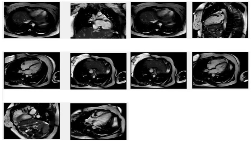
Figure 3 is The implementation of this command (the patient's MRI images).
Fig. 3 MRI recalling images of a heart patient
To begin with, we use Figure 4
C. Conversion operations Fourier
C.1. Fourier transform
I encountered a problem calling the image, which was that the whole image was an object.
Therefore, I need to shake the image to eliminate the noise around the image.
With the research that I did and with the test and error method, I came to the conclusion that using the Fourier transform is the best way to break off such an image.
Fig . 5 a) main picture b) fft2 picture c) fftshift picture d) log picture
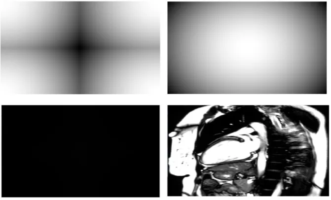
C.2. Gaussian High Pass Filter
The application of this filter is further explored in the field of video signals.
Video signals typically have a frequency between 0 and 50 MHz, which is the image's range of these frequencies.
Fig . 6 Images after Gaussian filter implementation
D. Main program
D.1. Preprocessing
After executing the Fourier transform, first, the image is called, then the image is converted to a gray image, and then we automatically use the binary command.
Then we use the edge detection command to eliminate the noise on the image.
Fig .8 Shape obtained after noise reduction operation
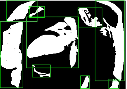
D.2. Extracting interconnected components
After completing the above steps, we get a binary image of the interconnected components.
Each part is an interconnected component. We need to get information about each component.
Then draw a rectangular box with the desired position.
In figure 9, A round rectangular box is drawn around each interconnected component.
Fig . 9 Drag the rectangular box around each connected component
D.3. Search for patient heart
D.4 . Isolation of the heart
At this point, the heart is removed, then the heart image is stored and displayed.
Fig . 10 Isolated image
D.5. Segmentation
Fig .11 a) Segment the boundaries of objects. b) Putting the boundary on the original image.
The final result of this algorithm is in Fig 11-b It has been shown that in fact the heart image has been removed and the segmentation operation has been performed.
E. Split operation
E.1. Preprocessing
Fig . 12 a) Optimize light intensity . b) Select the image spatial range. c) Remove border pixels. d) Matching the initial image on the image and segmenting the image.
As you can see, in the picture 12-d , the heart is completely detached and the heart image is marked out. This is the final result of this algorithm.
IV. The results of the proposed method
Recent research has shown that cardiovascular disease accounts for more than 31% of the world's total deaths, of which more than 3 million have been under the age of 60.
Only in the United States, every 34 seconds, a person suffers a heart attack, and about 200,000 people die from cardiovascular disease, accounting for 25% of the total death rate.
Therefore, any attempt to improve the screening, diagnosis and treatment of cardiovascular disease can reduce the mortality and side effects of these diseases in human society, it is important and valuable.
Magnetic cardiac resonance imaging is a non-invasive tool for diagnosing and treating heart disease (CVD). Despite the extensive use of MRI imaging In related research with CVD The use of this imaging instrument is clinically limited for two reasons: The first limitation, the patient's high role in output image quality.
More precisely, the patient's physical movement or breathing movement causes distortion in the MRI image and reduces its quality.
Of course, the problem today is greatly improved by the advancement of imaging devices or noise removal operations in the preprocessing of images.
In order to eliminate the obstacles encountered, the use of MRI images requires a primary and final processing process.
The primary processing for the partitioning of the images is to achieve the correct endocardial and epicard boundaries in each of the MRI frames, and at a more advanced stage, having the processed information of these frames and putting them together in a 3D animated form of the dynamic function of the heart muscle (External and internal) achieved.
In this way, the importance of having automated or semi-automatic methods for partitioning magnetic resonance images becomes more apparent.
The magnetic resonance images of the heart are faced with the main challenge, namely the wide distribution of brightness due to complex anatomy and variable parameters throughout the heart cycle.
So far, despite much research, the issue of segmentation of the heart holes is still an open issue.
V. Conclusion
Unfortunately, there were limited articles in this regard, which made my work very difficult.
In this research, Gaussian filter was used and then, using segmentation methods, we performed cardiac separation.
REFERENCES
[1] Chenghui Jin, Jingcheng Zhou, and Buket D. Barkana, " Application of Different Transformation Methods to Whole Heart Region Segmentation", Member, IEEE
[2] Caroline Petitjean, Jean-Nicolas Dacher, " A review of segmentation methods in short axis cardiac MR images", Elsevier B.V. All rights reserved. vol. 15, pp. 169-184, 2011.
[3] Olivier Ecabert, Jochen Peters, Hauke Schramm, Cristian Lorenz, Jens von Berg, Matthew J. Walker, Mani Vembar, Mark E. Olszewski, Krishna Subramanyan, Guy Lavi, and Jürgen Weese, " Automatic Model-Based Segmentation of the Heart in CT Images" , IEEE Transactions On Medical Imaging, Vol. 27, No. 9, September 2008
[4] Amit Chakraborty, Lawrence H. Staib, " Deformable Boundary Finding in Medical Images by Integrating Gradient and Region Information", IEEE Transactions On Medical Imaging, Vol.
15, No. 6, December 1996.
[5] Jochen Peters, Olivier Ecabert, Carsten Meyer, Hauke Schramm, Reinhard Kneser, Alexandra Groth, and J¨urgen Weese, " Automatic Whole Heart Segmentation in Static Magnetic Resonance Image Volumes", Philips Research Laboratories, X-Ray Imaging Systems, Weisshausstrasse 2, D-52066 Aachen, Germany
[6] Xiahai Zhuang, Kawal S. Rhode, Reza S. Razavi, David J. Hawkes, and Sebastien Ourselin, "A Registration-Based Propagation Framework for Automatic Whole Heart Segmentation of Cardiac MRI", IEEE Transactions On Medical Imaging, Vol. 29, No. 9, September 2010. [7] X`enia Alb`a, Marco Perea˜nez, Corn´e Hoogendoorn, Andrew J. Swift, Jim M. Wild, Alejandro
F. Frangi, " An Algorithm for the Segmentation of Highly Abnormal Hearts using a Generic Statistical Shape Model", Journal Of Transactions On Medical Imaging IEEE, Vol. Xx, No. X, Month 2015.
[8] Li Wang, Yurun Ma, Kun Zhan, Yide Ma, " Automatic Left Ventricle Segmentation in Cardiac MRI via Level Set and Fuzzy C-Means ", Proceedings Of 2015 RAECS UIET Panjab University Chandigarh 21-22nd December 2015.
[10] Dr. V.S. Jayanthi, D. Baskar, J. Divya Priyanka, "Extraction of Myocardial Wall from Cardiac CT Images", IEEE Sponsored 2nd International Conference on Innovations in Information Embedded and Communication Systems , 2015.
[11] Xinglong Liu1, Fei Hou1, Hong Qin, Aimin Hao1," Robust Optimization-based Coronary Artery Labeling from X-Ray Angiograms", IEEE Journal of Biomedical and Health Informatics, 2015. [12] Yunliang Cai,Ali Islam, Mousumi Bhaduri, Ian Chan, and Shuo Li, " Unsupervised Freeview
Groupwise Cardiac Segmentation using Synchronized Spectral Network" , Submitted To Ieee Transaction On Medical Imaging, 2015.
[13] Seena V, Jerrin.Yomas, "A Review On Feature Extraction And Denoising Of Ecg Signal Using Wavelet Transform" , 2nd International Conference on Devices, Circuits and Systems (ICDCS), 2014.
[14] Xiuying Cao, Zhe Li, " Denoising of ECG signal based on a comprehensive framework", IEEE, 2010.