Original Article
Effects of external fixation technique on hospitalization
time, fracture healing time and complication
rate of pediatric fracture
Shenghua Qiu1*, Jian Liu2*, Chengliang Liu1
Departments of 1Pediatric Surgery, 2Laboratory, Linyi Central Hospital, Linyi, Shandong Province, China. *Equal contributors and co-first authors.
Received June 3, 2017; Accepted July 11, 2017; Epub August 15, 2017; Published August 30, 2017
Abstract: Objective: To investigate the effects of external fixation on hospitalization time, fracture healing time and complication rate of pediatric fracture. Methods: Eighty children with fracture treated in our hospital from January 2015 to January 2017 were selected and divided into external fixation group (n=40) and internal fixation group (n=40) in accordance with therapeutic methods. Children in internal fixation group were treated by internal fixation while those in external fixation group were treated by external fixation. Hospitalization time, fracture healing time and postoperative total complication rates of children in two groups were statistically analyzed. Results: Hospital-ization time and fracture healing time of children in external fixation group were significantly shorter than those in internal fixation group (P<0.05). Postoperative complication rate in external fixation group (10.0%, 4/40) was significantly shorter than that in internal fixation group (20.0%, 8/40, P<0.05). Conclusion: External fixation is more effective than internal fixation in reducing the hospitalization time as well as fracture healing time and lowering the total complication rates of pediatric fracture, which is worthy to be popularized in clinic.
Keywords: External fixation, pediatric fracture, hospitalization time, fracture healing time, total complication rate
Introduction
The 20th century has witnessed the huge de-
velopment of internal fixation with steel plate, intramedullary nail, etc. as fixation materials. It
is widely used in fracture and orthopaedic treat-ments, but limited in clinical application to some extent because of its drawback of high infection rate when the normal tissues are cut
via surgery and the fixation materials are
enclosed in vivo [1]. In recent years, however,
influenced by the high causative factors of limb damage such as traffic accident, etc., trauma
tends to have increasingly complex forms and growingly serious injuries. The principle of
external fixation is to insert into steel needle on
the bone through soft tissues like skin, mean-while, assemble into some kind of mechanical construction like connecting rod with
extracor-poreal device; this mechanical configuration is firm and can effectively correct and treat frac -ture [2]. For better treatment on complex
trau-mas, biomechanics of external fixation is fur -ther researched in clinic and the mechanics of
external fixators is constantly designed and assessed at the same time; thus, external fixa -tion has been widely used with each passing
day since 1950s [3]. External fixation has
unique advantages in the treatment of high energy damage, long bone defect, etc. External
fixation plays a key role in the creations of suit -ably biomechanical and mechanobiological environment for fracture healing and bone
lengthening [4]. It is mainly applied in the fixa
-tion of fracture at first, and it relies on external fixators in the process of fixing the far end and
proximal end of fracture, which is more stable
than indirect external fixation such as splint and
gypsum [5]. In 1897, doctor Parkhill from
America designed the first modern external fix -ator [6]. For more than a century, it is
continu-ally updated, and today, circular fixation and multi plane fixation have become popular. External fixation makes full use of thinner men -tal needle or men-tal nail while penetrating into
the bone. Connecting with external fixator makes strong fixation in vitro and expands fixed
conducted on the basic of actual demand with features of minimally invasion, convenience,
etc., that is why it is more suitable in fixing com -plexly traumatic fracture and correcting bone malformation [7].
In clinical practice, manual reduction and
gyp-sum fixation can correct the fracture of most
children. The greatest advantage of plaster
immobilization is noninvasive. But if the frac -ture lacks of stability, for example, if there exists a trend of displacement and shortening at the fracture end of short oblique fracture of
tibia and fibula, it will be difficult to effectively
maintain the fracture reduction when only
relies on plaster immobilization. The older the
children get, the slower the fracture healing is. The joints of adjacent parts of fracture will become stiff under the impact of plaster
immo-bilization for a long time. Osteoporosis and dis -use muscle atrophy will occur when lower limbs are non-weight-bearing for a long time [8].
Internal fixation can achieve reliable fixation
and anatomic reduction, but the surgery will certainly further damage soft tissues and may cause infection and fracture nonunion, which
needs re-operation to take out internal fixator
and will increase children’s pain as well as the charge; what’s worse, it will leave big and per-manent surgical scar [9]. While closed
reduc-tion is the main approach of external fixareduc-tion
fracture reduction and anatomic reduction.
Percutaneous pinning is performed in fixing fracture and external fixator is fully used to
reduce the secondary damage to fracture site. Closed reduction provides good preconditions for fracture healing. Also, demolition of external
fixator is simple to operate and there is no need
to perform re-operation generally [10]. To bet-ter treat pediatric fracture and effectively im- prove the prognosis, this study compared the
therapeutic effects of external fixation with internal fixation on pediatric fracture, finding the former had more positive influences on hospitalization time, fracture healing time and
total complication rates. Reports are as fo- llows.
Materials and methods
General materials
Eighty children with pediatric fracture treated in our hospital from January 2015 to January 2017 were selected. Inclusion criteria: All chil-dren were diagnosed with fracture via X-ray and
CT examination. Themselves and all their par-ents were informed and signed informed con-sents. Exclusion criteria: Exclude those without complete clinical data. Divide these 80 children
into external fixation group (n=40) and internal fixation group (n=40) based on their therapeu -tic methods.
Methods
Children in internal fixation group were treated by internal fixation technique, and then per -formed conventional open reduction and
inter-nal fixation [11]. Epidural anesthesia or general anesthesia was performed for children first.
The fracture site was exposed by surgery, and
the fracture end was fixed. According to the dif
-ferent condition of fracture, various internal fix -ation was chosen to keep the position after restoration. Priority should be given to vascular compression or injury, and there was no soft tissue between fracture ends. Calf anterolater-al incision was made from the fracture end for lower 1/3 tibial fracture, periosteum was stripped, and soft tissues in the fracture end was cleaned. Fracture reduction was performed under the direct view, plate screws were
pres-sured to fix, and two gramme needles were
intersected for supracondylar fracture of hu- merus. After humerus fracture end was ex- posed, local hematoma was removed. Then, one gramme needle was inserted intomedial epicondyle, and one was into lateral epicon-dyle, directed towards the upper part of the lat-eral condyle, the internal and external fracture
blocks was prized to make reduction of inter -condylar fractures, and reduction condition was observed by X-ray. If it was good, two origi-nal gramme needles were inserted into proxi-mal fracture ends or two newgramme needles were inserted into proximal fracture ends from
medial and lateral epicondyles to cross fix inter -condylar fractures. And in the end, after total hemostasis, the incision was sutured. Children
in external fixation group were treated by exter
-nal fixation technique. Specific operation:
pa-tients underwent epidural anesthesia or intra-venous anesthesia. Perform closed manual
reduction at first to those with closed fracture. After that, put external fixator inside for traction and fixation. If closed reduction failed or frac
-ture displacement occurred, firstly open reduc -tion should be performed in principle and then
thorough-ly cleared. Fracture site was performed at the fracture broken end under direct vision and
fixed temporarily. In this process, self-tapping screws and external fixator for external fixation
were made full use. Then the fractured end was further adjusted in order to recover the length and biomechanical line of the fractured limbs. For the patients with open fractures, the pollut-ants, small pieces of fractured bones, etc. in their injury sites were removed completely, at the same time, their periosteum and soft issues at the fracture broken end were protected in priority. For the patients with humeral fractures, their radial nerves were protected effectively and injuring radial nerves was avoided strictly. For patients with large oblique bones existed in the fracture broken end, self-tapping screws were fixed in the main fracture segments at first, to promote the increase of stability of frac -ture; then situ suture fixation of small fractured fragments was carried out, and in this process, absorbable lines were used to twine fractures.
Postoperative treatments
A drainage tubes was placed into the postop-erative wound, and pulled out according to patients’ physical conditions after 2 days. At the same time, patient’s injured limb was raised higher than the levels of his heart to provide a good precondition for diminishing local edema. Patients were allowed to carry out knee joints and ankle joints exercises 2 days after opera-tion, and do weight-bearing exercise 2 weeks after operation. We did regularly X-ray examina-tion every two weeks according to their frac-tured degrees during this period, and inspected their fracture sites and healing degrees. In
gen-eral condition, 6-12 weeks later, external fixa -tion devices could be removed gradually ac- cording to patients’ fracture healing degrees strictly. Patients should active their distal or proximal joints of tibia fractures as early as possible so as to recover functions of joints. Observation indexes
The hospitalization time and fracture healing
time in the two groups were observed and recorded. Meanwhile, wound infections,
trau-matic arthritis, trautrau-matic ossification, ankylosis
and other postoperative complications in the two groups were collected.
Statistical analysis
SPSS20.0 statistical software was used to
ana-lyze data, postoperative complications and
other enumeration data were presented as
per-centage (%), Chi-square test was adopted to
test comparisons between the two groups; standard deviation (_x±s) was used to express
hospitalization time and fracture healing time
and other measurement data, t test was adopt-ed to test comparisons between the two
groups, and the test criterion was a=0.05.
Results
Comparison of the general data between the two groups
Among children in the external fixation group,
there were 25 males and 15 females from 3 to 12 years old, with an average age of (7.1±3.2) years old; their course of disease was 0.6h-3d, with an average course of (5.4±2.2) d. As for
classifications of fracture, 30 cases were
closed fracture, and 10 cases were open frac-ture; as for types of fracture, 15 cases were fracture of middle lower 1/3 segment of tibia, 11 cases were humeral shaft fracture, 9 cases were supracondylar fracture of humerus, 5 cases were intercondylar fracture of humerus; as for sites of fracture, 23 cases were one site, 17 cases were two sites or more; as for causes
of fracture, 26 cases were caused by traffic
accident injury, 10 cases were falling-down injury, 3 cases were crashing injury, 1 case was chopped injury. Among children in the internal
fixation group, there were 23 males and 17
females from 4 to 12 years old, with an average age of (7.6±3.1) years old; their course of dis-ease was 0.5 h-3 d, with an average course of
(5.2±2.3) d. As for classifications of fracture,
31 cases were closed fracture, and 9 cases were open fracture; as for types of fracture, 16 cases were fracture of middle lower 1/3 seg-ment of tibia, 10 cases were humeral shaft fracture, 8 cases were supracondylar fracture of humerus, 6 cases were intercondylar frac-ture of humerus; as for sites of fracfrac-ture, 24 cases were one site, 16 cases were two sites and more; as for causes of fracture, 25 cases
were caused by traffic accident injury, 11 cases
were falling-down injury, 2 cases were crashing injury, 2 cases were chopped injury. There was
course of disease, types of fracture in the two groups (P>0.05), so it was with comparability, see Table 1.
Comparisons of hospitalization time and frac-ture healing time between the two groups
The hospitalization time, fracture healing time of the external fixation group were significantly shorter than those of the internal fixation group (P<0.05), see Table 2 and Figure 1.
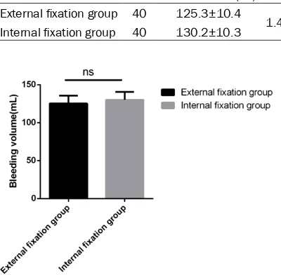
Comparison of intraoperative blood volume between the two groups
There was no statistical significance in intraop -erative blood volume between the two groups (P>0.05), see Table 3 and Figure 2.
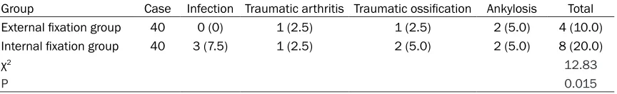
Postoperative complication rate in external fix
-ation group (10.0%, 4/40) was significantly lower than that in internal fixation group (20.0%, 8/40), (P<0.05), see Table 5.
Discussion
Pediatric fracture belongs to a kind of pediatric trauma which is common clinically. The main difference between pediatric fracture and adult fracture is epiphyseal plate. If there is no prop-er treatment for pediatric patients with epiphy-seal injury, it will lead to bone growth stagna-tion or deformity. Therefore, active and effective protection should be carried out to epiphyseal plate during the process of the treatment for pediatric fracture [12]. Plaster splint external
fixation and open reduction with internal fixa -Table 1. Comparison of the general data between the two groups
Variable Classification External fixation group (n=40) Internal fixation group (n=40) t/χ2 P
Gender 2.71 0.124
Male 25 (62.5) 23 (57.5)
Female 15 (37.5) 17 (42.5)
Age (year) 7.1±3.2 7.6±3.1 1.886 0.355
Course of disease (d) 5.4±2.2 5.2±2.3 1.638 0.256
Fracture classifications 4.61 0.655
Closed fracture 30 (75.0) 31 (77.5)
Open fracture 10 (25.0) 9 (22.5)
Fracture types 1.32 0.325
Fracture of middle lower 1/3 segment of tibia 15 (37.5) 16 (40.0)
Fracture of humeral shaft 11 (27.5) 10 (25.0)
Supracondylar fracture of humerus 9 (22.5) 8 (20.0) Intercondylar fracture of humerus 5 (12.5) 6 (15.0)
Fracture sites 2.77 0.352
One site 23 (57.5) 24 (60.0)
Two and more sites 17 (42.5) 16 (40.0)
Cause of injury 4.11 0.325
Traffic accident injury 26 (65.0) 25 (62.5)
Falling down injury 10 (25.0) 11 (27.5)
Crashing injury 3 (7.5) 2 (5.0)
Chopped injury 1 (2.5) 2 (5.0)
Table 2. Comparison of hospitalization time and frac -ture healing time between the two groups (_x±s)
Group Case Hospitalization time (d) Fracture healing time (d) External fixation group 40 7.5±2.8 61.3±12.8 Internal fixation group 40 14.2±6.2 75.6±20.1
t 4.303 3.182
P 0.023 0.026
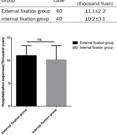
Comparison of hospitalization expenses between the two groups
There was no statistical significance in differences of hospitalization expenses
between the two groups. (P>0.05), see Table 4 and Figure 3.
tion are traditional methods of treating
pediat-ric fracture commonly used in clinic. Besides, plaster splint external fixation lacks precise fix -ation and has many complic-ations, so it easily causes fracture deformity on corresponding fracture site, thereby leading to obstacle of limb movement [13]. Open reduction with
inter-nal fixation has great damage to body and causes serious skeletal injury in some cases. Serious adverse effects will occur in pediatric skeletal growth and development once epiphy-seal plate is damaged. And due to fast healing speed of pediatric fracture, open reduction and
internal fixation easily causes damage to peri
-stability and flexible and diverse assembling
forms. It is extremely useful in all kinds of com-plex or simple fractures of the trunk and limbs, especially for severe bone defect, comminuted fracture, etc. It is effective in maintaining the length and force line of injured limbs, thereby providing effective prerequisite to necessary follow-up treatment and fracture healing.
Second, it has flexible fixed range. According to the design principle of external fixator, fixed
range can be selected based on actual
require-ments: fix a single bone or fix bones across joint. Over-articular fixation can increase fixa -tion stability in the course of treatment of intra-articular fracture and adjacent joint fracture.
Furthermore, it has reliable fixed effect. Modern external fixation fully utilizes conical screw nails fixation and other techniques to promote the
stability of system. Fracture angulation and bone displacement caused by unstable
exter-nal fixator rarely occur in the course of fixation.
[image:5.612.98.516.73.207.2]A stable system can effectively eliminate the shearing stress that leads to adverse effect of fracture healing and provides a good prerequi-site for micro-axial motion of fracture healing. Meanwhile, it effectively guarantees early post-operative activities and load-bearing needs of patients, as well as provides a good prerequi-site for rehabilitation and early return to society of patients. In addition, it is able to deal with open injury of soft tissues simultaneously. The
Figure 1. Comparison of hospitalization time and fracture healing time between the two groups. A. Comparison of hospitalization time between the two groups, *P<0.05; B. Comparison of fracture healing time between the two
groups, *P<0.05.
Table 3. Comparison of intraoperative blood volume between the two groups (_x±s)
Group Case blood volume (ml)Intraoperative t P External fixation group 40 125.3±10.4
1.476 0.255 Internal fixation group 40 130.2±10.3
Figure 2. Comparison of intraoperative blood volume between the two groups, ns, P>0.05.
osteum, so the healing speed of fractu-reis seriously affected [14].
In the treatment of trauma,
superiori-ties of external fixation are reflected in the following aspects specifically. First,
it has wide range of application. Modern
[image:5.612.93.293.314.510.2]results in this paper showed that the
hospital-ization time and fracture healing time in the external fixation group were significantly short
-er than those in the int-ernal fixation group (P<0.05). It may because the external fixator can provide the exact external fixation to pre -vent displacement of fracture segments effec-tively during the treatment of pediatric fracture. Meanwhile, longitudinal traction can enable stress occur on the longitudinal direction of fracture segment, to provide a good prerequi-site for fracture healing.
High-energy injury results in serious fracture and easily induces severe soft tissue injury, such as large area of skin stripping injury and open fractures. Additionally, relevant medical
studies have confirmed that the infection
[image:6.612.90.292.109.342.2]ra-tes of contaminated wounds increase to a large extent under the effect of metallic foreign body [15, 16]. The distance between the fixation needle of the external fixator and wound can be fairly far, which effectively avoids the direct infection of vulnerable parts. When percutane-ous fixation must be conducted, the external fixation needle can give a full play of drainage to significantly reduce the morbidity of severe intraosseous infection. Stable external fixation
which is the only surgical trauma of external
fixation, and patients almost have no blood
loss, so it won’t directly affect the fracture site.
Removal of the external fixator does not pro -mote the second surgical trauma [17, 18]. The results of this study showed that the postopera-tive total complication rate of pediatric patients
in the external fixation group was (10.0%, 4/40 significantly lower than that in the internal fixa
-tion group (20.0%, 8/40, P<0.05), indicating that the external fixation is more effective in
reducing the incidence of complications in pediatric fracture patients. This probably due to
the fact that external fixation can maximize the
protection of epiphyseal plate and periosteum so that it will not be damaged, which contrib-utes to the reduction of the impact of bone development in children and provides a good prerequisite to relatively fast healing speed of pediatric fracture [19]. Therefore, compared
with other treatment methods, external fixation treatment has a significantly treatment effect. Meanwhile, small surgical operation difficulty,
less trauma and less complication enable
external fixation technique extremely applica -ble in primary hospitals [20].
In conclusion, the external fixation technique is
valuable to be applied extensively in clinic, because it is more effective in shortening the
hospitalization time and fracture healing time
and reducing complication rate of pediatric
patients than the internal fixation technique. However, due to the small sample capacity of
this study, the results may not have representa-tiveness and universality; more samples should be added to further studies by relevant medical scholars.
Disclosure of conflict of interest
None.
Address correspondence to: Chengliang Liu, Depart- ment of Pediatric Surgery, Linyi Central Hospital, No.17 Jiankang Road, Linyi 276400, Shandong Province, China. Tel: 0539-2225007; Fax: +86-0539-2263541; E-mail: [email protected] Table 4. Comparison of hospitalization expenses between the
two groups (_x±s)
Group Case Hospitalization expenses (thousand Yuan) t P External fixation group 40 11.1±2.2
1.533 0.645 internal fixation group 40 10.2±3.1
Figure 3. Comparisons of hospitalization expenses between the two groups, ns, P>0.05.
References
[1] Al-Daghri NM, Aljohani N, Rahman S, Sabico S, Al-Attas OS, Alokail MS, Al-Ajlan A and Chrou-sos GP. Serum 25-hydroxyvitamin D status among Saudi children with and without a his-tory of fracture. J Endocrinol Invest 2016; 39: 1-6.
[2] Fatah RMN, Amin BRM, Mahmud HA and Yusif AJ. Assessment of the outcome of anterior ver-sus posterior approach in the management of displaced pediatric supracondylar humerus fracture. Open Journal of Orthopedics 2016; 06: 113-119.
[3] Hennrikus W, Slough J, Jensen K, Armstrong D, King S, Urish K. Measurement of radiation exposure when using the Mini C-Arm to reduce pediatric upper extremity fractures. J Pediatr Orthop 2016; 36: 122-125.
[4] Khaffaf RMN and Altaweel AH. Comparative Study between the Elastic Nail versus Hip Spi-ca Cast in Early Treatment of Pediatric Femoral Shaft Fractures. Open Journal of Orthopedi- cs, 2016; 6: 259-267.
[5] Naranje SM, Gilbert SR, Stewart MG, Rush JK, Bleakney CA, McKay JE, Warner WC Jr, Kelly DM, Sawyer JR. Gunshot-associated fractures in children and adolescents treated at two lev-el 1 pediatric trauma centers. J Pediatr Orthop 2016; 36: 1-5.
[6] Ali, Ahmed AA, Kabbash, Mansour M, Said, Samia MA, Shoeib, Mohamed A, Osman and Mohamed H. Use of biodegradable plates and screws in the treatment of pediatric facial bone fractures. Egyptian Journal of Oral & Max-illofacial Surgery 2016; 7: 86-93.
[7] Kumar P, Purohit NR and Gahlot TK. Clinical study on mandible fractures in dromedary camels (Camelus dromedarius). Trop Anim Health Prod 2014; 35: 64-66.
[8] Lee DJ and Elfar JC. External fixation versus open reduction with locked volar plating for ge-riatric distal radius fractures. Geriatr Orthop Surg Rehabil 2014; 5: 141-143.
[9] Iobst C and Liu R. A systematic review of inci-dence of pin track infections associated with external fixation. 2016; 2: 6-16.
[10] Ahmed A. An innovative external fixator for the management of trochanteric fractures of the femur. 2014; 49: 1-5.
[11] Lareau CR, Daniels AH, Vopat BG, Kane PM. Emergency department external fixation for provisional treatment of pilon and unstable ankle fractures. J Emerg Trauma Shock 2015; 8: 61-64.
[12] Barbier D, Neretin A, Journeau P and Popkov D. Gradual metatarsal lengthening by external fixation: a new classification of complications and a stable technique to minimize severe complications. Foot Ankle Int 2015; 36: 1369-1377.
[13] Chaudhuri A, Datta S, Roy DS, Singh AK, Ghosh S and Dutta S. Comparative analysis of exter-nal and interexter-nal fixation in lower radial articu-lar fractures. 2014; 7: 596-602.
[14] Jain A, Thompson JM, Brooks JT, Ain MC and Sponseller PD. Implant-related fractures in children with proximal femoral osteotomy: blade plate versus screw-side plate constructs. J Pediatr Orthop 2016; 36: e1-5.
[15] Cisneros LN, Gómez M, Alvarez C, Millán A, Caso JD and Soria L. Comparison of outcome of tibial plafond fractures managed by hybrid external fixation versus two-stage manage-ment with final plate fixation. Indian J Orthop 2016; 50: 123-130.
[16] Morelli I, Sabbadini MG and Bortolin M. Ortho-pedic injuries and their treatment in children during earthquakes: a systematic review. Pre-hosp Disaster Med 2015; 30: 478-485.
[17] Șerban Al, Obadă B, Turcu R, St A and Botnaru V. Distal tibial fracture treated by minimally in-vasive plate osteosynthesis after external fixa-tion Retrospective clinical and radiographic assessment. Ars Medica Tomitana 2014; 20: 44-49.
[18] Kao HK, Chen MC, Lee WC, Yang WE and Chang CH. A prospective comparative study of pin site infection in pediatric supracondylar hu-meral fractures: daily pin care vs. no pin care. Arch Orthop Trauma Surg 2014; 134: 919-923.
[19] Galal S. External fixation of paediatric subtro-chanteric fractures using calcar rather than neck pins. Strategies Trauma Limb Reconstr 2016; 11: 99-104.
[image:7.612.92.524.86.152.2][20] Kong H and Sabharwal S. External fixation for closed pediatric femoral shaft fractures: where are we now? Clin Orthop Relat Res 2014; 472: 3814-3822.
Table 5. Comparison of postoperative complications between the two groups (case, %)
Group Case Infection Traumatic arthritis Traumatic ossification Ankylosis Total External fixation group 40 0 (0) 1 (2.5) 1 (2.5) 2 (5.0) 4 (10.0) Internal fixation group 40 3 (7.5) 1 (2.5) 2 (5.0) 2 (5.0) 8 (20.0)
χ2 12.83