Segmentation and specification in the branchial region
of the head: the role of the Hox selector genes
FILIPPO M. RIJLI*, ANTHONY GAVALAS and PIERRE CHAMBON
Institut de Génétique et de Biologie Moléculaire et Cellulaire, CNRS/INSERM/ULP, Collège de France, Strasbourg, France
ABSTRACT Hox genes are segmentally expressed in the developing vertebrate hindbrain, neural crest cells and pharyngeal arches suggesting an important role in patterning these structures. Here we discuss the cellular and molecular mechanisms controlling segmentation and specification in the branchial region of the head. In addition, based on the recent phenotypical and molecular analysis of loss-of-function mutants in the mouse, we speculate that Hox genes may act like Drosophila selector genes in this system.
KEY WORDS:
homeotic genes, compartments, hindbrain, pattern formation, Hoxa1, Hoxa2
0214-6282/98/$10.00
© UBC Press Printed in Spain
*Address for reprints: Institut de Génétique et de Biologie Moléculaire et Cellulaire, CNRS/INSERM/ULP, Collège de France, BP 163 - 67404 Illkirch Cedex, C.U. de Strasbourg, France. FAX: 33 3 8865 3201. e-mail: rijli@igbmc.u-strasbg.fr
Introduction
The homeotic genes of the Hox complexes are transcriptional regulators encoding a 60-amino acid DNA-binding motif, the homeodomain, which are homologous to the homeotic genes of the Drosophila Antennapedia (ANT-C) and Bithorax (BX-C) com-plexes (reviewed in McGinnis and Krumlauf, 1992). Genetic analy-sis of Hox gene function both in invertebrates and vertebrates has shown that these genes are key developmental regulators which control morphological differences along the anteroposterior (A/P) body axis (reviewed in McGinnis and Krumlauf, 1992; Krumlauf, 1994; Favier and Dollé, 1997). In the mouse, Hox genes of paralog groups 1 to 4 (Fig. 1) have segmental expression domains in rhombomeres, rhombencephalic neural crest cells (NCC) and pharyngeal arch mesenchyme (reviewed in Krumlauf, 1993; Mark et al., 1995; Lumsden and Krumlauf, 1996) predicting an important role in patterning these structures. In this review, we discuss the cellular and molecular mechanisms controlling segmentation and specification in the branchial region of the head of vertebrates, with a focus on hindbrain development, and we draw a parallel with the establishment of compartments in Drosophila wing imaginal disc. In addition, based on the analysis of loss-of-function mutants in the mouse, we speculate that murine Hox genes may act similarly to Drosophila selector genes.
Learning from Hox gene function in Drosophila:
‘selectors’ of segmental identities
How a single protein present in a cell can change it from belonging to one pattern (e.g., the wing) to being part of another (a haltere)? The discovery of compartments in the wing and the study
of the phenotype of certain mutations in the BX-C complex led García-Bellido (1975,1977) to propose the concept of homeotic ‘selector’ genes as a rationale to explain Hox gene function. The fly wing originates from an initial group of about 50 cells, the imaginal disc, which undergo intensive proliferation throughout the larval period and pupation, after which visible cell differentiation begins. During differentiation, about 50,000 cells give rise to the cuticular processes typical of the adult wing pattern. One important feature, which was discovered by clonal analysis (García-Bellido et al., 1973), is that clones generated after a given time in development do not cross certain lines (boundaries) of the cuticular landscape, i.e., the wing disc become subdivided in compartments as prolif-eration proceeds (García-Bellido et al., 1973; see also for review Lawrence and Struhl, 1996). Compartments are sequentially gen-erated by a binary partition of a previously homogeneous cell population, unlike segmentation of the embryo in which segments are simultaneously generated on the head, thorax and abdomen. The basic property of cells segregating in different compartments is that they never mix with the cells of adjacent compartments, suggesting cell adhesion differences acting throughout develop-ment (e.g., García-Bellido and Lewis, 1976). This conclusion is also supported by the results of cell-aggregation experiments of dissociated cells from different disc regions. Therefore, cells in each compartment express properties of specific cell
differentia-Abbreviations used in this paper: ANT-C, Antennapedia Comples; BX-C Bithorax
tion long before cuticular pattern is visible. Each compartment will follow distinct developmental pathways giving rise to different parts of the wing. In this respect, there is a striking analogy between the compartimentalization and subsequent differentiation of the imagi-nal disc of Drosophila and the development of the hindbrain of vertebrates (see below).
Certain mutations in the BX-C Hox complex selectively affect disc compartments. For instance, two regulatory mutations in the ultrabithorax (Ubx) gene, bithorax (bx) and postbithorax (pbx), affect only anterior and posterior compartments respectively, i.e., transform anterior and posterior halves of the haltere in the corresponding parts of the wing (reviewed in García-Bellido, 1975). This suggests the existence of a common underlying ‘ground patterning program’ between cells of corresponding A/P positions of the wing and the haltere, but the interpretation depends on the presence of the Ubx gene which ‘selects’ differentiation as haltere rather than wing (e.g., by repressing an alternative developmental pathway leading to the wing). Thus, homeotic selector genes are those which control developmental pathways (García-Bellido, 1975). In concomitance with the topographical allocation of compartment founder cells, different combinations of selector genes, among which the Hox genes, are activated conferring a specific genetic ‘address’ or ‘code’ to each set of cells which is maintained through subsequent DNA replication and cell divisions. Several subse-quent binary decisions may be made by a typical group of cells each involving the activation of a ‘selector’ gene in a subset of the cells and its inactivation in the remainder. Selector genes promote the activation of a large battery of downstream ‘realizator’ genes which affect the cellular processes relevant to morphogenesis such as, for instance, proliferation rate, mitotic orientation, cell adhesion properties, cell differentiation, etc. Thus, while mutations in ‘selector’ genes should change the overall pattern of a develop-mental system (homeosis) without affecting normal cytodifferentia-tion mechanisms, mutacytodifferentia-tions in ‘realizator’ genes should affect gen-eral properties of cells relevant to morphogenesis. The discovery that Hox proteins act as transcription factors and the identification of a few of their direct downstream targets in Drosophila (reviewed in Graba et al., 1997) have provided strong support to this idea. Selection of alternative developmental pathways by Hox genes may be achieved through a functional hierarchy (Gonzalez-Reyes and Morata, 1990; Gonzalez-Reyes et al., 1990; Mann and Hogness, 1990) or competi-tive interactions (Heuer and Kaufman, 1992; Lamka et al., 1992; Castelli-Gair et al., 1994) among homeoproteins in the binding affinities to a common set of target genes (see also Rijli and Chambon, 1997).
The concept of ‘selector’ genes provides a conceptual frame-work to study Hox gene function in a number of other different animal systems, including vertebrates, and has been instrumental in the study of the functional role of the murine Hox genes in setting up and patterning the hindbrain compartments, the rhombomeres, and the segmented neural crest.
Hindbrain segmentation in vertebrates: of
compart-ments, cell affinities, and cell recognition molecules
During the development of the central nervous system (CNS), a large variety of neurons are generated at appropriate times and locations with respect to the principal axis of the system. An early ordered pattern of cell specification is crucial, given the
extraordi-nary complexity of neuronal functional connections in the mature brain. The possibility that at least part of the CNS is patterned from a reiterated set of repeated units has received much attention in the last few years with the discovery that hindbrain early development proceeds through a transient segmentation process which has been highly conserved in vertebrate evolution (Metcalfe et al., 1986; Hanneman et al., 1988; Lumsden and Keynes, 1989; Fraser et al., 1990; Lumsden, 1990).
The original observation that the neural tube of the chick embryo is progressively subdivided in bulges (rhombomeres) at the level of the rhombencephalon (Vaage, 1969), has been subsequently corroborated at the cellular level by the studies of Lumsden and colleagues (Fraser et al., 1990; Lumsden ,1990). The rhombomeres reflect an intrinsic segmentation of the neural tube which correlates with the subsequent differentiation of neurons in reiterated pat-terns (e.g., Lumsden and Keynes, 1989; Carpenter et al., 1993; Clarke and Lumsden, 1993). In addition, hindbrain partitioning may underlie segmental specification of NCC contributing to cranial sensory ganglia and branchial arches (Noden, 1983,1988; Lumsden et al., 1991; Serbedzija et al., 1992; Sechrist et al., 1993; Köntges and Lumsden, 1996) (see below).
Several observations, at both the cellular and molecular levels, suggest that rhombomeres behave as compartments of cell-lineage restriction which may share features with the insect com-partments. The hindbrain segments are not generated simultane-ously from the neural plate but sequentially, by binary partitions of previously homogeneous cell populations, and in an invariant order which does not follow an obvious rostro-caudal progression (Lumsden, 1990). Segmentation is marked by the appearance of a narrow line of specialized cells which form boundaries at the interface of two adjacent rhombomeres (Lumsden and Keynes, 1989; Heyman et al., 1993,1995), similarly to the Drosophila wing imaginal disc where, for instance, dorsal and ventral compart-ments are separated by a band of non-dividing cells (O’Brochta and Bryant, 1985). Boundaries may act as mechanical barriers to cell movements across rhombomere interfaces (Fraser et al., 1990), even though their role in restricting cell mixing has been recently questioned, as cells maintain their rostrocaudal restriction even in the absence of boundaries (Wingate and Lumsden, 1996; Nittenberg et al., 1997). In addition, a few cells do cross segment boundaries at the time of segmentation (Birgbauer and Fraser, 1994). It appears more likely that boundary formation reflects a secondary feature of a segmental organization intrinsic to the different compartments, in which restriction in precursor cell mixing along the A/P axis appears right before, or at the time of, rhombomere formation (Wingate and Lumsden, 1996).
are not generated simultaneously (Lumsden, 1990). In quail/chick grafts, for instance, cells from r3 disperse better in r3/r3 grafts than they do in r3/r5 grafts than they do in r3/r2 graft (Guthrie et al., 1993).
The above experiments suggest that different rhombomeres may express different repertoires of cell surface molecules. Differ-ent types of recognition molecules have been shown to be ex-pressed in a restricted manner in the developing hindbrain. Kuratani (1991) described the alternate staining in rhombomeres of the HNK-1 antibody which recognizes a sulfated glucuronic acid-containing carbohydrate epitope shared by several adhesion mol-ecules (including NCAM and L1). Other cell surface antigens, such as peanut agglutinin, are also expressed in a segmental manner in the hindbrain (Layer and Alber, 1990). Two members of the cadherin subfamily of cell adhesion molecules (CAMs), R-cadherin and cadherin 6 (cad6) (Ganzler and Redies, 1995; Matsunami and Takeichi, 1995; Redies, 1995; Inoue et al., 1997), also display
restricted expression patterns in specific rhombomeres and R-cadherin may be involved in differential segregation of cells (Matsunami and Takeichi, 1995). Another cadherin, F-cadherin, is expressed at boundaries in the Xenopus neural tube (Espeseth et al., 1995). The Eph-related receptor tyrosine kinases (RTKs) and their membrane-bound ligands are another subclass of cell recog-nition molecules with segmental expression patterns in the devel-oping brain (reviewed in Wilkinson, 1995; Lumsden and Krumlauf, 1996). Since their ligands are anchored in the plasma membrane, Eph-receptors may mediate cell contact dependent-signaling im-plicated in cell migration, axon pathfinding, and patterning mecha-nisms (Drescher et al., 1995; Pandey et al., 1995; Winslow et al., 1995; Xu et al., 1995,1996). In the hindbrain, receptors and their ligands are expressed in complementary domains suggesting that they may restrict intermingling of cells of different rhombomeres. Indeed, interference with the normal function of Sek1 by expres-sion of a dominant negative form resulted in cells with r3/r5 identity crossing irregularly into even-numbered rhombomeres (Xu et al., 1995). In addition, Eph-receptors and their ligands have been recently involved in restricting migration of specific population of trunk and cranial NCC suggesting a general role in regulating cell movement by a repulsion mechanism (Krull et al., 1997; Smith et al., 1997; Wang and Anderson, 1997).
Hindbrain segmentation and specification of the
seg-ment phenotype: the same set of selector genes at
work?
Relatively little is still known about the genetic hierarchy which controls hindbrain development. Clues to the hierarchy of the genetic control of hindbrain segmentation and specification of the segment identity come from the study of the expression patterns and function of the homologues of the fly Hox genes.
Paralogous genes in the 3' parts of the vertebrate Hox clusters (HoxA to D) are sequentially and segmentally expressed in the developing hindbrain, with sharp anterior expression boundaries coinciding with rhombomeric borders (Fig. 1) (e.g., Hunt et al., 1991; Murphy and Hill, 1991; Prince and Lumsden, 1994; see also for review Krumlauf, 1993; Keynes and Krumlauf, 1994; Wilkinson, 1995). It is important to note that Hox expression domains are established at early neural plate stages (E7.5-8.0 in the mouse), i.e., before the formation of definitive rhombomeres (occurring about one day later), and they are in general maintained up to late stages of hindbrain development well after morphological segmen-tation has disappeared (with the exception of the Hoxa1 gene; see below) (Krumlauf, 1993 and refs. therein; Wingate and Lumsden, 1996). A direct correlation exists between Hox gene expression and commitment to a rhombomere-specific fate. Grafts of chick neural plate-stage hindbrain neuroepithelium transplanted in more posterior locations express Hox genes and display morphological features appropriate for the new location (Grapin-Botton et al., 1995). In contrast, grafts transplanted just before or at the time of rhombomere formation maintain both specific Hox expression and their segmental identities (Guthrie et al., 1992; Kuratani and Eichele, 1993; Simon et al., 1995), even though their commitment may still be reversible under certain conditions (Itasaki et al., 1996; Grapin-Botton et al., 1997). Therefore, it appears that, after a period of plasticity, the definitive commitment to a specific segmen-tal fate is accompanied by the establishment of a unique genetic ‘address’ of Hox gene expression in a block of precursors cells Fig. 1. Hox gene expression domains in the mouse hindbrain and
which is maintained through subsequent cell divisions and directs the differentiation program of that segment.
An interesting corollary of these experiments is that definitive commitment to a specific fate coincides with the time of cell-lineage restriction within a specific rhombomere (see above), suggesting that segment formation and specification are tempo-rally linked and may be under the genetic control of the same set of Hox selector genes. Should this hypothesis be correct, then one would expect some classes of recognition molecules restrict-ing rostro-caudal cell movement and controllrestrict-ing cell contact-dependent signaling, such as CAMs and RTKs (see above), to be under direct or indirect control of the Hox genes. Thus, in the absence of functional compensation, mutations in a Hox gene should result in both segmentation and specification problems in the mutant hindbrain, which may be considered as two aspects of the same process. Early precursor cells which are not correctly specified according to their A/P axial level may not be restricted at their appropriate position and/or form a coherent block of cells, therefore not forming a normal segment, but becoming intermin-gled with cells of similar genetic constitutions at adjacent rostrocaudal positions, eventually acquiring the same fate as their neighbors possibly through a segmental community effect (Gurdon, 1988; Wilkinson, 1995).
The generation of Hox mutants in the mouse has provided an invaluable model system for understanding the molecular basis of hindbrain segmentation and patterning and to test some of the above hypotheses. To date, several Hox genes with expression domains in the developing hindbrain (paralog groups 1 to 4; Fig. 1) have been knocked out via homologous recombination in ES cells (Capecchi, 1989). Hindbrain alterations have been reported for the Hoxa1 (Carpenter et al., 1993; Dollé et al., 1993; Mark et al., 1993), Hoxb1 (Goddard et al., 1996; Studer et al., 1996), Hoxb2 (Barrow and Capecchi, 1996), and, recently, Hoxa2 (Gavalas et al., 1997) knockouts. In contrast, Hoxa3 appears to have primary functions only in mesenchymal neural crest and endoderm (Manley and Capecchi, 1995) (see below). The apparent absence of hindbrain segmentation and/or specification defects in paralog group 4 Hox gene mutants (reviewed in Machonochie et al., 1996) may reflect functional compensation by other paralogous and non-paralogous Hox genes (e.g., Rijli and Chambon, 1997). The Hoxb1 (Goddard et al., 1996; Studer et al., 1996) and Hoxb2 (Barrow and Capecchi, 1996) knockouts resulted in specification defects of distinct subpopulations of neurons originated in rhombomere 4 (r4) (see below). On the other hand, both Hoxb1 and Hoxb2 mutations apparently did not affect normal hindbrain segmentation. Although cell lineage restrictions have not been studied in these mutants to reveal subtle segmentation problems, one possibility is that both Hoxb1 and Hoxb2 inactivations are functionally compensated at early neural plate stages by the function of their paralogous genes Hoxa1 and Hoxa2 .
We speculate that the primary role of Hox genes in early hindbrain regionalization is to restrict proliferating neural precur-sors and their progeny in compartment-like blocks of cells (rhombomeres). Such compartments will follow developmental pathways, controlled by the same set of Hox selector genes, appropriate for their A/P axial levels. Thus, mutating a given Hox gene may not only cause (partial) respecification to alternative developmental pathways (homeosis), but result in segmentation defects due to altered adhesive and/or signalling properties of
group of cells. What is the current evidence for this model? Although still largely speculative, some indirect evidence for a role of Hox genes in both segmentation and segment specification comes from the recent analysis of both Hoxa1 (Lufkin et al., 1991; Chisaka et al., 1992) and Hoxa2 (Rijli et al., 1993) mutant mice.
The Hoxa1 case
The expression of Hoxa1, together with Hoxb1, provides the earliest sign of regionalization in the presumptive hindbrain at headfold stage (E7.5-7.75) (Murphy and Hill, 1991; Dupé et al., 1997). Its expression domain extends from the posterior end of the embryo up the presumptive r3/r4 border and is down-regulated before the formation of boundaries. It is noteworthy that the first constrictions appearing in the chick hindbrain are those delineating the future r4 and r5 territories as a pair (Lumsden, 1990), suggest-ing that these may be the earliest rhombomeres to be specified. Targeted disruption of Hoxa1 resulted in segmentation defects, cranial nerve and ganglia, and inner ear abnormalities (Carpenter et al., 1993; Mark et al., 1993; reviewed in Mark et al., 1995). Early molecular analysis (Carpenter et al., 1993; Dollé et al., 1993) revealed that r4, based on Hoxb1 activity, was severely reduced, while r5, as assessed by Krox20 expression, was either absent (Carpenter et al., 1993) or very reduced (Dollé et al., 1993). These early defects lead to a dramatic reorganization of the patterning of cranial nerves and sensory ganglia (derived from neurogenic NCC) which appear very similar in both mutants (Carpenter et al., 1993; Mark et al., 1993; Gavalas et al., 1998).
Two main possibilities could account for the apparent depletion of r4 and r5 cells in Hoxa1 mutant hindbrains (reviewed in Mark et al., 1995; Wilkinson, 1995). In the first scenario, Hoxa1, acting as a Drosophila gap gene, would be involved in delimiting the future region encompassing r4 and r5. It may activate selector genes which in turn would confer specific rhombomeric identities to r4 and r5. In this case, Hoxa1 inactivation should result in a lower rate of cell proliferation and/or increased cell death leading to the physical loss of the future r4-r5 region. In the second scenario, Hoxa1 may act as a selector gene providing an early specification to prospec-tive r4 and r5 cells, e.g., conferring a specific adhesive property to these cells, restricting them at their appropriate axial level. In addition, transient Hoxa1 expression might be required to activate other Hox and/or non-Hox selector genes to further specify the r4-r5 region in two distinct rhombomeres. In such a case, prospective r4-r5 cells lacking HOXA1 would not be lost but mix with cells from adjacent rostrocaudal levels and may eventually acquire the fate of their neighbors.
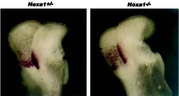
expression in the hindbrain of the Hoxa1 heterozygous mutant embryo shown in Figure 2A is restricted to a compact band of cells with a rather sharp posterior boundary (arrowheads). In the Hoxa1 homozygous mutant (Fig. 2B), the overall Wnt8 expres-sion levels appear reduced. Interestingly, expresexpres-sion in the ho-mozygous mutant appears extended along the rostrocaudal axis displaying less defined anteroposterior restriction (arrowheads in Fig. 2B), as compared to the heterozygous mutant. This result is intriguing, given that the expression of another r4 molecular marker, Hoxb1, is severely reduced and spatially restricted in the Hoxa1 mutants (Carpenter et al., 1993; Dollé et al., 1993), suggesting that a significant portion of the presumptive r4 domain may have been lost. One way to reconcile these observations is to assume that presumptive r4 (and possibly r5) Hoxa1 mutant cells are not lost at this stage, but just not properly restricted along the A/P axis. Thus, Wnt8 may provide a useful marker to follow the mixing of mutant presumptive r4 cells with adjacent territories resulting in an apparently enlarged Wnt8 expression domain. It is interesting to note that the expression domain of Krox20 in r3 appears posteriorly extended in Hoxa1 mutants (Carpenter et al., 1993; Gavalas et al., 1998), suggesting that: 1) r3 normal seg-mentation may indirectly depend on interactions at the interface with r4 Hoxa1-positive cells and 2) that r4 Hoxa1-mutant cells and r3 Krox20-positive cells may intermingle in the mutant back-ground. Support for this interpretation comes from the finding of mixing of facial and trigeminal motor neurons (Carpenter et al., 1993; Gavalas et al., 1998). It will be crucial to study whether some of the r4 mutant cells may be re-specified to an r3 or, perhaps, an r2 fate as a result of the interaction with the new environment. Hints that this may be the case may come from reinterpretation of the Hoxb1 expression pattern in the mutants. Since r4 presumptive cells may still be present, although redistrib-uted, it follows that Hoxb1 expression in these cells could depend on Hoxa1 activity, as is significantly reduced in the absence of Hoxa1 (Carpenter et al., 1993; Dollé et al., 1993). Indeed, evi-dence was recently obtained that Hoxb1 activation in vivo re-quires Hoxa1 during normal development (Studer et al., 1998). Mutant r4 cells not expressing Hoxb1 may well be respecified to
a more rostral phenotype. In fact, in mice lacking Hoxb1 activity (Studer et al., 1996), the failure of r4 markers to be upregulated and the associated ectopic expression of r2 markers suggest that r4 may adopt an altered identity. Consistent with this interpreta-tion, the migratory behavior of r4 facial branchiomotor neurons is abnormal, and similar to that of r2 trigeminal neurons (Goddard et al., 1996; Studer et al., 1996). In addition, another population of r4 neurons, the contralateral vestibuloacoustic efferent (CVA) neurons, may be incorrectly specified (Studer et al., 1996).
The Hoxa2 case
A recent phenotypical analysis of the Hoxa2 mutants (Gavalas et al., 1997) shows that this gene may provide an example of a selector gene involved in both restricting cells in compartments and directing their subsequent differentiation. Hoxa2 is the only Hox gene ex-pressed in r2 (Krumlauf, 1993; Prince and Lumsden, 1994) and, in r3, is only coexpressed with its paralog, Hoxb2 (Krumlauf, 1993; and refs. therein) (Fig. 1). Hoxa2 is also expressed in more posterior rhombomeres, in migrating NCC (except those derived from r2; Hunt et al., 1991; Prince and Lumsden, 1994) and in the mesenchyme of the second and more posterior pharyngeal arches (Fig. 1) (see below). Targeted inactivation of Hoxa2 results in lethality at birth and in a homeotic transformation of the skeletal elements of the second pharyngeal arch (Gendron-Maguire et al., 1993; Rijli et al., 1993; reviewed in Mark et al., 1995) (see below). The analysis of hindbrain patterning in these mutants reveals that r2 and r3 segmental identi-ties are altered, both at the molecular and morphological level (Gavalas et al., 1997). The expression patterns of two recognition molecules, members of the Eph-subfamily of RTKs, are selectively changed suggesting a modification of rhombomere-specific cell-signaling properties: Sek1(Nieto et al., 1992) expression is abolished in r2, and MDK1 expression is abolished in r3 and altered in r2 and r4 (Taneja et al., 1996). These findings together with the ectopic expression of two r1-specific markers, En2 (Davis and Joyner, 1988) and Sax1 (Schubert et al., 1995), in the r2-r3 region of the mutants, suggest a (partial) switch in cell fate of r2-r3 towards an r1 identity, which may therefore represent a ‘ground patterning program’ for Fig. 2. Wnt8 expression pattern in the
hindbrain of Hoxa1 mutant embryos. Lateral views of whole-mount heterozygous (A) and homozygous (B) Hoxa1 mutant embryos at about E8.25 probed with Wnt8 for the r4 presumptive domain. Arrowheads denote the rostrocaudal extent of the expression domains in the devel-oping neural tube. In (A), Wnt8 expression is restricted to a compact band of cells with a rather sharp posterior boundary. In the Hoxa1 homozygous mutant (B), the overall Wnt8 pression levels appear reduced, while the ex-pression domain is extended along the rostrocaudal axis displaying less defined antero-posterior restriction, as compared to the hetero-zygous mutant in (A). Note that heterohetero-zygous mutants may already display a partial spread of Wnt8 expression along the rostrocaudal axis, as compared to wild-type embryos (data not shown), particularly on the dorsal part of the neural folds.
hindbrain compartments (see above). In support of this hypothesis, morphological changes are observed in the brain of mutant fetuses. The cerebellar surface is enlarged, possibly because of an abnormal recruitment of exterminal germinal layer (EGL) cells, normally de-rived from r1 (and the isthmic region), from the alar plates of mutant r2 and r3. Concomitantly, the anterior portion of the longitudinal column of cochlear nuclei, normally derived from r3 and more posterior rhombomeres (Marin and Puelles, 1995), is missing in the pons of mutant fetuses. Interestingly, aspects of r2 and r3 motor neuron differentiation are altered as well. Trigeminal motor axons turn caudally and exit the hindbrain from the r4 facial nerve exit point and not from their normal exit point in r2. In addition, the hindbrain segmentation pattern is altered at E10.5: the r1/r2 boundary is missing and the r2/r3 border is affected, even though only partially. One possibility is that, in r3, Hoxb2 may partially compensate for the loss of Hoxa2, even though r3 patterning problems have not been reported for a Hoxb2 mutation (Barrow and Capecchi, 1996). Analy-sis of double Hoxa2/Hoxb2 mutants will be required to reveal potential functional redundancy between these genes. It is notewor-thy that some r2 features are conserved in the Hoxa2 simple mutants, such as the presence of a r2 nerve exit point. Thus, Hoxa2 may specify the fate of only a subset of cell populations and the remaining r2 territory may not be under the control of Hox genes.
Hox genes, neural crest cell specification, and patterning
of the branchial arches
Rhombomere-specific production of NCC is observed along the dorsal part of the hindbrain, resulting in a segmental pathway of migration (Lumsden et al., 1991; Sechrist et al., 1993). The seg-mented expression of Hox genes in the hindbrain is reflected in the NCC which express a complement of Hox genes characteristic of the axial level of their origin (Hunt et al., 1991). The even-numbered rhombomeres and r1 generate the vast majority of hindbrain crest cells, whereas r3 and r5 are massively depleted from crest cells through apoptosis (Graham et al., 1993,1994), generating small subpopulations migrating rostrally and caudally into the arches (Sechrist et al., 1993; Köntges and Lumsden, 1996). The hindbrain NCC migrate ventrally in three distinct streams at the axial levels of rhombomeres 2, 4 and 6, and populate the pharyngeal arches giving rise to cranial sensory ganglia, mesenchyme and contributing to the formation of skeletal and vascular structures (Le Lievre and Le Douarin, 1975; Noden, 1983; Bockman and Kirby, 1984; Couly et al., 1993; Köntges and Lumsden, 1996). In addition, transplantation experiments suggest that the pharyngeal arch neural crest is respon-sible for specifying the identity of muscles (Noden 1988) and deter-mining the muscle attachment points to the skeleton of the head (Köntges and Lumsden, 1996). The understanding of the nature of the patterning mechanisms of the pharyngeal arch neural crest-derived mesenchyme and the role played by the Hox genes is still in an early phase (reviewed in Mark et al., 1995). The main conclusion of Noden’s transplantation experiments (Noden, 1983) was that the morphogenetic fate of first arch osteogenic and chondrogenic NCC is specified at the neural plate stage, i.e., before the onset of migration. On the other hand, other experiments have pointed out the importance of the competence of surface ectoderm of the arch in patterning neural crest cell-derived structures (Mina and Kollar, 1987; Lumsden, 1988). The generation and analysis of loss-of-function alleles suggested that Hox genes play a major role in conferring segmental identity to the mesenchyme of the pharyngeal
arches (see below). In addition, the observation that subpopulations of NCC rapidly change Hox gene expression levels upon leaving the neural tube (Prince and Lumsden, 1994; Nieto et al., 1995; Saldivar et al., 1996) argue for an important role of the arch environment in the regulation of Hox gene expression in the NCC.
The targeted inactivation of Hoxa2 results in a homeotic transfor-mation of the second arch neural crest-derived skeletal elements into first arch derivatives (Gendron-Maguire et al., 1993; Rijli et al., 1993). This finding indicates the existence of a skeletogenic ‘ground patterning program’ common to the mesenchymal neural crest of (part of) the first and second arches (Rijli et al., 1993; reviewed in Mark et al., 1995), and that Hoxa2 acts to ‘select’ the developmental pathway appropriate to the second pharyngeal arch. The generation of a mirror-image rather than a tandem duplication is consistent with the presence of a signaling center common to first and second pharyn-geal arch mesenchymal cells, lying at their interface, and consistent with the hypothesis that Hoxa2 changes the interpretation of these signals (Rijli et al., 1993).
The Hoxa3 null mutants show specific deletions or hypoplasias of structures derived from the third arch. They are athymic, aparathyroid, have reduced thyroid and exhibit malformations of the throat carti-lage and musculature, the bones of the jaw and the heart (Chisaka and Capecchi, 1991; Manley and Capecchi, 1995). In these mutants, changes in overall production or migration of neural crest are not observed, arguing for an intrinsic patterning and/or proliferative defect once they have reached their destination (Manley and Capecchi, 1995). The failure of these mutants to upregulate Pax1 in the pharyngeal endoderm suggests that Pax-1 is a likely downstream target of Hoxa3 (Manley and Capecchi, 1995).
The Hoxa1 null mutants presented neural crest defects limited to the neurogenic derivatives of rhombomeres 6 and 7 (Carpenter et al., 1993; Mark et al., 1993), whereas Hoxb1 null mutants do not show any neural crest defects (Goddard et al., 1996; Studer et al., 1996). Strikingly, lack of expression of both Hoxa1 and Hoxb1 in the presumptive hindbrain leads to early disruption of the development of the second pharyngeal arch and subsequent loss of the second arch derived mesenchymal structures (Gavalas et al., 1998). Inter-estingly, the migration pathway and the amount of second arch neural crest migrating in the second arch of double mutants do not appear to be significantly altered, as assessed by analysis with molecular markers, compared to that of the Hoxa1 mutants. Since neither Hoxa1, nor Hoxb1 are expressed in migrating mesenchymal NCC (Murphy and Hill, 1991), the pharyngeal arch defects may therefore reflect an early specification defect of premigratory NCC, e.g., the lack of expression of a given receptor mediating the response to a mitogenic signal once the NCC have reached their final destination in the arch. Alternatively, the second arch defects may be due to a primary requirement of Hoxa1 and Hoxb1 in the pharyngeal endoderm, which in turn may provide patterning signals for the arch mesectoderm, similarly to the case of Hoxa3 (Manley and Capecchi, 1995). It is interesting to note that labial, the closest Drosophila homolog of Hoxa1 and Hoxb1, is required to specify the fate of a specific endodermal cell type (Hoppler and Bienz, 1994).
1994) are detected in the endoderm of the pharyngeal pouches, and the ectoderm of the pharyngeal arches. The repetitive expression of the Fgfs in regions between successive pharyngeal arches suggests that they may be part of the signals that control their growth and patterning.
Bmp-4 and Bmp-7 are expressed in the distal tip of the second pharyngeal arch and near the first pharyngeal cleft and the pharyn-geal clefts, respectively, of the developing chicken embryo at stages 14-18 (Wall and Hogan, 1995) whereas, at a comparable stage, expression of a Type I BMP receptor has been detected in the mesenchyme of the mandibular part of the first arch and the second arch of the developing mouse embryo (Dewulf et al., 1995).
The platelet-derived growth factor A (PDGF-A) and its receptor (PDGFR-A) are expressed in a complementary manner in the pharyngeal arches, the receptor been expressed in the mesen-chyme and PDGF-A in the cleft regions (Orr-Urtreger and Lonai, 1992). The analysis of fluorescently labeled neural crest tissue in Xenopus embryos suggested that NCC are the only source of PDGFR-A in the arch mesenchyme (Ho et al., 1994).
Endothelin-1 (ET-1) is also expressed in arch cleft regions and its functional inactivation leads to hypoplasia of both the first and second pharyngeal arches resulting in severe craniofacial abnormalities which include loss of the tympanic ring, all the middle ear ossicles and grossly underdeveloped auricles (Kurihara et al., 1994).
Given the spatial distribution of these signaling molecules it is tempting to speculate that they provide a morphogenetic field into which the mesenchymal cells grow and differentiate into the appro-priate structures. In this scenario Hox genes would provide the means, by activating a battery of downstream target genes, to perceive and correctly interpret these signals.
Acknowledgments
We are grateful to Sandra Metz for help in preparation of Figure 1. Work in the authors’ laboratory is supported by funds from the INSERM, CNRS, ULP, Collège de France, Association pour la Recherche sur le Cancer, Fondation pour la Recherche Medicale, and Ligue Nationale contre le Cancer.
References
BARROW, J.R. and CAPECCHI, M.R. (1996). Targeted disruption of the Hob-2 locus in mice interferes with expression of Hoxb-1 and Hoxb-4. Development 122: 3817-3828.
BIRGBAUER, E. and FRASER, S.E. (1994). Violation of cell lineage restriction compartments in the chick hindbrain. Development 120: 1347-1356.
BOCKMAN, D.E. and KIRBY, M.L. (1984). Dependence of thymus development on derivatives of the neural crest. Science 223: 498-500.
BOUILLET, P., OULAD-ABDELGHANI, M., WARD, S.J., BRONNER, S., CHAMBON, P. and DOLLÉ, P. (1996). A new mouse member of the Wnt gene family, mWnt-8, is expressed during early embryogenesis and is ectopically induced by retinoic acid. Mech. Dev. 58: 141-152.
CAPECCHI, M.R. (1989). Altering the genome by homologous recombination. Science 244: 1288-1292.
CARPENTER, E.M., GODDARD, J.M., CHISAKA, O., MANLEY, N.R. and CAPECCHI, M.R. (1993). Loss of Hox-A1 (Hox-1.6) function results in the reorganization of the murine hindbrain. Development 118: 1063-1075.
CASTELLI-GAIR, J., GREIG, S., MICKLEM, G. and AKAM, M. (1994). Dissecting the temporal requirements for homeotic gene function. Development 120: 1988-1995. CHISAKA, O. and CAPECCHI, M.R. (1991). Regionally restricted developmental defects resulting from targeted disruption of the mouse homeobox gene Hox-1.5. Nature 350: 473-479.
CHISAKA, O., MUSCI, T.S. and CAPECCHI, M.R. (1992). Developmental defects of the ear, cranial nerves and hindbrain resulting from targeted disruption of the mouse homeobox gene Hox-1.6. Nature 355: 516-520.
CLARKE, J.D. and LUMSDEN, A. (1993). Segmental repetition of neuronal phenotype sets in the chick embryo hindbrain. Development 118: 151-162.
COULY, G.F., COLTEY, P.M. and LE DOUARIN, N. (1993). The triple origin of skull in higher vertebrates: a study in quail-chick chimeras. Development 117: 409-429. CROSSLEY, P.H. and MARTIN, G.R. (1995). The mouse Fgf8 gene encodes a family
of polypeptides and is expressed in regions that direct outgrowth and patterning in the developing embryo. Development 121: 439-451.
DAVIS, C.A. and JOYNER, A.L. (1988). Expression patterns of the homeobox contain-ing genes En-1 and En-2 and the proto-oncogene int-1 diverge durcontain-ing mouse development. Genes Dev. 2: 1736-1744.
DEWULF, C., VERSCHUEREN, K., LONNOY, O., MORÉN, A., GRIMSBY, S., VANDE SPIEGLE, K., MIYAZONO, K., HUYLEBROECK, D. and TEN DIJKE, P. (1995). Distinct spatial and temporal expression patterns of two type I receptors for bone morphogenetic proteins during mouse embryogenesis. Endocrinology 136: 2652. DOLLÉ, P., LUFKIN, T., KRUMLAUF, R., MARK, M., DUBOULE, D. and CHAMBON, P. (1993). Local alterations of Krox-20 and Hox gene expression in the hindbrain suggest lack of rhombomeres 4 and 5 in homozygote null Hoxa-1 (Hox-1.6) mutant embryos. Proc. Natl. Acad. Sci. USA. 90: 7666-7670.
DRESCHER, U., KREMOSER, C., HANDWERKER, C., LOSCHINGER, J., NODA, M. and BONHOEFFER F. (1995). In vitro guidance of retinal ganglion cell axons by RAGS, a 25kDa tactal protein related to ligands for Eph receptor tyrosine kinases. Cell 82: 359-370.
DRUCKER, B.J. and GOLDFARB, M. (1993). Murine FGF-4 gene expression is spatially restricted within embryonic skeletal muscle and other tissues. Mech. Dev. 40: 155-163.
DUPÉ, V., DAVENNE, M., BROCARD, J., DOLLÉ, P., MARK, M., DIERICH, A., CHAMBON, P. and RIJLI, F.M. (1997) In vivo functional analysis of the Hoxa-1 3' retinoic acid response element (3' RARE). Development 124: 399-410. ESPESETH, A., JOHNSON, E. and KINTNER, C. (1995). Xenopus F-cadherin, a novel
member of the cadherin family of cell adhesion molecules, is expressed at boundaries in the neural tube. Mol. Cell. Neurosci. 6: 199-211.
FAVIER, B. and DOLLÉ, P. (1997). Developmental functions of mammalian Hox genes. Mol. Hum. Reprod. 3: 115-131.
FRASER, S., KEYNES, R. and LUMSDEN, A. (1990). Segmentation in the chick embryo hindbrain is defined by cell lineage restrictions. Nature 344: 431-435. GANZLER, S.I. and REDIES, C. (1995). R-cadherin expression during nucleus
formation in chick forebrain neuromeres. J. Neurosci. 15: 4157-4172.
GARCIA-BELLIDO, A. (1975). Genetic control of wing disc development in Drosophila. CIBA Found. Symp. 29: 161-183.
GARCIA-BELLIDO, A. (1977). Homoeotic and atavic mutations in insects. Am. Zool. 17: 613-629.
GARCIA-BELLIDO, A. and LEWIS, E.B. (1976). Autonomous cellular differentiation of homoeotic bithorax mutants of Drosophila melanogaster. Dev. Biol. 48: 400-410. GARCIA-BELLIDO, A., MORATA, G. and RIPOLL, P. (1973). Developmental compartmentalization of the wing disk of Drosophila. Nature New Biol. 245: 251-253.
GAVALAS, A., DAVENNE, M., LUMSDEN, A., CHAMBON, P. and RIJLI, F.M. (1997). Role of Hoxa-2 in axon pathfinding and rostral hindbrain patterning. Development 124: 3683-3691.
GAVALAS, A., STUDER, M., LUMSDEN, A., RIJLI, F.M., DRUMLAUF, R. and CHAMBON, P. (1998). Hoxa1 and Hoxb1 synergize in patterning the hindbrain, cranial nerves and second pharyngeal arch. Development 125: 1123-1136. GENDRON-MAGUIRE, M., MALLO, M., ZHANG, M. and GRIDLEY, T. (1993).
Hoxa-2 mutant mice exhibit homeotic transformation of skeletal elements derived from cranial neural crest. Cell 75: 1317-1331.
GODDARD, J.M., ROSSEL, M., MANLEY, N.R and CAPECCHI, M.R. (1996). Mice with targeted disruption of Hoxb-1 fail to form the motor nucleus of the VIIth nerve. Development 122: 3217-3228.
GONZALEZ-REYES, A., URQUIA, N., GEHRING, W., STRUHL, G. and MORATA, G. (1990). Are cross regulatory interactions between homeotic genes functionally significant? Nature 344: 78-80.
GRABA, Y., ARAGNOL, D. and PRADEL, J. (1997). Drosophila Hox complex down-stream targets and the function of homeotic genes. Bioessays 19: 379-388. GRAHAM, A. FRANCIS-WEST, P. and LUMSDEN, A. (1994). The signalling molecule
BMP-4 mediates apoptosis in the rhombencephalic neural crest. Nature 372: 684-686.
GRAHAM, A., HEYMAN, I. and LUMSDEN, A. (1993). Even-numbered rhombomeres control the apoptotic elimination of neural crest cells from odd-numbered rhombomeres in in the chick hindbrain. Development 119: 233-245.
GRAPIN-BOTTON, A., BONNIN, M.A. and LE DOUARIN, N.M. (1997). Hox gene induction in the neural tube depends on three parameters: competentce, signal supply and paralogue group. Development 124: 849-859.
GRAPIN-BOTTON, A., BONNIN, M.A., MCNAUGHTON, L.A., KRUMLAUF, R. and LE DOUARIN, N.M. (1995). Plasticity of transposed rhombomeres: Hox gene induction is correlated with phenotypic modifications. Development 121: 2707-2721. GURDON, J.B. (1988). A community effect in animal development. Nature 336:
772-774.
GUTHRIE, S. and LUMSDEN, A. (1991). Formation and regeneration of rhombomere boundaries in the developing chick hindbrain. Development 112: 221-230. GUTHRIE, S., MUCHAMORE, I., KURIOWA, A., KRUMLAUF, R. and LUMSDEN, A.
(1992). Neuroectodermal autonomy of Hox-2.9 expression revealed by rhombomere transpositions. Nature 356: 157-159.
GUTHRIE, S., PRINCE, V. and LUMSDEN, A. (1993). Selective dispersal of avian rhombomere cells in orthotopic and heterotopic grafts. Development 118: 527-538. HANNEMANN, E., TREVARROW, B., METCALFE, W.K., KIMMEL, C.B. and WESTERFIELD, M. (1988). Segmental pattern of development of the hindbrain and spinal cord of the zebrafish embryo. Development 103: 49-58.
HEIKINHEIMO, M., LAWSHE, A., SHACKLEFORD, G.M., WILSON, D.B. and MACARTHUR, C.A. (1994). Fgf-8 expression in the post-gastrulation mouse suggests roles in the development of the face. Mech. Dev. 48: 129-138. HEUER, J.G. and KAUFMAN, T.C. (1992). Homeotic genes have specific functional
roles in the establishment of the Drosophila embryonic peripheral nervous system. Development 115: 35-47.
HEYMAN, I., FAISSNER, A. and LUMSDEN, A. (1995). Cell and matrix specialisationof rhombomere boundaries. Dev. Dynamics 204: 301-315.
HEYMAN, I., KENT, A. and LUMSDEN, A. (1993). Cellular morphology and extracel-lular space at rhombomere boundaries in the chick embryo hindbrain. Dev. Dynamics 198: 241-253.
HO, L., SYMES, K., YORDAN, C., GUDAS, L.J. and MERCOLA, M. (1994). Localiza-tion of PDGF A and PDGFRa mRNA in Xenopus embryos suggests signalling from neural ectoderm and pharyngeal endoderm to neural crest cells. Mech. Dev. 48: 165-174.
HOPPLER, S. and BIENZ, M. (1994). Specification of a single cell type by a Drosophila homeotic gene. Cell 76: 689-702.
HUNT, P., GULISANO, M., COOK, M., SHAM, M.H., FAIELLA, A., WILKINSON, D., BONCINELLI, E. and KRUMLAUF, R. (1991). A distinct Hox code for the branchial region of the vertebrate head. Nature 353: 861-864.
INOUE, T., CHISAKA, O., MATSUNAMI, H. and TAKEICHI, M. (1997). Cadherin-6 expression transiently delineates specific rhombomeres, other neural tube subdi-visions, and neural crest subpopulations in mouse embryos. Dev. Biol. 183: 183-194.
ITASAKI, N., SHARPE, J., MORRISON, A. and KRUMLAUF, R. (1996). Reprogram-ming Hox expression in the vertebrate hindbrain: influence of paraxial mesoderm and rhombomere transposition. Neuron 16: 487-500.
KEYNES, R. and KRUMLAUF, R. (1994). Hox genes and regionalization of the nervous system. Annu. Rev. Neurosci. 17: 109-132.
KÖNTGES, G. and LUMSDEN, A. (1996). Rhomboencephalic neural crest segmenta-tion is preserved throughout craniofacial ontogeny. Development 122: 3229-3242. KRULL, C.E., LANSFORD, R., GALE, N.W., COLLAZO, A., MARCELLE, C., YANCOPOULOS, G.D., FRASER, S.E. and BRONNER-FRASER, M. (1997). Interactions of Eph-related receptors and ligands confer rostrocaudal pattern to trunk neural crest migration. Curr. Biol. 7: 571-580.
KRUMLAUF, R. (1993). Hox genes and pattern formation in the branchial region of the
vertebrate head. Trends.Genet. 9: 106-112.
KRUMLAUF, R. (1994). Hox genes in vertebrate development. Cell 78: 191-201. KURATANI, S.C. (1991). Alternate expression of the HNK-1 epitope in rhombomeres
of the chick embryo. Dev. Biol. 144: 215-219.
KURATANI, S. C. and EICHELE, G. (1993). Rhombomere transplantation repatterns the segmental organization of cranial nerves and reveals cell-autonomous expres-sion of a homeodomain protein. Development 117: 105-117.
KURIHARA, Y., KURIHARA, H., SUZUKI, H., KODAMA, T., MAEMURA, K., NAGAI, R., ODA, H., KUWAKI, T., CAO, W-H., KAMADA, N., JISHAGE, K., OUCHI, Y., AZUMA, S., TOYODA, Y., ISHIKAWA, T., KUMADA, M. and YAZAKI, Y. (1994). Elevated blood pressure and craniofacial abnormalities in mice deficient in endothelin-1. Nature 368: 703-710.
LAMKA, M.L., BOULET, A.M. and SAKONJU, S. (1992). Ectopic expression of UBX and ABD-B proteins during Drosophila embryogenesis: competition, not a func-tional hierarchy, explains phenotypic suppression. Development 116: 841-854. LAWRENCE, P.A. and STRUHL, G. (1996). Morphogens, compartments, and pattern:
lessons from Drosophila? Cell 85: 951-961.
LAYER, P.G. and ALBER, R.A. (1990). Patterning of the chick brain vesicles as revealed by peanut agglutinin and cholinesterases. Development 109: 613-624. LE LIEVRE, C.S. and LE DOUARIN, N.M. (1975). Mesenchymal derivatives of the
neural crest: analysis of chimaeric quail and chick embryos. J. Embryol. Exp. Morphol. 34: 125-154.
LUFKIN, T., DIERICH, A., LE MEUR, M., MARK, M. and CHAMBON, P. (1991). Disruption of the Hox-1.6 homeobox gene results in defects in a region correspond-ing to its rostral domain of expression. Cell 66: 1105-1119.
LUMSDEN, A. (1988). Spatial organization of the epithelium and the role of neural crest cells in the initiation of the mammalian tooth germ. Development 103: 155-169. LUMSDEN, A. (1990). The cellular basis of segmentation in the developing hindbrain.
Trends Neurosci. 13: 329-335.
LUMSDEN, A. and KEYNES, R. (1989). Segmental patterns of neural development in the chick hindbrain. Nature 337: 424-428.
LUMSDEN, A. and KRUMLAUF, R. (1996). Patterning the vertebrate neuraxis. Science 274: 1109-1115.
LUMSDEN, A., SPRAWSON, N. and GRAHAM, A. (1991). Segmental origin and migration of neural crest cells in the hindbrain region of the chick embryo. Development 113: 1281-1291.
MACONOCHIE, M., NONCHEV, S., MORRISON, A. and KRUMLAUF, R. (1996). Paralogous Hox genes: function and regulation. Annu. Rev. Genet. 30: 529-556. MAHMOOD, R., MASON, I.J. and MORRISS-KAY, G.M. (1996). Expression of Fgf-3 in relation to hindbrain segmentation, otic pit position and pharyngeal arch morphol-ogy in normal and retinoic acid-exposed mouse embryos. Anat. Embryol. 194: 13-22.
MANLEY, N. R. and CAPECCHI, M. R. (1995) The role of Hoxa-3 in mouse thymus and thyroid development. Development 121: 1989-2003.
MANN, R.S. and HOGNESS, D.S. (1990). Functional dissection of Ultrabithorax proteins in D. melanogaster. Cell 60: 597-610.
MARIN, F. and PUELLES, L. (1995). Morphological fate of rhombomeres in quail/chick chimeras: a segmental analysis of hindbrain nuclei. Eur. J. Neurosci 7: 1714-1738. MARK, M., LUFKIN, T., VONESCH, J.L., RUBERTE, E., OLIVO, J.C., GORRY, P., LUMSDEN, A. and CHAMBON, P. (1993). Two rhombomeres are altered in Hoxa-1 mutant mice. Development Hoxa-1Hoxa-19: 3Hoxa-19-338.
MARK, M., RIJLI, F.M. and CHAMBON, P. (1995). Alteration of Hox gene expression in the branchial region of the head causes homeotic transformations, hindbrain segmentation defects and atavistic changes. Semin. Dev. Biol. 6: 272-284. MATSUNAMI, H. and TAKEICHI, M. (1995). Fetal brain subdivisions defined by R- and
E-cadherin expression: Evidence for the role of cadherin activity in region-specific, cell-cell adhesion. Dev. Biol. 172: 466-478.
MC GINNIS, W and KRUMLAUF, R. (1992). Homeobox genes and axial patterning. Cell 68: 283-302.
METCALFE, W.K., MENDELSON, B. and KIMMEL, C.B. (1986). Segmental homologies among reticulospinal neurons in the hindbrain of the zebrafish larvae. J. Comp. Neurol. 251: 147-159.
Biol. 32: 123-127.
MURPHY, P. and HILL, R.E. (1991). Expression of the mouse labial-like homeobox-containing genes, Hox 2.9 and Hox 1.6, during segmentation of the hindbrain. Development 111: 61-74.
NIETO, M.A., GILARDI-HEBENSTREIT, P., CHARNAY, P. and WILKINSON, D.G. (1992). A receptor protein tyrosine kinase implicated in the segmental patterning of the hindbrain and mesoderm. Development 116: 37-50.
NIETO, M.A., SECHRIST, J., WILKINSON, D.G. and BRONNER-FRASER, M. (1995). relationship between spatially restricted Krox-20 gene expression in branchial neural crest and segmentation in the chick hindbrain. EMBO J. 14: 1697-1710. NISWANDER , L. and MARTIN, G.R. (1992). Fgf-4 expression during gastrulation,
myogenesis, limb and tooth development in the mouse. Development 114: 755-768.
NITTENBERG, R., PATEL, K., JOSHI, Y., KRUMLAUF, R., WILKINSON, D.G., BRICKELL, P.M., TICKLE, C. and CLARKE, J. (1997). Cell movements, neuronal organisation and gene expression in hindbrains lacking morphological boundaries. Development 124: 2297-2306.
NODEN, D.M. (1983). The role of the neural crest in patterning of avian cranial skeletal, connective, and muscle tissues. Dev. Biol. 96: 144-165.
NODEN, D.M. (1988). Interactions and fates of avian craniofacial mesenchyme. Development 103 (Suppl.): 121-140.
O’BROCHTA and BRYANT (1985). A zone of non-proliferating cells at a lineage restriction boundary in Drosophila. Nature 313: 138-141.
ORR-URTREGER, A. and LONAI, P. (1992). Platelet-derived growth factor-A and its receptor are expressed in separate, but adjacent cell layers of the mouse embryo. Development 115: 1045-1058.
PANDEY, A. LINDBERG, R.A. and DIXIT, V.M. (1995). Receptor orphans find a family. Curr. Biol. 5: 986-989.
PRINCE, V. and LUMSDEN, A. (1994). Hoxa-2 expression in normal and transposed rhombomeres: independent regulation in the neural tube and neural crest. Devel-opment 120: 911-923.
REDIES, C. (1995). Cadherin expression in the developing vertebrate CNS: From neuromeres to brain nuclei and neural circuits. Exp. Cell Res. 220: 243-256. RIJLI, F.M. and CHAMBON, P. (1997). Genetic interactions of Hox genes in limb
development: learning from compound mutants. Curr. Opin. Genet. Dev. 7: 481-487.
RIJLI, F.M., MARK, M., LAKKARAJU, S., DIERICH, A., DOLLE, P. and CHAMBON, P. (1993). A homeotic transformation is generated in the rostral branchial region of the head by disruption of Hoxa-2, which acts as a selector gene. Cell 75: 1333-1349. SALDIVAR, J.R., KRULL, C.E., KRUMLAUF, R., ARIZA-MCNAUGHTONN L. and BRONNER-FRASER, M. (1996). Rhombomere of origin determines autonomous versus enviromentally regulated expression of Hoxa3 in the avian embryo. Devel-opment 122: 895-904.
SCHUBERT, F.R., FAINSOD, A., GRUENBAUM, Y. and GRUSS, P. (1995). Expres-sion of the novel murine homeobox gene Sax-1 in the developing nervous system. Mech. Dev. 51: 99-114.
SECHRIST, J., SERBEDZIJA, G.N., SCHERSON, T., FRASER, S.E., and BRONNER-FRASER, M. (1993). Segmental migration of the hindbrain neural crest does not
arise from its segmental generation. Development 118 : 691-703.
SERBEDZIJA, G.N., BRONNER-FRASER, M. and FRASER, S.E. (1992). Vital dye analysis of cranial neural crest migration in the mouse embryo. Development 116: 297-307.
SIMON, H., HORNBRUCH, A. and LUMSDEN, A. (1995). Independent assignement of antero-posterior and dorso-ventral positional values in the developing chick hindbrain. Curr. Biol. 5: 205-214.
SMITH, A., ROBINSON, V., PATEL, K. and WILKINSON, D.G. (1997). The EphA4 and EphB1 receptor tyrosine kinases and ephrin-B2 ligand regulate targeted migration of branchial neural crest cells. Curr. Biol. 7: 561-570.
STUDER, M., LUMSDEN, A., ARIZA-MCNAUGHTON, L., BRADLEY, A. and KRUMLAUF, R. (1996). Altered segmental identity and abnormal migration of motor neurons in mice lacking Hoxb-1. Nature 384: 630-634.
STUDER, M., GAVALAS, A., MARSHALL, H., ARIZA-McNAUGHTON, L., RIJLI, F.M., CHAMBON, P. and KRUMLAUF, R. (1998). Genetic interactions between Hoxa1 and Hoxb1 reveal new roles in regulation of early hindbrain patterning. Develop-ment, 125: 1025-1036.
TANEJA, R., THISSE, B., RIJLI, F.M., THISSE, C., BOUILLET, P., DOLLÉ, P. and CHAMBON, P. (1996). The expression pattern of the mouse receptor tyrosine kinase gene MDK1 is conserved through evolution and requires Hoxa-2 for rhombomere-specific expression in mouse embryos. Dev. Biol. 177: 397-412. VAAGE, S. (1969). The segmentation of the primitive neural tube in chick embryos
(Gallus domesticus). A morphological, histochemical and autoradiographical inves-tigation. Adv. Anat. Embryol. Cell Biol. 41: 1-88.
WALL, N.A. and HOGAN, B.L.M. (1995). Expression of bone morphogenetic protein-4 (BMP-protein-4), bone morphogenetic protein-7 (BMP-7), fibroblast growth factor-8 (FGF-8) and sonic hedgehog (SHH) during branchial arch development in the chick. Mech. Dev. 53: 383-392.
WANG, H.U. and ANDERSON, D.J. (1997). Eph family transmembrane ligands can mediate repulsive guidance of trunk neural crest migration and motor axon outgrowth. Neuron 18: 383-396.
WILKINSON, D.G.(1995). Genetic control of segmentation in the vertebrate hindbrain. Perspect. Dev. Neurobiol. 3: 29-38.
WILKINSON, D.G., PETERS, G., DICKSON, C. and MCMAHON, A.P. (1988). Expres-sion of the FGF-related proto-oncogene int-2 during gastrulation and neurulation in the mouse. EMBO J. 7: 691-695.
WINGATE, R. and LUMSDEN, A. (1996). Persistence of rhombomeric organisation in in the postsegmental avian hindbrain. Development 122: 2143-2152.
WINSLOW, J.W., MORAN, P., VALVERDE, J., SHIH, A., YUAN, J.Q., WONG, S.C., TSAI, S.P., GODDARD, A., HENZEL, W.J., HEFTI, F., BECK, K.D., and CARAS, I.W. (1995). Cloning of AL-1, a ligand for an Eph-related tyrosine kinase receptor involved in axon bundle formation. Neuron 14: 973-981.