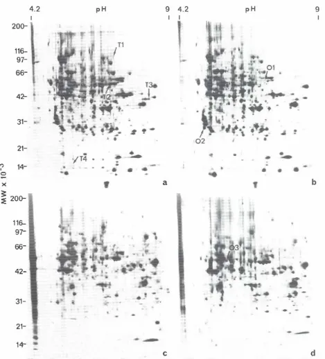
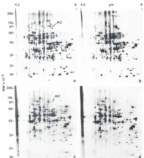
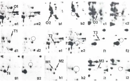
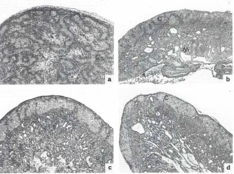
Detection of sex specific proteins in chick embryo gonads and mesonephros: effects of estradiol benzoate or tamoxifen on their expression
9
0
0
Full text
Figure

+2
Related documents
Therefore, the influence of the spirit of Ubuntu on Africans as well as the underlying cultural dispositions of South African Indians, based on their Indian origin, were
In this paper I take as a case study Microsoft’s Azure “Face Cognitive Service.” MS Face offers facial emotion recognition that categorizes the array of facial expressions
In case of single structure, to arrive at the optimum slip force in the friction damper, the variation of peak displacement , peak acceleration of SDOF structure are plotted with
Results — The analysis of phase dynamics of the studied oscillatory systems revealed that the spinal anesthesia suppresses the coupling between the rhythms of autonomic control
The realization of European Economic and Monetary Union has made it obvious that certain fundamental principles of the EC Treaty have be- come part of Germany's
9 All of the authors recognize that students en- rolled in clinical programs can learn skills as they apply critical lawyer theory, the theoretics of practice, and
After an analysis of the provisions of CERCLA, PRPs who join forces with the United States Environmental Protection Agency (EPA) in undertaking a response action
As survival and anti-apoptotic pathways are activated by huMIF in an autocrine manner (15, 33, 34) and TvMIF secretion is induced during serum starvation (Fig. 3), we
