Abstract
JUN YAN. Using Boronic Acid as the Recognition and Signaling Moiety for Sugar Sensing. (Under the direction of Dr. Binghe Wang.)
4;.
/' \r~,. .~
Jl,~aL~
Binghe Wang
Chair of the Advisory Committee
Daniel L. Comins
Dedication
To my family: Chengxian Yan Shifen Guo Li Yan Yinghua Gao
and my lovely niece, Zhihan Gao.
Biography
Acknowledgments
I would like to thank my family.
I would like to thank my research advisor Professor. Binghe Wang for his guidance, patience and encouragement during my graduate work.
The members of my committee made suggestions and comments during my graduate studies. I am very grateful to my committee members, Professor Daniel L. Comins, Professor George H. Wahl and Professor Bruce Eaton.
I would like to thank Dr. Wenqian Yang, Dr. Greg Sprensteen and Susan Deeter for their contributions and help in my research.
I would like to thank the Wang group members, especially Dr. Hao Fang, Dr. Xingming Gao, Dr. Charles E. Ballard and Dr. Weijuan Ni for all the helpful discussions and ideas, and also all of the former and current group members for their help in research and my life. I do enjoy the time I worked with them.
I am grateful to Professor. Edmond F. Bowden, Ms. Cynthia Wertz and Ms. Crissy Williams Brown, for the advice, suggestions and assistance.
Table of Contents
page
List of Schemes vii
List of Tables ix
List of Figures x
List of Abbreviations xii
Chapter I. Introduction
1.1. The Binding between a Boronic Acid and a Diol-the Chemistry Involved.
1 2 1.1.1. The Equilibriums in the Boronic Acid-Diol
Aqueous Solution
1.1.2. Methods for Binding Constants Determination 1.1.3. The Structures of Boronic Acid-Diol Complex 1.1.4. Factors that Influence the Binding
2 5 11 15 1.2. The Design of spectroscopic Boronic Acid Reporter
Compounds
1.2.1. Work of the Czarnik group 1.2.2. Work of Shinkai group 1.2.3. Work of Heagy group 1.2.4. Work of Lackowicz group 1.2.5. Work of James group 1.2.6. Work of the Wang group 1.3. Conclusions
References 18 18 19 25 26 27 28 28 30 Chapter II Relationship among pKa, pH, and Binding Constants in the
Interactions between Boronic Acids and Diols. 2.1. Introduction
2.2. Results and Discussions
2.2.1. Effect of Substituents on the Apparent pKa of Boronic Acids
2.3. Conclusions 2.4. Experimental
2.4.1. General Methods 2.4.2. Apparent pKa
2.4.3. Binding Constant Determination References 43 44 44 44 45 45 Chapter III 8-Quinolineboronic Acid-a Novel Type of
Fluorescent Carbohydrate Reporter. 3.1. Introduction
3.2. Design
3.3. Results and Discussions
3.3.1. Fluorescence Studies upon Biding to Sugars. 3.3.2. pKa Assignment.
3.3.3. Fluorescence Mechanism
3.3.4. Evaluation of 8-QBA as a Potential Diol Sensor 3.4. Conclusions
3.5. Experimental
3.5.1. General Methods 3.5.2. pKa Determination 3.5.3. Binding Constant
3.5.4. Quantum yield determination References 48 48 49 51 51 54 57 58 62 63 63 63 63 65 65 Chapter IV The Design and Synthesis of 8-QBA-based Carbohydrate
Sensors
4.1. Introduction
4.2. Results and Discussion
4.2.1. The Design of Diboronic Acid
Compounds for Carbohydrate Sensing 4.2.2. Synthesis
4.2.3. Building Block Binding Study 4.2.4. Binding Study of Diboronic Acids 4.3. Conclusions 4.4. Experimental 67 67 69 69 76 82 86 86 87 4.4.1. General Methods
4.4.2. pKa Determination
4.4.3. Binding Constant Determination 4.4.4. IR Study
List of Schemes
Page Scheme 1.1. The Binding process between Phenylboronic Acid and a diol. 2
Scheme 1.2. The overall binding constant Keq. 3
Scheme 1.3. Saccharide detection through a competitive boronic acid
receptor. 9
Scheme 1.4. Alizarin Red S. (ARS) binding with Boric Acid. 9 Scheme 1.5. Structures of D-glucose Complexes Formed with Trigonal
p-Tolylboronic Acid, Tetrahedral p-Tolylboronic Acid and a Diboronic Acid.
13
Scheme 1.6. Structural assignment of additional D-fructose boronate esters formed at higher boronic acid:carbohydrate ratios. 14 Scheme 1.7. Complex of Sorbitol and (S,S)-2-(N,N-dimetyl-
1-amoniethyl)ferroceneboronic Acid. 14 Scheme 1.8. Boronic acid reporters from Czarnik group. 19 Scheme 1.9. Possible mechanisms for the fluorescence intensity changes. 21 Scheme 2.1. The relationships between phenylboronic acid and its diol
ester.Keq-tert and Keq-trig are the equilibrium constants of
tetrahedral and trigonal forms of the boronic acid
38
Scheme 3.1. The ionization steps of 8-QBA and its esters. 57 Scheme 4.1. Conceptual design of bisboronic acid-based sensors. 69 Scheme 4.2. Synthesis of momoboronic acid building block. 76 Scheme 4.3. Synthesis of symmetric 8-quinolineboronic acid 41, 42 and
43. 77
Scheme 4.4. An alternative approach to symmetric diboronic acids 41, 42
and 43. 78
Scheme 4.5. Synthesis of 44. 78
Scheme 4.8. Synthesis of 49 and 50. 80
List of Tables
page
Table 1.1. Association constants with PBA. 4
Table 1.2. Fluorescent intensity change of ARS in the presence of a
series of aryl boronic acids. 11
Table 1.3. Fluorescence properties of sensor 16.
26 Table 2.1. The pKa’s of a series of substituted phenylboronic acid
compounds. 37
Table 2.2. Optimal pH for binding between some henylboronic acids
and Alizarin Red S 40
Table 2.3. Binding Constants a of Boronic Acids and Sugars
42 Table 3.1. The lowest excited singlet of quinoline derivatives in
different solvents. 50
Table 3.2. pKa of pyridine and quinoline derivatives.
55 Table 3.3. Association constants (M-1) of 8-QBA and PBA with various
sugars. 60
Table 3.4. Association constants (Keq) of the fructose ester with PBA
and 8-QBA at various pH, in 0.10 M phosphate buffer. 60 Table 3.5. Quantum yield determinations
61 Table 4.1. The binding constants of 8-QBA and 51 with sugars.. 82
List of Figures
Figure 1.1. ICT reporter from Shinkai group.
24 Figure 1.2. Porphyrin boronic acid reporter from Shinkai group.
25 Figure 1.3. Reporter from Heagy group.
26 Figure 1.4. ICT reporter from Lackowicz group.
27 Figure 1.5. Reporters from James group.
28 Figure 2.1. The pKa of phenylboronic acid.
35 Figure 2.2. pKa vs σ for substituted arylboronic acid compounds
38 Figure 2.3. Binding constant determination for 3,4,5-trifluoroPBA
with fructose at different pH’s. 41
Figure 3.1. 8-quinolineboronic acid.
51 Figure 3.2. Fluorescence response of 8-QBA (6.3 × 10⁻⁵ M) in 0.10 M
phosphate buffer at pH 7.4 in the presence of D-fructose 51 Figure 3.3. Fluorescence intensity of 8-QBA (6.3 × 10⁻⁵ M) in 0.10 M
phosphate buffer at pH 7.4 in the presence of sugars 52 Figure 3.4. Fluorescence intensity pH profile of 8-QBA (6.3 × 10⁻⁵ M)
in 0.10 M phosphate buffer: [saccharide] = 0.5 M, 53 Figure 3.5. Absorbance intensity pH profile of 8-QBA (6.3 × 10⁻⁵ M)
in 0.10 M phosphate buffer. 54
Figure 3.6. Absorbance intensity pH profile of 8-QBA (6.3 × 10⁻⁵ M)
in 0.10 M phosphate buffer. 54
Figure 3.7. Hydrogen bond between boron hydroxyl group and
protonated nitrogen. 56
Figure 3.8. The fluorescent spectra of 8-QBA (6.0 × 10⁻⁵ M) in the presence of D-fructose (100 mM) under different storage conditions.
62
Figure 3.9. Plot for binding constant determination.
64 Figure 4.1. Structures of some cell surface carbohydrates.
68 Figure 4.2. Monoboronic acid building block.
71 Figure 4.3. Symmetric diboronic acid 41, 42 and 43.
Figure 4.4. Unsymmetrical diboronic acid 44, 45 and 46.
72 Figure 4.5. Unsymmetrical diboronic acid 47 and 48.
72 Figure 4.6. Favorable conformation of 41, 43 and 44.
73 Figure 4.7. 8-QBA-based Glucose Sensor.
74 Figure 4.8. Conformation of the complex of D-glucose and 49 after
the first boronic acid moiety binds to D-glucopyranose. Spartan (molecular mechanics, MMFF).
75
Figure 4.9. Conformation of the Complex of D-glucose and 50 after the first boronic acid moiety binds to D-glucopyranose. Spartan (molecular mechanics, MMFF).
75
Figure 4.10. Conformation of the Complex of D-glucose and 51 after the first boronic acid moiety binds D-glucofuranose. Spartan (molecular mechanics, MMFF).
76
Figure 4.11. 8-QBA building blocks.
82 Figure 4.12. UV absorbance of compound 40 and its fructose complex
vs. pH. at 314 nm in 0.10 M sodium phosphate buffer. 83 Figure 4.13. Interactions between the functional groups in 40.
84 Figure 4.14. IR Spectrum of 8-QBA and 25 (trigonal).
84 Figure 4.15. IR Spectrum of 8-QBA and 25 (tetrahedral).
84 Figure 4.16. Hydrogen bond between boronic acid moiety and
List of Abbreviations
Abs.: Absorbance ARS: Alizarin Red S. BOC: tert-butoxycarbonyl
DCC: 1,3-dicyclohexylcarbodiimide DCM:Dichloromethane
DMF: N,N-dimetylformamide DMSO: Dimethylsulfoxide
EDCI: 1-[3-(dimethylamino)propyl]-3-ethylcarbodiimide hydrochloride HEPES: N-2-hydroxyethylpiperazine-N’-2-ethanesulfonic acid
ICT: internal Charge Transfer If: Fluorescence Intensity
Ka-acid : Acid dissociation constant of the boronic acid
Ka-ester : Acid dissociation constant of the boronic ester
Keq-trig : Equilibrium constant of the trigonal boronic acid with the diol
Keq-tet : Equilibrium constant of the tetrahedral boronic acid with the diol
NMR: nuclear magnetic resonance PBA: Phenylboronic acid
PET: Photoinduced electron transfer Ph: phenyl
QBA: quinolineboronic acid rt: room temperature
s: singlet t: triplet
TEA: triethylamine TFA: trifluoroacetic acid THF: tetrahydrofuran
Chapter I. Introduction
It is well known that mammalian cell surfaces are coated with saccharides in the
forms of glycoproteins and glycolipids
.
1-9These glycosylated biomolecules play very
important roles in various physiological and pathological processes.
10,11Critical to
essentially all the saccharide-mediated events is the binding of a biomolecule to certain
cell-surface saccharide or its conjugates.
12-14For example, cell-cell adhesion in
inflammatory processes involves lectin-carbohydrate interactions, which mediate
leukocyte latching to inflamed tissues;
15,16HIV infection is mediated by glycoprotein
binding with cell surface receptors; embryonic development at early stages is known to
rely on Lewis X for cell-cell adhesion;
17and certain cancer metastasis also involves
saccharide-mediated events.
18,19Furthermore, binding of certain cell-surface
carbohydrates by a class of carbohydrate-binding proteins, lectins, is known to trigger
various biological events.
20For example, galectin-3 binding to its target has been shown
to trigger apoptosis.
21Therefore, small molecule mimics of proteins that can bind
cell-surface carbohydrates (artificial lectins) could have a variety of biological and biomedical
applications ranging from diagnostics to medicinal agents. In designing such artificial
lectins, boronic acids occupy a special place. This is due to the tight and reversible
complexation of the boronic acid moiety with 1,2- or 1,3-diols commonly found on
carbohydrates.
22-24This dissertation work focuses on (1) the examination of various
factors that affect the diol-boronic acid binding, (2) the development of a water-soluble
fluorescent boronic acid reporter compound that responds to the diol binding by changing
fluorescent properties, and (3) the development of new synthetic methods that allow for
discovered. Some background information on boronic acid-diol interactions, and
previous effort on developing fluorescent boronic acid reporter compounds are described
in this introductory chapter to put the dissertation work in perspective.
1.1. The Binding between a Boronic Acid and a Diol-the Chemistry Involved.
1.1.1. The Equilibriums in the Boronic Acid-Diol Aqueous Solution
The interaction between a boronic acid and a diol is known to be one of the
strongest single-pair reversible functional group interactions in an aqueous environment.
Critical to the effort of using boronic acid as the recognition motif is the understanding of
the chemistry involved. However, before any further discussions of the binding, it is
important to note that the acidity of the boronic acid is not reflected in its deprotonation
as is commonly defined in carboxylic acids, rather its reaction with a water molecule that
leads to the formation of the boronate species (
3
) with the release of a proton (Scheme
1.1). By the same token, the boronic ester (
2
) is also an acid and can react with water to
release a proton. Because of this unique feature that both the boronic acid and boronic
ester are acids, and both the complexed (
2
and
4
) and un-complexed (
1
and
3)species
B(OH)2 B
HO OH OH
B HO O
O B
O O
Keq-trig Keq-tet
1 3
2 4
HO OH R2 R1
HO OH R2 R1
R1
R2 R1
R2
-2 H2O -2 H2O
+H2O, -H+
+H2O, -H+
exist in two ionization states, there are several ways to describe the binding between a
boronic acid and a diol.
23These include (1) K
trigdescribing the equilibrium between the
two boronic species (
1
and
2,
Scheme 1.1); (2) K
tetdescribing the equilibrium between
the two boronate species (
3
and
4
); and (3) K
eqdescribing the overall equilibrium
between the un-complexed (
1
and
3
) and the complexed species (
2
and
4
) (Scheme 1.2).
23
B(OH)2
B O OH
OH
B HO O
O B O
O Keq
Diol
R1
R2
R1 R2
1
3
2
4 2
Scheme 1.2. The overall binding constant Keq.
The ability of boronic and boric acids (
1,
Scheme 1.1) to bind with diols was first
recognized over a century ago. In 1842, it was reported that sugars increased the acidity
of boric acid solutions.
25Later publications included comparative studies of different
diols and their binding abilities with boric and boronic acids.
26,27In 1959, Lorand and
coworkers published the first comprehensive study of the binding constants between
various diol-containing compounds and phenylboronic acid (PBA) using the so-called
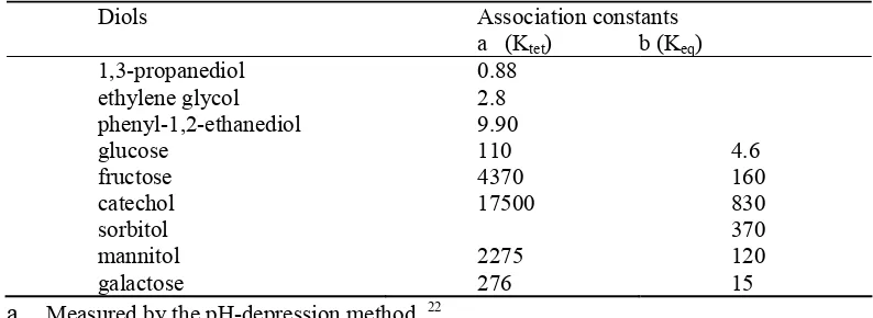
pH-depression method (Table 1.1).
22The study demonstrated that different diols have
different affinities for the boronic acid group and lower the acidity of the boron species to
different degrees. The numbers listed in Table 1.1, although referred to as binding
constants, are very different from the binding constants of other monoboronic acids
determined later using spectroscopic methods, sometimes by over 20-fold. A careful
of the meaning of the term “binding constants.” The “binding constants” determined
using the pH-depression method are K
tetinstead of K
eq.
23This was because the
pH-depression method assumed that the boronic ester did not exist (or existed in negligible
amount). Therefore, the K
trigpart of the equation in Scheme 1.1 was omitted. Since the
relationship between
1
and
3
is an acid-base equilibrium, then the only unknown that was
determined was the K
tet. With this clarification, it became easy to understand why the
discrepancy in the literature.
Table 1. 1. Association constants with PBA.
Diols Association constants
a (Ktet) b (Keq) 1,3-propanediol 0.88
ethylene glycol 2.8
phenyl-1,2-ethanediol 9.90
glucose 110 4.6
fructose 4370 160
catechol 17500 830
sorbitol 370
mannitol 2275 120
galactose 276 15
a.
Measured by the pH-depression method. 22b.
Measured by the ARS competition method at physiological pH. 23Springsteen and Wang in 2002 published the first systematic examination of the
K
eqbetween various diol-containing compounds and phenylboronic acid (Table 1.1).
These numbers are much lower than the K
tetdetermined using the pH-depression method.
Springsteen and Wang also pointed out that buffer has an effect on the binding constants
between a boronic acid and a diol. Therefore, the binding constants determined are really
apparent binding constants under the specific experimental conditions described. The
understanding of the buffer effect is very important for biological applications. James
and co-workers have recently published a detailed study on the effect of buffer
28which
Springsteen and Wang also described the relations among K
trig, K
tet,and K
eq(Eq
1-6).
23,29Such relationship allows for the comparison of various “binding constants”
determined using different methods.
Keq = (([BD-][H+] / Ka-ester) + [BD-]) / (([B-][H+] / Ka-acid) + [B-]) ([D] + [D-]))
(1)
Keq-tet = [BD-] / [H+]([D-]/[D]+[D-])[B-][Diol]
(2)
Keq-trig = [BD] / [H+]([D-]/[D]+[D-]) [B][Diol]
(3)
eq K ester -a K ] [H 1 acid -a K ] [H 1 tet -eq
K + ×
+ + + =
(4)
ester -a K acid -a K eq K ester -a K ] [H 1 acid -a K ] [H 1 trig -eqK + ×
+ + + =
(5)
tet eq K ester trig eq K acid eqK =% × − +% × −
(6)
B = Trigonal Boronic Acid, BD = Complex of Trigonal Boronic Aicd and Diol B-= Tetrahedral Boronic Acid BD- = Complex of Tetrahedral Boronic Acid and Diol
1.1.2. Methods for Binding Constants Determination
In order to examine factors that affect the binding between boronic acids and diols,
there need to be sensitive methods for the determination of their binding constants. Early
methods include the pH-depression method
22,3031-34and the
11B-NMR method.
35-40These
methods, although served the purpose for the early determination of binding constants
between boronic acids and diols, are insensitive, require a large amount of sample, and
take long experiment times. Therefore, these two methods most of the time do not meet
the need in modern day sensor and boronolectin (boronic acid-based artificial lectins)
rapid, and require very little sample. Below is a detailed description of the different
methods used in the determination of the binding between boronic acids and diols.
1.1.2.1. The pH-Depression Method
The very first method used for the systematic determination of boronic acid-diol
binding constants was the so-called pH-depression method.
22,30-34This is based on the
finding that most the time diol binding lowers the pKa of boronic acid. Therefore,
addition of a diol to a boronic acid solution would lower its pH. The extent of the pH
lowering effect of a particular diol can be correlated with the “binding constant” between
these two species. In the mathematical derivation of the “binding constant,” it was
assumed that the trigonal boronic ester (
2
, Scheme 1.1) did not exist. This was based on
the idea that the decreased pKa of the boron upon ester formation would ensure that it is
converted to its tetrahedral anionic form (
4
).
Therefore, the equation was simplified to
K
tet. In addition to the fact that the “binding constants” determined using the
pH-depression method is the K
tet, our own experiments later on have also shown that the
assumption that the neutral boronic ester did not exist is incorrect.
23For example, the
apparent pKa of the phenylboronic ester of glucose is about 6.8. At physiological pH,
about 20% is in the free ester form. Although the exact pH under which the
pH-depression experiments were conducted is not known, it is reasonable to expect that the
pH of a particular boronic ester be close to its pKa. Therefore, one would expect that the
boronic ester exist in over 20-30% under some of the experimental conditions. In
conclusion, the pH-depression method gives an approximate estimation of the K
tet, and
this method requires a large amount of the boronic acid sample.
Another method that had been used was that of
11B-NMR.
35-40This is based on the
same concept that the boronic ester pKa is lower than that of the free boronic acid.
Therefore, addition of a diol to a boronic acid solution would result in an increased
portion of the boron being converted from the neutral trigonal form to the anionic
tetrahedral form. This change can be detected using B-NMR because of the significant
chemical shift differences (about 30 ppm for the neutral form and 7 ppm for the anionic
form of boronic acids). The
11B-NMR approach uses a similar mathematical derivation,
and, therefore, also gives the K
tet. For example, the binding constants of boric acid with
glucose and galactose were determined using NMR method.
36They are generally in the
range of 100 M
-1, which are in line with the K
tetnumbers determined using the
pH-depression method. When the distinct species can be directly detected, this offers a direct
and excellent approach to the determination of the K
tet. However, the
11B-NMR method
suffers from low sensitivity, difficulties with peak resolution, and the requirement for
high concentration of the sensor compound. Such restrictions make the
11B-NMR
method less useful in the development of boronic acid sensors. Therefore, if sample
quantity is an issue, the utility of this method would be limited.
1.1.2.3. Spectroscopic Methods
It is well-known that spectroscopic methods are generally more sensitive than
methods using NMR or pH determination, although not always. Therefore, it is easy to
understand that there has been much effort in developing spectroscopic methods for the
determination of the binding constants between boronic acids and diols. However, for
this to happen, the binding event needs to trigger a change in the spectroscopic properties
fluorescent property changes upon diol binding. We term them as boronic acid
spectroscopic/fluorescent reporter compounds. For those compounds, spectroscopic
determination is a very sensitive and convenient way for determining the binding
constants. In this regard, spectroscopic methods used for the binding constant
determination include CD spectroscopy
4142-49absorption spectroscopy,
50-55and
fluorescence spectroscopy.
23,56-67For example, Yoon and Czarnik determined the
dissociation constant
of
anthrylboronic acid with D-fructose at pH 7.4 using fluorescence
spectroscopy.
68Titration of anthrylboronic acid (0.75 µM) at pH 7.4 with a polyol gives
a dissociation constant of about 3.7 mM with D-fructose. Similarly, UV absorption
titration was used for the K
eqdetermination in nitrophenol-based boronic acid color
sensors. The association constant was obtained for a couple of sugar boronic esters.
55CD
is another detection method for boronic acid based sugar sensors. The Shinkai group
developed a porphyrin based boronic acid sensor system. Upon binding to sugars,
changes in CD were observed, presumably due to the immobilization of two porphyrins
by a saccharide bridge. From the plot of CD intensity vs saccharide concentration was
obtained the association constant.
41-49When the boronic acids possess the appropriate
spectroscopic properties for direct measurement, such methods work very well. However,
most boronic acids do not change their spectroscopic properties sufficiently and/or
consistently enough for their binding constants to be determined this way. The section
will discuss in detail how a fluorescent reporter compound can be used for the sensitive
determination of boronic acid-diol binding constants in a competition assay.
Because of the limitations of the various methods available, there was a need in the
binding constants between boronic acids and diols, regardless of whether the boronic
acids are fluorescent or not. To address this issue, our group developed a generally
applicable, highly sensitive method for the determination of the binding constants
between boronic acids and diols. This method uses Alizarin Red S. (ARS) as a reporter
compound in a three-component competition assay (Scheme 1.3).
Boronic acid + Fluorophore Boronic acid + cis Diol + Fluorophore Strong Fluorescence Non-fluorescence
cis Diol
Scheme 1.3. Saccharide detection by using a competitive boronic acid receptor.
ARS is a dye commonly used in the textile industry.
69It has been used as a reagent
for the fluorometric detection of boric acid.
69ARS is not fluorescent, because of the
possible excited state proton transfer from one of the catechol dihydroxyl groups to the
carbonyl oxygen (Scheme 1.4,)
70O
O OH
OH SO3Na
O
O O
O SO3Na
B HO
R
RB(OH)2
5 6
OH
O O
OH SO3Na excited state
proton transfer
5a
R1 R2 OH
R1 R2 O O
B
R HO
nonfluorescent fluorescent
Scheme 1.4. Alizarin Red S. (ARS) binding with a Boronic Acid.
transfer process, which is responsible for the fluorescence quenching. This should result
in significant fluorescent intensity increases upon boronic acid binding. Based on this
assumption, we tested the binding of a series of 6 boronic/boric acids to ARS. All those
tested caused a very significant fluorescent intensity change of ARS (30-90 fold, Table
1.2).
29Even boric acid induced about 25 fold fluorescent intensity increase. It is also
important to note that the most sensitive region is around physiological pH and ARS is
very soluble in water. These are two important criteria for the study of binding under near
physiological conditions. In addition, this method also allows for the determination of the
binding constants at a given pH and in various buffers, both are factors that influence the
apparent binding constants. One may argue that the “intrinsic” binding constants should
be independent of external factors such as buffer. However, for what we and many other
groups are interested, the application of boronolectins and boronic acid-based sensors in
near physiological conditions, the apparent binding constants under certain designated
conditions are more important than that of the “intrinsic” affinities. One can draw an
analogy between this and enzyme-inhibitor binding, where the “intrinsic” binding affinity
without the influence of external factors has little practical meaning because of the
intended applications, although the theoretical studies of such “intrinsic” affinities may
help understand the fundamental questions in binding and catalysis. Therefore, when one
designs a study, it has to be very clear as to what the purpose is for such a study in order
to remain focused on the problem one intends to address.
Using this method, we have determined the apparent binding constants of a series of
diol-containing compounds with phenylboronic acid. This was carried out by first
addition of the interested diol (Scheme 1.4). The addition of a diol-containing molecule
perturbs the ARS-boronic acid equilibrium, which provides a measure of the strength of
the boronic acid-diol association. This method allows for the determination of the K
eqas
well as K
tetand K
trigas defined in Scheme 1.1 (Equations 4 and 5). Again, we should
note that these numbers are the apparent binding constants under these specific conditions.
The development of the ARS method allowed for the first time the sensitive
determination of K
eqbetween a boronic acid and a diol regardless of the spectroscopic
properties of the boronic acid portion. It is also easy to use and rapid. It allowed us to
understand the relationship between K
eq, K
tet, and K
trig. Although this method is not free
from interference by factors such as the nature of the buffer and its concentration, it
allows for the determination of binding constants relevant to the intended applications, as
long as one carefully designs the experiments.
Table 1.2. Fluorescent intensity change of ARS in the presence of a series of aryl boronic acids. 29
Boronic acids Fluorescence Intensity Increase (fold)
methylpyridinium-3-boronic acid 30
phenylboronic acid 59
boric acid 26
2,4,6-trifluoroboronic acid 88
Pyridine-3-boronic acid 63
4-fluoro-2-methylphenyl boronic acid 53
1.1.3. The Structures of Boronic Acid-Diol Complex
Although the high affinity of boronic acid for sugars was recognized in the
1950’s,
22,26,71the structural studies of the sugar-boronic acid complexes came at a later
in 1960’s and 1970’s using
1H and
13C NMR spectroscopy.
72-74However, with the
continuous wavelength instrument at relatively low field strength, the studies could only
be carried out in organic solvents that provide the high solubility needed in order to see
the signal. For carbohydrates sensor development, it is more important to understand the
structural features under aqueous conditions. With the availability of high-field FT-NMR
instrument, studies in aqueous environments became feasible. Most of the studies
focused on using either mono- or diboronic acids and glucose or fructose as the model
sugar. Along this line, several groups have devoted much effort.
75-80The conformational
and structural studies were mostly based on the determination of C-H and H-H coupling
constants which are different depending on the ring size and conformation of the sugar in
the complex. For a while, there was some debate as to whether the complex between
glucose and phenylboronic acid exists in the pyranose and furanose form.
79,81,82In 1995,
Eggert and Norrild studied the complex formation between boronic acid and D-glucose
using
1H and
13C NMR spectroscopy under both neutral non-aqueous and alkaline
aqueous conditions. A furanose complex structure was deduced by comparing the
chemical shifts and coupling constants of the boronic acid-glucose complex with those of
the boronic acid-modified glucofuranose complex.
79Their studies showed that in both
neutral non-aqueous and alkaline aqueous conditions
,
the first binding site was position
(1, 2) of the furanose form of D-glucose. Under neutral non-aqueous conditions the
second boronic acid was bound to positions (3, 5) (
7,
Scheme 1.5). Under alkaline
aqueous conditions the second monoboronic acid was bound to positions (3, 5, 6) (
9a
and
9b
scheme 1.5) in a tris coordinated manner, and a diboronic acid sensor (
8)
binds its
O O O O B
HO
H3C
B O CH3 O O O O B
H3C
B O CH3 O O O O B
H3C
B O CH3 O O OH HO O O OH O B O O B HO OH CH3O
CH3O trigonal boronic acid/D-glucose
tetrahedral boronic acid/D-glucose
diboronic acid/D-glucose
endo exo
7 8
9a 9b
Scheme 1.5. Structures of D-glucose complexes formed with trigonal p-tolylboronic acid, tetrahedral p-tolylboronic acid and a diboronic acid. 79
In 1996, Norrild reported the structural studies of the fructose complex with
phenylboronic acid by using 2D NMR.
80In a ratio of 1:1, boronic acid and fructose,
under alkaline aqueous condition, form a 2, 3, 6 tridentate complex as the major
product (
10
, Scheme 1.6, 82%). A small amount of 2, 3 exo and endo isomers (
11a
and
11b
, Scheme 1.6) were present while fructose was in furanose form in all cases. Four
new complexes were observed at high boronic acid/fructose ratios, such as 2:1 and 4:1
(Scheme 1.6). Two boronic acids can bind to positions (2, 3) and (4, 5) of the
fructopyranose, respectively, result in four endo/exo diastereomers as shown in Scheme
O O O O O OH O O O O O OH O O O O O OH O O O O O OH B O O O B
OH OH OH
O HOHO B
O OH OO B OH HO OH OH B HO B OH B HO B OH B OH O O O B B OH OH 10 11a
exo isomer 11b endo isomer
12a 12b 12c 12d
OH
Scheme 1.6. Structural assignments of additional D-fructose boronate esters formed at higher boronic acid: carbohydrate ratios. 80
Sorbitol is another sugar that has been reported to form a trivalent complex with a
boronic acid. (Scheme 1.7
)
In 2001, Norrild studied the complex structure between
D-sorbitol
and (S, S)-2-(
N,N
-dimetyl-1-amoniethyl) ferroceneboronic acid using
1H,
13C and
11
B NMR. A 2,3,5-bound sorbitol complex (
13a
, Scheme 1.7) was deduced.
83Since
sorbitol and fructose both bind to a sugar, when possible, in a tridentate fashion, it
becomes easy to understand why they normally have higher affinities to boronic acids
than other commonly seen sugars such as glucose, mannose, etc.
23Fe B
O OO
HO OH
OH
N(CH3)2
H
CH2OH
CH2OH
H OH HO H H OH H OH Fe B OH OH N(Me)2 CH3
13 sorbitol 13a
1.1.4. Factors that Influence the Binding
One of the most important things in designing boronic acid-based sensors and
boronolectins is the understanding of how various factors influence the binding between a
simple boronic acid and a diol. It is generally believed that pH, the nature and
concentration of the buffer, the pKa of the boronic acid, the pKa of the boronic ester, the
dihedral angle of the diol, and of course temperature. Below is a summary discussion of
all these issues.
1.1.4.1. The Apparent pKa Values of the Boronic Esters and the Stability of the
Boronic Acid-Diol Complex.
It is well known that the pKa values of boronic esters are generally lower than
that of the boronic acids. As discussed earlier, different diols have different affinity for a
given boronic acid. It seems that the binding constant is generally correlated with the
apparent pKa value of the boronic ester, with the exception of those engaged in trivalent
binding such as sorbitol.
23The lower the apparent pKa of the boronic ester, the higher
the binding constant. Therefore, any factor that increases the boron acidity should
increase the binding constants as well. Theoretically, the change in pKa values from
boronic acid to boronic esters could be the result of either electronic or steric factors.
However, the added alkyl groups in boronic esters, compared with the free boronic acids
(
2
, Scheme 1.1), are electron-donating, and would be expected to increase the electron
density on the boron atom, which should result in a decrease in the acidity of the ester.
29This is apparently contrary to experimental results. Consequently, electronic factor is
probably not the dominant factor. On the other hand, the different affinity among various
the boron to go from trigonal to the tetrahedral form can be directly related to the boronic
ester pKa and the diol-boronic acid binding constants. A key factor in affecting the
conversion from trigonal to tetrahedral form is the dihedral angle of the diol. Smaller
dihedral angles correlate with a smaller distance between the two diol oxygen, which in
turn favors a smaller O-B-O bond angle that closely resembles the
sp
3form (109 degree)
and moves away from the 120 degree needed for the boron to stay in the trigonal form.
This is the reason why diols with small and constrained dihedral angles tend to bind to
boronic acids with higher affinities.
291.1.4.2 Buffer Effects
The use of buffer is very common in evaluating boronolectins and boronic acid-based
sensors. For biological applications, phosphate is a commonly used buffer in simulating
physiological conditions at pH 7.4. In order to get a good sense of how results obtained
using certain buffer can be extrapolated to near physiological conditions, one needs to
have a good understanding of the buffer effect on the binding constants between a
boronic acid and a diol. Although studies in this area are limited, one thing is sure, i.e.,
buffer does play an important role in influencing the binding constants determined. This
is understandable since boronic acid is a Lewis acid. The presence and concentration of
Lewis bases such as phosphate, chloride, fluoride, and bromide are expected to affect the
complexation state of the boron atom, and therefore its ability to interact with other
Lewis bases such as water.
Our lab has shown that the nature and concentration of the buffer do affect the
binding affinity.
23For example, when determined in phosphate buffer, the binding
concentration range of 0.05 – 1 M. On the other hand, the binding constants between
fructose and phenylboronic acid did not change as much. It is not clear why this
difference in binding constants between these two diols. Compared with phosphate
buffer, the concentration of HEPES buffer, within the range tested, seemed to have very
little effect on the binding constants. There are two possible reasons that could explain
the lack of any significant effect of buffer concentration when HEPES was used. In the
first scenario, HEPES buffer may not interact with boronic acid as a Lewis base. Under
such a situation, one could envision a lack of buffer effect on the binding. A more likely
explanation is with the second scenario, when at the lowest concentration tested, the
buffer could have reached its maximal effect. This is very logical since HEPES has a
1,2-aminoalcohol structure, which could have a high affinity with a boronic acid.
Furthermore, the binding constants determined in HEPES buffer are lower than that
determined at the highest concentration of phosphate buffer, which is consistent with the
higher effect of the HEPES. Clearly, there is much more to do in order to understand the
effect of buffer on the binding constants between a boronic acid and a diol. In a very
recent study, James and co-workers have studied the buffer (phosphate) effect in a
quantitative fashion.
28The same approach can be very useful for the quantitative
de-convolution of the effect of other Lewis bases (buffer components) on the binding
constants between a boronic acid and a diol.
1.1.4.3. pH and pKa of Boronic Acids
It’s well known that pH affects the affinities of boronic acids toward diols, the higher
the pH, the higher the binding constants between boronic acid and diol. It is also
Our group’s study on the relationships between pKa, pH and binding constant indicates
that this is not always true.
23It’s clear that pKa and pH play important role in the binding
process. However, there has not been a systematic examination on the relationships
among all those factors so far. Any information on this subject would undoubtedly assist
the efforts to optimize the binding affinity and specificity of potential fluorescent sensors.
Chapter 2 discusses in detail on the finding from our studies on this subject.
1.2. The Design of Fluorescent Boronic Acid Reporter Compounds
A key component in the design of fluorescent sensor is the fluorescent reporter
unit that changes fluroescent properties upon binding. Therefore, it is not surprising that
there has been much effort in the development of such reporter boronic acids. In this area,
several groups have made very important contributions using various mechanisms.
75,79,81,87-89
In those fluorescent reporter systems, the binding event between a boronic acid
and diols acts as an on-off or off-on switch to generate detectable fluorescence changes.
In rare exceptional cases, the fluorescent properties change at more than one wavelength
and therefore give a ratiometric sensing mechanism.
90This section will give a brief
description of literature fluorescent reporter compounds and their important properties.
Because the work in this area cannot be easily categorized based on either mechanism,
they are grouped based on the principal investigators.
1.2.1. Work of the Czarnik Group
In 1992, Czarnik and co-workers reported the first boronic acid system that
showed significant fluorescent intensity changes upon sugar binding.
Specifically, 2- and
sugar or a catechol resulted in decreased fluorescence intensity.
68,91As discussed earlier,
since the boronic ester has a lower pKa, the addition of the sugar converted the boron
from trigonal neutral form to the anionic tetrahedral form. For example, the fructose-
7
complex has an apparent pKa of about 5.9, which means that it exists in the anionic
tetrahedral form at physiological pH. Although the mechanism through which the
observed fluorescence intensity change happens was not extensively examined, it was
thought that boronate anion (
7a
) quenches the anthracene fluorescence. This was a
seminal study that stimulated much more work later on in using boronic acid for the
preparation of fluorescent sensors for carbohydrates.
B(OH)2
B(OH)2
7 8
B(OH)2 B(OH)3
HO HO diol or catechol
BO O
HO HO diol or catechol
BO O OH
+ +
pH = 8.8
pH < 8.8
High Fluorescence Low Fluoresscence 7a
7b 7c
Scheme 1.8. Boronic acid reporters from the Czarnik group.
1.2.2. Work of the Shinkai Group
During the last decade, the Shinkai lab played a very prominent role in the field
of using boronic acid as the recognition moiety for the preparation of sensors for
various saccharides. Of course, at the center of such research activities is the availability
of reporter compounds that change spectroscopic properties upon saccharide binding.
1.2.2.1. The Anthracence-based Systems
Shinkai’s group also used the anthracene system in designing fluorescent reporter
9
(Scheme 1.9). In this system, an amino group is positioned in a 1,5-relationship with
the boronic acid. It had previously been demonstrated by Wulff
92that such an
arrangement promotes B-N bond formation under certain conditions. When this happens,
it helps to lower the apparent pKa of the boronic acid, which was presummed to help
increase binding as well as B-N strength. Indeed,
9
showed much higher binding affinity
to fructose and glucose compared with phenylboronic acid
24,60,93This system (
9
) also
exhibits a very significant fluorescence intensity change upon sugar binding. This system
and its analogs have been used by many labs,
61,62,94including our own,
59,63,95for the
synthesis of fluorescent sensors for saccharides.
Initially, the mechanism through which the fluorescence intensity changes was
thought to be by the modulation of the excited state photoinduced electron transfer (PET)
through the formation or strengthening of the B-N bond (Scheme 1.9). Specifically, the
lone pair electrons on the benzylic amine moiety is known to quench the anthracene
fluorescence. Consequently,
9
in the absence of any saccharide is only weakly
fluorescent. Upon ester formation, due to the increased acidity of the boron,
29it was
proposed that B-N bond forms or strengthens, which helps to “tie up” the lone pair
electrons, and therfore prevent fluorescence quenching through PET. The end result is an
increased fluorescent intensity upon sugar binding. Although this mechanism seems to
9a N B OH HO H 9b N B OH HO 9c N B -OH HO HO N B H O O R R' N B H O O R R' HO + N B O O R R' HO OH HO R' R pKa1 pKa1 pKa2 pKa2 N B H O O R R' N B O O R R' + -N B O O R R' HO -OH HO R' R pKa2
-2 H2O
10a 10b 10c
10a 10c 10d + + +
-H2O
pKa1
-Scheme 1.9. Possible mechanisms for the fluorescence intensity changes for the Shinkai anthracene
system.
telltale signs that the mechanism through which such a system works may not have to
involve B-N bond formation as originally proposed. Because of the enormous role that
compound
9a
has played in the field of boronic acid-based sensor design, our lab
undertook an effort to examined the mechanistic detail of the fluorescent intensity
changes of this system upon sugar binding. Theoretically, one can write two reasonable
mechanisms through which fluorescence intensity changes can happen upon binding to a
sugar (Scheme 1.9). In addition to the B-N bond mechanism, which is shown in the top
portion of Scheme 1.9, one can also write a hydrolysis mechanism (lower portion of
B-N bond alredy exsiting in the free acid form (
9b
). In the B-N bond mechanism, the
strengthening of the B-N bond could help to “tie up” the amine nitrogen lone pair
electrons and prevent PET. The diminished PET should result in increased fluorescence
intensity. In the hydrolysis mechanism, the protonation of the nitrogen could “tie up” the
nitrogen lone pair electrons and result in the same fluorescence intensity increase. We
have analyzed these two possible mechanisms and designed a series tests to see which is
the more likely mechanism. Along this line, we (1) studied the pH profile of the reporter
system in the presence and absence of a sugar, (2) examined the maximal fluorescence
intensity changes induced by various sugars to see whether there would be any difference
or not, (3) tested the effect of sugars capable of trivalent binding to boronic acid, and (4)
calculated the B-N bond strength.
55All such anslyses are consistent with the hydrolysis
mechanism, and inconsistent with the B-N bond formation mechanism. Specifically, the
pH profiles of the free boronic acid (
9a
) shows a singificant fluorescence intensity
decrease assoicated with the first pKa, but not the second. With the ester (
10a
), the
situation is just opposite. This is only consistent with the hydrolysis mechanism because
the B-N mechanism would give the ester the same pH profile as the free acid itself. The
studies with different sugars also led to the same conclusion. It is well known that the
pKa values of the boronic ester of different sugars are different. This would mean that
the the B-N bond strength would be different for
10b
, which should in turn affect the
quenching efficience by the lone pair electrons and consequently the maximal fluorescent
intensity for a paricular ester. However, all sugars gave the same maximal fluorescence
intensity, which is inconsistent with the B-N bond mechanism, but consistent with the
featurs regardless of which sugar is used. In addition, if the B-N bond is the reason for
the fluorescence intensity changes, sugars capable of trivalent binding to a boronic acid
should not be able to induce fluorescent intensity changes. The same fluorescent
intensity changes observed with sorbitol and fructose, both of which are capable of
binding in a trivalent fashion,
80,83are again inconsistent with the B-N bond mechansim,
but are consistent with the hydrolysis mechanism. Lastly, the B-N bond strength has
been estimated to be around 3 kcal/mol,
96far smaller than what is needed to “tie up” the
lone pair electrons to prevent PET. With all this evidence, it is reasonable to say that the
Shinkai system most likely undergoes fluorescent intensitiy changes upon sugar binding
through a hydrolysis mechanism (Scheme 1.9,
10d
). Specifically, in the free form the
sensor can exist as
9b
. Upon sugar addition, because of the lowered intrinsic pKa of the
boron, the first pKa becomes the reaction of the boron with a water molecule to give
10d
.
The existence of the anionic boron next to the amine also helps to stabilize the protonated
form, which increase its pKa. The conversion of the weak B-N bond form in
9b
to the
amine-protonated form (
10d
) allows for the masking of the lone pair electrons, which
abolishes the PET quenching of the anthracene fluorescence and results in an increase
fluorescence intensity.
551.2.2.2. ICT sensor
The Shinkai group also reported the use of stilllen boronoic acid
11
as
fluorescent carbohydrate sensor in 1995.
97Lackwoicz group later on characterized this
system as an ICT (internal charge transfer) sensor (Figure 1.1). Therefore, this part will
the Shinkai group synthesized another boronic acid
12
with the coumarin chromophore as
an ICT sensor.
98However, compound
12
only showed a very small shift in fluorescence
intensity and wavelength upon addition of sugars.
N O O
B(OH)2
11
B(OH)2
N
12
Figure 1.1. ICT fluorescent reporters from the Shinkai group.
1.2.2.3. Porphyrin systems and others
Using a similar concept to the anthracene system, the Shinkai group reported
another PET sensor, tetraphenyl porphyrinato)tin(iv)
13
(Figure 1.2), which is an
“on-off” reporter upon binding with carbohydrates.
99Molecular dynamic simulation results
suggested that the tertiary nitrogen and boron atom were close to each other in the free
receptor form. However, in the complexed form, it was suggested that the distance
between the boron and nitrogen atom was longer than its acid form due to steric effect.
Such an effect was proposed to decrease the B-N interaction. Consequently, lone pair
electrons on nitrogen were free to quench the fluorescence. The fluorescence profile of
the compound
13
and reference compound (
14
,
15
) showed that the fluorescence
intensity of
13
in the free reporter form is 30% of
14
, but is nearly nine fold of
15
. This
implied that the B-N interaction in free receptor form prevented the tertiary amine from
quenching the fluorescence to some extent. In light of the new mechanism described in
the previous section, it is not clear as to what the exact mechanism is for the fluorescent
N N N N
N Ph
B HO OH
Sn Cl
Cl
13
N N N N
Sn Cl
Cl
14
N N
N N
N Ph
Sn Cl
Cl
15
Figure 1.2. Porphyrin boronic acid reporter from Shinkai group.
1.2.3. Work of the Heagy group
Aimed at improving water solubility of the fluorescent saccharide reporter
moiety, the Heagy group synthesized and evaluated the
N
-phenylnaphthalimide sensors
16a-c,
which were soluble in aqueous solution at least up to micromolar concentrations.
100-102
The fluorescence intensity of the
16a
and
16b
decreased with the addition of
saccharides due to chelation enhanced quenching. Molecular modeling and quantum
mechanics calculations suggested that the naphthalimide group (fluorophore) and
phenylboronic acid (reporter) are not in same plane. After sensor (
16a-b)
binding to a
carbohydrate, boron hybridization changed to the sp
3tetrahedral form and the orbital
energy of the HOMO for the phenyl boronic
π
-system was altered via occupation of the
next highest molecular orbital. Compound
16a
showed the greatest fluorescence intensity
changes in the presence of fructose with a dissociation constant of 1 mM. With the
attachment of nitro group to the naphathalic anhydride ring,
16b
displayed dual
glucose over fructose. After reducing the nitro to an amino group,
16c
exhibited the
ability to enhance fluorescence intensity upon addition of galactose at low pH.
N O
O
B(OH)2
16
R1 R2
R3
16a R1=H; R2=H; R3=H
16b R1=H; R2=H; R3=NO2
16c R1=H; R2=H; R3=NH2
Figure 1.3. Fluorescent reporters from
Heagy group.
Table 1.3. Fluorescence properties of sensor 16.
1.2.4. Work from the Lackowicz group
The fluorescent boronic acid carbohydrate sensors based on ICT (internal
charge transfer) have been studied in the last ten years since Shinkai’s group reported
stillbene boronic acid
11
in 1995.
97Generally, an ICT system contains an electron donor
group and an electron acceptor group in the same fluorophore. The boronic acid acts as
electron acceptor in the neutral form. When the boronic acid group turns into its anionic
form (tetrahedral form) at certain pH upon binding with a sugar, it is no longer an
electron acceptor. This leads to the spectral changes due to the perturbation of the charge
transfer nature of the excited state and trigger the change of the fluorescence spectrum.
103
Four stillbene boronic acid analogs
17a-c
and
11
were synthesized and evaluated by
the Lackowicz group. Compounds
17b
and
11
has an electron-donating group and
17c
has an electron-withdrawing cyano group. Changing the pH from low to high induced a
Sensor λex (nm) λem (nm) φ
16a 345 400 0.01
16b 337 430/550 0.006
blue shift in the emission spectrum of
17b
and
11
, and an increased intensity by about
one fold in the presence of sugar. This was thought to be the result of converting the
electron-withdrawing properties of neutral boron at low pH to the anionic form upon
sugar addition. Similarly, the polyene derivatives
18
,
104diphenyloxazole derivatives
19
105,106and chalcone derivatives
20
107were prepared and tested for binding with sugars.
The fluorescence intensity changed only by a maximum of five fold in these ICT systems.
Furthermore, these compounds are not very water-soluble, which limit their applications
in certain situations.
17 R
B(OH)2
R=H, STBA 17a
R=CH3O-, MSTBA
17b
R=CN, CSTBA 17c
R=N(CH3)2 , DSTBA
11
N
B(OH)2
18
N
O N
B(OH)2
19
O
(HO)2B N
20
Figure 1.4. ICT reporter from Lackowicz group.
1.2.5. Work of the James group
In 2001, James and co-workers prepared monoboronic acid
21a
as a fluorescent
reporter.
108It was found that the emission
λ
maxshifted 40 nm from 404 to 365 nm with
the addition of a saccharide. As expected based on the intrinsic binding affinity of
monoboronic acids, D-fructose has higher binding affinities with
21a
than D-galactose
and D-glucose. More recently, the James group published new results of
21a
compared
with reference compound
21b
and
21c
.
28The authors proposed that
21a
contains an
and TICT (twisted internal charge transfer) states. The other two systems (
21b
and
21c
)
were said to have no B-N bond and only showed fluorescence based on the LE state and
did not show
λ
maxshift upon binding with sugar. Again, the mechanism of such systems
may need to be re-examined in light of the new mechanism proposed for the original
anthracene system (Section 1.2.2.1).
NH
21a. B
NH
OH HO
B(OH)2
NH B(OH)2
21b 21c _
Figure 1.5. Fluorescent reporters from the James group.