Original Research Article
Visual inspection of cervix with acetic acid in early diagnosis of cin and
early cervical cancer
Jyoti Bindal, Sanju Agrawal*
INTRODUCTION
Cervical cancer is the second most common cancer worldwide.1
The cervix is the commonest site of female genital tract cancer worldwide. According to statistics, every year 8,000,000 new cases of carcinoma cervix are diagnosed and about 5,000,000 people die from disease. Out of all cervical cancers cases seen in the world, 14% occur in the developed countries and about 86% occur in developing countries. Carcinoma cervix continues to be the most
common genital cancer encountered in clinical practice in India.2
Cancer cervix has a long pre-invasive stage, giving an opportunity for its early detection and treatment. Preventable but not prevented remains the reality for women in developing countries. It is preventable and also curable, provided it is diagnosed early enough. Health education programmes couples with early detection, appropriate management and availability of standard therapy have helped to reduce the morbidity and mortality up to 80% in many developed countries.3
ABSTRACT
Background: The cervix is the commonest site of female genital tract cancer worldwide. Cancer cervix has a long pre-invasive stage, giving an opportunity for its early detection and treatment.
Methods: The present study conducted on 400 patients in OPD and Indoor admitted patients during the period of June 2015 to Sept. 2016. Pap's smears were studied and reported according to the Bethesda classification. On next visit patient attended with cytology report and VIA, VILI was performed and then subjected to colposcopy.
Results: The mean age of our population and VIA positivity were 38.2 and 36.8 years. Majority of VIA positive cases had white discharge or pain in lower abdomen as their presenting complaints. The majority of patients who were VIA positive either had cervical ectropion or a hypertrophied cervix. 22 cases were positive on Pap smear and 66 cases were positive on VIA. Only 2 cases positive on Pap smear were missed by VIA. VIA is more sensitive than pap smear. 93.33% of cases, positive on colposcopy were detected by VIA testing. VIA is more sensitive and pap smear more specific in screening of cancer cervix sensitivity ratio of VIA with pap smear is 1.55%.
Conclusions: VIA is an attractive test in low resource settings like India. It is simple, inexpensive, low technology test that requires minimal infrastructure for use. It is comparable to cytology in detecting low as well as high grade lesions in terms of sensitivity, specificity and positive predictive value.
Keywords: Cancer cervix, Colposcopy, VIA
Department of Obstetrics and Gynecology, G.R. Medical College, Gwalior, Madhya Pradesh, India
Received: 05 June 2017
Accepted: 29 June 2017
*Correspondence:
Dr.Sanju Agrawal,
E-mail: sanjugrmc@gmail.com
Copyright: © the author(s), publisher and licensee Medip Academy. This is an open-access article distributed under the terms of the Creative Commons Attribution Non-Commercial License, which permits unrestricted non-commercial use, distribution, and reproduction in any medium, provided the original work is properly cited.
Natural history of carcinoma cervix begins with minor histological atypicalities arising at the squamocolumnar junction over a period of years under preventable influencing factors, the degree of atypia increases and slowly progresses through a spectrum of changes from minor dysplasia to carcinoma in situ to frank cancer.
Cytology based screening programmes have been difficult to implement on a large scale basis, in developing countries with little impact on the incidence of cervical cancer and mortality. This has led to the development of multiple low cost, based technologies, as: Down staging Gynoscopy, visual inspection of the cervix after application of acetic acid and lugol's iodine. These would overcome the barriers posed by cytological screening.4
Visual inspection of cervix with acetic acid has several
screening advantages; minimal requirement of
infrastructure and equipments, immediate results, preliminary screening of high risk cases for referral and also several studies indicate that visual inspection of cervix with acetic acid has an ability, nearly equivalent to or better than that of cervical cytology to detect cervical cancer at the earliest.5-7
METHODS
The present study was carried out in Gajra Raja Medical
College in the Department of Obstetrics and
Gyanecology, OPD and Indoor admitted patients during the period of June 2015 to September 2016. Study included 400 cases.
All married women above the age of 18 years are included in the study. Unmarried women, women with frank invasive cancer cervix. (With visible growth on cervix), women with bleeding per vagina and pregnant women were excluded
After taking history as per proforma and consent, the women were made to lie down in modified lithotomy position. After proper positioning, she was observed for any vaginal discharge, the external genitalia and perineal region was examined.
Then a sterile vaginal speculum was introduced to observe the size and shape of the cervix. On 1st/2nd visit, two smears were made after removing the excessive discharge, with the help of cotton swab. The slide was sent for cytology in the Pathology department GRMC, Medical College, Gwalior.
Pap's smears were studied and reported according to the Bethesda classification. On next visit patient attended with cytology report and VIA, VILI was performed and then subjected to colposcopy and documentation was done in proforma. 5% Acetic acid was applied using a cotton swab, soaked in acetic acid and then cervix was carefully inspected for white lesions, particularly in the
transformation zone. The results were recorded after one minute of application of acetic acid. If VIA turns out to be positive the patients were subjected to further investigations as colposcopy and biopsy and further evaluation was done by the histopathological report.
The results of visual inspection of cervix with acetic acid (VIA) were correlated with that of pap smear on the basis of sensitivity, specificity, positive predictive value.
RESULTS
The mean age of our population is 38.2 years; the mean age of VIA positivity is 36.8 years. (S.D. - 6.7).
Table 1: Distribution of VIA positive cases according to age.
Age in years Total % VIA positive %
18-24 46 11.5 2 3.03
25-34 122 30.5 24 36.36
35-44 182 45.5 26 39.39
45-54 32 8 10 15.15
[image:2.595.315.545.397.528.2]>55 18 4.5 4 6.06
Table 2: Distribution of women according to education.
Education No. of
women %
VIA positive
case %
Illiterate 232 58 48 72.72
Primary 54 13.5 6 9.09
Middle 34 8.5 4 6.06
High school 32 8 4 6.06
Higher
secondary 32 8 2 3.03
Graduation 12 3 2 3.03
Post
graduate 4 1 - -
Table 3: Distribution of parity and VIA test results in the population.
Parity VIA
positive %
VIA
negative Total
P0 2 3.03 18 20
P1 6 9.09 48 54
P2 14 21.21 116 130
P3 16 24.24 104 120
P4 16 24.24 28 44
P5 8 12.12 14 22
P6 4 6.06 6 10
Table 3 shows that VIA positive cases were common in women who were multiparous or women with two
children. High parity has high percentage of VIA positive cases.
Table 4: Distribution of cases according to presenting complaints and correlation with VIA.
VIA Complaints Total
1 2 3 4 5 6 7 8 9 10
Positive 18 14 2 8 4 2 2 4 2 10 66
Negative 88 78 18 42 16 - - 16 22 54 334
Total 106 92 20 50 20 2 2 20 24 64 400
1-White discharge, 2-Pain in lower abdomen, 3-Backache, 4-Irregular menses, 5-Urinary complaints ( Frequency of micturition, burning micturition), 6-Blood mixed discharge, 7-Post menopausal bleeding, 8-Itching in private parts, 9-For check up. (asymptomatic), 10-Others (Secondary infertility, something coming out P/V, dysmenorrhea, amenorrhea etc.)
Table 5: Distribution of cases according to per speculum examination and correlation with VIA.
VIA Per speculum examination Total
1 2 3 4 5 6 7 8
Positive 4 30 18 2 4 4 2 2 66
Negative 152 92 50 4 - - 18 18 334
Total 156 122 68 6 4 4 20 20 400
1-Cervix vagina healthy, 2-Cervical ectropion, 3-Cervix hypertrophied, 4-Cervical growth/polyp, 5- Cervix flushed with vagina, 6-Cervix atrophied, 7-Curdy white discharge, 8-Others
Majority of VIA positive cases had white discharge or pain in lower abdomen as their presenting complaints.
Table 6: Comparison and correlation of VIA with pap smear.
Pap smear VIA
Positive Negative Total
Positive 20 2 22
Negative 46 332 378
Total 66 334 400
Table 5 presents that the majority of patients who were VIA positive either had cervical ectropion or a hypertrophied cervix.
The Table 6 shows that 22 cases were positive on Pap smear and 66 cases were positive on VIA. Only 2 cases positive on Pap smear were missed by VIA. Thus, VIA is more sensitive then pap smear. The difference between the two tests is statistically significant.
Colposcopy was performed on 80 cases out of which 66 were VIA positive and 14 cases were those of abnormal or unhealthy cervix, 29 cases had positive findings on colposcopy, 27 had VIA positive, 2 had inconclusive VIA.
[image:3.595.53.553.637.698.2]93.33% of cases, positive on colposcopy were detected by VIA testing.
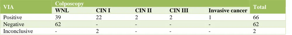
Table 7: Correlation of VIA reporting with Colposcopy findings.
VIA Colposcopy Total
WNL CIN I CIN II CIN III Invasive cancer
Positive 39 22 2 2 1 66
Negative 62 - - - - 62
Inconclusive - 2 - - - 2
14 biopsy proven cases were detected by VIA and the 2 which were not detected had an inconclusive VIA test. Comparing the results of VIA and Pap smear, the
positive predictive value of pap smear is greater than VIA. The negative predictive value for VIA is 99.40% Vs 98.15% for Pap smear.
[image:4.595.52.550.163.264.2]VIA is more sensitive and pap smear more specific in screening of cancer cervix sensitivity ratio of VIA with pap smear is 1.55%.
Table 8: Correlation of VIA and histopathology report (biopsy).
HPR VIA Total
Positive Negative Inconclusive
Chronic cervicitis with atypical cells 2 - - 2
Mild dysplasia 5 - 2 7
Moderate dysplasia 4 - - 4
Severe dysplasia 2 - - 2
Invasive cancer 1 - - 1
Total 14 2 16
Table 9: Comparison of VIA with cytology according to sensitivity, specificity and positive predictive value.
Test Sensitivity Specificity Positive predictive Negative predictive
VIA 87.5% 56.84% 21.21% 99.40%
Pap smear 56.25% 96.61% 40.90% 93.15%
DISCUSSION
In present study, mean age of population was 38.2 years correlating with the study of Saleh HS et al (mean age 35.1 years).8
In present study, 75.5% population were illiterate correlating with the study of Saleh HS et al (60%).8
The main complaints in this study were white discharge and pain in lower abdomen. Saleh HS et al found in their study chief complaints were white discharge in 61.5%
and lower abdominal pain (14%).8
In current study, on per speculum examination cervical ectropion and hypertrophy of cervix were main abnormalities of cervix correlation with study done by Saleh HS et al.8
983.3% of cases positive on colposcocy were detected by VIA testing in our study. Study done by Saleh HS et al showed sensitivity of VIA 91.3% which is correlating with our study.8
In present study, positive predictive value of VIA is less than pap's smear (21.21%, 40.90%) while negative predictive value of these tests were comparable (99.4%, 93.15%). Similar results were found in study done by Saleh HS et al (Positive predictive value - 40.11%, 75.12% and negative predictive value - 98.05%, 97.09%).8 Sensitivity and specificity of VIA in our study
was 87.5% and 56.8%. Similar results were found in study done by Goel A et al (sensitivity - 96% and
specificity - 36%) and Cronje et al(sensitivity - 79% and specificity - 49%).9,10
CONCLUSION
VIA is an attractive test in low resource settings like India. It is simple, inexpensive, low technology test that requires minimal infrastructure for use. It is comparable to cytology in detecting low as well as high grade lesions in terms of sensitivity, specificity and positive predictive value. It is a real-time test in the sense that the results are available immediately, making it possible to institute further diagnostic investigations for test positive women, as well as plan and offer treatment during the same visit.
Funding: No funding sources Conflict of interest: None declared Ethical approval: Not required
REFERENCES
1. Schiffman MH, Brinton LA. The epidemiology of
cervical carcinogenesis. Cancer. 1995;76(S10):1888-901.
2. Seung KIM. Role of colposcopy and cervicography
in screening and management of precancerous lesions of uterine cervix. J Obstet Gynecol India. 2000;50:139-144.
3. Hakama M, Miller AB, Day NE. Screening for
[image:4.595.52.544.294.331.2]4. Lunt R. Worldwide early detection of cervical cancer. Obstet Gynaecol. 1984;63:708-13.
5. Sankaranarayanan R, Pisani P. Prevention measures in the third world: are they practical? In Franco E, Monsonega J.(eds) New developments in cervical cancer screening and prevention. Oxford Blackwell. Science. 1997;70:83.
6. Gaffikin L, Bluementhal PD, Davis C, Birchin SJG (eds). Alternatives for cervical cancer screening and treatment in low resource settings: workshop proceedings: Baltimore: JHPIEGO;1997.
7. Stensword J, Eddy D, Luthra U, Stanley K. Plotting a
new course for cervical cancer screening in
developing countries. World Health Forum
1987;8:42-5.
8. Saleh HS, El Hameid AAA, Mowafy HE, Sherif HE,
Abdelsalam WA.) Visual inspection of the cervix
with (acetic acid or Lugol’s iodine) for cervical cancer screening. Cervical Cancer. 2016;1(1):111.
9. Goel A, Gandhi G, Batra S, Bhambhani S, Zutshi V.
Visual inspectino of the cervix with acetic acid for cervical intraepithelial lesions. Int J Gynaecol Obstet. 2005;88:25-30.
10. Chronje HS, Parham GP, Cooreman BF, de Beer A,
Divall P, Bam RH. A comparison of four screening methods for cervical neoplasia in a developing country. Am J Obstet Gynecol. 2003;188:395-400.