Original Research Article
Study of serum calcium and vitamin D levels with hormonal profile
along with biochemical profile in women with
polycystic ovary syndrome
Ilangovan Subashree*, Umakant Ramchandra Valvekar, Geetha Prasad
INTRODUCTION
It is showed that, insulin resistance is responsible in developing polycystic ovaries in PCOS women though obesity is said to be the major cause.1 Hence pathogenic
determinants of PCOS include insulin resistance and obesity. Therefore, women with PCOS had an increased
risk for type 2 diabetes. The sequel of PCOS reach beyond reproductive health, as women affected with PCOS at long-term are at increased risk of adverse lipid profiles, type II diabetes and hypertension, as well as cardiovascular or cerebrovascular morbidity.1 Vitamin D
deficiency might be a causal factor in the pathogenesis of insulin resistance and the metabolic syndrome in
ABSTRACT
Background: The polycystic ovary syndrome (PCOS) is one of the commonest human endocrinopathies and is increasingly recognized as a variant of the metabolic syndrome in women with the characteristic features of insulin resistance, central obesity, impaired glucose metabolism, dyslipidemia, and hypertension.
Methods: This study is mainly focused on study of parameters like gonadotropin hormonal profile, serum vitamin D and calcium levels in polycystic ovary disease (PCOD). The study comprised 45 clinically proven polycystic ovary disease patients in the age range of 19-34 years. The biochemical estimations carried out in the study were – Fasting Blood sugar, LH, FSH, prolactin, 25- OH vitamin D and calcium along with anthropometric data. The values obtained were compared with age matched equal number of healthy control female subjects from the same population.
Results: The serum concentration of calcium and vitamin D levels are decreased significantly (P <0.001) when compared to controls. Insulin resistance is predominantly seen in PCOS subjects. The study outlines the importance of insulin resistance, dyslipidemia, decreased serum calcium and vitamin D levels in PCOS subjects may be a cause for the progression of polycystic ovary syndrome.
Conclusions: In the present study vitamin D deficiency is highly prevalent in PCOS women from this area compared to control women. We also relations of vitamin D status with insulin sensitivity, HDL-C, and C-reactive protein in PCOS patients, which support the increasing evidence that vitamin D deficiency is associated with multiple metabolic risk factors in PCOS women. A high prevalence of vitamin D deficiency and low calcium levels were observed in PCOS women from our population when compared to controls. Insulin resistance was predominantly seen in PCOS subjects when compared with controls, indicating the association of vitamin D levels with insulin resistance.
Keywords: 25-OH vitamin D, Gonadotropin hormones, Hypovitaminosis, Polycystic ovary syndrome, Serum calcium
Department of Obstetrics and Gynecology, Karpaga Vinayaga Institute of Medical Sciences and Research Centre, Madhurantagam, Tamil Nadu, India
Received: 11 July 2017
Accepted: 05 August 2017
*Correspondence:
Dr.Ilangovan Subashree,
E-mail: subashreeobg@gmail.com
Copyright: © the author(s), publisher and licensee Medip Academy. This is an open-access article distributed under the terms of the Creative Commons Attribution Non-Commercial License, which permits unrestricted non-commercial use, distribution, and reproduction in any medium, provided the original work is properly cited.
PCOS.2,3 In obese people vitamin D is deposited in
adipose tissues, making it unavailable for the body to use. As a result, obese people are expected to have low levels of serum vitamin D. Vitamin D deficiency is associated with multiple health conditions such as diabetes, cardiovascular diseases including stroke, depression, dementia and other conditions.4
It is shown that the relationship between vitamin D and insulin resistance is compensated by various mechanisms; for example, low serum vitamin D levels by calcium mobilization due to elevated parathyroid hormone (PTH) secretion.5,6 The cellular effect of vitamin D is mediated
through the intra-nuclear vitamin D receptor (VDR). VDR is expressed in variety of tissues other than the skeleton, including intestines, parathyroid glands, immune cells, pancreas, and more recently the hypothalamic pituitary axis and reproductive tract. The presence of VDR in the ovary, uterus, placenta and testis suggest a regulatory role of vitamin D in reproductive physiology vitamin D deficiency is hypnotized to contribute to a spectrum of gynecological disorders of which PCOS appears the best studied.5-7
A small number of observational studies identify the inverse association of serum 25-OH vitamin D and with insulin resistance, features of hyperandrogenism in women and circulating androgens in women with PCOS. Serum levels of vitamin D are reported to predict ovarian response in women undergoing ovulation induction with clomiphene citrate.5,6,8 This was independent of the BMI.
Low levels of vitamin D were found to be associated with lower rate of follicular development and pregnancy after ovarian stimulation with 50 mg clomid.5,9
Several data converge towards beneficial effect of vitamin D in metabolic disturbances in women with PCOS. Recent studies supporting the contribution of vitamin D deficiency to metabolic disturbances in women with PCOS, including insulin resistance (IR), obesity, hypertension and menstrual dysfunction have been done in western, European and Middle East countries.4-9 It is
shown that the relationship between vitamin D and insulin resistance is compensated by various mechanisms; for example, low serum vitamin D levels by calcium mobilization due to elevated parathyroid hormone (PTH) secretion.6
Recent studies supporting the contribution of vitamin D deficiency to metabolic disturbances in women with PCOS, including insulin resistance (IR), obesity, hypertensionand menstrual dysfunctionhave been done in western, European and Middle East countries.3,6-13
Only few studies have been done so far in India to confirm the relationship of vitamin D with insulin resistance. Therefore, our aim of this study was to evaluate the association of vitamin D deficiency along with glucose, insulin and insulin resistance in Indian women with polycystic ovary syndrome.
METHODS
The study was conducted at Karpaga Vinayaga Institute
of Medical Sciences and Research Centre,
Madhurnatagam, Tamil Nadu, India. The study comprises of 45 women with PCOS and equal number of age matched normal females without any present or previous history of PCOS were selected to serve as controls. PCOS patients with age range 19 to 34 years were selected for the study. All women with PCOS were come as out patients to Gynecology and Obstetrics department of Karpaga Vinayaga Institute of Medical Sciences and Research Centre, Madhurnatagam, Tamil Nadu.
Women of the control group were healthy volunteers. All the subjects were enrolled in the study after being explained the purpose of the study by concerned physician and informed consent was obtained from them. The research study was approved by institutional ethical committee. PCOS diagnosis was done on the basis of the Rotterdam criteria.13 According to this criterion, PCOS
was defined by the presence of menstrual dysfunction i.e. oligomenorrhea (fewer than six menstrual periods in the preceding year) or amenorrhea (absence of periods for more than 6 months), and clinical hyperandrogenism (i.e.
hirsutism: Ferriman-Gallwey score >6) and/or
hyperandrogenemia. Furthermore, patients with any other cause of oligomenorrhea or hyperandrogenism, such as nonclassic congenital adrenal hyperplasia, androgen
secreting tumours, Cushing’s syndrome, or
hyperprolactinaemia were excluded. Patients with congenital adrenal hyperplasia, Cushing’s syndrome, hyperprolactinemia, androgen-secreting tumors and intake of any medication affecting endocrinal parameters were excluded from the study. PCOS women who were pregnant or on any contraceptive pills or oral hypoglycemic agents were also excluded from the study.
About 5 ml of blood is collected from the antecubital vein. Fasting blood samples were collected in plain, sodium fluoride and heparin tubes. Serum is separated by centrifugation. Blood samples are centrifuged at 3500 rpm for 10 min to separate serum. Serum glucose, serum total cholesterol, triglycerides, HDL, and calcium levels were analyzed using Biosystems reagent kits available for semiautomated biochemistry analyzer.
Hormones Insulin, LH, FSH, Prolactin, were estimated by enzyme linked immuno fluorescent assay (ELFA) method using minividas (Germany) fully automated hormone analyzer. For adequate quality control both normal, abnormal reference control serum solutions and calibrators were run before analyzing each test sample. Other factors affecting the results like proper functioning of instrument, glassware, cuvettes and distilled water are thoroughly checked before using.
RESULTS
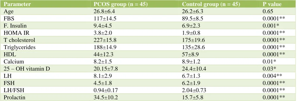
Comparison of biochemical and hormonal parameters of women with PCOS and controls was given in Table 1. All the subjects included in the study were assessed for fasting blood sugar, fasting insulin, calcium, phosphorous and Vitamin D.
Comparisons were made between the two groups (controls and PCOS women). The biochemical findings made during the course of the study are represented in
Table 1. Mean age of the control women was 26.2±6.3 and that of PCOS patients was 26.8±6.3 (p = 0.65).
[image:3.595.51.546.252.421.2]There was no significant statistical difference with respect age of the PCOS women and control women. In the present study fasting glucose showed a significant increase (p ˂0.0001) in PCOS women compared to controls. Calcium and 25 – OH vitamin D levels were decreased in PCOS women (p <0.01; and p <0.03 respectively) when compared to control women (Table 1).
Table 1: Comparison of biochemical and hormonal parameters in PCOS women and healthy controls.
Parameter PCOS group (n = 45) Control group (n = 45) P value
Age 26.8±6.4 26.2±6.3 0.65
FBS 117±14.5 89.5±8.5 0.0001**
F. Insulin 9.4±4.5 6.9±2.3 0.001*
HOMA IR 3.8±2.0 1.9±0.8 0.0001**
T cholesterol 227±15.8 175±19.6 0.0001**
Triglycerides 188±14.9 135±28.6 0.0001**
HDL 44±12.3 57±8.9 0.0001**
Calcium 8.2±1.5 8.9±1.2 0.01*
25 – OH vitamin D 20.15±7.8 24.4±10.4 0.03*
LH 8.1±2.9 6.7±1.3 0.004**
FSH 4.5±1.8 6.2±1.9 0.0001**
LH/FSH 0.94±0.17 2.04±0.73 0.0001**
Prolactin 34.5±10.2 15.7±5.8 0.0001**
[image:3.595.53.543.473.572.2]FBS – Fasting Blood Sugar; F. Insulin – Fasting insulin; HDL –High Density Lipoprotein; LH – Leutinizing Hormone; FSH – Follicle Stimulating Hormone. *, **p value is less than 0.05 which is statistically significant.
Table 2: Correlation study for biochemical and hormonal parameters among PCOS women.
F. Glucose F. Insulin HOMA IR LH FSH LH/FSH Prolactin
F. Glucose 1 0.703** 0.811** 0.256** -0.533** 0.615** 0.699**
F. Insulin 1 0.981** 0.021 -0.410** 0.409** 0.542**
HOMA IR 1 0.049 -0.469** 0.466** 0.603**
LH 1 0.244** 0.556** 0.089
FSH 1 -0.571** -0.481**
LH/FSH 1 0.469**
Prolactin 1
It is evident from Table 1 that total cholesterol, triglycerides, HDL showed a highly significant increase (p ˂0.001) and decrease in HDL (p < 0.05) in cases compared to controls indicating that PCOS women had dyslipidemia. Fasting glucose, fasting insulin and insulin resistance showed a significant increase (p ˂0.001) in cases compared to controls. Gonadotropin hormones LH and FSH showed mild increase (p <0.0001) in PCOS women when compared to healthy controls.
Correlation study (Table 2) revealed significant positive correlation of fasting glucose and fasting insulin with insulin resistance (HOMA IR) (r = 0. 811 and r = 0.981 p ˂0.01) and significant negative correlation with FSH (r=
-0.533 and r = -0.41, p ˂0.01). Fasting glucose showed positive correlation with LH/FSH ratio and prolactin as well (r = 0.615; r = 0.699; respectively and p < 0.01). LH showed positive correlation with LH/FSH ratio (r = 0.556; p<0.01). FSH showed negative correlation with LH/FSH ratio (r = -0.571; p <0.01). LH/FSH ration showed positive correlation with prolactin (r = 0.469; p <0.01).
DISCUSSION
ovary on ultrasonography, which affects 4 to 16% of women in reproductive age.15-17 It is frequently associated
with insulin resistance and compensatory
hyperinsulinemia. Most common abnormalities seen in PCOS are increased BMI, low HDL levels and elevated triglycerides.16 In the present study abnormal lipid profile
was found in women with Polycystic Ovary Syndrome. The findings of elevated TC, TG and LDL-C were in agreement with those of Mattson et al, Wild et al and Talbott et al, Saha S et al.17-20 PCOS may represent a
major segment of the female population at a risk of cardiovascular disease which may be related to increased VLDL levels.21 As shown by other research workers, this
increase in VLDL is basically due to insulin resistance.21
Insulin normally inhibits the expression of microsomal triglyceride that is responsible for apo-B and VLDL secretion. Hence, insulin resistance may be responsible for increased VLDL in PCOS individuals.22 In the present
study we observe 70% of PCOS patients exhibiting an abnormal lipid profile and the mean values of cholesterol, TGL, HDL, LDL and, VLDL are increased (Table 1).
The mechanism by which Vitamin D affects insulin secretion is not known, but one of the propositions is activation of human insulin gene transcription.6-9 It is not
surprising to see Vitamin D to affect insulin metabolism as it regulates calcium metabolism and calcium plays an essential role in insulin secretion.14-17 Vitamin D has
many diverse actions, its role as an immunomodulatory molecule may provoke an inflammatory response in the presence of Vitamin D deficiency and inflammation is known to predispose to IR.19,21
In this study, we demonstrated that women with PCOS had low serum vitamin D levels when compared to age matched controls. Previous studies also demonstrated lower levels of Vitamin Dor no difference in women with PCOS when compared to age matched controls.17,22,8,20 In
accordance with previous reports, we detected Vitamin D levels that correlated negatively with insulin levels in
women with PCOS.14,15
The present study includes 45 PCOS patients and 45 controls. Cut off value of HOMA IR is taken as >2.5.7,13,22-26 In the present study serum insulin and
HOMA IR in PCOS patients is increased when compared with controls and is highly significant (P < 0.001) and this is in accordance with the previous studies.15-19,22,27 In
the present study the mean values of cholesterol, TGL, LDL and, VLDL are increased, whereas mean value of HDL was decreased.
Insulin resistance, hyper insulinemia are the factors to play an important role in the pathogenesis of PCOS. In the present study, our results showed that there is
predominant insulin resistance, hypothyroidism,
dyslipidemia, and increased LH/FSH ratio in women with POCS patients when compared with control women.23-26
The direct effect that testosterone plays on adipocytes was investigated and induction of androgen receptor
mediated insulin resistance via testosterone was established.27 The hyperandrogenism is due to increased
LH and low to normal FSH levels. Due to the increase in LH and hence estrogen, FSH is negatively inhibited.
Theca cell hyperplasia ensues leading to
hyperandrogenemia that clinically presents as hirsutism. In our study, hirsutism is taken as one of the clinical features to diagnose hyperandrogenism. Currently, BMI has been negatively associated with the baseline levels of LH, LH pulse amplitude, and the pituitary response to
GnRH.27,28 The negative correlation between BMI,
baseline levels of LH in PCOS patients, observed in the present study, supports studies published before.27,28
In our study, we observed mild increase the prolactin level (Table 1) in 70% cases, which is similar to the previous studies, Kalsum and Jalali, where 69.51% of subfertile women suffered from hyperprolactinaemia.29
Nizam et al. also showed that hyperprolactinaemia is a major cause of subfertility, and treatment with drugs which lowers prolactin level showed that 24% infertile women became pregnant.30 This finding is consistent
with our study.
CONCLUSION
This study showed that there is predominant insulin resistance, dyslipidaemia, and increased incidence of hypothyroidism in case of PCOS women, which appears to place them at a higher risk of developing diabetes as well as cardiovascular diseases. Normal gonadotropin-ovarian axis is disturbed in PCOS women. This is reflected by the higher levels of LH, lower FSH levels and elevated level of LH: FSH ratio. The ratio of LH/FSH greater than 2.0 can be the significant in the diagnosis of PCOS women in our population. We have also observed that the mean LH levels elevated significantly, mild hyperprolactinemia and decrease in FSH, suggesting the importance of regulation of these hormones in the management and treatment of PCOS.
This study concludes that women with PCOS show high prevalence of insulin resistance and low 25-OH vitamin D levels which in turn may be responsible for the metabolic and endocrine disturbances. Hence, we suggest vitamin D supplementation might improve menstrual frequency and metabolic disturbances in women with polycystic syndrome. The present study also indicates that insulin resistance was an independent risk factor for the presence of vitamin D deficiency in women with PCOS. Further, we recommended long term follow up studies to identify the role vitamin D supplementation in patients with PCOS to confirm the beneficial role of vitamin D.
Funding: No funding sources Conflict of interest: None declared
REFERENCES
1. Kumar AN, Naidu JN, Satyanarayana U,
Ramalingam K, Anitha M. Metabolic and endocrine characteristics of Indian women with polycystic ovary syndrome. Int J Fertil Steril. 2016;10(1):22-8.
2. Hahn S, Haselhorst U, Tan S, Quadbeck B, Schmidt
M, Roesler S, et al. Low serum 25-hydroxyvitamin D concentrations are associated with insulin resistance and obesity in women with polycystic ovary
syndrome. Exp Clin Endocrinol Diabetes.
2006;114:577-83.
3. Da Silva Feuser CS, Barbosa JS, da Silva EB, de Medeiros SF. Current insights into gonadotropic pituitary function in the polycystic ovary syndrome. Asia Pac J Reprod. 2014;3(1):64-70.
4. Dunaif A, Segal KR, Futterweit W, Dobrjansky A. Profound peripheral insulin resistance, independent of obesity, in the polycystic ovary syndrome. Diabetes. 1989;38(9):1165-74.
5. Holick MF. Vitamin D deficiency. N Engl J Med
2007;357:266-81.
6. Li HW, Brereton RE, Anderson RA. Vitamin D
deficiency is common and associated with metabolic risk factors in patients with polycystic ovary syndrome. Metab Clin Exp. 2011;60:1475-81.
7. Holick MF. Vitamin D: importance in the prevention
of cancers, type 1 diabetes, heart disease, and osteoporosis. Am J Clin Nutr. 2004;79:362-71.
8. Naidu JN, Swapna GN, Kumar AN, Krishnamma M,
Anitha M. Importance of elevated insulin resistance, dyslipidemia and status of antioxidant vitamins in polycystic ovary disease. Free Radicals and Antioxidants. 2013;3(1):17-9.
9. Kumar AN, Naidu JN, Stayanarayanac U, Anitha M.
Past, Present and Future of Insulin Gene and Its Related Genes In Relation To Polycystic Ovary Syndrome. J Mol Genet Med. 2014;8:107.
10. Yildizhan R, Kurdoglu M, Adali E. Serum
25-hydroxyvitamin D concentrations in obese and non-obese women with polycystic ovary syndrome. Arch Gynecol Obstet. 2009;280:559-63.
11. Lamberg BA. Glucose metabolism in thyroid
disease. Acta Med Scand. 1965;178:351-62
12. Kumar AN, Satyanarayana U, Naidu JN,
Ramalingam K, Anitha M. Comparison of Lipid Profile, Thyroid Profile, Glycaemic Status, Sex Hormonal Levels, 25-OH Vitamin D and Oxidative Stress Status in Obese and Non Obese Women with Polycystic Ovary Syndrome before and after Treatment with Metformin. J Pharm Biomed Sci. 2015;05(07):572-82.
13. The Rotterdam ESHRE/ASRM-Sponsored
Consensus Workshop Group. Revised 2003
consensus on diagnostic criteria and long-term health risks related to polycystic ovary syndrome (PCOS). Hum Reprod. 2004;19(1):41-7.
14. Wehr E, Trummer O, Giuliani A. Vitamin D
associated polymorphisms are related to insulin
resistance and vitamin D deficiency in PCOS. Eur J Endocrinol. 2011;164:741-9.
15. Kumar AN, Satyanarayana U, Naidu JN,
Ramalingam K, Anitha M. Vitamin D receptor gene polymorphism in obese and non-obese Indian women with polycystic ovary syndrome. Int J Current Res. 2015;7(8):19686-91.
16. Shoelson SE, Herrero L, Naaz A. Obesity,
inflammation, and insulin resistance. Gastroenterol. 2007;132:2169-80.
17. Mattson L, Culberg G, Hamberger L, Samsioe G,
Silfverstolpe G. Lipid metabolism in women with polycystic ovary syndrome: possible implications for an increased risk of coronary heart disease. Fertil Steril. 1984;42:579-84.
18. Wild R, Painter P, Coulson P, Carruth K, Ranney G.
Lipoprotein lipid concentrations and cardiovascular risk in women with polycystic ovary syndrome. J Clin Endocrinol Metab. 1985;61:946-51.
19. Talbott E, Clerici A, Berga SL, Kuller L, Guzick D,
Detre K, et al. Adverse lipid and coronary heart disease risk profiles in young women with polycystic ovary syndrome: results of a case-control study. J Clin Epidemiol. 1998;51:415-22.
20. Saha S, Sarkar C, Biswas SC, Karim R. Correlation between serum lipid profile and carotid intima-media thickness in polycystic ovarian syndrome. Indian J Clinical Biochem. 2008;23(3):262-6.
21. Maestro B, Molero S, Bajo S. Transcriptional
activation of the human insulin receptor gene by 1,25-dihydroxyvitamin D3. Cell Biochem Funct. 2002;20:227-32.
22. Mahmoudi T, Gourabi H, Ashrafi M. Calciotropic
hormones, insulin resistance, and the polycystic ovary syndrome. Fertil Steril. 2010;93:1208-14.
23. Milner RD, Hales CN. The role of calcium and
magnesium in insulin secretion from rabbit pancreas studied in vitro. Diabetologia. 1967;3:47-9.
24. Kayaniyil S, Vieth R, Harris SB. Association of 25(OH)D and PTH with metabolic syndrome and its traditional and nontraditional components. J Clin Endocrinol Metab. 2011;96:168-75.
25. Maestro B, Davila N, Carranza MC, Calle C.
Identification of a vitamin D response element in the human insulin receptor gene promoter. J Steroid Biochem Mol Biol. 2003;84:223-30.
26. Wehr E, Pilz S, Schweighofer N, Giuliani A, Kopera
D, Pieber T, et al. Association of hypovitaminosis D with metabolic disturbances in polycystic ovary syndrome. European J Endocrinol. 2009;161:575-82.
27. Srouji SS, Pagan YL, D Amato F, Dabela A, Jimenez
Y, Supko JG, et al. Pharmacokinetic factors contribute to the inverse relationship between luteinizing hormone and body mass index in polycystic ovarian syndrome. J Clin Endocrinol Metab. 2007;92(4):1347-52.
29. Kalsum A, Jalali S. Role of hyperprolactinemia in fertility. Pak J Med Res. 2002;41:1-5.
30. Nizam K, Memon N, Devrajani BR. Outcome of
treatment with lisuride in hyperprolactinemic infertile women. J Liaquat Univ Med Hlth Sci. 2008;7(2):120-3.
Cite this article as: Subashree I, Valvekar UR,
Prasad G.Study of serum calcium and vitamin D
levels with hormonal profile along with biochemical profile in women with polycystic ovary syndrome. Int J Reprod Contracept Obstet Gynecol