Original Research Article
Risk factors of thrombocytopenia in pregnancy
Ayisha Begam, Sujatha TL, Bindu Nambisan*, Vasanthakumari KP
INTRODUCTION
Thrombocytopenia in pregnancy, may result from several etiologies which include those that are unique to pregnancy and others which may occur in non pregnant settings.1 They include gestational thrombocytopenia,
hypertensive disorders of pregnancy like preeclampsia, eclampsia and HELLP syndrome, liver diseases including acute fatty liver of pregnancy, drug induced thrombocytopenia, immune thrombocytopenia, thrombocytopenia due to viral infections, SLE, APLA syndromes. Awareness of these causes, facilitates proper diagnosis and management of thrombocytopenia in the
pregnant women. Normal pregnancy is associated with a physiologic fall in the platelet count. The reason for this decline is not known, although it has been speculated that these changes may reflect dilution, decreased platelet production, or increased platelet turnover during pregnancy.2 The fall in the platelet in some pregnant
women results in platelet counts that fall into the thrombocytopenic range.3
Thrombocytopenia is classically defined as a platelet count of less than 150×109 /L.4 Counts to 100 to 150× 109
/L are considered mildly depressed, 50 to 100×109 /L are
moderately depressed and less than 50 × 109 /L are
Department of Obstetrics and Gynecology, Government Medical College, Trivandrum, Kerala, India
Received: 12 December 2016
Accepted: 06 January 2017
*Correspondence:
Dr. Bindu Nambisan,
E-mail: bindu.nambisan1971@gmail.com
Copyright: © the author(s), publisher and licensee Medip Academy. This is an open-access article distributed under the terms of the Creative Commons Attribution Non-Commercial License, which permits unrestricted non-commercial use, distribution, and reproduction in any medium, provided the original work is properly cited.
ABSTRACT
Background: Thrombocytopenia in pregnancy occurs due to several etiologies which include both pregnancy specific and non pregnancy related causes. It is second only to anemia as the most common hematological abnormality encountered in pregnancy. Better antenatal care has led to increased detection. Once diagnosed, it is important to further evaluate and to determine the cause to optimize management. Several studies have been undertaken across the world; however there are very few studies in Kerala hence this study. The aim of this study was to determine the risk factors of thrombocytopenia in pregnancy.
Methods: This was a case control study undertaken in the Department of Obstetrics and Gynecology, Medical college Hospital, Trivandrum, Kerala. Using purposive sampling, a sample size of 96 cases and 96 controls were included in this study. “Cases” were antenatal women with thrombocytopenia and “controls” were consecutive women without it. Study period was 18 months and analysis was done using SPSS version 22.
Results: In 49% of subjects ,the cause was identified as gestational thrombocytopenia,39.5% cases were due to hypertensive disorders of pregnancy.10.4% was due to ITP. SLE, AFLP, Dengue infection, HUS and APLA were rare causes of thrombocytopenia in our hospital. Of the 96 cases enrolled in the study,88 were diagnosed during pregnancy. Amongst the hypertensive disorders,16.7% was due to gestational hypertension,10.4% due to preeclampsia and 7.4% were due to HELLP syndrome.82.3% of patients with thrombocytopenia in this study were asymptomatic.
Conclusions: Thrombocytopenia should be evaluated by making a practice of routinely checking the platelet count and peripheral smear in early pregnancy and also in third trimester to enable early diagnosis since most cases may be asymptomatic.
severely depressed.5 Thrombocytopenia is second only to
anemia as the most common hematologic abnormality during pregnancy.6 The overall incidence of
thrombocytopenia in pregnancy is 8%, but when patients with obstetric or medical conditions are excluded, the incidence drops to 5.1%.6 The most common cause is
gestational thrombocytopenia which accounts for around 70% cases. Hypertensive disorders account for 21% which includes preeclampsia, eclampsia, and HELLP syndrome. Immune-mediated thrombocytopenia, including idiopathic thrombocytopenic purpura is responsible for 4.1% of cases, which is a proportionally small number. These conditions, however, can cause considerable morbidity and mortality, and they must be managed closely. Thrombocytopenia can have a wide range of prognostic implications, from completely benign to life threatening.7
Gestational thrombocytopenia, which is seen in about 5% pregnancies, is a benign phenomenon. In this condition however platelet counts usually remain above 100×109/L,
but can fall to 70×109 /L. This condition usually occurs in
the late second or third trimesters of pregnancy. The aetiology may be due to increased peripheral consumption of platelets. An immune mechanism is also postulated. It is usually a diagnosis of exclusion. It usually requires routine prenatal care and monitoring of platelet count every 4 weeks. Mode of delivery is as per maternal and fetal indications. Epidural anaesthesia is safe at counts above one lakh. Recurrence of gestational thrombocytopenia in subsequent pregnancies is common and patient should be properly counselled about this. It has features overlapping ITP. History of thrombocytopenia before 28 weeks and a platelet count less than 50×109/L is highly suggestive of ITP.
Gestational thrombocytopenia resolves spontaneously after delivery within 6 weeks postpartum. In ITP most often there is a history of bleeding outside pregnancy and low platelet counts prior to pregnancy. Management in ITP is directed towards raising platelet counts to prevent spontaneous bleeding and ensure safe delivery. Treatment of ITP in pregnancy is mainly steroids and IVIG though the response is lesser in pregnant than non-pregnant patients. Generally treatment is required only in the third trimester and aim is to keep the values above 50×109 /L.
Due to the increased risk of neonatal thrombocytopenia instrumental delivery should be avoided in cases of maternal ITP.
Immune thrombocytopenia due to production of IgG antiplatelet antibodies against own platelet membrane glycoprotein’s in characterized by, moderate thrombocytopenia, is an early gestation platelet count below one lakh, and increased megakaryocyte level on bone marrow biopsy. Pregnancy does not worsen the course of this condition but cases of severe thrombocytopenia can cause severe morbidity and mortality to the fetus.8 This condition usually requires
treatment with IVIG and steroids. Platelet transfusions
may be less effective in view of ongoing platelet destruction.
Preeclampsia is a common cause of thrombocytopenia in third trimester of pregnancy. The risk of development of thrombocytopenia is about 18% in preeclampsia, 10-12% in DIC and 4-15% in HELLP syndrome.9 The general
approach in these cases would be medical stabilization and expeditious delivery after 34 weeks. If earlier then maternal well being has the priority.
Thrombocytopenia may be related to liver disease and hypersplenism in pregnancy. Most liver disease in pregnancy is attributable to 1 of the 5 liver diseases unique to pregnancy: hyperemesis gravidarum, intrahepatic cholestasis of pregnancy, preeclampsia, HELLP syndrome, and acute fatty liver of pregnancy. Of these disorders, only HELLP, preeclampsia, and acute fatty liver of pregnancy are associated with thrombocytopenia. A recent study showed that 47% of patients had platelet count less than 100×109 /L.
Management of AFLP is immediate termination of pregnancy. Coagulation studies should be optimized by transfusion prior to delivery.
Systemic lupus erythematosus (SLE) predominantly affects women of childbearing age. Disease flares during SLE pregnancy pose challenges with respect to distinguishing physiologic changes related to pregnancy from disease related manifestations. So a multidisciplinary approach with close medical, obstetric, and neonatal monitoring is necessary to optimize both maternal and fetal outcomes.10 Thrombocytopenia less
than 100 × 10 9 /L is one of the diagnostic criteria of SLE.
The thrombocytopenia of SLE rarely becomes severe. If treatment is required in severe thrombocytopenia, patients may respond to hydroxychloroquine, glucocorticoids, or other immunosuppressive medications used for other manifestations of SLE.11 Heparin induced
thrombocytopenia (HIT) occurs in 1% to 5% of patients receiving unfractionated heparin within the previous 5 to 10 days. HIT is an extremely thrombotic process, despite the low platelet count.
Analgesics like aspirin, acetaminophen, antibiotics like penicillin and drugs like heparin, methyldopa, digitalis, and cyclosporine may cause thrombocytopenia. . It is recommended to obtain a baseline platelet count before starting therapy, then repeat platelet counts on Days 7 and 14 of therapy. HIT should be suspected if the platelet count falls by approximately 50% or more. If the repeat platelet counts are normal, no further testing is necessary.
Antiphospholipid antibodies cause obstetric accidents by a thrombocytopenia phenomenon.12
METHODS
This was a case control study conducted in a tertiary referral Medical College Hospital in, Trivandrum, Kerala, India in the Department of Obstetrics and Gynecology.
Study population: Antenatal women attending SAT hospital, Trivandrum.
Sample size: Sample size was calculated using the prevalence noted in study conducted in Israel, where prevalence of HELLP syndrome amongst cases was 18% and in control group was 4.7%.13 Incidence of HELLP in
OBG department in SAT hospital is 1%. The Odds ratio calculated was 4.45%.Using these assumptions sample size was calculated as 90 patients as cases and 90 patients as controls. Sample size was calculated using the formula:
𝒏
= {Z1− ∝
2
√2P̅∗(1 − P̅∗) + Z
1− β√P1∗(1 − P1∗) + P2∗(1 − P2∗)} 2
(P1∗− P2∗)2
Where,
P1∗=
(OR)P2∗
(OR)P2∗+ (1 − P2∗)
P
̅∗=P1∗+ P2∗
2
P1*: Probability of exposure given disease present
P2*: Probability of exposure given disease absent
OR: Anticipated odds ratio
α: Significance level
1-β: Power
Where P1 is 18, P2 is 4.75, Power is 80%, Odd’s ratio is
4.45%, and Alpha error is 5%
Sampling technique: Purposive sampling
Inclusion criteria: Antenatal women attending SAT hospital at any gestational age with platelet count below 150 ×109/L including referred patients.
Exclusion criteria: Those who are not willing to participate in the study and patients with normal platelet count.
Study duration: 18 months
Definition of terms: Cases were defined as antenatal women of any gestational age with platelet count less than 150 ×109/L, mild cases defined as when platelet
count is between 100 and 150 ×109/L, moderate when
platelet count is between 50 and 100 ×109/L, severe when
count is below 50×109/L69.
Controls were defined as consecutive antenatal women of any gestational age with normal platelet count
Data collection tools: Structured questionnaire
Data collection process: After informed consent, pregnant women were interviewed for detailed socioeconomic, demographic, past, personal and family history. Complete blood count was done by using automated cell counters in haematology lab of SAT hospital; same was confirmed by doing peripheral blood smear examination. For referred patients to SAT hospital thrombocytopenia was confirmed in haematology lab by doing complete blood count and peripheral blood smear examination.
Data analysis: Qualitative variables were expressed in frequency distribution. Data analysis was performed using SPSS version 22.Between groups comparison of qualitative variables were analyzed by chi-square test. A p value of 0.05 was taken as the level of significance.
RESULTS
Sociodemograpic characteristics: There were 96 patients in the cases group and 96 in the control group. In this study age of the patient, socioeconomic class and gestational age at presentation did not prove to be statistically significant. So they were not found to be a risk factor of thrombocytopenia.
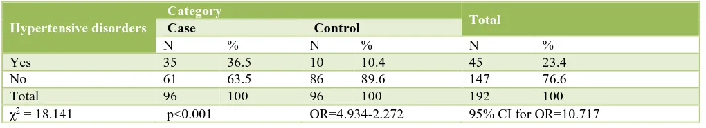
Table 1: Distribution of hypertensive disorders amongst cases and controls.
Hypertensive disorders
Category
Total
Case Control
N % N % N %
Yes 35 36.5 10 10.4 45 23.4
No 61 63.5 86 89.6 147 76.6
Total 96 100 96 100 192 100
Parity: 55 patients were of primiparity and 41 were multiparous amongst “cases” and 60 patients were primi and 36 were multiparous amongst the “controls”. This was not statistically significant and parity was not a risk factor for thrombocytopenia.
Past obstetric history: 22 cases and 21 controls had previous abortions. History of IUD was noted in 5 patients in the control group and none in the case group. There was no history of still birth among patients with thrombocytopenia but 5 patients in the control group had history of still birth. Past obstetric history was not found to be a risk factor for thrombocytopenia.
Table 2: Hypertensive disorders sub classified amongst cases and controls.
Maternal diseases Category Total
Case Control
N % N % N %
None 61 63.5 86 89.6 147 76.6
Gestational HTN 16 16.7 6 6.3 22 11.5
Preeclampsia 10 10.4 1 1 11 5.7
C/C Hypertension 2 2.1 3 3.1 5 2.6
HELLP 7 7.3 0 0 7 3.6
Total 96 100 96 100 192 100
Table 3: Distribution of ITP between cases and controls.
Known case of ITP
Category
Total
Case Control
N % N % N %
Yes 10 10.4 2 2.1 12 6.3
No 86 89.6 94 97.9 180 93.8
Total 96 100 96 100 192 100
χ2 =5.689 p=0.017 OR=5.465 95% CI for OR=1.164-25.648
Table 4: Platelet count at admission in cases.
Presence of hypertensive disorders: Among 96 cases of thrombocytopenia, around 36.5% (n=35) of patient had hypertensive disorder of pregnancy. Among the control group 10.4% of (n=10) had hypertensive disorder of pregnancy. Hence hypertensive disorder of pregnancy was as a risk factor for thrombocytopenia (p<0.001) in this study (Table 1). Of the 35 cases with hypertensive disorders, 16 cases (16.7%) were due to gestational hypertension, 10 cases (10.4%) were due to preeclampsia, 7 cases (7.3%) were due to HELLP, and 2 case (2.2%) were due to chronic hypertension (Table 2). Presence of ITP: There were 10 patients with ITP in the cases group and only 2 patients in the control group. History of ITP
was a significant risk factor of thrombocytopenia in pregnancy (p<0.017) (Table 3).
History of thyroid disorders: 11.5% of cases and 17.7% of control group had history of hypothyroidism which was not a statistically significant risk factor. There were only 2 women with hyperthyroidism among cases.
History of GDM: There were 7 women among cases 2 among the control group who had GDM. Rest was not having this history. Presence of gestational diabetes was not a significant risk factor for thrombocytopenia.
History of hypertension: Past history of hypertension was not a significant risk factor.
Presence of gestational thrombocytopenia: 47 out of 96 women had gestational thrombocytopenia. So this was the commonest cause of thrombocytopenia in this study.
Platelet count at admission: 47.9% of patients had mild thrombocytopenia, 40.6% had moderate thrombocytopenia and 11.5% had severe Platelet count at
admission
Case
N %
<50000 11 11.5
51000-99999 39 40.6
100000-150000 46 47.9
thrombocytopenia. Using peripheral smear examination 51% had mild thrombocytopenia, 33.3% of patients had moderate thrombocytopenia, 9.4% had severe thrombocytopenia and 6.3% had thrombocytopenia with haemolysis. (Table4 and 5)
Treatment modalities: 26 out of 96 cases with thrombocytopenia required some form of treatment. 8 patients needed platelet transfusion, 6 patients needed
oral prednisolone, one needed I/V methyl prednisolone, 3 patients needed both steroid and platelet transfusion. There was one patient who needed parenteral as well as oral prednisolone along with platelet transfusion.
History of heparin intake: 9 patients out of 96 had a history of heparin intake. Among the control group 4 patients had a history of heparin intake. This was not found to be statistically significant risk factor.
Table 5: Platelet counts sub classified according to severity in peripheral smear.
Peripheral smear Category Total
Case Control
N % N % N %
Mild thrombocytopenia 49 51 0 0 49 25.5
Moderate thrombocytopenia 32 33.3 0 0 32 16.7
Severe thrombocytopenia 9 9.4 0 0 9 4.7
Thrombocytopenia and haemolysis 6 6.3 0 0 6 3.1
Not applicable 0 0 93 96.9 93 48.4
Total 96 100 96 100 192 100
History of intake of antibiotics: There is no significant difference between cases and control group when we compare h/o antibiotic intake causing thrombocytopenia (p=0.112).
Other medical disorders: In this study there were 7 cases of APLA, 5 cases of SLE,2 cases of dengue infection and one case each of Rheumatoid arthritis, AFLP and HUS was there which was not a statistically significant risk factor in this study probably due to small numbers.
DISCUSSION
This study sought to determine the risk factors of thrombocytopenia among the antenatal patients attending our hospital.
Although most cases of thrombocytopenia in pregnancy do not result in adverse outcomes, the underlying pathology can sometimes be life threatening so once thrombocytopenia is diagnosed in pregnancy woman should undergo further clinical and laboratory evaluation to determine the cause. Careful analysis of time of onset of thrombocytopenia and clinical manifestations is critical to provide timely diagnosis and appropriate maternal and fetal care.
In our study, gestational thrombocytopenia was the most common cause of thrombocytopenia (49%). Around 17% of cases were due to Preeclampsia-HELLP syndrome and 10.4% of patients had ITP. In a study by Vyas et al in Ahmadabad, of the 4818 patients studied, gestational thrombocytopenia was the commonest aetiology
(44.6%).14 Incidence of thrombocytopenia due to severe
preeclampsia and HELLP syndrome was 22%. Incidence of ITP was 4.4%. These results were consistent with the results of our study. However, in the above mentioned study pregnant women with mild thrombocytopenia were taken as the control group whereas in our study normal pregnant women were taken as the control group. .In another study conducted by Parnat et al in Soroka University Medical Centre, Israel the main causes of thrombocytopenia were gestational thrombocytopenia (GT) (59.3%), immune thrombocytopenic purpura (ITP) (11.05%), preeclampsia (10.05%), and HELLP syndrome (12.06%).13 There was only one case of APLA. Their
study results were consistent with our study findings. However in their study patients with mild thrombocytopenia were not included in the study group.
95% of cases in our study presented in the third trimester and gestational thrombocytopenia (46.5%) was the most common cause in this trimester. There was only 1 case of AFLP. In a study done by Mamatha S et al in M. S. Ramaiah Medical College, Bangalore, 46.6% of cases were due to gestational thrombocytopenia and 46.6% of cases were due to HELLP and preeclampsia and 2 cases were due to AFLP. About 60% of the patients presented in third trimester, closely followed by 40% in second trimester. In this difference could be due to the very small sample size (n=30) in the study done by Mamatha S et al compared to the sample size in our study (n=96).
thrombocytopenia were preeclampsia (16%) and idiopathic thrombocytopenic purpura (ITP) (3%). This study results were consistent with our study findings which shows that gestational thrombocytopenia is the most common cause of thrombocytopenia in pregnancy followed by hypertensive disorder of pregnancy (36.5%).
In this study 51% of patients had mild thrombocytopenia, 33.3% of patient had moderate thrombocytopenia and 9.4% of patient had severe thrombocytopenia. In a study by Katke RD et al, 70.9% patients had moderate, 29.1% patients had severe thrombocytopenia.17 This difference
might be due to the large number of patients included in their study and also could be due to the non-inclusion of patients with mild thrombocytopenia in their study. In their study, 35% cases were primipara, 32% cases were gravida 2, 33% cases were gravida 3 to 5. In our study 57% of cases were primipara and 43% were multipara. As the difference is not significant, parity was not proven as a risk factor for thrombocytopenia according to our study. Gestational thrombocytopenia was the most common etiological factor with 30.1% cases. 27.2% cases were due to hypertensive disorders, 18.4% cases due to Lucknow, gestational thrombocytopenia was seen in 64.2%, followed by thrombocytopenia due to hypertensive disorders of pregnancy. The results of this study were consistent with our study findings.18
In a study done in Ege University in Turkey assessing thrombocytopenia in pregnancy, around 43.5% of thrombocytopenia was due to HELLP syndrome followed by gestational thrombocytopenia (39.5%) and ITP was around 16%.19 Fifty percent of patients were primigravida
and 14% of patients had advanced maternal age. In our study as well as several other studies gestational thrombocytopenia was the most common cause of thrombocytopenia.
All cases of thrombocytopenia require an early detection and appropriate treatment according to the diagnosis. Babies also need to be evaluated for neonatal thrombocytopenia. The risk of neonatal thrombocytopenia in babies born to mothers with gestational thrombocytopenia is considered negligible.
CONCLUSION
Gestational thrombocytopenia is the most common cause of thrombocytopenia (49%). Most of the gestational thrombocytopenia is incidental findings. 82.3% of patients with thrombocytopenia were asymptomatic. Hypertensive disorder of pregnancy were the second
most cause of thrombocytopenia in pregnancy (39.5%), of which 16.7% were due to GHTN,10.4% were due to preeclampsia and 7.4% were due to HELLP syndrome. 10.4% of cases were due to ITP. 51% of patients had mild thrombocytopenia, 33.3% had moderate thrombocytopenia and 9.4% had severe thrombocytopenia. SLE, AFLP, Dengue infection, HUS and APLA are the rare causes of thrombocytopenia in reappraisal of the threshold. Obstet Gynecol. 2000;95:29-33
3. Provan D, Stasi R, Newland AC, et al. International consensus report on the investigation and management of primary immune thrombocytopenia. Blood. 2010;115:168-86.
4. Shehata N, Burrows RF, Kelton JG. Gestational thrombocytopenia. Clin Obstet Gynecol. 1999;42:327-34.
5. Magann EF, Martin JN Jr. Twelve steps to optimal management of HELLP syndrome. Clin Obstet Gynecol. 1999;42(3):532-50.
6. Sullivan CA, Martin JN Jr. Management of the obstetric patient with thrombocytopenia. Clin Obstet Gynecol. 1995;38:521-34.
7. BockenstedtP.et al.Thrombocytopenia in Pregnancy. Hematology/Oncology Clinics of North America. 2011;25(2);293-310.
8. Jeffrey A, Levy CPT, MC, FS, and Lance D. Murphy, MD, Thrombocytopenia in pregnancy. Clinical review JABFP. 2002;15(4):290-7.
9. Lefkou E. Bleeding disorders in pregnancy. Obstetrics, Gynecology and Reproductive Medicine. 2008;18(8):217-23.
10. Bermas BL, Smith NA, Pisetsky DS, Curtis MR. Pregnancy in women with systemic lupus erythematosus.uptodate.com.
11. Adams TM, Allaf MB, Vintzileos AM. Maternal thrombocytopenia in pregnancy. Clinics in Laboratory Medicine. 2013;33(2):327-41.
12. Arvieux J, Hachulla, E. Le syndrome des antiphospholipides. Annales de Cardiologieetd' Angéiologie. 2002;5(3):146-51.
14. Vyas R, Shah S,Yadav P, Patel U. Comparative study of mild versus moderate to severe thrombocytopenia in third trimester of pregnancy in a tertiary care hospital. NHL Journal of Medical Sciences. 2014;3(1).
15. Mamatha S. Thrombocytopenia during pregnancy. Journal of Evolution of Medical and Dental Science. 2010;3(57):12956-60.
16. Salnlo. Maternal thrombocytopenia at term: a population-based study. Acta Obstetriciaet Gynecologica Scandinavica. 2000;79(9).
17. Katke RD. Thrombocytopenia during pregnancy: an institutional based study. Int J Reprod Contracept Obstet Gynecol. 2014;3(4):947-51.
18. Nisha. Prevalence and Characterization of Thrombocytopenia in Pregnancy in Indian Women. Indian J of hematology and blood transfusion. 2012;28(2):77-81.
19. Asmaa M. Thrombocytopenia in Iraqi Pregnant Women. J Fac Med Baghdad. 2011;53(2).