Scholarship@Western
Scholarship@Western
Electronic Thesis and Dissertation Repository
5-24-2012 12:00 AM
Complementary and Self-Complementary Hydrogen Bond Double
Complementary and Self-Complementary Hydrogen Bond Double
Helical Complexes
Helical Complexes
Bhanu P. Mudraboyina
The University of Western Ontario Supervisor
James A. Wisner
The University of Western Ontario Graduate Program in Chemistry
A thesis submitted in partial fulfillment of the requirements for the degree in Doctor of Philosophy
© Bhanu P. Mudraboyina 2012
Follow this and additional works at: https://ir.lib.uwo.ca/etd
Part of the Materials Chemistry Commons, and the Organic Chemistry Commons
Recommended Citation Recommended Citation
Mudraboyina, Bhanu P., "Complementary and Self-Complementary Hydrogen Bond Double Helical Complexes" (2012). Electronic Thesis and Dissertation Repository. 548.
https://ir.lib.uwo.ca/etd/548
This Dissertation/Thesis is brought to you for free and open access by Scholarship@Western. It has been accepted for inclusion in Electronic Thesis and Dissertation Repository by an authorized administrator of
(Spine title: Complementary and Self-Complementary Double Helical Complexes)
(Thesis format: Integrated Article)
by
Bhanu Prakash Mudraboyina
Graduate Program in Chemistry
A thesis submitted in partial fulfillment of the requirements for the degree of
Doctor of Philosophy
The School of Graduate and Postdoctoral Studies The University of Western Ontario
London, Ontario, Canada
ii
THE UNIVERSITY OF WESTERN ONTARIO School of Graduate and Postdoctoral Studies
CERTIFICATE OF EXAMINATION
Supervisor
______________________________ Dr. James A. Wisner
Examiners
______________________________ Dr. Ken Maly
______________________________ Dr. Brian L. Pagenkopf
______________________________ Dr. Leonard G. Luyt
______________________________ Dr. Gerasimos Moschopoulos
The thesis by
Bhanu Prakash Mudraboyina
entitled:
COMPLEMENTARY AND SELF-COMPLEMENTARY HYDROGEN
BONDED DOUBLE HELICAL COMPLEXES
is accepted in partial fulfillment of the requirements for the degree of
Doctor of Philosophy
iii
Abstract
The design of artificial or synthetic strands that self-assemble to form double-helical
complexes have been of great interest to chemists and researchers since the discovery of the
double helical DNA structure in 1953 by Watson and Crick. Most of the complexes were
self-complementary double-helical homodimers and while few heterodimer complexes are
also known. The present thesis describes the design, synthesis and characterization of
complementary and self-complementary hydrogen bond arrays built from heterocycles such
as pyridine, thiazine dioxide and indole connected in different sequences. The
sequence-based stabilities, insolubility issues, substitution and preorganization effects in these arrays
have been studied in detail.
The design and syntheses of four self-complementary oligomers that contain an
underlying AADD hydrogen bond Donor/Acceptor sequence are presented and their
self-association examined in the solution and solid states. Substitution with electron donating and
withdrawing groups and the influence of preorganization had a large effect on the overall
stabilities of the complexes studied. A wide range (>105 M-1) of stabilities were demonstrated
and in the most extreme case, the dimerization constant measured (Kdimer ≥ 4.5 x 10⁷ M-1) is
comparable to the most stable homodimers of neutral coplanar AADD arrays reported to
date.
Two sets of DDD hydrogen bond arrays were synthesized that form triply hydrogen
bonded double helical complexes with an AAA array when combined in CDCl3 solution. In
contrast to the detrimental effect of appended alkyl chain arrays containing tethers between
iv
The effect of introduction of a hexyl chain on the solubility of an originally insoluble
(in CDCl3) DDD array based on three thiazine dioxides was studied. The association
constants measured based on NMR titrations and ITC titrations demonstrate formation of a
highly stable double-helical pair with a Ka value of 1.4 x 10⁵ M-1. A self-complementary
double helical complex based on six hydrogen bond AAADDD array was also synthesized
and displays very strong dimerization (Kdimer > 4.5 x 10⁷ M-1 in CDCl3) examined by NMR
dilution and mixed solvent studies. These findings establish the high potential of the DDD
array and the AAADDD array as monomer components to build supramolecular architectures
or polymers.
Keywords
Complementary, Self-Complementary, Hydrogen Bond, Double Helix, Substituent Effect,
Preorganization, NMR Dilutions, NMR Titrations, Isothermal Titration Calorimetry, X-ray
v
Co-Authorship Statement
Dr. Jiaxin Li and Dr. Hong-Bo Wang have synthesized 4-1 and 3-1a-h, respectively.
Doug Hairsine (manager, mass spectrometry) has collected all the mass spectrometry data.
vi
Acknowledgments
I would like to take this opportunity to thank everyone who has helped me to reach
this point and travel through my graduate program. Firstly, I acknowledge my supervisor
Prof. James A Wisner, for providing me with an opportunity to pursue my doctoral studies,
and his guidance through the years. He has been very supportive during the entire period of
my Ph.D. program, especially through the “not so productive” time and has given me the
freedom to extend my research project in right direction. He patiently demonstrated valuable
lab techniques from simple flash column chromatography to running sensitive titrations on
ITC calorimeter for which I am very grateful. I am fortunate to work with him as he is not
only a great supervisor but also a marvellous being. I have learnt a lot from him regarding
chemistry and the academic world.
I want to thank my examiners, Dr. Ken Maly, Dr. Brian L. Pagenkopf , Dr. Len G.
Luyt and Dr. Gerasimos Moschopoulos for reviewing my thesis work.
I am thankful to all the past and present graduate students of the Wisner lab; Dr.
Barry Blight, Dr. Jiaxin Li, Dr. Hong-Bo Wang, Fatimah Al-garni, Jeff Pleizier, Iamnica
Linares Mendez. Being an international student, I greatly appreciate my lab mates for
providing a friendly atmosphere. Besides passing on the techniques, I have learnt about being
a good team leader and a colleague while supervising few 490 students during the term of my
Ph.D. program. I extend my thanks to all the 490 students; Melissa McDonald, Stephanie
Holborne and Brian Ngo.
I am grateful to Prof. Nicholas Payne for teaching me X-ray crystallography and to
vii
solve our research lab X-ray data. I am thankful to Prof. Stephen Loeb for arranging to
collect one of my crystal data when the X-ray facility at UWO ceased to work for a while.
Special thanks to Dr. Hudson and Dr. Workentein for permissions to use their UV, Mass and
HPLC spectrophotometers.
I would like to extend my appreciation to all the departmental staff members,
especially: Mathew Willans for helping me out with several NMR experiments, Doug
Hairsine for the timely mass spectrums; Yves Rambour for fixing the broken glassware;
Darlene McDonald for her duties as a Graduate Coordinator; Monika Chirigel,
Don Yakobchuk, Marylou Hart and Sherrie McPhee at the chem-bio-stores for their
respective services.
I am indebted to my family; Dad and Ma for giving me the freedom to make my own
decisions and encouraging me to pursue my dreams; brothers Madhukar and Anil for their
loving support. My wife, Preeti has been with me all through my journey as a graduate
student and before. I am thankful for having such a delightful person as best friend and wife,
who‟s constant support has made my life easier and cheerier. Without her, my life would not
be as wonderful. I also take the opportunity to thank my parent in-laws and all my family and
friends in India and abroad for their continuous love, support and understanding. Last but not
least, I thank God for giving me the determination and strength to diligently pursue my goals.
viii
Table of Contents
CERTIFICATE OF EXAMINATION ... ii
Abstract ... iii
Co-Authorship Statement... v
Acknowledgments... vi
Table of Contents...viii
List of Tables ... xiii
List of Figures ... xiv
List of Schemes ... xxii
List of Abbreviations and Symbols... xxiv
Chapter 1 1 Introduction ... 1
1.1 Significance and Occurrence of Hydrogen Bonds ... 2
1.2 Hydrogen Bonding ... 5
1.2.1 Definition ... 5
1.2.2 Characteristics of Hydrogen Bonds………..7
1.2.2.1 Strength of a Single Hydrogen Bond………...7
1.2.2.2 Directionality of Single Hydrogen Bonding………9
1.2.2.3 Specificity of Hydrogen Bonds……….11
1.3 Hydrogen Bonded Complexes ... 12
ix
1.3.1.1 Functional Groups and Substituents………..14
1.3.1.2 Preorganization………..17
1.3.1.3 Tautomers………..20
1.3.1.4 Solubility………....22
1.3.1.5 Fidelity………...25
1.3.1.6 Number of Hydrogen Bonds and Secondary Interactions……….27
1.3.2 Complementary and Self-Complementary Hydrogen Bonded Complexes . ..28
1.3.2.1 Self-Complementary or Homodimer Complexes………..29
1.3.2.1.1 ADAD Complexes………..30
1.3.2.1.2 AADD Complexes………..31
1.3.2.1.3 Six-Membered Self-Complementary Complexes………35
1.3.2.2 Complementary or Heterodimer Complexes……….………36
1.3.2.2.1 AADDDA Complementary Complexes……….36
1.3.2.2.2 AAADDD Complementary Complexes……….37
1.3.2.2.3 ADDADAAD Complementary Complexes………...38
1.3.2.2.4 AAAADDDD Complementary Complexes………...39
1.3.2.2.5 Unusual Complementary Complexes………..41
1.3.3 Double-Helical Complexes ... 42
1.3.3.1 Design of Double-Helical Arrays………..47
1.4 Scope of the Thesis ... 48
1.5 References ... 49
Chapter 2 2 Synthesis and Self-Association of Double Helical AADD Arrays ... 56
x
2.2 AADD Arrays as Components of Hydrogen Bonded Materials ... 58
2.3 Design of Double-Helical AADD Arrays ... 61
2.4 Synthesis of Double Helical AADD Arrays ... 66
2.5 X-ray Analysis of AADD Arrays ... 84
2.6 Solution Characterization of the Dimerization of 2-1a-d ... 95
2.6.1 Analysis of Complex Stability ... 96
2.6.2 1H NMR Studies of 2-1a-d ... 100
2.7 Conclusion ... 107
2.8 Experimental ... 108
General. ... 108
2.8.1 1H NMR Dilution Procedure ... 109
2.8.2 General Synthetic Methods ... 110
2.8.3 Synthetic Methods... 111
2.9 References ... 132
Chapter 3 3 The Effect of Sterics and Preorganization on Stability in Double-Helical AAA-DDD Complexes. ... 140
3.1 Contiguous Arrays for Hydrogen Bonded Complex Formation ... 140
3.2 Results and Discussion ... 145
3.2.1 Design of the donor Arrays ... 145
3.2.2 Synthesis of 3-3a-c Donor Arrays ... 147
3.2.3 Synthesis of Single Trimethylene Tethered DDD Arrays ... 149
3.2.4 Synthesis of Dissymetric Soluble DDD arrays 3-5a-b and 3-6a-b ... 152
xi
3.4 NMR Titration Studies of DDD Arrays ... 160
3.4.1 NMR Titration Studies of DDD Array 3-4b. ... 162
3.4.2 NMR Studies of Soluble DDD Arrays 3-5a,b and 3-6a,b: ... 165
3.5 Conclusion ... 169
3.5.1 Experimental ... 169
General ... 169
3.5.2 1H NMR Titration Procedure ... 170
3.5.3 Synthetic Procedures ... 171
3.6 References ... 183
Chapter 4 4 Synthesis and Binding Studies of a Complementary DDDAAA Complex and a Self-Associated Double-Helical AAADDDDDDAAA Complex ... 187
4.1 Synthesis of Alkylated DDD Array 4-4 ... 190
4.2 NMR Titration Studies of the AAADDD Array... 192
4.3 Synthesis of a Double Helical Self-Complementary AAADDD Array... 196
4.4 Self-Association of the Double-Helical AAADDDDDDAAA Complex ... 200
4.5 Conclusion ... 202
4.6 Experimental ... 203
4.6.1 Isothermal Titration Calorimetry Procedure ... 203
4.6.2 Synthetic Procedures ... 204
4.7 References ... 215
Chapter 5 5 Conclusions ... 219
xii
5.2 References ... 223
Appendices...224
Permission to Reproduce Material from the Literature ... 224
xiii
List of Tables
Chapter 2
Table 2-1 Cyanide reagents and stoichiometries used and yields observed in the cyano-
dehalogenation reaction of 2-3. Reaction conditions as in Figure 2-6…….…....71
Table-2-2 Trials of the bromination of 2-9a using different reaction conditions………….73
Table 2-3 Crystallographic parameters for 2-1a2-1a and 2-1b2-1b crystals………..85
Table 2-4 Crystallographic data for 2-1c2-1c and 2-1d2-1d...86 Table 2-5 Summary of bond distances and angles of 2-1b2-1b, 2-1c2-1c and 2-1d2-1d
from their X-ray crystal structure data………...94
Chapter 3
Table 3-1 Trials of reactions attempted for last step of scheme 3-1………150
Table 3-2 Four sets of complementary AAADDD complexes (3-5a3-2a, 3-5b3-2a, 3-6a
3-2a and 3-6b3-2a) compared with the association constants, free energies, their
differences and net difference in energies with that of 3-1a3-2a and 3-1b3-2a
xiv
Table of Figures
Chapter 1
Figure 1-1 Interaction of enzyme with substrate by a lock and key mechanism to give an
enzyme–substrate complex.. ... 1
Figure 1-2 A cartoon representation of DNA double helical structure with specific base
pairing projected as an inset. Keratin (below) displaying -helix and a-pleated sheet secondary structures………....3
Figure 1-3 A two dimentional array of melamine and cyanuric acid hydrogen bonding
assembly. ... 4
Figure 1-4 Hydrogen bonding between two electronegative heteroatoms X and Y.. ... 5
Figure 1-5 Molecular electrostatic potential surfaces plotted on the van der Waals‟ surface
of the molecule calculated by using AM1. ... 9
Figure 1-6 Hydrogen bonding between the acceptor (A) and the hydrogen of donor (D).. . 10
Figure 1-7 Arrangement of complementary sites leads to specifically attractive or repulsive
interactions as demonstrated using 2-aminopyridine molecules. ... 11
Figure 1-8 Common complementary hydrogen bonding arrays developed that resemble base
pairs. ... 12
Figure 1-9 (i) Hydrogen bonding in an anti-parallel sheet and (ii) Bing Gong‟s hydrogen
bonded complex which resembles the sheet. ... 13
Figure 1-10 AAA-DDD Model system investigated by Boyd and coworkers. ... 15
Figure 1-11 Substituent effects of the heterocyclic based array AAA-DDD complementary
system on the association constants. ... 15
Figure 1-12 Possible tautomeric and conformational configurations of arrays 1-3 and 1-4..16
Figure 1-14 Conformational equilibrium of ethoxynaphthyridine 1-5 and its complex with
xv
Figure 1-15 Imide-urea strands that pair into self-complementary duplexes 1-81-8 via
bifurcated hydrogen bonds.. ... 19
Figure 1-16 The tautomeric and self-association equilibria observed in a solution of 2-
ureido-4-pyrimidone 1-9 ... 21
Figure 1-17 Zimmerman‟s AADD array 1-10a that can only form a tautomeric AADD array
1-10b thereby maximising the association constant values possible for the
complex. ... 22
Figure 1-18 Intermolecular interactions in solution are a competition between solute–
solvent interactions in the free state, and solute–solute and solvent–solvent interactions in the bound state... 22
Figure 1-19 Results of 31P NMR titration experiments displaying the association constant for
formation of a 1:1 complex between perfluoro-tert-butyl alcohol and tri-n-butyl phosphine oxide. ... 24
Figure 1-20DANUG complex formed due to high fidelity between the two arrays. ... 26 Figure 1-21 Secondary interactions shown in four different modules, DADADA,
ADDDAA and DDDAAA motifs ... 28
Figure 1-22 Dimers of acylated diaminotriazine and diaminopyrimidine ADAD
modules………….………...30
Figure 1-23 Diacylpyrimidine 1-11 and ureidoacylpyrimidine 1-12 as ADAD bonding
motifs according to Meijer and co-workers. ... 31
Figure 1-24 Meijer‟s AADD motifs displaying extreme complex stabilities. ... 31
Figure 1-25 Amidinourea-based self-complementary modules with preorganized linear
hydrogen-bonding arrays.. ... 32
Figure 1-26 A highly selective, neutral, fluorescent sensor based on 2-ureido-4[1H]-
xvi
Figure 1-27 Design and self-assembly of general-purpose Bis-DeAP module. ... 33
Figure 1-28 Structure of an AADD array based on ureidoimidazo[1,2-a]pyrimidine UImp-2,
forming a stable dimer. ... 35
Figure 1-29 The design of the self-complementary duplex forming a AADADD-DDADAA
complex in the solution state... 35
Figure 1-30 Assorted complexes containing the AADDDA motif. The Ka values are measured in CDCl3. ... 36
Figure 1-32 Extremely stable complexes of both neutral and ionic types. ... 38
Figure 1-33 Highly stable complexes exhibiting ADDADAAD hydrogen bond arrays. .... 38
Figure 1-34 Formation of the putative heterodimer AAAADDDD with four hydrogen
bonds and six attractive secondary interactions……….39
Figure 1-351H NMR spectra of DDDD+, complex DDDD+AAAA and AAAA ………...40
Figure 1-36 Oligoamide strands containing both benzene and naphthalene spacers sharing
AADD sequences that heterodimerize. ... 42
Figure 1-37 Structure of an oligopyridinecarboxamide and the crystal structures of its single
helix foldamer and double helix dimer. ... 43
Figure 1-38 Schematic illustration of the heteroduplex formation of 9merH with
oligosaccharides ... 44
Figure 1-39 Structures of m-terphenyl-based molecular strands bearing amidine and/or
carboxyl groups. ... 45
Figure 1-40 Fluoro-substituted quinoline oligoamide that forms cross-hybridized double
helical complex.. ... 46
Figure 1-41 A and D subunits form components of a supramolecular “toolbox” which can be
xvii
Chapter 2
Figure 2-1 A plot of the relation between association constant Ka and the degree of
polymerization of idealized monomers at two different concentrations. ... 57
Figure 2-2 Changing the macroscopic properties of a telechelic poly(ethylene/ butylene)
copolymer (left) by end-group modification ... 58
Figure 2-3 Examples of AADD arrays and designs with Kdimer values and supporting solvents at room temperature. ... 59
Figure 2-4 Biomimetic linear modular polymer based on the Upy AADD array, mimicking
titin, a skeletal muscle protein. ... 60
Figure 2-5 Basic design of the arrays with a simple donor (D) and acceptor (A) pair leading
to an oligomeric strand... 62
Figure 2-6 The AADD complex design as a hybrid of alternating ADADA and contiguous
AAADDD sequences. ... 63
Figure 2-7 Design attributes of the AADD array outlined using different colors. ... 64 Figure 2-8 Four different AADD arrays 2-1a-d highlighting the progressive changes made
to the basic design that may lead to increased stability of the complexes.. ... 65
Figure 2-9 1H NMR spectrum of the substitution reaction using Zn(CN)2 as cyanide reagent, displaying resonances corresponding to the two products 2-4 and 2-5 andthe
starting material 2-3.. ... 69
Figure 2-10 1H NMR of the mixture of mono and dibrominated acetyl skatole before (a) and
after (b) washing with ice cold methanol shows considerable reduction of the dibromide contaminant compared to the mono brominated product. ... 80
Figure 2-11 Stick representation of X-ray crystal structure of array 2-1a with intermolecular
xviii
Figure 2-12 Stick representation of the X-ray crystal structure of array 2-1b with
intermolecular (dashed orange lines) hydrogen bonds indicated. ... 88
Figure 2-13 Stick representation of the X-ray crystal structure of dimer 2-1c2-1c with
intermolecular hydrogen bonds indicated (dashed orange lines). ... 90
Figure 2-14 Stick representation of the X-ray crystal structure of dimer 2-1d2-1d with
intermolecular hydrogen bonds indicated (dashed orange lines). ... 92
Figure 2-15 (A) Schematic diagram of ITC; (B) An example of isotherms obtained and
plotting of the isotherm for determination of G, H and S. ... 97
Figure 2-16 1H NMR spectra displaying the concentration dependant behaviour of 2-1a in
CDCl3.. ... 100
Figure 2-17 NMR dilution curve of array 2-1a (following N-Hb) with Kdimer value and free energies calculated from fitting of the data to a 1:1 dimerization model. ... 101
Figure 2-18 Stacked plot of 1H NMR dilutions of 2-1b in CDCl3 at concentrations of 6.0 x
10-3 M, 1.5 x 10⁻³ M, 7.2 x 10⁻⁴ M, 2.8 x 10⁻⁴ M, 8.6 x 10⁻⁵ M ... 102
Figure 2-19 Dilution curve of array 2-1b (following N-Hb) in CDCl3 with Kdimer value and
free energy calculated from fitting of the data to the 1:1 dimerization
model………..………..103
Figure 2-20 Stacked plot of 1H NMR dilutions of 2-1c in CDCl3 at concentrations of 26.0 x 10⁻³ M, 4.2 x 10⁻³ M, 6.5 x 10⁻⁴ M, 1.3 x 10⁻⁴ M, 9.8 x 10⁻⁵ M ... 104
Figure 2-21 Dilution curve of array 2-1c (following N-Hb) with Kdimer value and free energy
calculated from fitting of the data to a 1:1 dimerization model. ... 105
Figure 2-22 (i) 600 MHz 1H NMR spectrum of 2-1d at 2.5 x 10⁻³ M in CDCl3; (ii) and (iii)
xix
Chapter 3
Figure 3-1 Early experimental examples of AA•DDD and AAA•DDD hydrogen bonding
arrays reported by Zimmerman‟s group (left and middle) and Bell and Anslyn
(right). ... 141
Figure 3-2 Two examples of complementary AAAA•DDDD+ hydrogen bonding complexes and their Ka values determined in CDCl3 (left side)5 and CH2Cl2 (right side)………142
Figure 3-3 Averystable complementary double helical AAA•DDD hydrogen bond complex with a Ka value > 105 M-1 in CDCl3 ... 143
Figure 3-4 Complexes 1a-h2a-c displaying theincrease in the association constants by up to a factor of 30 in the cases studied ... 144
Figure 3-5 DDD arrays originally designed for the current study ... 146
Figure 3-6 Synthesis of methyl 2-acetyloctanoate, for the incorporation of a pentyl chain at the 3-position of the indole heterocycle……….…………....155
Figure 3-7 Stick representations of X-ray crystal structure of DDD array 3-4a. ... 155
Figure 3-8 Stick representation of X-ray crystal structure of array 3-4b... 157
Figure 3-9 Stick representations of X-ray crystal structure of array 3-5b. ... 159
Figure 3-10 Titration curves measured at six different percentages of added CH3OH (v/v) in CDCl3 ... 163
Figure 3-11 (i) Ka and G values measured in solutions with different percentages of added CH3OH for complex 3-4b3-2a; Plots of Ka (ii) and G values (iii) vs. % CH3OH in CDCl3……….164
xx
Chapter 4
Figure 4-1 An AAADDD complementary complex with high stability (Ka≥ 105 M-1) in
CDCl3. Stick representation of X-ray crystal structure of 4-14-2 displaying the
double helical arrangement of the complex. ... 187
Figure 4-2 (i) Thiazine dioxide and indole containing DDD arrays. (ii) Crystal structure of
the insoluble array 4-3a ... 188
Figure 4-3 (i) Terthiazine dioxide based DDD array 4-4 including a hexyl chain attached to
the central donor heterocycle to induce solubility in CDCl3; (ii) a six-hydrogen bonded self-complementary AAADDD array 4-5. ... 189
Figure 4-4 Stacked plot of the downfield region of the 1H NMR spectra of alkylated donor
array 4-4, acceptor array 4-2 and the complex 4-44-2 in CDCl3 at room
temperature ... 193
Figure 4-5 Titration curve displaying the concentration dependent chemical shifts of the
amine proton of thiazine dioxide of 4-4 when titrated with 4-2 ... 194
Figure 4-6 ITC data for the binding of 4-4 and 4-2 in CDCl3 at 22 C.. ... 195
Figure 4-7 A double-helical six hydrogen bonded self-complementary system 4-54-5 based
on thiazine dioxide, indole and pyridine heterocycles. ... 196
Figure 4-8 1H NMR spectrum of the double-helical AAADDDDDDAAA 4-54-5
Complex, in CDCl3 at room temperature. ... 200
Figure 4-9 1HNMR dilution curve of array 4-5 with a Kdimer value of 1.2 x 10⁴ M-1,
calculated from fitting of the data to a 1:1 dimerization model with 5% DMSO in CDCl3………...…...201
Figure 4-10 Examples of dimers with their definitive Kdimer values determined in 5% DMSO
xxi
Chapter 5
Figure 5-1 Propyl chain incorporated DDD arrays………..221
Figure 5-2 N,N’-dimethylamine functionalized and preorganized DDD arrays. ... 221 Figure 5-3 : Supramolecular polymers formed on the basis of DDD-Linker-DDD and AAA-
xxii
List of Schemes
Chapter 2
Scheme 2-1 Retrosynthetic pathways leading from the series of AADD arrays…………..67 Scheme 2-2 Synthesis of acceptor components 2-10a-c………...….69
Scheme 2-3 Direct incorporation of bromide and tributyltin groups to 4-picoline and 3,4-
lutidine………...………...……..72
Scheme 2-4 Synthesis of -haloacylpyridyl fragments 2-11a,b of the AADD arrays…...73
Scheme 2-5 Synthesis of 2-11a,b from 2,6-dibromo-3,5-lutidine………...……..74 Scheme 2-6 Direct acetylation of skatole using AlCl3 as Lewis acid resulting in mixture of
by-products………...77
Scheme 2-7 Synthesis of the indole containing fragments of AADD arrays……….77
Scheme 2-8 Synthetic schemes of nitro substituted donor units reflecting poor yields and
synthetic difficulties obtaining acyclic nitro skatoles compared to the facile formation of cyclic nitroindole 2-16………...78
Scheme 2-9 Mechanistic details of the diazonium salt of nitroaniline undergoing Japp-
Klingemann reaction followed by Fisher Indole cyclization………..………...79
Scheme 2-10 Final steps in the synthetic route to AADD arrays 2-1a-d………..…….83
Chapter 3
Scheme 3-1 Retrosynthetic pathway leading from the preparation of doubly di/trimethylene
tethered DDD arrays 3-3a-c to commercially available anilines and cyclic
ketones………..…148
xxiii
Chapter 4
Scheme 4-1 Synthetic pathway for the preparation of alkylated DDD array 4-4 which
undergoes complementary helical complex formation with 4-2………...192
Scheme 4-2 Synthetic scheme leading to preparation of acceptor component 4-19 ... 198
Scheme 4-3 Synthetic scheme leading to 4-2………...…………199
xxiv
List of Abbreviations and Symbols
A acceptor
alpha
AcOH acetic acid AT adenine-thymine
beta
nBuLi nbutyl lithium
oC celsius degree
CG cytosine-guanine
δ chemical shift D donor
d doublet
DMAP 4-dimethylaminopyridine DMF dimethylformamide DMSO dimethylsulfoxide DNA deoxyribonucleic acid EI electron impact ionization ESI electrospray ionization
ES enzyme- substrate
et al.
and others
gamma
g gram
xxv
h hour
HRMS high resolution mass sepectrometry Hz hertz
IN indole
ITC isothermal titration calorimetry K kelvin
Ka association constant
Kdimer dimerization constant
kCal kilo Calorie kJ kilo Joule
KSAc potassium thioacetate Lu lutidine
m multiplet M-1 (mol/liter)-1
mCPBA metachloroperoxybenzoic acid
MeCN acetonitrile min minute mL milliliter mmol millimole mol-1 /mol
m.p. melting point
N/A not available/not applicable NMR nuclear magnetic resonance
NOESY nuclear overhauser effect spectroscopy
xxvi
Ph phenyl
ppm part per million
Py pyridine
RNA ribonucleic acid
σ Hammett substituent constant s singlet
∆S entropic change t triplet
T temperature
TFAA trifluoroacetic anhydride THF tetrahydrofuran
TH thiazine dioxide
UHP urea-hydrogen peroxide
Chapter 1
1 Introduction
The concept that the complementarity of interacting sites forms the basis for
molecular recognition was first introduced by the Dutch chemist Emil Fischer,1 who
proposed in 1894 that an enzyme and substrate fit together "like a lock and key". A
contemporary view on molecular recognition, termed induced fit, considers that the
interacting molecules are flexible and can change their shape during the recognition
process (Figure 1-1).2 Induced fit has been observed experimentally for many
protein-ligand interactions. At the molecular level, the factors that contribute to the
complementarity between two molecules include the shape of the interacting sites, their
various non-covalent interactions and the chemical as well as physical environment.
Figure 1-1 (i) Interaction of an enzyme with a substrate by a lock and key mechanism to
The essential processes of life such as self-replication, information transportation
and metabolisms occur largely by site-specific interactions between biological molecules.
Therefore understanding how molecules recognize each other is one of the fundamental
issues in biochemical processes. Molecules can be engineered to self-assemble into
higher order complexes by arranging the “codes” or information placed on these sites via
non-covalent bonds. Although single and discrete non-covalent interactions are usually
weak in nature and often do not withstand the thermal collisions of molecules that keep
them apart, the effects of their cumulative strength are evident in both natural and
synthetic materials. The profound effect of these secondary interactions that are used to
build „smart materials‟3 has a range of interacting energies that gives rise to a flexible
array of interesting properties in contexts as diverse as the vital functions of living
organisms to data storage in novel materials.4 While molecular recognition is broadly
based on non-covalent interactions such as hydrogen bonding, π-π stacking,5
pre-organizational effects,6 ion-dipole interactions,7 hydrophobic8 and lipophilic interactions,9
it is hydrogen bonding that often forms the basis for a recognition process that requires
specificity, directionality and stability in complex formation.
1.1 Significance and Occurrence of Hydrogen Bonds
Indeed, hydrogen bonds provide directional interactions that support not only
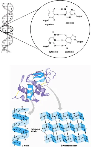
molecular recognition but a wide range of self-assembly. The cores of most proteins are
composed of hydrogen bonded secondary structures such as -helices and -sheets
(Figure 1-2 (below)).10 Among natural or designed substrate receptors, complementary
DNA is one central and remarkable example of two different oligomeric molecular
strands coming together in an intertwined, highly specific and reversible manner.
Figure 1-2 (above) A cartoon representation of DNA double helical structure with
If not for hydrogen bonding, water would not have its special role as a solvent that
boils at a high temperature of 100 oC. The hydrogen bonding makes the water molecules
“stick” together. In contrast to the desirable qualities, unwanted effects can be seen when
cyanuric acid and melamine are brought together (Figure 1-3) leading to kidney stone
formation and renal damage.11 Thus this concept of weak interactive forces is very
powerful as a cumulative effect and needs to be well studied and understood.
Figure 1-3 A two dimensional array of melamine and cyanuric acid assembled due to the
intermolecular attractions of hydrogen bonding that form an insoluble crystal lattice.
Hydrogen bonding can be used to construct larger molecules from smaller ones
and thus can be used as „molecular Velcro‟ to glue molecules together in a highly specific
manner. The stability of such supramolecules in turn rests on the strength of the net
hydrogen bonding, the type of modules taking part in hydrogen bonding to bringing about
1.2 Hydrogen Bonding
Hydrogen bonding is a complex interaction that consists of at least four types of
chemical characteristics: electrostatics (acid/base), polarization (soft/hard), van der Waals
(repulsion/dispersion), and covalency (charge transfer).12 The division into these
components has been well studied and reported though polarization is not completely
independent of the other three components.
1.2.1 Definition
Pauling, in 1939, stated in his book The Nature of the Chemical Bond “under
certain conditions an atom of hydrogen is attracted by rather strong forces to two atoms,
instead of only one, so that it may be considered to be acting as a bond between them”.
Thus an H atom is the key element that brings a hydrogen bonding donor (X) and
hydrogen bonding acceptor (Y) together (Figure 1-4). Depending on the nature of X and
Y, the energy of hydrogen bond lies in the range of 2.1 to 167 kJ mol-1. The strongest
hydrogen bonds are stronger than the weakest covalent bonds while the weakest
hydrogen bonds are practically indistinguishable from van der Waals interactions.
Figure 1-4 Hydrogen bonding between two electronegative heteroatoms X and Y
mediated by a hydrogen atom.
In the recent past (1997), IUPAC defined hydrogen bonding in its Gold Book.
The definition states that a hydrogen bond is “… a form of association between an
electronegative atom. It is best considered as an electrostatic interaction, heightened by
the small size of hydrogen, which permits proximity of the interacting dipoles or charges.
Both electronegative atoms are usually (but not necessarily) from the first row of the
Periodic Table, i.e., N, O or F. Hydrogen bonds may be intermolecular or
intramolecular. With a few exceptions, usually involving fluorine, the associated energies
are less than 20–25 kJ mol−1(5–6 kcal mol−1) …”. The evidence for hydrogen-bond
formation may be experimental or theoretical, or ideally, a combination of both. Some
criteria useful as evidence and some typical characteristics for hydrogen bonding, not
necessarily exclusive, are listed in a recent essay by Desiraju, in detail.13,14
These two definitions do serve to describe hydrogen bonds in their own manner
but in simpler parlance we will define it as: the attractive force between the
electropositive hydrogen interceding between two electronegative species such as X and
Y. Usually the electronegative species X and Y are heteroatoms such as oxygen, nitrogen,
or fluorine, which have a partial negative charge and the hydrogen a partial positive
charge. In supramolecular terms, the electronegative heteroatom to which the hydrogen is
covalently bound is called the hydrogen bond donor (denoted by D). The other
electronegative atom must have one or more unshared electron pairs as in the case of
oxygen and nitrogen, have a negative partial charge and will be called a hydrogen bond
acceptor (denoted by A). The hydrogen on the donor, which has a partial positive charge
binds to another atom of oxygen or nitrogen with excess electrons to share and is
attracted to the partial negative charge of the acceptor. This forms the basis for a
1.2.2 Characteristics of Hydrogen Bonds
Hydrogen bonding is ubiquitous in nature and is characterized mainly by three
qualities : strength, directionality and specificity. These unique qualities distinguish it
from other types of non-covalent interactions that are generally lacking in at least one of
these characteristics. The variance of these qualities in single hydrogen bonds as well as
on hydrogen bonded complementary complexes are discussed in the further sections of
this chapter.
1.2.2.1 Strength of a Single Hydrogen Bond
The strength of a single hydrogen bond depends on the electronegativity of D and
A heteroatoms and the influence of the adjacent functional groups they are connected to.
This results in a wide range of energies observed starting from < 2 kJ mol-1 to > 170 kJ
mol-1.15 A weak hydrogen bond can be characterized by bond energies less than 16 kJ
mol-1, an angle less than 110 and a very long bond length between the heteroatoms (>
3.6 Å). On the other hand strong hydrogen bonds are easy to distinguish with energies >
40 kJ mol-1, short bond distances (< 3.2 Å) and angles from 150 180.16 The strength of
a hydrogen bonding is a direct outcome of both the electronegativities of the donor and
the acceptor as well as the linearity of the hydrogen bond.
Physical organic chemists have determined the association constants Ka for a very
large number of intermolecular interactions in the solution phase. For simple molecules,
Abraham has developed an equation that relates the log K for a hydrogen bond
interaction between two functional groups and their empirically determined donor or
where log K = C1 α2H β2H + C2
C1 is a solvent dependent constant and C2 is the entropic cost of bringing two neutral
molecules together (approximately 6 kJ mol-1).17a
The relationship between pKa values of a given functional group and the ability to
hydrogen bond is not directly applicable to all the functional groups. The relationship
may hold among a set of similar functional group derivatives but not for comparisions
between different functional groups. For example, relative to alcohols, thiols are fairly
acidic. Butanethiol has a pKa of 10.5 vs 15 for butanol. Thiophenol has a pKa of 6 vs 10
for phenol. However, alcohols are fairly good hydrogen bond donors whereas thiols are
very poor hydrogen bond donors.
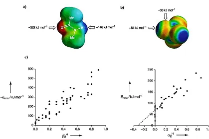
In 2004, Hunter developed a new pair of quantities ( and )17b that describe
hydrogen bond donor and acceptor ability respectively:
α = Emax/52 kJ mol-1 = 4.1(α2H + 0.33)
β = Emin/52 kJ mol-1 = 10.3(β2H + 0.06)
where Emax and Emin are the potential minima and maxima on the molecular electrostatic
potential surfaces of the molecules as determined by AM1 calculations (Figure-5). Based
on Abraham‟s examples of simple common molecules participating in hydrogen bonding,
Hunter has published scatter plots of α2H and β2H values vs Emin and Emax per kJ mol-1 and
correlated and with α2H and β2H values. The results are surprisingly linear and form
the basis for a reasonably accurate estimation of the strength of a hydrogen bond between
Figure 1-5 Molecular electrostatic potential surfaces plotted on the van der Waals‟
surface of the molecule calculated by using AM1 and a positive point charge in a vacuum as the probe. a) N-methyl acetamide; b) Carbon tetrachloride. Positive regions are shown
in blue, negative regions are shown in red and green is neutral and c) The maxima (Emax) and minima (Emin) in the AM1 molecular electrostatic potential surfaces of a range of simple molecules containing only one functional group plotted against the corresponding experimentally determined values of α2H and β2H from Abraham‟s examples.17c
1.2.2.2 Directionality of Single Hydrogen Bonding
Directionality is one widely accepted aspect of hydrogen bonding. Although
secondary interactions in a system may force the angle D–H…A away from linearity, it is
the directionality in hydrogen bonding that develops from an anisotropic intermolecular
potential that separates it from the more general van der Waals forces, which are likely to
directional aspect and they do not arise from point charges as in case of ionic interactions.
The forces are the direct result of the tendency of charge separation between an
electronegative atom and the hydrogen connected to it. The hydrogen being partially
electropositive at a point on its electrostatic surface directly opposite to the donor
heteroatom, seeks association with a partially electronegative area of interaction located
on A. Linkage through these electronegative regions provides the directionality, as only a
particular area and orientation (Figure 1-6) is actually available for interaction and not the
entire spherical space around A. The hydrogen bond is strongest when the hydrogen
forms the „bridge‟ between the two electronegative atoms in aligned linearly with an
angle , close to 180 with a short bond length. There may be slight deviation from
linearity but cannot form or hydrogen bond where < 110 as it will lead an acute angle
with lesser compatibility for hydrogen bond formation. Ideally the angles > 150 are
generally considered best for hydrogen bonding even though the bond lengths may be
longer than usual.18
A
A
Figure 1-6 Hydrogen bonding between the acceptor (A) and the hydrogen atom of the
1.2.2.3 Specificity of Hydrogen Bonds
Specificity may be defined as the ability to distinguish or discriminate between
the arrangement of complementary surfaces and their complementary sites on these
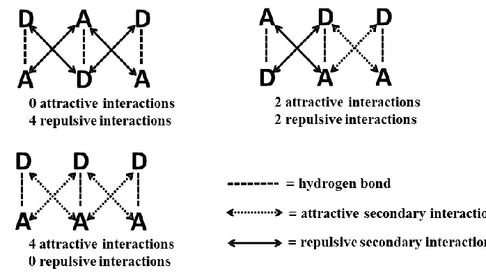
surfaces based on strength and orientation of interactions (Figure 1-7). Due to the
presence of partial charges that give rise to intermolecular interactions, recognition may
occur in a highly specific manner. The partial charges act as electrostatic map i.e. a
partially electronegative species or atoms will selectively attract a partially
electropositive species or atoms. The specificity of the interaction (similar to a binary
code) is enhanced by the proper alignment which gives the interaction a high degree of
selectivity in terms of binding.
Figure 1-7 Arrangement of complementary sites leads to specifically attractive or
repulsive interactions as demonstrated using 2-aminopyridine molecules.
Depending on the nature and strength of electronegative species participating in
hydrogen bonding, these interactions lead to wide variations in selectivity. Proximity of
the available partial charges also can affect the specific nature of binding when the
partially charged species form side chains on a covalent main chain moiety thus leading
to sequence specificity through hydrogen bonding. Sequence specificity was pointed out
by Watson and Crick in their double helical DNA model where base pairs display near
specific hydrogen bond interactions to form complexes where the components are
arranged in a specific manner.
1.3 Hydrogen Bonded Complexes
Figure 1-8 Common complementary hydrogen bonding arrays developed that resemble
base pairs.
Almost six decades after the discovery of the complementary double-helical DNA
complex structure, supramolecular chemists have gained enough information and access
to design, synthesis and study the binding patterns of artificial complementary complexes
mimicking the base pairs of DNA. Hydrogen bonding serves as the basis for
complementary complex formation. Numerous examples of complementary complexes
have been reported and various aspects of these complexations have been subjected to
complexes with binding constants on the order of 104 M-1 and above.20 These motifs are
often used in the construction of supramolecular architectures.
Meijer and Zimmerman are two pioneers in the field of development of molecular
motifs for complementary hydrogen bonding. Ureido-pyrimidones (UPy),20-21 the
butylurea of guanosine (UG)22 and diamido-naphthyridine (DAN)21b,23 derivatives are
well known motifs that form hydrogen bonded arrays (Figure 1-8). Most of the existing
synthetic hydrogen bond arrays are a result of inspiration from natural complementary
complexes such as the DNA base pairs or pleated sheets of proteins. Gong and
coworkers have reported numerous examples that are mimics of the pleated sheets
(Figure 1-9).24
Figure 1-9 (i) Hydrogen bonding in an anti-parallel sheet and (ii) Bing Gong‟s
hydrogen bonded complex which resembles a sheet.
1.3.1 Design Parameters of Hydrogen Bonded Complexes
The primary interest in supramolecular systems is the examination of complex
formation. Small monomer components that are built to self-assemble hold the
synthetic scheme which is often simple and consists of only a few steps. It also takes into
account all the plausible geometrical issues of the complexes whether they be steric or
electronic effects that dictate the interactions and stabilities of such assemblies.25 The
goal of a designer has always been to create these assemblies with minimum number of
synthetic steps and generate complexity by assembly of monomer components using
molecular recognition into materials with the desired properties. In order to maximize the
effects of hydrogen bonding, various types of complexes such as cleft structures, linear or
helical complexes have been designed, studied and manipulated to understand the
stabilities of these complexes. Often, the starting materials are commercially available
and inexpensive. We will discuss several aspects that are important for a well-designed
complex system such as functional groups, preorganization, tautomer formation,
solubility, fidelity and number of hydrogen bonds and secondary interactions.
1.3.1.1 Functional Groups and Substituents
Hydrogen bonding is often highly sensitive to the nature of the substituents
connected to the donor and acceptor components. An electron withdrawing group
connected to a donor subunit in an array can make the donor hydrogen atom(s) more
highly electropositive. The connection can be in the form of resonance through
conjugation or inductively through the - framework. Similarly electron donating groups
that are connected to an acceptor subunit in an array may improve the acceptor character
and tuning of these factors together can significantly improve the overall stabilities of the
resulting complexes. Thus a basic design with an accommodation to incorporate
functional groups at optimal positions can have a significant effect on stabilities of
triply hydrogen bonded system whose binding strengths were studied as a function of
various electron withdrawing groups on the donor components and electron donating
groups on acceptor components.26 The largest effects were seen when the withdrawing
groups acted through resonance (Figure 1-10).
Figure 1-10 AAA-DDD Model system investigated by Boyd and coworkers including
the gas phase binding energies stated as function of withdrawing groups on DDD components.
Figure 1-11 Substituent effects in the complementary AAA-DDD arrays on the
association constants (measured in CDCl3) which are displayed on right hand side; N/A = data not available.
R‟ Binding Energy (kJ mol-1)
NH2 31.8
OH 32.2
H 32.6
F 35.6
Cl 41.0
CN 66.9
CHO 73.6
Recently our research group has reported substituent effects on a triply hydrogen
bonded system27 (Figure 1-11) incorporating withdrawing substituents such as halogens,
esters and nitrile groups on indole hydrogen bond donors. The association constants
could be raised from 3.1 x 10³ M-1 to 4.8 x 10⁵ M-1 (i.e. by a factor of 30 or 12 kJ mol-1
difference) when titrated against substituted (methyl and amino) terpyridyl based
acceptor components in CDCl3.
Wilson and coworkers have developed AADDDA type heterodimers28 based on
amidoisocytosine and ureidoimidazole moieties, respectively, that illustrated that remote
substituent effects control dimerization affinity in a predictable manner.
Figure 1-12 Possible tautomeric and conformational states of arrays 1-3 and 1-4.
The ureidoimidazole motif 1-4 is suitable for studying remote electronic substituent
effects because although the hydrogen-bonding array may adopt two tautomeric
adopted as a consequence of the enforced intramolecular hydrogen bonding presents a
DDA array (Figure 1-12). Similarly two tautomeric forms are possible for
amidoisocytosine (1-3)both stabilized by intramolecular hydrogen bonding among which
only one presents the required AAD array. A series of complexes were synthesized with
different substituents in the para position of the aromatic ureido/amido ring system.
1.3.1.2 Preorganization
Preorganization is a central factor that affects the stability of complexation during
molecular recognition. In order to form a complex, the orientation of the non-covalent
interactions depends on the geometrical arrangement of the individual components and
the way they come together. The degree of freedom to rotate over single bonds in a
molecule is what determines its range of conformations. It generally requires energy to
bring individual functional groups into the right alignment to form a stable complex.
Hence, preorganization can have a great effect in terms of conserving energies which
otherwise would be spent bringing the array to its optimal geometrical alignment for
complexation. As a design parameter preorganization can be introduced to the
participating groups via intramolecular interactions, contributing to the net stability of
complex formation.
While preorganization in metal driven coordinated complexes has been extensively
studied,29 non-metallic complexation assemblies with preorganizational effects have been
gaining importance in recent times. In 1990, Etter framed a set of rules30 as “hydrogen
for compounds with various functional groups, which can be utilized while designing a
complex based from them.
1. All good proton donors and acceptors are used in hydrogen bonding.
2. Six-membered-ring intramolecular hydrogen bonds form in preference to inter-
molecular hydrogen bonds.
3. The best proton donors and acceptors remaining after intramolecular hydrogen-
bond formation form intermolecular hydrogen bonds to one another.
Figure 1-14 Conformational equilibrium of ethoxynaphthyridine 1-5 and its complex
with array 1-6. Array 1-7 contains an oxy substituent but is constrained in a ring.
Association constants are measured in CDCl3.
Along with intramolecular hydrogen bonding, other conformational issues may
affect the level of preorganization. Hamilton found that ethoxynaphthyridine 1-5 bound
triacetyl guanosine 1-6 with a Ka = 126 M-1 (Figure 1-14),31 which is at least two orders
of magnitude lower than the Ka of a very similar complex, 1-61-7. In the example
(Figure 1-14) Murray and Zimmerman proposed that the ethoxy group of 1-5 suffers
from steric interactions with the guanine amino group. Thus there is an energy cost for
producing the less stable conformation of the ethoxy group in 1-51-6. Evidence for this
is “tied back” in a lactone ring. In contrast to 1-51-6 the Ka of 1-61-7 is > 104 M-1,
which is expected of a DDAAAD type complex.32
Although intramolecular hydrogen bonding is a very useful tool to arrange a
molecule in a desired conformation, there are also complications that can arise with
preorganization via intramolecular hydrogen bonding. The example above demonstrates
the unwanted effects of an intramolecular hydrogen bond by clipping a ureido donor to a
pyridyl group and bypassing the ADD system intended to produce a DA array instead.
This is an undesired result of poor design that introduces intramolecular hydrogen
bonding that has a negative effect on the stability of the resulting complex. Thus the
example emphasizes the powerful nature of preorganization and how it can be determined
by design.
Figure 1-15 Imide-urea strands that pair into self-complementary duplexes 1-81-8 via
In a recent example,33 a quadruply hydrogen-bonded duplex (1-81-8), based on
an imide-urea structure preorganized partially by three-center hydrogen bonds was
reported to associate via bifurcated hydrogen bonds (Figure 1-15). 1H NMR dilution
experiments revealed the high stability of the homodimer in a non-polar solvent (Kdimer >
105 M–1 in CDCl3) and enhancement of the association due to electron-withdrawing
substituent effects (eg. –CF3 in Figure 1-15).
1.3.1.3 Tautomers
Tautomers are isomers differing only in the positions of hydrogen atoms and
electrons
.
The carbon skeleton of the compound is unchanged. A reaction which involvessimple proton transfer in an intramolecular fashion is called a tautomerism. When
designing a complex it is a good idea to pay special attention to the locations of double
bonds and functional groups such as carbonyl, amide, amine and lactams so as to avoid
unnecessary tautomerism which may affect the overall stability of a complex. In fact, it
has been proposed that mutations in DNA may occur as a consequence of mispairing of
minor tautomers of the four natural bases,34 indicating the importance of the concept.
Meijer and coworkers have reported a DDA array of 2-ureido-4-pyrimidone
(UPy, 1-9) which tautomerizes to an AADD array (1-9a) and ADAD array (1-9b) and
undergoes self-association (Figure 1-16).21a This is a very good example that
demonstrates the role of tautomer formation in determining the overall stability of the
complex. The AADD array displays greater stability (over two orders of magnitude)
interactions. Some tautomers may not even allow the formation of a complex by altering
the sequence of the hydrogen bonding array.
Figure 1-16 The tautomeric and self-association equilibria observed in a solution of
2-ureido-4-pyrimidone (1-9). The dimerization values were measured in CDCl3.
Zimmerman andcoworkers have reported an AADD array 1-10a that by design
formation of a less favourable array (Figure 1-17).35 Hence, these types of tautomer
conflicts can be controlled through careful design if necessary.
Figure 1-17 Zimmerman‟s AADD array 1-10a that can only form a tautomeric AADD
array 1-10b thereby maximising the association constant values possible for the complex.
1.3.1.4 Solubility
Figure 1-18. Intermolecular interactions in solution are a competition between solute–
solvent interactions in the free state, and solute–solute and solvent–solvent interactions in the bound state. For simple functional groups, the primary mode of interaction is hydrogen-bond contacts between the maxima (black) and minima (grey) in the electrostatic potential surfaces of the molecules.36
Solubility plays a central role in many chemical transformations and in the field
of molecular recognition, desolvation can be a dominant factor in the stability of
non-covalently interacting systems. Polar solvents can bind competitively to hydrogen bond
Solvent Solvent
A DH HD
A Solvent
Solvent
arrays and may cause a significant decrease in the stability of any complex formation.
The analysis of many self-assembled systems are thus restricted to operation in
non-competitive, non-polar solvents such as chloroform, toluene and cyclohexane. Solubility
is one of the most commonly faced hurdles in terms of the physical properties of
supramolecular complexes.
In the solution phase, there is a competition between solute–solute, solvent–
solvent, and solute–solvent interactions (Figure 1-18) and Hunter‟s universal
hydrogen-bond scale can be used to predict the free energy of hydrogen-hydrogen-bonding interactions (ΔG
H-bond in kJ mol−1) in most solvents.36-37
-RT lnK =∆GH-bond = - (α-αs) (β-βs)
α and β are hydrogen-bond donor and hydrogen-bond acceptor constants for the solute
molecules, and αS and βS are the corresponding hydrogen-bond donor and hydrogen-bond
acceptor constants for the solvent. The new parameters, α and β correspond to normalized
versions of Emax and Emin [α = Emax/52 = 4.1(α2H + 0.33), β = - Emin/52 = 10.3(β2H + 0.06)]
determined from AM1 electrostatic potential surfaces, as discussed earlier.
Hunter and coworkers have also studied a system for which the association
constants were reported in various solvents to highlight the role of competitive solvents
and non-competitive solvents play in the determination of the stability of the
complexes.17b One of the most polar hydrogen-bond donors known is perfluoro-tert-butyl
alcohol and one of the best hydrogen-bond acceptors known is tri-n-butylphosphine oxide
(Figure 1-19). Experiments on the complexation between these two compounds in
tetrachloride suggest that the complex should exhibit extraordinary stability, thereby
allowing quantification of the hydrogen bond interaction in competitive polar solvents.37
The results demonstrate the predictable drop in association constants, Ka values with
increasing solvent competition/polarity.
Solvent Ka in M-1 n-decanol 1.6 x 10⁻¹
DMSO 6.8 x 10⁻¹
NMF 8.9 x 10⁻¹
Pyridine 6.5 x 100 pyrrole 1.3 x 101 acetone 6.5 x 101 acetonitrile 1.6 x 10² tetrahydrofuran 2.4 x 10² nitromethane 1.5 x 10³ CHCl3 2.7 x 10³ Benzene 1.9 x 10⁴
CCl4 7.6 x 10⁴
Cyclohexane > 105
Figure 1-19 Results of 31P NMR titration experiments displaying the association constant
for formation of a 1:1 complex between perfluoro-tert-butyl alcohol and tri-n-butyl phosphine oxide at 295 K as a function of solvent properties. Errors in Ka are 20%, except for the values in N-methylformamide (NMF), dimethyl sulfoxide (DMSO), and n
-decanol, where only 30–40% of the binding isotherm was accessible and the values are accurate to within an order of magnitude.
There are different ways to overcome solubility issues without disrupting the
primary design of a complex. Lengthy alkyl chains, polyethylene glycol units or sterically
hindering groups can be incorporated that may improve the solubility of otherwise
insoluble components. Alternatively, mixed solvent systems can be used to measure the
association constants or for comparative studies of specific interactions. Both of these
1.3.1.5 Fidelity
Fidelity is defined in various fields in different manners. It is the degree or quality
of faithfulness toward a particular interaction. The genetic information that is passed on
from double helical DNA strands to RNA and subsequently on to proteins is based on the
specific recognition of complementary base pairing and such specificity is highly
desirable when mimicking nature in order to develop materials for perticular applications.
In supramolecular terms, fidelity is minimal competition from other recognition events
during the process of complex formation. Fidelity has been defined as the ratio of
concentration of the desired complexes to the concentration of all associated species.
Thus fidelity F, can range from 0 F ≥ 1, where F = 1 indicates exclusive formation of
the desired complex and F = 0 indicates exclusive formation of other undesired
complexes.
Zimmerman and coworkers have reported several triple and quadruple hydrogen
bonded motifs that form heterodimer complexes with very high fidelity.22, 38
Orthogonality has been studied in several examples and a few of them stand out
displaying high fidelity. The concept is well demonstrated by complex formation of
2,7-diamido-1,8-naphthyridine (DAN) and the butylurea of guanosine (UG) in chloroform as
DANUG. The complex is exceptionally strong due to high fidelity between the
participating arrays. The association constant for the DANUG complex was found to be
5 × 10⁷ M-1 by fluorescence energy transfer from the naphthyridine unit of DAN to
coumarin 343 covalently linked to UG (Figure 1-20) and is among the highest reported
(Kdimer < 10 M-1) and UG (Kdimer = 300 M-1) strongly suggests that the DANUG complex
forms with unparalleled fidelity.
Figure 1-20 (i) DANUG complex formed due to the high fidelity interaction between
the two arrays; (ii) Fluorescence emission of DANUG complex displaying the
fluorescence energy transfer from the naphthyridine unit of DAN to a coumarin 343
covalently linked UG with dilution in chloroform (background subtracted); (iii)
Fluorescence intensity (arbitrary units) plotted against concentration for the association pictured in (ii).
In the above case, F is calculated using the equation: