Clinical Ophthalmology
A case involving an Ahmed™ glaucoma valve
transferred from the vitreous into the anterior
chamber of the eye with a silicone oil tamponade
for the treatment of neovascular glaucoma
Michiko Miki Mari Ueki
Tetsuya Sugiyama Shota Kojima Tsunehiko Ikeda
Department of Ophthalmology, Osaka Medical College, Takatsuki, Japan
Correspondence: Tetsuya Sugiyama Department of Ophthalmology, Osaka Medical College, 2-7 Daigaku-machi, Takatsuki 569-8686, Japan Tel +81 726 83 1221 Fax +81 726 81 8195
Email tsugiyama@poh.osaka-med.ac.jp
Purpose: To report the short-term efficacy and safety of the transfer of an Ahmed™ glaucoma
valve (AGV™) tube from the vitreous into the anterior chamber, in a patient with neovascular glaucoma who had undergone pars plana AGV™ implantation and ultimately needed a silicone
oil tamponade.
Case: A 41-year-old male with proliferative diabetic retinopathy in both eyes was referred to us
for treatment in December 2009. Although the patient previously underwent several surgeries, he ultimately lost vision in his right eye. His left eye suffered from neovascular glaucoma after undergoing a pars plana vitrectomy for tractional retinal detachment. After several vitreous and glaucoma surgeries, the patient underwent implantation of a pars plana AGV™. Postoperatively, although his intraocular pressure was stabilized at approximately 10 mmHg, he had repeated vitreous hemorrhage and hyphema without improvement. He ultimately underwent PPV with a silicone oil tamponade and at the same time, the AGV™ tube was pulled out from the vitreous and inserted into the anterior chamber in order to avoid complications caused by the silicone oil.
Results: At 19 months postoperative, the patient’s intraocular pressure had stabilized at
10 mmHg with no recurrence of vitreous hemorrhage and hyphema. Eventually, he lost vision in his left eye because of cerebral hemorrhage.
Conclusion: The findings show that insertion of a pars plana AGV™ tube into the anterior chamber
in a patient undergoing a silicone oil tamponade is both effective and safe in the short-term.
Keyword: tube implantation, glaucoma surgery, tube transfer, pars plana, proliferative diabetic
retinopathy, intraocular pressure
Introduction
Although tube-based surgeries for the treatment of glaucoma are prevalent in Europe and America, they have only recently been approved for use in Japan. The results of a questionnaire survey conducted by the American Academy of Ophthalmology revealed that the number of surgeons opting to perform trabeculectomy is apt to reduce, while the number of surgeons opting to perform tube implantation is apt to increase. The recent “Tube versus Trabeculectomy study,” a comparative, randomized, multicenter study of tube implantation into the anterior chamber versus trabeculectomy conducted in the United States, showed that patients who underwent tube implantation obtained better
intraocular pressure (IOP) control and suffered overwhelmingly fewer complications.1
Thus, it is expected that the number of surgeons opting to perform the tube implanta-tion procedure will greatly increase in the future.
Dove
press
C A S E r E P O rT open access to scientific and medical research
Open Access Full Text Article
Clinical Ophthalmology downloaded from https://www.dovepress.com/ by 118.70.13.36 on 21-Aug-2020
For personal use only.
Number of times this article has been viewed
This article was published in the following Dove Press journal: Clinical Ophthalmology
Pars plana implantation, also referred to as the “fourth generation of implant tube–based surgery,” was developed to prevent complications that can occur with implantation into the anterior chamber, such as a shallow anterior cham-ber, corneal decompensation, and adhesion of the iris, and is done for cases of secondary glaucoma, such as neovas-cular glaucoma (NVG). In a pseudophakic eye treated with a silicone oil tamponade, insertion of the glaucoma valve tube into the anterior chamber seems to result in a better clinical course, due to the fact that the effect of silicone oil on the tube can probably be prevented. Therefore, it would be advantageous if transfer of the tube from the vitreous into the anterior chamber is possible, especially in cases of pseudophakic eye.
The purpose of this present case report was to report a
case involving the transfer of an Ahmed™ Glaucoma Valve
(AGV™) (New World Medical, Inc, Rancho Cucamonga,
CA, USA) from the vitreous cavity into the anterior cham-ber, in a patient with NVG who had undergone pars plana
AGV™ implantation and ultimately needed a silicone oil
tamponade.
Case report
This case report involved a 41-year-old male patient who first became aware of visual loss in both eyes in the summer in 2009. He later visited an ophthalmologist in December 2009, and was diagnosed with proliferative diabetic retin-opathy (PDR) in both eyes. The patient was subsequently referred to us in the same month of that year, and he also visited a general physician and was diagnosed with diabetes mellitus on the same day (blood sugar: 501 mg/dL, glycated
hemoglobin [HbA1c]: 14.0%). Consultation with the patient
revealed that his father had undergone dialysis treatment for kidney disease, yet the patient’s own past medical history had nothing in particular to reveal.
At the initial examination, the patient’s visual acuity was 30 cm/HM in his right eye and 0.5 in his left eye. His IOP was 16 mmHg in both eyes. Fundus examination of the patient revealed PDR with fibrovascular membrane, preretinal hemorrhage, and tractional retinal detachment in his right eye; and PDR with fibrovascular membrane in his left eye (Figure 1).
In the right eye of the patient, vision was lost despite twice undergoing pars plana vitrectomy (PPV), in February and July, 2010. In October 2010, we performed panretinal pho-tocoagulation on his left eye; however, the tractional retinal detachment in that eye progressed. Therefore, we considered PPV to be indicated for this case. Examination of the patient
in October 2010 revealed that visual acuity was 0.09 and IOP was 12 mmHg in his left eye. In addition, iris neovasculariza-tion, ectropion uvea, and tractional retinal detachment were found, and peripheral anterior synechia (PAS) index was approximately 60% in his left eye (Figure 2).
Later in December 2010, the patient underwent PPV com-bined with additional photocoagulation and cataract surgery. However, 1 month after surgery, IOP rose to 50 mmHg and the PAS index was 100%, in his left eye. He thus underwent PPV combined with trabeculectomy in December 2010. The vitreous hemorrhage was removed and fibrovascular membrane of the peripheral retina was resected, followed by additional photocoagulation. At 1 month postoperative, the IOP was again elevated for the filtering bleb dysfunction, due to repeated vitreous hemorrhage. Thus, the surgical inter-vention was no longer effective against IOP elevation. The
patient then underwent pars plana implantation of an AGV™
(model AGV-PC7) in January 2011. The implant tube was inserted at the 2 o’clock position. Postoperatively, although
Figure 1 Ocular fundus findings at the initial examination of the patient showed (A) Right eye: PDR with fibrovascular membrane, preretinal hemorrhage, and tractional retinal detachment; and (B) Left eye: PDR with fibrovascular membrane.
Abbreviation: PDr, proliferative diabetic retinopathy.
Figure 2 Slit-lamp biomicroscopy and gonioscopy findings at the visit on October 16, 2010.
Note: The left eye showed iris neovascularization, PAS index: 60%.
Abbreviation: PAS, peripheral anterior synechia.
Dovepress
Miki et al
Clinical Ophthalmology downloaded from https://www.dovepress.com/ by 118.70.13.36 on 21-Aug-2020
his IOP was stabilized at approximately 10 mmHg, the patient had repeated vitreous hemorrhage without improvement. He
subsequently underwent PPV + silicone oil tamponade +
insertion of a pars plana AGV™ tube into the anterior chamber
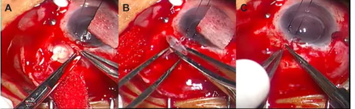
in May 2011. During that operation, the tube was pulled out from the vitreous cavity and inserted into the anterior cham-ber, in order to avoid complications caused by the silicone oil (Figure 3, Supplementary material).
Four months after the last operation, the patient’s visual acuity was 0.01 and IOP stabilized at 10 mmHg. Corneal
endothelial cells were 1970/mm2 and corneal
transpar-ency was preserved. He suffered no recurrence of vitreous hemorrhage and no complications, such as disorder of eye movement (Figure 4).
The patient suffered from cerebral hemorrhage in October 2011 and lost vision in his left eye. In December 2012, the patient’s IOP was still stabilized at 10 mmHg and there was no silicone oil migration into the anterior chamber, although fundus examination revealed tractional retinal detachment in the superior part of the fundus.
Discussion
It has been reported that 3%–15% of diabetic retinopathy
cases that needed vitrectomy were complicated by NVG.2
The findings of other studies have shown that 10%–15% of PDR cases that underwent vitrectomy had repeated
vitreous hemorrhage.3–5 Moreover, 7.5% of cases that
underwent vitrectomy for PDR reportedly required a
silicone oil tamponade.6 Therefore, it can be considered
that cases of severe diabetic retinopathy complicated by NVG have a high possibility of needing a silicone oil tamponade. Reported results of pars plana tube implanta-tion for the treatment of NVG showed a good success rate (72%–94.4%) at 6 months to 14.2 months, and if tube-based surgeries are performed widely in the future, many cases will require the insertion of a tube for an eye that
has undergone silicone oil tamponade after vitrectomy, or require a silicone oil tamponade for an eye that has already
undergone tube implantation.7
The AGV™ and the Baerveldt® Pars Plana Glaucoma
Implant (Model BG 102-350; Abbot Laboratories Inc, Abbott Park, IL, USA) are the two main types of valves
that are currently used. The AGV™ Model PC7 joins a
pars plana clip to the usual AGV™ Model FP7 tube for the
anterior chamber, and this pars plana clip is movable (for
positioning); on the other hand, the Baerveldt® Pars Plana
Glaucoma Implant uses a Hoffman elbow at the tip of the tube for insertion into the pars plana and is fixed. Thus, the
AGV™ tube can be transferred from the vitreous cavity into
the anterior chamber.
When tubes are implanted in eyes that have undergone a silicone oil tamponade, there is a high possibility of dete-rioration of the intraocular inflammation and subsequent blockage of the tube caused by the silicone oil. Therefore, having undergone a silicone oil tamponade is a risk
fac-tor for implantation.8 Our method of implanting a tube in
the superior temporal location is used to avoid silicone oil leakage and blockage of the tube by silicone oil when we implant a tube for an eye that has undergone a silicone oil tamponade. Several studies have reported that a massive amount of silicone oil moved into the subconjunctival space and orbit of the eyes through the tubes when implanted tubes were exposed to silicone oil, such as in cases of aphakic
eyes or eyes with pars plana–implanted tubes.9–11 Thus, we
considered that it would be possible to put an implant tube in the superotemporal location in a pseudophakic eye, such as in the case presented in this study, because silicone oil only slightly migrates into the anterior chamber. In fact, the IOP in this patient was stabilized, and there have been no complications to date.
In conclusion, the findings of this case show that insertion
of a pars plana AGV™ tube into the anterior chamber in a
Figure 3 Transfer of a pars plana AGV™ tube into the anterior chamber. (A) The tube being inserted into the vitreous body; (B) The tube being pulled out from the vitreous
cavity; and (C) The tube being inserted into the anterior chamber.
Abbreviation: AGV™, Ahmed™ glaucoma valve.
Dovepress An Ahmed™ glaucoma valve transferred into the anterior chamber
Clinical Ophthalmology downloaded from https://www.dovepress.com/ by 118.70.13.36 on 21-Aug-2020
patient undergoing silicone oil tamponade is both effective and safe in the short term. However, further investigation is needed to elucidate the long-term efficacy and safety of this treatment.
Disclosure
The authors report no conflicts of interest in this work.
References
1. Gedde SJ, Schiffman JC, Feuer WJ, Herndon LW, Brandt JD, Budenz DL; the Tube Versus Trabeculectomy Study Group. Three-year follow-up of the tube versus trabeculectomy study. Am J Ophthalmol. 2009;148(5): 670–684.
Figure 4 Slit-lamp biomicroscopy and ocular fundus findings after the patient’s final surgery. (A) A pars plana AGV™ tube inserted into the anterior chamber; and
(B) PDr was stable, with no recurrence of vitreous hemorrhage.
Abbreviations: AGV™, Ahmed™ glaucoma valve; PDr, proliferative diabetic retinopathy.
2. Summanen P. Neovascular glaucoma following vitrectomy for diabetic eye disease. Acta Ophthalmol (Copenh). 1988;66(1):110–116. 3. Steel DH, Connor A, Habib MS, Owen R. Entry site treatment to
prevent late recurrent postoperative vitreous cavity haemorrhage after vitrectomy for proliferative diabetic retinopathy. Br J Ophthalmol. 2010;94(9):1219–1225.
4. Yan H, Cui J, Lu Y, Yu J, Chen S, Xu Y. Reasons for and manage-ment of postvitrectomy vitreous hemorrhage in proliferative diabetic retinopathy. Curr Eye Res. 2010;35(4):308–313.
5. Lo WR, Kim SJ, Aaberg TM Sr, et al. Visual outcomes and incidence of recurrent vitreous hemorrhage after vitrectomy in diabetic eyes pretreated with bevacizumab (avastin). Retina. 2009;29(7):926–931. 6. Helbig H, Kellner U, Bornfeld N, Foerster MH. Rubeosis iridis after
vitrectomy for diabetic retinopathy. Graefes Arch Clin Exp Ophthalmol. 1998;236(10):730–733.
7. Luttrull JK, Avery RL. Pars plana implant and vitrectomy for treatment of neovascular glaucoma. Retina. 1995;15(5):379–387.
8. Federman JL, Schubert HD. Complications associated with the use of silicone oil in 150 eyes after retina-vitreous surgery. Ophthalmology. 1988;95(7):870–876.
9. Hyung SM, Min JP. Subconjunctival silicone oil drainage through the Molteno implant. Korean J Ophthalmol. 1998;12(1):73–75.
10. Nazemi PP, Chong LP, Varma R, Burnstine MA. Migration of intraocu-lar silicone oil into the subconjunctival space and orbit through an Ahmed glaucoma valve. Am J Ophthalmol. 2001;132(6):929–931. 11. Chan CK, Tarasewicz DG, Lin SG. Subconjunctival migration of
silicone oil through a Baerveldt pars plana glaucoma implant. Br J Ophthalmol. 2005;89(2):240–241.
Dovepress
Miki et al
Clinical Ophthalmology downloaded from https://www.dovepress.com/ by 118.70.13.36 on 21-Aug-2020
Clinical Ophthalmology
Publish your work in this journal
Submit your manuscript here: http://www.dovepress.com/clinical-ophthalmology-journal Clinical Ophthalmology is an international, peer-reviewed journal covering all subspecialties within ophthalmology. Key topics include: Optometry; Visual science; Pharmacology and drug therapy in eye diseases; Basic Sciences; Primary and Secondary eye care; Patient Safety and Quality of Care Improvements. This journal is indexed on
PubMed Central and CAS, and is the official journal of The Society of Clinical Ophthalmology (SCO). The manuscript management system is completely online and includes a very quick and fair peer-review system, which is all easy to use. Visit http://www.dovepress.com/ testimonials.php to read real quotes from published authors.
Supplementary material
Video presentation of the operation performed in May 2011. The tube of the pars plana AGV™ was pulled out from the vitreous cavity and inserted into the anterior chamber, before silicone oil tamponade.
Abbreviations: AGV™, Ahmed™ glaucoma valve.
Dovepress
Dove
press
An Ahmed™ glaucoma valve transferred into the anterior chamber
Clinical Ophthalmology downloaded from https://www.dovepress.com/ by 118.70.13.36 on 21-Aug-2020