1071-412X/96/$04.0010
Copyrightq1996, American Society for Microbiology
Regulation of Early Complement Components C3
and C4 in the Synovium
TIMOTHY COLLINS,
1JERRY A. WINKELSTEIN,
1AND
KATHLEEN E. SULLIVAN
1,2*
Division of Allergy and Immunology, Department of Pediatrics, Johns Hopkins University School of Medicine,
Baltimore, Maryland 21205,
1and Division of Allergy, Immunology, and Infectious Diseases, Department
of Pediatrics, Children’s Hospital of Philadelphia, Philadelphia, Pennsylvania 19104
2Received 15 June 1995/Returned for modification 28 August 1995/Accepted 12 September 1995
To determine the cytokine inducibility of early complement component (C3 and C4) expression in the
synovium, explant tissue was maintained in culture for 7 days. C3 and C4 production was measured by specific
enzyme-linked immunosorbent assay, and RNA was evaluated by semiquantitative PCR. The effects of
leuke-mia inhibitory factor (LIF), gamma interferon (IFN-
g
), IFN-
a
, and estrogen on C3 and C4 expression were
evaluated. C3 levels were unaffected by 7 days of LIF, IFN-
g
, or IFN-
a
treatment. In contrast, C4 levels were
significantly induced in synovial samples treated for 7 days with either IFN-
g
or IFN-
a
. LIF had no effect on
C4 levels in this system. Estrogen was found to down-modulate the induction of expression due to IFN-
g
. These
data provide evidence for cytokine regulation of C4 expression in the synovium and for estrogen modulation
of those effects.
The complement system plays an important role in host
defense and immunologically mediated inflammation (43).
Ac-tivation of the complement cascade produces biologically
ac-tive peptides capable of increasing vascular permeability,
stim-ulating chemotaxis, enhancing phagocytosis, and directly
inducing cellular injury. While most serum-derived
comple-ment is produced by the liver, many other cells are capable of
producing complement components in local inflammatory
re-actions (15). The synovium is one of the tissues capable of
producing multiple complement components (33, 35). The
mechanisms by which complement production is regulated in
the synovium have not been previously investigated.
In systemic lupus erythematosus (SLE), it is believed that
deposition of immune complexes in end organs with
subse-quent complement activation mediates most of the pathology
(27). Synovial pathology in SLE is typically characterized by
increases in synovial lining cells and a leukocytic infiltration
and is believed to be mediated by complement and
polymor-phonuclear leukocytes (26). Nevertheless, it is not clear what
role complement plays in the pathogenesis of synovitis in SLE.
Interestingly, two biologic agents capable of markedly inducing
complement gene expression have been implicated in
drug-induced SLE. Both alpha interferon (IFN-
a
) and IFN-
g
have
been associated with drug-induced SLE (8, 12, 21, 34).
Cytokines that are believed to play a role in inflammatory
synovitis include tumor necrosis factor alpha, interleukin-1,
interleukin-6, leukemia inhibitory factor (LIF),
granulocyte-macrophage colony-stimulating factor, and transforming
growth factor
b
(1, 5, 9, 11, 19, 36, 44). Levels of LIF, in
particular, have been demonstrated to be markedly elevated in
synovitis, and LIF has been demonstrated to be capable of
independently inducing synovial inflammation (6, 32, 41).
Lo-cally produced complement factors may be important in
in-flammation, in which cytokine regulation of complement
com-ponent expression may increase their levels substantially (17).
Other immunomodulatory factors, such as sex hormones,
may play a role in modulating inflammatory responses. Many
rheumatologic disorders associated with synovitis occur with
greater frequency in females than in males, and the course of
SLE may vary with the menstrual cycle (37). Synovial tissue has
recently been demonstrated to contain estrogen binding sites
(13) and could be metabolically influenced by that hormone.
The effect of estrogen on complement component expression
has not previously been investigated.
Local synovial production of complement components
ac-counts for much of the intra-articular complement (35), and
the capacity of complement to mediate inflammation suggests
that local production may play a role in inflammatory synovitis.
In the present study, we examined the regulation of early
complement component expression in the synovium by IFN-
a
,
IFN-
g
, LIF, and estrogen in an effort to understand the
path-ways by which complement component expression might be
influenced in the synovium.
MATERIALS AND METHODS
Synovial membrane cultures.Synovial samples were obtained from discarded tissue from patients undergoing total knee replacement for osteoarthritis. None of the patients were taking corticosteroids. Synovial villi were dissected from the surrounding adipose tissue and cut into full-thickness squares (2 by 2 mm). These synovial membrane cultures were maintained in minimal essential medium sup-plemented with 10% fetal bovine serum (Gibco-BRL, Gaithersburg, Md.). The culture media were changed daily and reagents were added as indicated below for the individual experiments. Triplicate or quadruplicate samples from each of eight patients were evaluated. At the end of the test period, the explant tissue was dried and weighed. To examine a pure population of synoviocytes, adherent synoviocytes were grown as monolayers for four passages in minimal essential medium supplemented with 10% fetal bovine serum (Gibco-BRL) by previously described purification techniques (32).
Biologic agents.IFN-gwas a generous gift from Genentech (South San Fran-cisco, Calif.). 17b-estradiol, IFN-a, and LIF were purchased from Sigma (St. Louis, Mo.), Hoffmann-La Roche (Nutley, N.J.), and Genzyme (Cambridge, Mass.), respectively. Time course and dose-response experiments using the sy-novial membrane cultures were performed with each biologic agent, and the doses and time points in the experiments were selected on the basis of the maximal responses. LIF was used at 10 ng/ml to be representative of levels that can be achieved within the joint space (22, 40).
ELISA.Polyclonal antisera directed against C4 (goat anti-human C4 from Calbiochem, San Diego, Calif., and rabbit anti-human C4 from Behring Diag-* Corresponding author. Mailing address: Division of Allergy,
Im-munology, and Infectious Diseases, Department of Pediatrics, Chil-dren’s Hospital of Philadelphia, 34th St. and Civic Center Blvd., Phil-adelphia, PA 19104. Phone: (215) 590-4685. Fax: (215) 590-3044.
5
on August 17, 2020 by guest
http://cvi.asm.org/
nostics, Somerville, N.J.) were used in an indirect enzyme-linked immunosorbent assay (ELISA) with horseradish peroxidase-labeled goat anti-rabbit immuno-globulin G from Miles-Yeda (Tel Aviv, Israel). Polyvinyl chloride microtiter plates were coated overnight with a 1:10,000 dilution of goat anti-human C4 in carbonate buffer. Supernatants from synovial cultures were added to the plates at various dilutions in phosphate-buffered saline–Tween with 2% bovine serum albumin. On each plate, dilutions of a standard human control serum containing a known amount of C4 were used for quantification of C4 protein. A 1:1,000 dilution of rabbit anti-human C4 was used as a second antibody, and alkaline phosphatase-labeled goat anti-rabbit immunoglobulin G was used as the final antibody. The plates were developed with o-phenylenediamine dihydrochloride (Sigma). The reaction was stopped with a 1 M HCl solution. A modification of the C4 ELISA was used to quantify C3. Goat anti-human C3 at 1:500 (Accurate Antibody, Westbury, N.Y.) was used as the capture antibody. The second anti-body was biotinylated goat anti-human C3 at 1:750. A 1:1,000 dilution of avidin-horseradish peroxidase was used in the next step, and the plates were developed as described above.
Reverse transcriptase PCR assay.Total RNA was prepared from synovial samples and monolayer cultures by using guanidinium isothiocyanate (10). Two micrograms of RNA was used as a template for first-strand cDNA synthesis, using oligo(dT) and avian myeloblastosis virus reverse transcriptase (Boehringer Mannheim, Indianapolis, Ind.). The cDNA samples were then used as templates for simultaneous PCR amplifications with actin and C4-specific primers. Actin is an internal control which is not altered by exposure to cytokines. The actin primers (exon 4, 59-TGACGGGGTCACCCACACTGTGCCCATCTA-39; exon 6, 59-CTAGAAGCATTGCGGTGGACGATGGAGGG-39) amplified a product of 661 bp, while the C4 primers bracketed exons 38 to 41 and amplified a product of 420 bp (exon 38, 59-GAAGTGCCCTCGCCAGCGTCG-39; exon 41, 59-CCT GGCACCCCTGAGTGCCATAC-39). Amplification of any potentially contam-inating DNA template is not possible with these primer pairs with our standard cycling protocol. Kinetic analysis was performed to ensure that the product was obtained during the linear phase of amplification. Quantitation was performed by using a densitometer and standardization to the actin signal.
Statistical analysis.The results in Fig. 1 are the averaged values for the patients; error bars correspond to the standard errors of the means. Statistical comparisons of the effects of estrogen and IFN-gon synovial C4 production were performed by the Wilcoxon matched-pairs test, comparing triplicate or quadru-plicate control sets with treatment sets for each patient.
RESULTS
To validate the membrane culture system and examine
whether cytokine effects on early complement component
ex-pression would be detectable in this system, we initially
mea-sured C4 production by synovial membrane cultures in the
presence or absence of IFN-
g
during the first week after
ex-planation. The effect of IFN-
g
was maximal at day 7 in culture.
The synovial cells remained viable throughout this time period
as demonstrated by synoviocyte purification and trypan blue
exclusion (5). Therefore, the remainder of the experiments
with synovial membrane cultures were performed on day 7.
To examine the effects of three cytokines believed to play a
role in different types of inflammatory reactions, we exposed
synovial organ cultures to physiologic levels of 10 ng of LIF per
ml, 1,000 U of IFN-
g
per ml, or 1,000 U of IFN-
a
per ml.
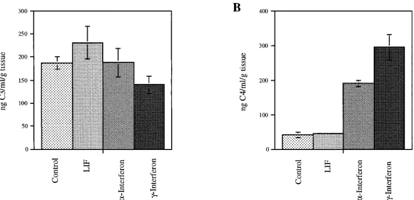
Figure 1 demonstrates the effect of these cytokines on C3 and
C4 expression. Examination of C3 expression by ELISA did
not reveal any response to LIF, IFN-
g
, or IFN-
a
. In contrast,
IFN-
g
and IFN-
a
increased C4 expression sevenfold (P
,
0.0001) and fivefold (P
5
0.0007), respectively. LIF did not
affect C4 expression in these synovial membrane cultures.
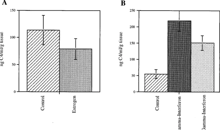
To evaluate whether estrogen could directly affect
comple-ment component expression, we treated synovial explants with
estrogen, IFN-
g
, or both. Estrogen clearly down-modulated
the induction of C4 expression by IFN-
g
(Fig. 2). This effect
was observed in samples from both male and female donors.
To demonstrate that the synoviocytes themselves produced
C4 and to confirm that the synovial samples synthesized C4 de
novo, semiquantitative PCR studies were performed to
dem-onstrate the presence of C4 message. Figure 3 shows results for
simultaneous PCR amplifications of total cDNA isolated from
day-7 membrane cultures with primers specific for C4 and with
actin as an internal control. The C4-specific bands appear in all
of the sample lanes, confirming that the synovium actively
transcribes C4 message. To demonstrate that purified
synovio-cytes produce C4, RNA was isolated from these cultures and
assayed by reverse transcriptase PCR. Purified synoviocytes
also expressed C4 message, confirming that synoviocytes are
capable of C4 production. In both the membrane culture
sys-tem and the synovial monolayer cultures, C4 message was
upregulated two- to threefold in response to IFN-
g
. Treatment
of the monolayer culture with IFN-
a
also resulted in increased
C4 message levels. These studies confirm that the synovium
produces C4 and that synoviocytes participate directly in the
production of C4. Expression of C4 in the synovium was shown
to be regulated by IFN-
a
and IFN-
g
, and the increases in
protein production detected by ELISA were paralleled by
in-FIG. 1. Cytokine induction of C3 and C4 expression. Triplicate and quadruplicate synovial explant cultures from patients were treated with LIF at 10 ng/ml, IFN-a at 1,000 U/ml, or IFN-gat 1,000 U/ml for 7 days. Each day the supernatant was removed and replaced with fresh medium and treatment agent. (A) C3 production was not affected. (B) C4 production was significantly increased by IFN-g(P,0.0001) and IFN-a(P50.0007).on August 17, 2020 by guest
http://cvi.asm.org/
creases in C4 message level. Semiquantitative PCR was not
sensitive enough to determine whether the effect of estrogen
was also paralleled by changes in C4 transcript levels.
DISCUSSION
It is known that the synovium is capable of producing large
amounts of individual complement components and that
com-plement appears to be upregulated in rheumatoid arthritis (19,
35). The mechanisms by which complement production is
reg-ulated in the synovium have not been previously investigated.
Inflammatory synovitis has been artificially reproduced in a
number of animal model systems. Chronic antigenic challenges
with collagen (38), streptococcal cell wall antigen (42), and
mycobacterial heat shock protein (39) are all capable of
induc-ing inflammatory synovitis. Certain viral infections are capable
of inducing synovitis, and exposure to high levels of
inflamma-tory cytokines may also induce an inflammainflamma-tory state with
arthritis (24). How these different initiators might all be able to
induce a common pathologic lesion is not known. The
com-plement system, by virtue of its ability to interface with the
humoral immune system, may play a significant role.
Complement components are not coordinately regulated. In
addition, the cytokine regulation of complement component
expression depends not only on the individual component but
also on the tissue type. Cytokine regulation of C3 expression
appears to be very tissue specific (3, 4, 7, 14, 16, 18, 28–31).
Cytokine regulation of C4 expression has been less extensively
studied but has been found to be upregulated in monocytes
and scleral fibroblasts in response to IFN-
g
(23, 25). In an
effort to determine how C3 and C4 expression in the synovium
is regulated, we exposed synovial cultures to IFN-
g
and IFN-
a
.
C3 expression was unchanged by either cytokine. C4
expres-sion was upregulated in response to both cytokines. Both the
explant cultures and the synovial monolayers responded to
IFN-
g
and IFN-
a
, and these responses were paralleled by the
transcripts as determined by reverse transcriptase PCR.
Inter-estingly, in humans SLE with active synovitis has been induced
by exposure to IFN-
a
and IFN-
g
(8, 12, 21, 34).
To examine whether another cytokine which has been
dem-onstrated to play an active role in inflammatory synovitis was
capable of inducing C3 or C4 expression, synovial cultures
were exposed to LIF (6, 32, 41). There was no evidence of
induction of either component by this cytokine. C3 is known to
be induced by the cytokine interleukin-6 (17), which shares
FIG. 2. Estrogen modulation of IFN-geffects. (A) Triplicate and quadruplicate synovial explant cultures were treated with 1025M 17b-estradiol for 7 days. The difference between the control cultures and those treated with estrogen approached significance (P50.08). (B) Cultures were treated with 1025M 17b-estradiol, 1,000 U of IFN-gper ml, or both for 7 days. The difference between the control cultures and those treated with IFN-gis significant (P50.002), and the difference between the IFN-g-treated cultures and those which received both IFN-gand estrogen is significant (P50.01).FIG. 3. Reverse transcriptase PCR of C4 transcripts in the synovium. RNA was isolated from synovial explant tissue after treatment for 7 days. Lane 1, no treatment; lane 2, 1,000 U of IFN-gper ml; lane 3, 1,000 U of IFN-gper ml with 1025
M 17b-estradiol. RNA was isolated from pure synoviocyte monolayer cultures treated for 3 days. Lane 4, no treatment; lane 5, 1,000 U of IFN-gper ml; lane 6, 1,000 U of IFN-aper ml; lane 7, 1,000 U of IFN-gper ml with 1025 M 17b-estradiol. Molecular weight markers (M) are shown to the left. cDNA was produced from the RNA and was amplified by PCR with oligonucleotides spe-cific for actin and C4. The actin bands are 661 bp in length and are the upper band in each lane. The C4-specific bands are 420 bp in length and are the lower band in each lane. A negative control (lane 8) was amplified in parallel without a template. The bands were scanned with a densitometer and standardized to the actin signal for semiquantitative comparisons. The relative intensity of the C4 signal, standardized to that of the actin signal, is shown below each lane.
on August 17, 2020 by guest
http://cvi.asm.org/
many characteristics and a subunit of its receptor with LIF
(20). Nevertheless, C3 expression was unchanged by LIF at any
of the concentrations tested.
Because many rheumatologic disorders associated with
sy-novitis occur with greater frequency in females than in males,
it is important to understand the effects of sex steroid
hor-mones on various aspects of the inflammatory response. In the
synovial cultures, estrogen markedly down-modulated the
in-duction of C4 expression by IFN-
g
and appeared to decrease
basal expression as well. This is the first demonstration of an
estrogen effect on the production of a complement component.
The transcription control element governing estrogen
induc-tion of transcripinduc-tion has been identified (2), although the C4
promoter region does not appear to contain this transcription
control element. At present, the mechanism of the estrogen
effect is not known. Semiquantitative PCR was not able to
identify a change in transcript level associated with estrogen
treatment of either membrane cultures or synovial monolayers.
This may be due to a limited sensitivity of this assay system, or
the effect of estrogen on C4 production may occur through
posttranslational mechanisms.
ACKNOWLEDGMENTS
This work was supported by NIH grants AI07007 and HL47191 and a MAPS-CHRC (HD 28815) scholar award.
We gratefully acknowledge the valuable contributions of Carl A. Johnson, Associate Professor of Orthopedic Surgery, Johns Hopkins University School of Medicine.
REFERENCES
1. Arend, W. P., and J.-M. Dayer. 1990. Cytokines and cytokine inhibitors or antagonists in rheumatoid arthritis. Arthritis Rheum. 33:305–315. 2. Beato, M. 1989. Gene regulation by steroid hormones. Cell 56:335–344. 3. Botto, M., D. Lissandrini, C. Sorio, and M. Walport. 1992. Biosynthesis and
secretion of complement component (C3) by activated human polymorpho-nuclear leukocytes. J. Immunol. 149:1348–1355.
4. Brooimans, R. A., A. A. J. van der Ark, W. A. Buurman, L. van Es, and M. Daha.1990. Differential regulation of complement factor H and C3 produc-tion in human umbilical vein endothelial cells by IFN-gand IL-1. J. Immu-nol. 144:3835–3840.
5. Bucala, R., C. Ritchlin, R. Winchester, and A. Cerami. 1991. Constitutive production of inflammatory and mitogenic cytokines by rheumatoid synovial fibroblasts. J. Exp. Med. 173:569–574.
6. Campbell, I. K., P. Waring, U. Novak, and J. A. Hamilton. 1993. Production of leukemia inhibitory factor by human articular chondrocytes and cartilage in response to interleukin-1 and tumor necrosis factor-a. Arthritis Rheum. 36:790–794.
7. Celada, A., M. J. Klemsz, and R. A. Maki. 1989. Interferon-gactivates multiple pathways to regulate expression of the genes for major histocom-patibility class III, tumor necrosis factor and complement component C3 in mouse macrophages. Eur. J. Immunol. 19:1103–1109.
8. Chazerain, P., O. Meyer, and M. F. Khan. 1992. Rheumatoid arthritis-like disease after alpha-interferon therapy. Ann. Intern. Med. 116:427. 9. Chen, E., E. C. Keystone, and E. N. Fish. 1993. Restricted cytokine
expres-sion in rheumatoid arthritis. Arthritis Rheum. 36:901–910.
10. Chirgwin, J. M., R. J. Przybyla, R. J. MacDonald, and W. J. Rutter. 1979. Isolation of biologically active ribonucleic acid from sources enriched in ribonuclease. Biochemistry 18:5294–5299.
11. Chu, C. Q., M. Field, S. Allard, E. Abney, M. Feldmann, and R. N. Maini. 1992. Detection of cytokines at the cartilage/pannus junction in patients with rheumatoid arthritis: implications for the role of cytokines in cartilage de-struction and repair. Br. J. Rheumatol. 31:653–661.
12. Conlon, K. C., W. J. Urba, J. W. Smith, R. G. Steis, D. L. Longo, and J. W. Clark.1990. Exacerbation of symptoms of autoimmune disease in patients receiving alpha-interferon therapy. Cancer 10:2237–2242.
13. Cutolo, M., S. Accardo, B. Villaggio, P. Clerico, M. Bagnasco, D. Coviello, G. Carruba, M. Lo Casto, and L. Castagnetta.1993. Presence of estrogen binding sites on macrophage-like synoviocytes and CD81, CD291, CD45Ro1T lymphocytes in normal and rheumatoid synovium. Arthritis Rheum. 36:1087–1097.
14. Dauchel, H., N. Julen, C. Lemercier, M. Daveau, D. Ozanne, M. Fontaine, and J. Ripoche.1990. Expression of complement alternative pathway
pro-teins by endothelial cells. Differential regulation by interleukin-1 and glu-cocorticoids. Eur. J. Immunol. 20:1669–1675.
15. Falus, A. 1990. Regulation of complement biosynthesis by tissue-specific and hormonal factors. Immunol. Lett. 24:227–230.
16. Falus, A., K. Feher, E. Walcz, M. Brozic, G. Fust, T. Hidvegi, T. Feher, and K. Meretey.1990. Hormonal regulation of complement biosynthesis in hu-man cell lines. I. Androgens and gamma interferon stimulate the biosynthe-sis and gene expression of C1 inhibitor in human cell lines U937 and HepG2. Mol. Immunol. 27:191–195.
17. Falus, A., H. Rokita, M. Brozic, T. Hidvegi, and K. Meretey. 1990. Hormonal regulation of complement biosynthesis in human cell lines. II. Upregulation of the biosynthesis of complement components C3, factor B and C1 inhibitor by interleukin-6 and interleukin-1 in human hepatoma cell line. Mol. Immu-nol. 27:197–201.
18. Falus, A., E. Walcz, M. Brozic, H. Rokita, G. Fust, A. Hajnal, and K. Meretey.1989. Stimulation of histamine receptors of human monocytoid and hepatoma derived cell lines and mouse hepatocytes modulates the produc-tion of the complement components C3, C4, factor B, and C2. Scand. J. Immunol. 30:241–248.
19. Firestein, G. S., M. M. Paine, and B. H. Littman. 1991. Gene expression (collagenase, tissue inhibitor of metalloproteinases, complement and HLA-DR) in rheumatoid arthritis and osteoarthritis synovium. Arthritis Rheum. 34:1094–1105.
20. Gearing, D. P., M. R. Comeau, D. J. Friend, S. D. Gimpel, C. J. Thut, J. McGourty, K. K. Brasher, J. A. King, S. Gillis, B. Mosley, S. F. Ziegler, and D. Cosman.1992. The IL-6 signal transducer, gp130: an oncostatin M re-ceptor and affinity converter for the LIF rere-ceptor. Science 255:1434–1437. 21. Graninger, W. B., W. Hassfeld, B. B. Pesau, K. P. Machold, C. C. Zielinski,
and J. S. Smolen.1991. Induction of systemic lupus erythematosus by inter-feron-gamma in a patient with rheumatoid arthritis. J. Rheumatol. 18:1621– 1622.
22. Hamilton, J. A., P. M. Waring, and E. L. Filonzi. 1993. Induction of leukemia inhibitory factor in human synovial fibroblasts by IL-1 and tumor necrosis factor-a. J. Immunol. 150:1496–1502.
23. Harrison, S. A., B. J. Mondino, and F. J. Mayer. 1990. Scleral fibroblasts. Human leukocyte antigen expression and complement production. Invest. Ophthalmol. Visual Sci. 31:2412–2419.
24. Keffer, J., L. Probert, H. Cazlaris, S. Georgopoulos, E. Kaslaris, D. Kioussis, and G. Kollias.1991. Transgenic mice expressing human tumor necrosis factor: a predictive genetic model of arthritis. EMBO J. 10:4025–4031. 25. Kulics, J., H. R. Colten, and D. H. Perlmutter. 1990. Counterregulatory
effects of interferon-gand endotoxin on expression of the human C4 genes. J. Clin. Invest. 85:943–949.
26. Labowitz, R., and H. R. Schumacher. 1974. Articular manifestations of SLE. Ann. Intern. Med. 74:911–922.
27. Lachman, P. J. 1990. Complement deficiency and the pathogenesis of auto-immune auto-immune complex disease. Chem. Immunol. 49:245–263. 28. Lappin, D. F., G. D. Birnie, and K. Whaley. 1990. Interferon mediated
transcriptional and post-transcriptional modulation of complement gene ex-pression in human monocytes. Eur. J. Biochem. 194:177–184.
29. Lappin, D. F., G. D. Birnie, and K. Whaley. 1990. Modulation by interferons of the expression of monocyte complement genes. Biochem. J. 268:387–392. 30. Lappin, D. F., D. Guc, A. Hill, T. McShane, and K. Whaley. 1992. Effect of interferon-gamma on complement gene expression in different cell types. Biochem. J. 281:437–442.
31. Lappin, D. F., and K. Whaley. 1990. Interferon-induced transcriptional and post-transcriptional modulation of factor H and C4 binding-protein synthesis in human monocytes. Biochem. J. 271:767–772.
32. Lotz, M., T. Moats, and P. M. Villeger. 1992. Leukemia inhibitory factor is expressed in cartilage and synovium and can contribute to the pathogenesis of arthritis. J. Clin. Invest. 90:888–896.
33. Moffat, G. J., D. Lappin, G. D. Birnie, and K. Whaley. 1989. Complement biosynthesis in human synovial tissue. Clin. Exp. Immunol. 78:54–60. 34. Ronnblom, L. E., G. V. Alm, and K. E. Oberg. 1991. Autoimmunity after
alpha-interferon therapy for malignant carcinoid tumors. Ann. Intern. Med. 115:178–183.
35. Ruddy, S., and H. R. Colten. 1974. Rheumatoid arthritis, biosynthesis of complement proteins by synovial tissues. N. Engl. J. Med. 290:1284–1288. 36. Seitz, M., P. Loetscher, B. Dewald, H. Towbin, M. Ceska, and M. Baggiolini.
1994. Production of interleukin-1 receptor antagonist, inflammatory chemo-tactic proteins, and prostaglandin E by rheumatoid and osteoarthritic syno-viocytes—regulation by IFN-gand IL-4. J. Immunol. 152:2060–2065. 37. Steinberg, A. D., and B. J. Steinberg. 1985. Lupus disease activity associated
with menstrual cycle. J. Rheumatol. 12:816–817.
38. van den Berg, W. B., L. A. Joosten, M. Helsen, and F. A. van de Loo. 1994. Amelioration of established murine collagen-induced arthritis with anti-IL-1 treatment. Clin. Exp. Immunol. 95:237–243.
39. Van Eden, W., J. Holoshitz, Z. Nevo, A. Frenkel, A. Klajman, and I. R. Cohen.1985. Arthritis induced by a T lymphocyte clone that responds to mycobacterium. Proc. Natl. Acad. Sci. USA 82:5117–5120.
40. Waring, P., K. Wycherly, D. Cary, N. Nicola, and D. Metcalf. 1992. Leukemia
on August 17, 2020 by guest
http://cvi.asm.org/
inhibitory factor levels are elevated in septic shock and various inflammatory body fluids. J. Clin. Invest. 90:2031–2037.
41. Waring, P. M., G. J. Carroll, D. A. Kandiah, G. Buirski, and D. Metcalf. 1993. Increased levels of leukemia inhibitory factor in synovial fluid from patients with rheumatoid arthritis and other inflammatory arthritides. Ar-thritis Rheum. 36:911–915.
42. Wilder, R. L., J. B. Allen, L. M. Wahl, G. B. Calendra, and S. M. Wahl. 1983. The pathogenesis of group A streptococcal cell wall-induced polyarthritis in the rat: comparative studies in arthritis resistant and susceptible inbred rat
strains. Arthritis Rheum. 26:1442–1449.
43. Winkelstein, J. A., K. E. Sullivan, and H. R. Colten. 1994. Complement, p. 3911–3942. In C. R. Scriver, A. L. Beaudet, W. S. Sly, and D. Valle (ed.), The metabolic basis of inherited disease, 7th ed., vol. III. McGraw-Hill, Inc., New York.
44. Wood, N. C., J. A. Symons, E. Dickens, and G. W. Duff. 1992. In situ hybridization of I1-6 in rheumatoid arthritis. Clin. Exp. Immunol. 87:183– 189.