Scholarship@Western
Scholarship@Western
Electronic Thesis and Dissertation Repository
9-12-2018 2:30 PM
Arabidopsis TT8 orthologue of pinto bean (Phaseolus vulgaris L.)
Arabidopsis TT8 orthologue of pinto bean (Phaseolus vulgaris L.)
regulates proanthocyanidin genes and seed coat darkening
regulates proanthocyanidin genes and seed coat darkening
Nishat Shayala Islam
The University of Western Ontario Supervisor
Dhaubhadel, Sangeeta
The University of Western Ontario Co-Supervisor Kohalmi, Susanne
The University of Western Ontario Graduate Program in Biology
A thesis submitted in partial fulfillment of the requirements for the degree in Master of Science © Nishat Shayala Islam 2018
Follow this and additional works at: https://ir.lib.uwo.ca/etd
Part of the Agriculture Commons, and the Molecular Genetics Commons
Recommended Citation Recommended Citation
Islam, Nishat Shayala, "Arabidopsis TT8 orthologue of pinto bean (Phaseolus vulgaris L.) regulates proanthocyanidin genes and seed coat darkening" (2018). Electronic Thesis and Dissertation Repository. 5685.
https://ir.lib.uwo.ca/etd/5685
This Dissertation/Thesis is brought to you for free and open access by Scholarship@Western. It has been accepted for inclusion in Electronic Thesis and Dissertation Repository by an authorized administrator of
i
Postharvest darkening of seed coat in pinto bean (Phaseolus vulgaris L.) is an undesirable
trait that hinders its market potential. Darkening is more rapid in the cultivars like CDC
Pintium than the newly developed slow darkening cultivar 1533-15. A single gene,
SLOW DARKENING (Sd), is responsible for the slow darkening in pinto beans and the
trait co-segregates with two simple sequence repeat (SSR) markers. The objective of this
research is to identify and characterize the Sd gene to understand the slow darkening
mechanism in pinto bean seed coat. A search for Sd within the linkage distance from the
SSR markers has identified a basic Helix-Loop-Helix (bHLH) transcription factor gene,
PvbHLH333 as a candidate. PvbHLH333 from CDC Pintium and 1533-15 has shown
sequence and functional variations. The ability of PvbHLH333 from 1533-15 to rescue
the tt8 mutant phenotype in Arabidopsis suggests that PvbHLH333-1533-15 is the
orthologue of TT8 which regulates proanthocyanidin biosynthesis in Arabidopsis.
Keywords
Arabidopsis, genetic complementation, pinto bean, postharvest seed coat darkening,
ii
First, I would like to express my sincere gratitude to my supervisor Dr. Sangeeta
Dhaubhadel for keeping faith in me and giving me continuous support and opportunities.
Her vision and guidance helped to write this thesis. I would like to thank my
co-supervisor, Dr. Susanne Kohalmi who I knew would always be willing to help whenever
I needed. I found her so kind to help me to overcome my constraints. I would also like to
thank my advisory committee members, Dr. Frédéric Marsolais and Dr. Jim Karagiannis,
for their helpful advice and guidance throughout my project.
I was very lucky to have wonderful lab mates in past two years. Dr. Jaeju Yu, Dr. Gary
Tian, Dr. Arun Kumaran Anguraj Vadivel, Arjun Sukumaran, Smarth Narula, Kevin
Krysiak and Kelsey Pannunzio are the people with great soul, and all of them contributed
in their way to this project. I cannot thank enough Ling Chen, who was always there to
have my back during both technical and personal difficulties.
I owe a special thanks to the laboratory of Dr. Krzysztof Szczyglowski and Dr. Yuhai Cui
for providing technical support. Thanks to Alex Molnar for helping with photographs,
illustrations and gracing this thesis with his incredible artistic abilities.
Thanks to all my friends at Agriculture Canada specially Alpa Puri, for making the
environment so wonderful to stay and work hard. The wonderful work environment at
Agriculture Canada always motivated me to reach my destination and to be like the great
scientists working here.
iii
Abstract ... i
Keywords ... i
Acknowledgments ... ii
Table of Contents ... iii
List of Figures ... vi
List of Tables ... viii
List of Abbreviations ... ix
INTRODUCTION ... 1
Common beans ... 1
Common bean (Phaseolus vulgaris L.) has diverse market classes ... 1
Nutritional values of common beans ... 4
Pinto bean ... 5
Pinto bean cultivation in Canada ... 5
Postharvest seed coat darkening of pinto bean: a major issue ... 6
Postharvest darkening in other crops ... 8
Seed coat darkening mechanisms in pinto bean ... 9
Biochemistry of seed coat darkening ... 9
Genetics of seed coat slow darkening ... 15
Seed coat darkening mechanism in Arabidopsis ... 19
Seed coat darkening mechanisms in other plants ... 22
iv
Plant materials and growth conditions ... 25
Bacterial strains and growth conditions ... 26
In silico analysis ... 27
Genomic DNA extraction from leaves ... 27
RNA extraction and RT-PCR ... 28
Gel electrophoresis ... 29
Cloning and transformation procedure ... 29
Cloning into the Gateway entry vector... 29
Cloning into Gateway destination vectors ... 29
Bacterial transformations ... 30
Subcellular localization ... 31
Transient expression of proteins in N. benthamiana leaves ... 31
Confocal microscopy ... 32
Arabidopsis transformation ... 32
Seed staining for PA detection ... 33
RESULTS ... 36
Comparison of physical and linkage distances identifies a candidate gene ... 36
PvbHLH333 transcripts accumulate in flower buds, flowers and pods ... 40
PvbHLH333 contains TT8-specific conserved motifs and forms a clade with legume TT8s ... 44
v
1533-15 ... 50
Differential subcellular localization of PvbHLH333 from CDC Pintium and 1533-15 ... 60
Complementation of tt8 with PvbHLH333-1533-15 and PvbHLH333-CDC Pintium ... 65
DISCUSSION ... 74
PvbHLH333 identified as a potential candidate Sd ... 75
Polymorphisms in PvbHLH333 might be responsible for the SD and RD traits .... 77
Differential subcellular localization of PvbHLH333 from CDC Pintium and 1533-15 ... 80
PvbHLH333-1533-15 rescues Arabidopsis tt8 phenotype ... 81
Future directions ... 83
REFERENCES ... 84
vi
Figure 1.1 Variations in dry bean seeds. ... 2
Figure 1.2 Postharvest seed coat darkening in CDC Pintium and 1533-15. ... 10
Figure 1.3 Proposed proanthocyanidin biosynthesis in pinto bean... 13
Figure 1.4 SD trait is associated with two SSR markers in recombinant inbreed lines (RIL) and F2 population derived from CDC Pintium and 1533-15... 17
Figure 1.5 Arabidopsis transparent testa (tt) mutant seeds. ... 20
Figure 3.1 Schematic diagram showing locations of genes present within the linkage region of Pvsd-1158. ... 37
Figure 3.2 Tissue-specific transcript levels of genes present within 50 kb of Pvsd-1158... 42
Figure 3.3 Multiple sequence alignment of PvbHLH333 and other TT8s found highly conserved motifs. ... 46
Figure 3.4 Phylogenetic analysis of characterized TT8s. ... 48
Figure 3.5 PvbHLH333 was annotated as two different genes. ... 51
Figure 3.6 Sequence variation of PvbHLH333 in CDC Pintium and 1533-15. ... 54
Figure 3.7 Protein sequence comparison of PvbHLH333 between 1533-15 and CDC Pintium. ... 56
vii
1533-15. ... 61
Figure 3.10 Subcellular localization of PvbHLH333 in CDC Pintium and 1533-15 at different time points. ... 63
Figure 3.11 Arabidopsis tt8 has a T-DNA insertion mutation. ... 66
Figure 3.12 PvbHLH333 affects DFR and ANR expression in Arabidopsis. ... 69
viii
Table 1.1 PA biosynthesis regulated by MBW complex in plants ... 23
Table 2.1 List of primers used for cloning, gene expression and gene identification ... 34
Table 3.1 List of genes present within 50 kb regions of Pvsd-1158 with their annotated
functions ... 39
ix Note: SI units are not listed.
4CL 4-coumarate-CoA ligase
ACT Aspartate kinase, chorismate mutase and tyrosine A.
ANR Anthocyanidin reductase
ANS Anthocyanidin synthase
At Arabidopsis thaliana
ATP Adenosine triphosphate
att Attachment
BAN BANYLUS
bHLH Basic Helix-Loop-Helix
BLAST Basic local alignment search tool
Bo Brassica oleracea
bp Base pair
Br Brassica rapa
C4H Cinnamate-4-hydroxylate
CDC Crop Development Center
cDNA Complementary DNA
CFP Cyan fluorescent protein
CHI Chalcone isomerase
CHS Chalcone synthase
cM Centi Morgan
CoA Coenzyme A
x
DMACA 4-dimethylamino-cinnamaldehyde
EDTA Ethylenediaminetetraacetic acid
Es Epimedium sagittatum
F3’H Flavonoid 3’-hydroxylase
Fh Freesia hybrida
FLS Flavonoid 3-hydroxylase
g Gravity
Gh Gossypium hirsutum
HPLC High-performance liquid chromatography
IFS Isoflavone synthase
In Ipomoea nil
kb kilo base pairs
LB Lysogeny broth
Lj Lotus japonicus
MATE Multidrug and toxic extrusion
Mb Mega base pairs
MES 2-(N-morpholino) ethanesulfonic acid
Mt Medicago truncatula
MYB MYELOBLASTOSIS
ND Non-darkening
NLS Nuclear localization signal
OD Optical density
xi
PA Proanthocyanidin
PCR Polymerase chain reaction
Ph Petunia hybrida
Pp Prunus persica
Ptr Populus trichocarpa
RD Regular darkening
RIL Recombinant inbreed line
RPM Revolutions per minute
RT-PCR Reverse transcription PCR
SD Slow darkening
SDIP-1 Slow Darkening Idaho Pinto 1
SNP Single nucleotide polymorphism
SOC Super Optimal Broth with Catabolite Repression
SSR Simple sequence repeats
TAE Tris-acetate-EDTA
TBE Tris-borate-EDTA
T-DNA Transfer-DNA
Vv Vitis vinifera
v/v Volume per volume
WDR WD40 repeat
w/v Weight per volume
YFP Yellow fluorescent protein
INTRODUCTION
Common beans
Common bean (Phaseolus vulgaris L.) has diverse market classes
Common beans are an ancient crop, domesticated about 10,000 years ago in Central and
South America (Bellucci et al., 2014). This dicotyledonous plant belongs to the Fabaceae
and it has evolved from a wild-growing vine ancestor. Initially, beans were grown as a
nitrogen-fixing crop during intercropping and soon became popular as a major food crop.
According to the Food and Agriculture Organization (FAO), in 2016 the world
production of common bean was 26.83 million tons hence it is considered as one of the
highly cultivated crops (http://www.fao.org/faostat/en/#data/QC, accessed 19 August
2018). Common beans are mainly grown in the United States, Mexico, China, India,
Myanmar and Brazil. Canada produced 322 thousand tonnes of dry beans in the year of
2016-17 and is ranked as one of the leading producers of common beans (Jones and
Mejia, 1999). A major attraction of common beans are the availability of many varieties
(Singh et al., 1991). There are 62 commercial market classes of common beans, each has
enormous collections of different varieties (in total > 40,000 varieties) (Siddiq and
Uebersax, 2013). These varieties differ genetically and morphologically depending on
their origin (Singh et al., 1991). Beans are eaten either as fresh vegetables like snap beans
or mature seeds like dry beans. Market classes of dry beans are available with variable
seed types, such as background color, pattern color, size and shape which include small
red, pink, great northern, black, navy, kidney, cranberry and pinto beans (Figure 1.1). The
research in this thesis is focused on pinto beans. This bean has a medium sized seed
Figure 1.1 Variations in dry bean seeds.
A photograph showing several market class dry beans including from navy to light red
kidney beans. Two Mesoamerican and 2 Andean beans are shown on the left having
smaller seed sizes. The reference line, G19833 and pinto bean are indicated by red
Nutritional values of common beans
Beans have an excellent nutritional profile and considered ‘nearly balanced’ diet for
human (Jones and Mejia, 1999). The market classes of dry beans slightly differ in taste
but have almost similar nutrient composition. Beans are a meat substitute in the
vegetarian diet due to their high protein content. They have around 15-20% of crude
protein with higher lysine and lower sulphur-containing amino acids (Suárez-Martínez et
al., 2016). They are also a good source of gluten-free, fibre rich carbohydrates (60-65%)
which are mostly composed of mono- and di-saccharides (Bravo et al., 1998). Beans have
a low glycemic index as they release glucose slowly into the blood which delays the
hunger sensation and therefore are ideal for diabetic patients (Bravo et al., 1998). All dry
beans are an excellent source of vitamins and minerals, however, the levels can vary in
different market classes (Hu et al., 2006). Except for the red and brown beans, they have
high iron and copper content. A daily intake of half a cup of beans is recommended to
recover from iron deficiency and to fight micronutrient malnutrition (Petry et al., 2015).
Pinto beans are a good source of molybdenum and folate which is beneficial for fetal
development and reducing the risk of breast and colon cancer (Campos-Vega et al., 2013;
Feregrino-Pérez et al., 2008). The high polyphenol content in the seed coat of dry beans
adds an antioxidant activity that prevents free radical-mediated damage (Suárez-Martínez
et al., 2016). Compared to other legumes, beans have a lower fat content with 85%
unsaturated fats. Moreover, the fat of beans is cholesterol free and thus can decrease the
risk of coronary heart disease, obesity which is a major concern in the Western world.
Common beans are considered an excellent food source as they can help fulfil nutritional
Pinto bean
Pinto bean cultivation in Canada
Pinto beans are one of the most popular market classes of dry beans in the United States,
Mexico and Canada. In the US, it is the most cultivated dry bean (Parr et al., 2018). Pinto
beans require long summer days, cold nights that make North and South America a
suitable location for their cultivation. The mature bean seeds are harvested, dried and
processed mostly for human consumption. However, for the beneficial protein sources,
they are also used as animal food. In Canada, Ontario, Manitoba, Saskatchewan, Alberta
and Quebec are the major pinto bean producing provinces. After meeting Canadian
requirements, approximately 70% of the pinto beans are exported to Europe, USA, Asia
and Africa (Statistics Canada, Census of Agriculture, 2011 www.statcan.gc.ca). Starting
from the mid-19th century until recently, pinto bean production in Canada has increased
significantly except for a few years from 2007 to 2011 (reported by Agriculture and
Agri-Food Canada, 2016
https://www150.statcan.gc.ca/n1/pub/96-325-x/2014001/article/14041-eng.htm). Over 250 thousand tons of pinto bean were harvested
every year since 2012. Provinces like Manitoba and Alberta have increased the acreage of
colored bean cultivation including pintos in 2016-17 due to its high demand. However,
with the increase in pinto bean production, long-term storage becomes a major concern as
seed color changes to dark brown over time. Like other colored beans, pintos become
darker with time. This color change often conflicts with consumer preference as they
chose beans based on their appearance. Thus, the pinto bean seed coat color has become
Postharvest seed coat darkening of pinto bean: a major issue
In many colored beans, environmental factors and storage time accelerate the browning
of the seed coat (Park and Maga, 1999). High temperatures (≥ 25oC) and higher than 45%
relative humidity accelerates the darkening process (Almeida et al., 2017). In addition,
the presence of oxidizing agents in the atmosphere and UV light contributes to the change
in seed coat color after harvest (Junk-Knievel et al., 2007). High carbon dioxide and
oxygen coupled with low nitrogen in the atmosphere increases the rate of seed coat
darkening (Nasar-Abbas et al., 2008). Other market classes of dry beans with a pinto like
seed appearances such as cranberry, red kidney and carioca also have this postharvest
darkening problem (Beninger and Hosfield, 1999; Freixas et al., 2017). But seed coat
darkening does not happen in white colored beans such as navy, otebo or white kidney.
Postharvest seed coat darkening in pinto beans can be reduced by maintaining certain
storage conditions, such as low temperature (≤ 25oC), inert atmosphere with a little
higher percentage of nitrogen level, dry and dark places (Barron et al., 1999). However,
maintaining such conditions for a long period of time is expensive.
The Canadian Grain Commission graded a ‘good natural color’ higher over ‘off colored’
beans as the dark color is often an indicator of age (Elia et al., 1997). In older seeds, the
seed coat and cotyledons become harder and less water permeable which affects cooking
time (Shehata, 1992). Unfortunately, the early darkening beans give an impression of
being old and consumer’s tendency to avoid these beans has been observed. However, no
beans (Alvares et al., 2014). Even there is no particular difference in nutrition and taste
between these early darkened and fresh pinto beans (Plhak et al., 1989).
Although most of the commercially available cultivars of pinto beans have the
postharvest darkening phenomena, breeders have developed cultivars that darken slowly,
and some never darken during storage. Based on the postharvest seed coat darkening
phenotype, pinto beans are characterized into three categories: 1) Non-darkening (ND) 2)
Slow darkening (SD) and 3) Regular darkening (RD) (Elsadr et al., 2011). So, ND pinto
beans never change seed coat color during storage. The only known ND pinto bean
cultivar is witrood, which is a cranberry-like bean (not exactly a pinto bean). SD pintos
change color during storage but not as much as the RD cultivars (Elsadr et al., 2011).
There are few SD pinto cultivars available in the market. The first market released SD
cultivar is ‘Pinto Saltillo’ which was the result of a collaborative project between the
Mexican institute, Centro Internacional de Agricultura Tropical (CIAT) and the Instituto
Nacional de Investigaciones Forestales y Agropecuarias (INIFAP), and it is now one of
the most popular type of pinto bean in Mexico (Sanchez-Valdez et al., 2004).
Immediately after Pinto Saltillo, the Crop Development Center (CDC), at the University
of Saskatchewan, Canada, released another commercial SD line named 1533-15, which
was released as CDC WM-1 in 2009 (Beninger et al., 2005). Other released SD lines are
‘Slow Darkening Idaho Pinto 1’ (SDIP-1), Kimberly, Shoshone and ND-Palomino.
(Osorno et al., 2018; Singh et al., 2008). However, all remaining pinto cultivars have the
RD phenotype.
The newly harvested RD pinto beans cannot be distinguished from SD and ND type
picking freshly harvested beans. As a result, it affects the overall market price. It has been
observed that the SD are now the cultivar of choice for both consumers and bean growers
due to the higher color stability over RD lines (Junk-Knievel et al., 2008).
Postharvest darkening in other crops
Like common beans, other legumes are also affected by the postharvest darkening
phenomena. Some cultivars of faba bean (Vicia faba L.) have seed coats darkening when
stored at room temperature for six months or longer, while other cultivars do not darken
at all (Crofts et al., 1980). Most market classes of lentils (Lens culinaris Medik.) change
color upon ageing, and seed viability is reduced in dark lentils (Dueñas et al., 2002).
Kabuli chickpeas (Cicer arietinum L.) and cowpeas (Vigna unguiculata L.), also become
darker when exposed to higher than room temperature and higher levels of humidity
(Aveling and Powell, 2005; Reyes et al., 2000).
Cereal crops like rice, barley, wheat turn yellow to brown upon storage which has a
negative impact on their market value (Chrastil, 1990; Edney et al., 1998). Storage of
these crops at a lower temperature has been found effective to prevent discoloration but
increases the costs. In fruits and beverages, the browning can play different roles. For
coffee, prunes, raisins, wine, cocoa and tea color and flavour is good for consumer
choice. While in apples, grapes, peaches, pears, strawberries etc. browning is indicative
Seed coat darkening mechanisms in pinto bean
Biochemistry of seed coat darkening
Seed coat darkening over time is due to a change in the biochemistry of seed coats. Gesto
and Vazquez (1976) first reported that an increase in phenol and phytate levels might be
responsible for seed coat darkening. However, other studies found that phenols decrease
with ageing (Hincks and Stanley, 1986). Beninger et al. (2005) resolved this controversy.
In their study, they compared CDC Pintium, a regular darkening cultivar and 1533-15, a
slow darkening cultivar for their seed coat metabolites. A significantly higher
proanthocyanidin (PA) levels were found in CDC Pintium compared to 1533-15 just after
harvesting when the seed coat color is still similar (Figure 1.2). PA makes brown pigment
when it is oxidized (Dixon et al., 2005). Kaempferol, a precursor in PA biosynthesis, is
also high in CDC Pintium compared to 1533-15. The kaempferol level was found
decreasing in CDC Pintium with aging but no significant change was observed in
1533-15 (Beninger et al., 2005). Furthermore, in both aged cultivars, high kaempferol-catechin
adducts were identified which indicate oxidative degradation of PA during ageing or
storage. Recently, Freixas et al. (2017) showed that the non-darkening recombinant
inbred lines (RILs) of cranberry beans do not accumulate PA in any of the early,
intermediate and mature seed stages while the regular darkening beans produce a
Figure 1.2 Postharvest seed coat darkening in CDC Pintium and 1533-15.
A. Seeds of CDC Pintium (RD) and 1533-15 (SD) just after harvesting (left)
and after storing 6 months at room temperature and 40-50% humid
conditions (right).
B. High PA levels in the seed coat of CDC Pintium in respect to 1533-15 in
seed coat darkening and suggest the importance of PA pathway enzymes in the regulation
of this undesirable trait.
PA is the oligomeric or polymeric form of the monomer flavan-3-ols (catechin and
epicatechin), and one of the end products of the flavonoid biosynthetic pathway (Dixon et
al., 2005). PA biosynthesis is well described for M. truncatula, V. vinifera and
Arabidopsis. A proposed PA biosynthesis pathway in pinto bean seed coat is shown in
Figure 1.3. It starts with phenylalanine, a product of the shikimate pathway (Duwadi et
al., 2018). Phenylalanine ammonia-lyase (PAL) catalyzes the production of cinnamic
acid which is then hydrolyzed by cinnamate 4-hydroxylase (C4H) and then converted to
p-coumaroyl coenzyme A (CoA) by 4-coumarate CoA ligase (4CL). From this point, the
phenylpropanoid pathway branches to produce a wide variety of metabolites including
flavonoids i.e. anthocyanin, PA and isoflavonoids (Ferreyra et al., 2012). In the presence
of three molecules of malonyl-CoA, chalcone synthase (CHS) condenses p-coumaroyl
CoA to naringenin chalcone. Naringenin-chalcone is further converted by chalcone
isomerase (CHI) to naringenin. The hydroxylation of naringenin to dihydroquercetine and
dihydrokaempferol is catalyzed by flavonoid 3-hydroxylase (F3H). Dihydrokaempferol
and dihydroquercetin can be converted to produce kaempferol and quercetin,
respectively, by flavonol synthase (FLS). Both dihydrokaempferol and kaempferol can be
hydroxylated by flavonoid 3’-hydroxylase (F3’H) to produce dihydroquercetin and
quercetin, respectively. While the major turn towards PA synthesis is directed by
dihydroflavonol 4-reductase (DFR) to produce leucocyanidin, which is further converted
Figure 1.3 Proposed proanthocyanidin biosynthesis in pinto bean.
Multiple arrows indicate multiple steps and dashed arrows indicate speculated steps.
Metabolites and genes downregulated in 1533-15 compared to CDC Pintium are
shown in orange and purple, respectively. Transcripts of the enzymes, that are
regulated by the MBW complex in Arabidopsis PA biosynthesis pathway are shown
in brown boxes. Empty boxes indicates unknown metabolites. Red asterisks are the
metabolites that are found 3 to 4 times higher levels in CDC Pintium compared to
1533-15. Abbreviations are as follows: 4CL, 4-coumarate-CoA ligase; ANR,
anthocyanidin reductase; ANS, anthocyanidin synthase; CHI, chalcone isomerase;
CHS, chalcone synthase; C4H, cinnamate hydroxylase; DFR, dihydroflavonol
4-reductase; F3H, flavanone 3-hydroxylase; F3’H, flavonoid 3’-hydroxylase; F3’5’H, ,
flavonoid 3’, 5’ –hydroxylase; FLS, flavonol synthase; LAR, leucoanthocyanidin
reductase; PAL, phenylalanine ammonia-lyase, MATE, multidrug and toxic
leucoanthocyanidin reductase (LAR). Cyanidin is reduced further by anthocyanidin
reductase (ANR) to produce epicatechin. PA biosynthesis up to this level occurs in the
cytoplasm (Zhao and Dixon, 2009). The catechin and epicatechin monomers are
transported to the vacuole by multidrug and toxic extrusion (MATE) transporters in an
ATP-dependent manner. PA polymerization occurs in the vacuole (Zhao and Dixon,
2009), The PA polymer in the vacuole is a colorless compound. When the PA is
transported to the apoplastic space of the cell wall it gets oxidized by polyphenol oxidase
(PPO) and it turns brown.
Genetics of seed coat slow darkening
Little is known about the molecular mechanism of the postharvest darkening trait in
common beans. Studies on seed coat color in common bean have identified 9 genes at the
loci P, C, R, J, D, G, B, V, and Rk to be responsible for the seed coat (Emerson, 1909;
McClean et al., 2002). Bassett et al. (2002) confirmed that of these 9 loci, only J controls
the postharvest darkening trait in beans. J can work independently and also epistatically
to form variation in color and color patterns of seeds. Two alleles of the J locus, J and j,
determine seed coat darkening where homozygous jj results in the ND phenotype.
However in 2002, when CDC at the University of Saskatchewan developed a new SD
variety (1533-15), they discovered a second unlinked locus, Sd, associated with seed coat
postharvest darkening in pinto beans (Junk-Knievel et al., 2008). The recessive alleles of
Sd (sdsd) cause the SD phenotype (Elsadr et al., 2011). Furthermore, the J locus is
the rate or extent of darkening depending on environmental factors during postharvest
storage.
Due to the recessive alleles, developing SD lines of pinto beans through breeding is
challenging (Junk-Knievel et al., 2008). To get homozygous SD plants, at least two
generations of inbred lines are needed and the SD trait can be confirmed after observing
the seed phenotype. This process is time consuming. To facilitate breeding for SD pintos,
simple sequence repeat (SSR) markers linked to the trait were identified (Blair et al.,
2003; Felicetti et al., 2012; Grisi et al., 2007). The use of markers gives advantage to
identify the recessive trait in F1 plants with or without knowing the color of the seeds. By
analyzing the recombination frequencies, Felicetti et al. (2012) identified two SSR
markers Pvsd-1157 and Pvsd-1158 on chromosome 7 to be tightly linked with the Sd
locus. A linkage map was built by calculating the distance between Sd and the SSR
markers using recombinant inbreed lines (RIL), derived from CDC Pintium (RD) and
1533-15 (SD). For the RIL populations, the map was built from previously identified
BM210 and PvBR35 framework markers. In that map (Figure 1.4), BM210 was
considered at 0.0 cM distance, which placed Pvsd-1157, Pvsd-1158 and Pvsd-0028 at
12.7 cM, 13.2 cM and 16.7 cM, respectively. However, to show the linkage in the F2
population (Figure 1.4), the map was built by placing Pvsd-1157 at 0.0 cM which placed
Pvsd-1158 and Pvsd-0028 in the distance of 1.3 cM and 4.0 cM, respectively. In both
RIL and the F2 populations, Sd was found to be tightly linked with Pvsd-1158.
Identification of these linked SSR markers has helped bean breeders to develop SD lines
as the gene is not yet discovered. Furthermore, with the availability of the common bean
Figure 1.4 SD trait is associated with two SSR markers in recombinant inbreed
lines (RIL) and F2 population derived from CDC Pintium and 1533-15.
SSR markers BM210, Pvsd-1157, Pvsd-1158 and Pvsd-0028 are mapped with RILs
(left) and F2 population (right) on chromosome 7 of P. vulgaris. Green bar is the
Seed coat darkening mechanism in Arabidopsis
The brown color in Arabidopsis seeds are due to the accumulation of PA in the seed coat
(Debeaujon et al., 2003). PA biosynthesis in Arabidopsis involves the same core
intermediate and final products as proposed for pinto bean (Figure 1.3). Over 20 genes
have been found to be responsible for PA biosynthesis in Arabidopsis (18 shown in
Figure 1.5) (Appelhagen et al., 2014). Mutations in these genes result in yellow seeds as
the seed coats (testa) becomes transparent making the interior yellow cotyledons visible.
These mutants are called transparent testa (tt) and the seed coat color change differs
depending on the altered gene (Figure 1.5) (Debeaujon et al., 2003). PA biosynthesis in
Arabidopsis mostly depends on the expression of the late biosynthetic genes of the
flavonoid pathway which encodes the enzymes F3H, DFR and ANR (also called
BANYULS, BAN) (Nesi et al., 2000). From these late biosynthetic enzymes, DFR and
ANR are found to be the major enzymes for a seed coat coloration as their transcript
accumulation starts just before seed development (Debeaujon et al., 2003; Devic et al.,
1999). In all tt lines, no DFR and ANR expression can be detected (Appelhagen et al.,
2014). Moreover, DFR and ANR have similar DNA sequence and gene structure while
forming a separate clade in the phylogenetic tree, indicating that they have a common
ancestor (Devic et al., 1999). Both DFR and ANR in Arabidopsis are regulated by a
ternary protein complex containing a myeloblastosis (MYB) proto-oncogene homolog
protein, a basic Helix-Loop-Helix (bHLH) and a WD40 repeat protein (Debeaujon et al.,
2003). This MBW complex is used as a model for an evolutionarily conserved gene
Figure 1.5 Arabidopsis transparent testa (tt) mutant seeds.
Seeds are grouped based on their impaired functions in biosynthesis, transport and
regulation. Abbreviations: autoinhibited H+ ATPase 10 (aha10) , banyuls (ban),
tannin deficient seeds (tds), transparent testa (tt), transparent testa glabra (ttg) and
WT. Seed colour is shown in comparison to WT. Modified from Appelhagen et al.
are very tightly regulated and can be altered in response to different environmental
signals (Albert et al., 2014). In this complex, MYB and bHLH are transcription factors
and directly interact with the promotors of DFR and ANR (Nesi et al., 2000; Nesi et al.,
2001). Among the TTs identified in Arabidopsis, TT2, TT8 and TT GLABRA1 (TTG1)
loci encode MYB, bHLH and WD40 repeat proteins, respectively. For the normal PA
accumulation in the seed coat of Arabidopsis, expression of all 3 TTs are necessary (Nesi
et al., 2000; Walker et al., 1999).
Seed coat darkening mechanisms in other plants
PA biosynthesis in the model legume barrel medic (M. truncatula) also follows a similar
mechanism where the MBW complex regulates the expression of the PA biosynthetic
genes (Li et al., 2016a). In monocots like maize (Zea mays), PA biosynthesis is also well
studied (Carey et al., 2004; Grotewold et al., 2000; Nesi et al., 2001). Genes identified in
the maize PA biosynthesis pathway are orthologous to the ones from Arabidopsis. For
example, COLORLESS 1 (C1), RED (R) and PALE ALEURONE COLOR 1 (PAC1) in
maize are the orthologues of the Arabidopsis TT2, TT8 and TTG1, respectively.
Orthologues of the PA biosynthetic and regulatory genes are identified on a regular basis
for the breeding purposes and to understand the complex regulatory network of this
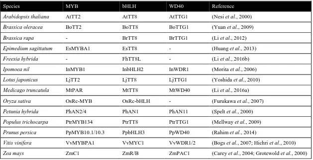
pathway in other plants (Zhang and Schrader, 2017). Table 1.1 shows the list of
Table 1.1 PA biosynthesis regulated by MBW complex in plants
Species MYB bHLH WD40 Reference
Arabidopsis thaliana AtTT2 AtTT8 AtTTG1 (Nesi et al., 2000)
Brassica oleracea BoTT2 BoTT8 BoTTG1 (Yuan et al., 2009)
Brassica rapa - BrTT8 BrTTG1 (Li et al., 2012)
Epimedium sagittatum EsMYBA1 EsTT8 - (Huang et al., 2013)
Freesia hybrida - FhTT8L - (Li et al., 2016b)
Ipomoea nil InMYB1 InbHLH2 InWDR1 (Morita et al., 2006)
Lotus japonicus LjTT2 LjTT8 LjTTG1 (Yoshida et al., 2010)
Medicago truncatula MtPAR MtTT8 MtWD40 (Li et al., 2016a)
Oryza sativa OsRc-MYB OsRc-bHLH - (Furukawa et al., 2007)
Petunia hybrida PhAN2/4 PhAN1 PhAN11 (Spelt et al., 2000)
Populus trichocarpa PtrMYB134 PtrTT8 PtrTTG1 (Mellway et al., 2009)
Prunus persica PpMYB10.1/10.3 PpbHLH3 PpWD40 (Rahim et al., 2014)
Vitis vinifera VvMYBPA1 VvMYC1 VvWDR1/2 (Bogs et al., 2007; Hichri et al., 2010)
Hypothesis and objectives
A single gene controls the SD trait which is associated with two SSR markers on
chromosome 7 of pinto beans (Felicetti et al., 2012; Junk-Knievel et al., 2008).
Moreover, 18 genes of PA biosynthesis pathway have been identified to be responsible
for the seed coat darkening in Arabidopsis (Appelhagen et al., 2014). Based on these
findings, I hypothesize that an orthologue of one of the 18 Arabidopsis PA biosynthetic
genes resides within the linkage distance of the SSR markers in pinto bean, and is a
potential Sd candidate gene. The objectives of my research are:
1. Analysis of the SSR marker region to identify potential candidate gene(s) for
postharvest seed coat darkening
2. Sequence analysis of the candidate gene(s) for gene structure, conserved domains
and evolutionary relationships.
3. Functional characterization of the candidate gene(s) and protein(s) by-
a. Determining the subcellular localization
b. Genetic complementation assay of the candidate gene(s) in respective
Arabidopsis mutant lines
c. Expression analysis of the PA late biosynthetic genes in Arabidopsis
MATERIALS AND METHODS
Plant materials and growth conditions
Nicotiana benthamiana plants were grown in a growth chamber set at 16-hour light at
25°C and 8 hours dark at 20°C cycle with 50% relative humidity. An average light
intensity of 70 μmol photons m-2s-1 was maintained. N. benthamiana seeds were
sprinkled onto the surface of wet PRO-MIX® BX MYCORRHIZAETM soil
(Rivière-du-Loup, Canada), and grown for one week. Individual seedlings were then transplanted into
single pots of soil and watered twice a week with a nutrient mixture of nitrogen (20%),
phosphorous (8%) and potassium (20%).
Pinto bean cv. CDC Pintium and 1533-15 seeds were surface sterilized with 70% ethanol
containing 3% H2O2 for 2 minutes and then rinsed six times with distilled water before
planting in soil. The growth chamber condition was the same as described above with an
average light intensity of 315 μmol photons m-2s-1. Plants were watered twice a week
with a nutrient mixture of nitrogen (20%), phosphorous (20%) and potassium (20%).
Wild-type (WT) A. thaliana accession Wassilewskija (Ws-2) and tt8 (line:
DEBTV122T3) mutant seeds were obtained from the Institut Jean-Pierre Bourgin (IJPB),
Institut National de la Recherche Agronomique (INRA, Versailles, France). Seeds were
cold treated by soaking in water for 2 days at 4°C. Upon spreading the seeds on wet soil,
the pot was covered with a plastic bag and kept in the growth chamber until the seedlings
appear. The growth conditions were as described for N. benthamiana with the average
light intensity of 120 μmol photons m-2s-1. Seedlings with 2 true leaves (approximately
mixture of nitrogen (20%), phosphorous (20%) and potassium (20%) was applied by
mixing with water.
To grow Arabidopsis under sterile conditions, seeds were surface sterilized with a 20%
(v/v) bleach (Lavo Inc., Canada) and 0.1% (w/v) sodium dodecyl sulphate
(Sigma-Aldrich, Germany) solution for 15 minutes with gentle shaking and then rinsed with
sterile distilled water for at least 6 times to remove bleach. The plant culture media was
prepared with 1X Murashige and Skoog (MS) basal salt (PhytoTechnology Laboratories,
USA) supplemented with 3% (w/v) sucrose (Sigma-Aldrich, Germany), 0.8% (w/v)
phytoagar (PhytoTechnology Laboratories, USA) and appropriate antibiotics (when
needed). For spreading, seeds were mixed with sterilized water and dispersed on the
plates. The sealed plates were kept in the dark at 4°C for 2 days and then placed at 24°C
with 16/8 hours light/dark cycle with an average light intensity of 80 µmol photons m-2s-1
until transplanted to the soil (after the appearance of 2 true leaves).
Bacterial strains and growth conditions
Escherichia coli strain DH5α (Invitrogen) and Agrobacterium tumefaciens strain GV3101
were grown in lysogeny broth (LB) or super optimal broth with catabolite repression
(SOC) media containing appropriate antibiotics at 37°C and 28°C, respectively, in a
shaker incubator at 225 RPM. Bacterial stocks transformed with desired plasmids were
In silico analysis
Pinto bean sequences were retrieved from the Phytozome database (P. vulgaris v2.1,
DOE-JGI and USDA-NIFA, http://phytozome.jgi.doe.gov/). Primers for SSR markers
were obtained from Felicetti et al. (2012) and listed in Table 2.1. Sequence similarities
were determined using the NCBI, basic local alignment search tool (BLAST) (Altschul et
al., 1990). Multiple sequence alignments were carried out using Clustal Omega followed
by BOXSHADE 3.21 (https://www.ch.embnet.org/software/BOX_form.html) (Sievers et
al., 2011). Phylogenetic trees were built using neighbour-joining program with the
bootstrap set to 1000, by MEGA7 software (Kumar et al., 2016). Protein sequence
alignments for the phylogenetic tree was done by ClustalW as a built-in program. Protein
motifs and molecular weight prediction were performed using InterProScan
(https://www.ebi.ac.uk/interpro) and ExPASy (https://web.expasy.org/translate/),
respectively. Tissue-specific RNA-seq data were retrived from the Phytozome database
and analyzed in Excel.
Genomic DNA extraction from leaves
Genomic DNA from Arabidopsis leaves was extracted by cetyl-trimethylammonium
bromide (CTAB) method (Porebski et al., 1997). Two frozen leaf discs were ground with
metal beads in MM300 MixerMill tissue disrupter (Qiagen) and mixed with 600 μL of
extraction buffer (1.4 M NaCl, 2% [w/v] CTAB, 50 mM Tris-HCl (pH 8.0), 10 mM
Ethylenediaminetetraacetic acid (EDTA) and 0.1 mg/mL RNase). Samples were
incubated at 65°C for 20 minutes and then cooled down to room temperature. After that,
g for 3 minutes. The aqueous phase was transferred into a fresh tube, and 0.6 volume of
isopropanol was added and mixed well. The sample was centrifuged at 15000 g for 10
minutes, supernatant removed, and DNA pellets were washed with 1 mL 70% (w/v)
ethanol. The pellets were air dried and dissolved in sterile MilliQ water. The quality of
extracted DNA was checked by gel electrophoresis (Section 2.6).
RNA extraction and RT-PCR
Total RNA was extracted from the seed coat (30-50 mg) and flower tissues of pinto bean
and leaves of Arabidopsis using the RNeasy Plant Mini kit (Qiagen). On-column DNA
digestion was performed using DNase I (Promega). RNA was quantified with a
NanoDrop 1000 spectrophotometer (Thermo Fisher Scientific). RNA quality was
evaluated by analyzing its A260/A280 ratio in NanoDrop and by gel electrophoresis
(Section 2.6). cDNA was synthesized from 0.5-1.0 µg of RNA using the ThermoScript™
RT-PCR System (Life Technologies). PCR amplification of the protein coding region
was performed using gene-specific primers (Table 2.1). Platinum® High Fidelity Taq
DNA Polymerase was used for PCR under following conditions: denaturation at 95°C for
1 minute, annealing for 30 seconds, extension at 68°C for 2 minutes (varied depending on
amplicon size), with a total of 25-40 cycles (varied) and final 5 minutes extension at
68°C. Upon gel electrophoresis, PCR amplicons were purified using the EZ-10 Spin
Column DNA Gel Extraction Kit (Bio Basic Inc.) and quantified using NanoDrop 1000
Gel electrophoresis
Different concentrations (0.8% for ≥ 3 Kb and genomic DNA, 1% for 1 Kb to 3 Kb, 1.5%
for RNA and 2% for ≤ 500 bp) of agarose gel based on the size of analyzing fragments
were made in 0.5X Tris-borate-EDTA (TBE) (for DNA) or 1X Tris-acetate-EDTA (TAE)
(for RNA) buffer. The liquid gel was mixed with RedSafe (iNtROn Biotechnology) stain
before it solidifies. 0.5X TBE (DNA) and 1X TAE (RNA) were also used as running
buffer and 100 kV voltage were used for electrophoresis. Nucleic acids were visualized
using Bio-Rad Gel DocTM.
Cloning and transformation procedure
Cloning into the Gateway entry vector
All primers used for cloning in Gateway compatible system were designed with attB1
adaptor sequence (5’-GGGGACAAGTTTGTACAAAAAAGCAGGCT-3’) for the
forward primer and attB2 adaptor sequence
(5’-GGGGACCACTTTGTACAAGAAAGCTGGGT-3’) for the reverse primer. PCR
products, amplified with Gateway primers (Table 2.1) were recombined into pDONR/Zeo
(Invitrogen) using Gateway BP Clonase® II Enzyme mix (Invitrogen). pDONR/Zeo
plasmid confers resistance to zeocin in E. coli (strain: DH5α).
Cloning into Gateway destination vectors
Entry clones were recombined into destination vectors: pEarleyGate101 (pEG101) and
pMDC32. pEG101 contains selectable marker conferring resistance to kanamycin in
a fusion reporter and it allows the inserted gene expression under Cauliflower mosaic
virus (CaMV) 35S promoter (Earley et al., 2006). pMDC32 contains selectable marker
conferring resistance to kanamycin in bacteria and hygromycin in plants. However, it
allows the inserted gene expression under dual 35S promoter (Curtis, 2003). The
recombination between the entry clone and respective destination vector was completed
by Gateway® LR Clonase™ II Enzyme mix (Invitrogen).
Bacterial transformations
For E. coli transformation, electro-competent DH5α cells were used. The BP or LR
reaction mix were used to transform E. coli via electroporation for 3-5 milliseconds at 1.8
kV in a Gene Pulser® Cuvette (Bio-Rad Laboratories, Inc.) with a 0.1 cm electrode gap
by MicroPulserTM (Bio-Rad Laboratories, Inc.). The cells were grown in SOC media for
1 hour and plated on respective antibiotic containing LB agar plates. Positive
transformants were identified by colony PCR using vector-specific and/or gene-specific
primers (Table 2.1).
For plasmid DNA extraction, EZ-10 Spin Column Plasmid DNA kit (Bio Basic Inc.) was
used. Extracted plasmid DNA was quantified using NanoDrop 1000 spectrophotometer
(ThermoScientific). Insertion of the correct DNA was confirmed by DNA sequencing,
carried out at the London Research and Development Center, Agriculture and Agri-Food
Canada in London, Ontario and Eurofins Genomics LLC, USA. For sequence analysis,
the DNASTAR® Lasergene software was used.
For A. tumefaciens transformation, electro-competent GV3101 cells were used. These
previously for E. coli except for 2.18 kV for 5-6 milliseconds. The transformation
reaction was plated on LB agar containing rifampicin (10 μg/mL), gentamycin (50
μg/mL), and kanamycin (50 μg/mL). Bacterial transformants were screened by colony
PCR using gene-specific primers (Table 2.1).
Subcellular localization
Transient expression of proteins in N. benthamiana leaves
A single colony of A. tumefaciens harboring an expression clone was grown at 28˚C in
infiltration culture media (LB broth containing 10 mM 2-N-morpholino-ethanesulfonic
acid (MES) (pH 5.6) and 100 μM acetosyringone with kanamycin (50 μg/mL), rifampicin
(10 μg/mL), and gentamycin (50 μg/mL)) until an OD600 of 0.5 - 0.8 was reached.
Bacterial cells were pelleted in a microcentrifuge at 1000 g for 30 minutes and
re-suspended in Gamborg’s solution (3.2 g/L Gamborg’s B5 and vitamins, 20 g/L sucrose,
10 mM MES (pH 5.6), and 200 μM acetosyringone) to obtain a final OD600 of 1.0. To
activate the virulence genes, Agrobacteria containing plasmid of interest were incubated
at room temperature for 1 hour with gentle agitation.
For co-infiltration, the A. tumefaciens (GV3101) containing nuclear localization signal
(NLS) fused with cyan fluorescent protein (CFP) in pEG100 vector was used (Earley et
al., 2006). Agrobacteria containing the gene of interest and NLS constructs were mixed
in 1:1 (v/v) for infiltration.
Leaves of 4-6 week old N. benthamiana were infiltrated with bacterial culture by placing
pressure. Plants were returned to the growth room at normal growth condition as
described in section 2.1 until visualization by confocal microscopy.
Confocal microscopy
The epidermal cell layers of the infiltrated N. benthamiana leaves were visualized 48
hours post-infiltration, using a Leica TCS SP2 inverted confocal microscope with a 63X
water immersion objective lens. For YFP visualization, the excitation wavelength was set
to 514 nm, and emission was collected at 530-560 nm. For CFP visualization, the
excitation wavelength was set to 434 nm and emission was collected at 470-500 nm. To
visualize the co-localization of the YFP and CFP signals, the ‘Sequential Scan’ tool was
used.
Arabidopsis transformation
A. tumefaciens harbouring appropriate expression clones were grown in 200 mL LB broth
containing kanamycin (50 μg/mL), rifampicin (10 μg/mL), and gentamycin (50 μg/mL)
until an OD600 of 0.8 – 1.0 was reached. Cells were harvested at 4000 g for 20 minutes at
4°C in Sorvall™ RC 6 Plus Centrifuge (Thermo Scientific™). Cell pellets were
re-suspended in infiltration media (0.5X MS salts, 0.5g/L MES, 5% sucrose, 0.03% silwet
L-77 and pH 5.75) to make a final dipping solution with OD600 of 1.0 (Clough and Bent,
1998).
Arabidopsis plants were grown (Section 2.1) until the flower bolts were around 10-15 cm
long and the first silique appeared. To ensure efficient transformation, siliques and
the infiltration media for 1 minute and were occasionally swivelled. After dipping, plants
were covered with plastic bags to maintain high humidity and kept in a dark chamber at
18°C for two days. The plants were then grown under normal growth condition for seed
collection (Section 2.1).
T1 seeds were screened on MS media containing hygromycin (50 mg/L) following the
method explained in section 2.1, and transformed plants were grown in soil to collect T2
seeds for further analysis.
Seed staining for PA detection
Arabidopsis seeds were treated with 4-dimethylamino-cinnamaldehyde (DMACA)
reagent to confirm the presence of PA as described by Zhao and Dixon (2009). Seeds
were stained in DMACA solution (0.5% (w/v) DMACA in methanol-3N HCl) for 36
hours in the dark (Li et al., 1996). Then seeds were de-stained by washing 6 times with
70% ethanol and stored in 70% ethanol. Both stained and unstained seeds were analyzsed
under a dissecting microscope, Nikon SMZ1500 (Nikon, Japan) with same light
Table 2.1 List of primers used for cloning, gene expression and gene identification
Primer name Sequence (5’ to 3’) Purpose
bHLH333GWF-CDC GGGGACAAGTTTGTACAAAAAAGCAGGCTTCATGGCTGCACCACTAGG
Amplification of full length PvbHLH333
from CDC Pintium bHLH333GWR-CDC GGGGACCACTTTGTACAAGAAAGCTGGGTCTGTCACCAAAATCAACTTTATA
bHLH333GWF-CDC-S GGGGACCACTTTGTACAAGAAAGCTGGGTCAGT CACCAAAATCAACTTTATA
bHLH333GWF-1533 GGGGACAAGTTTGTACAAAAAAGCAGGCTTCATGGCTGCACCACTAGGCAATA
Amplification of full length PvbHLH333
from 1533-15 bHLH333GWR-1533 GGGGACCACTTTGTACAAGAAAGCTGGGTCGTGGACAGCGTGGGGAATGATTCG
bHLH333GWR-1533-S GGGGACCACTTTGTACAAGAAAGCTGGGTTCAGTGGACAGCGTGGGGAATG
bHLH333F6 AGAGGACCTAACAGAATCCG
Sequencing
PvbHLH333 protein coding regions bHLH333R6 GAATAGCTCGTGAAAATGTTTTGC
bHLH333F2 GTTGGCTACATCACATATTCCCC
bHLH3337R TTCTTCCCGTTCGCATGTT
bHLH333LF TTCAACTACCAGCTTCAGCCCCTCATCTA
bHLH333LR TTCAACTACCAGCTTCAGCCCCTCATCTA
bHLH333-P2R CTAATAGATGAGGGGCTGCAA
deb122RB1 AGGAAGACAACTCAACCAGC
Arabidopsis specific (genotyping and transcript expression) deb122LB2 TCATCAGAATACAATTCTCAAATCT
deb122LB3 CTCCACGTGGCAAACGATGATTGG
Table 2.1 Continued
Primer name Sequence (5’ to 3’) Purpose
AtDFRR GTCTTATGATCGAGTAATGCGC Arabidopsis specific
(genotyping and transcript expression)
AtBANF AACAACTAAATCTCTATCTCTGTA
AtBANR GAATGAGACCAAAGACTCATATAC
AtEF1αA4 ATGCCCCAGGACATCGTGATTTCAT Positive control in A.
thaliana
AtEF1αA4 TTGGCGGCACCCTTAGCTGGATCA
M13F GTAAAACGACGGCCAGTCTTAA
Vector specific primers
M13R TGCCAGGAAACAGCTATGAC
pEGF TGACGCACAATCCCACTATCC
pEGR AAATATCATGCGATCATAGGCG
PvUbQF ACAGCTGGAGGATGAAAGGA Positive control in P.
vulgaris
PvUbQR GTCCGAACTCTCCACCTCAA
Pvsd-1157F AATGGGGAAGATGGTTGGTT
Primers used to locate SSR markers (in silico)
Pvsd-1157R GTGAGGGTTGAAAATTGCGT
Pvsd-1158F GCAATTGACAAAAAGCTTCG
Pvsd-1158R TTGTCATGCGGTTTT
Pvsd-0028F TGAAACGCCTAGATAAAATTTAAAAC
RESULTS
Comparison of physical and linkage distances identifies a candidate gene
Two SSR markers Pvsd-1157 and Pvsd-1158 were found to be linked with the Sd gene in
pinto bean SD cultivars (Felicetti et al., 2012). The markers and Sd were mapped onto
chromosome 7 based on their linkage distances and showed that Sd is located between
Pvsd-1157 and Pvsd-1158 markers (Figure 1.4). The genome of the common bean
Andean landrace, G19833, is recently sequenced (Schmutz et al., 2014). As G19833 and
pinto beans are two different market classes of common beans with the same Andean
origin, hence it is expected that their genomic sequences are similar. The whole genome
sequence of G19833 was used as a reference to identify the candidate Sd.
Using the primer sequences provided by Felicetti et al. (2012), the sequence and the
location of Pvsd-1157, Pvsd-1158 and Pvsd-0028 markers were identified on
chromosome 7. As shown in Figure 3.1, Pvsd-1158 is located closer to the centromere
and the physical distance between Pvsd-1157 and Pvsd-1158 is 57713 bp. The Pvsd-0028
marker is located further downstream from the Pvsd-1157 (Figure 3.1). A search for open
reading frames (ORFs) between the Pvsd-1158 and Pvsd-1157 markers found 2 genes;
FANTASTIC FOUR (FAF) and RAS GTPase (RAS) (Figure 3.1). The gene functions
retrieved from the published literatures for these two genes did not identify any link with
seed coat darkening (details described in Section 4.1). The search was extended further to
the 50 kb upstream and downstream regions of Pvsd-1158 as this is tightly linked marker
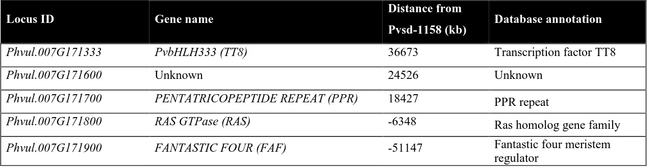
with the SD trait. Within this area, 3 genes PENTATRICOPEPTIDE REPEAT (PPR), an
Figure 3.1 Schematic diagram showing locations of genes present within the
linkage region of Pvsd-1158 in pinto bean.
Comparison of linkage and physical distances of the Pvsd-1158 and Pvsd-1157
markers on chromosome 7 identifies 5 genes in the 50 kb region surrounding the
Pvsd-1158. The candidate PvbHLH333 (red) is located in the centromere side of the
Table 3.1 List of genes present within 50 kb regions of Pvsd-1158 with their annotated functions in pinto bean
-ve values indicate the locations on other side from Pvsd-1158
Locus ID Gene name Distance from
Pvsd-1158 (kb) Database annotation
Phvul.007G171333 PvbHLH333 (TT8) 36673 Transcription factor TT8
Phvul.007G171600 Unknown 24526 Unknown
Phvul.007G171700 PENTATRICOPEPTIDE REPEAT (PPR) 18427 PPR repeat
Phvul.007G171800 RAS GTPase (RAS) -6348 Ras homolog gene family
Phvul.007G171900 FANTASTIC FOUR (FAF) -51147 Fantastic four meristem
functional analysis (Section 4.1), TT8 was the most appropriate candidate Sd, as it plays
an important role in the seed darkening in Arabidopsis. In this thesis, Arabidopsis TT8
orthologue in pinto bean is named as PvbHLH333. By assuming that PvbHLH333 is Sd,
the linkage and physical distances were recalculated. Table 3.2 shows the linkage and
physical distances between the SSR markers and the previously identified PvbHLH333 in
F2 population. The distance between Pvsd-1157 and PvbHLH333 is 0.9 cM (linkage) or
94.38 kb (physical). The Pvsd-1158 and PvbHLH333 have linkage distance of 0.4 cM
and physical distance of 36.04 kb. Similar study has reported previously to correlate
linkage and physical distance (Kong et al., 2004). However, the linkage and physical
distances between Pvsd-1157 and Pvsd-1158 does not follow the correlation and their
location proposed by Felicetti et al. (2012) appear to be in the opposite orientation with
respect to the marker Pvsd-0028. Based on this analysis, I propose that, the SSR markers
Pvsd-1157 and Pvsd-1158 flank the Sd on the same side of the chromosome and the
revised map is shown in Figure 3.1.
PvbHLH333 transcripts accumulate in flower buds, flowers and pods
To determine the tissue-specific transcript level of the 5 genes located within the 50 kb up
and downstream of the Pvsd-1158 marker, their available transcriptome data were
retrieved from the Phytozome database (Schmutz et al., 2014). Transcript accumulation
data for flowers, flower buds, young pods, green mature pods, stems, leaves, roots,
nodules, and young trifoliate leaves at different developmental stages were obtained.
Table 3.2 Linkage and physical distances between the markers and PvbHLH333 pinto bean
Markers and gene Linkage distance in
RIL (cM)
Linkage distance in F2
(cM)
Physical distance
(kb)
Pvsd-1157 and Pvsd-1158 0.5 1.3 57.73
Pvsd-1157 and PvbHLH333 0.5 0.9 94.38
Figure 3.2 Tissue-specific transcript levels of genes present within 50 kb of
Pvsd-1158 in common bean/ pinto bean.
RNA-seq data across various tissues were retrieved from the Phytozome database. The
relative transcript levels are in units of fragments per kilo base per million mapped
pods and young pods while no transcripts were detected in leaves, nodules and roots
(Figure 3.2). PvbHLH333 in tissues that lead to the formation of seeds is consistent with
a possible role in seed coat coloring. Phvul.007G171900 (FAF) has also displayed a
moderate transcript level in flower buds, but no transcript was detected in the pods which
makes FAF a weak candidate for the Sd. The transcript levels of Phvul.007G171800
(RAS), Phvul.007G171700 (PPR) and Phvul.007G171600 (unknown gene) were
abundant in vegetative tissues but not in seed coat related tissues, indicating they are
possibly not related to the seed coat darkening in pinto bean.
PvbHLH333 contains TT8-specific conserved motifs and forms a clade with
legume TT8s
An analysis of the PvbHLH333 protein sequence identified four distinct motifs: an N
terminal MYB interaction region (MIR), followed by a poly-glutamate (Poly E) site, a
signature bHLH and an ACT-like domain on the C-terminus (Figure 3.3A). These motifs
are also present in other characterized TT8 proteins. A multiple sequence alignment of
the PvbHLH333 with TT8 orthologues from A. thaliana,E. sagittatum, L. japonicus and
M. truncatula showed highly conserved regions in these protein motifs (Figure 3.3B).
Among the four motifs, Poly E is the most variable in sequence.
A phylogenetic tree was constructed for PvbHLH3333 and known TT8s from A. thaliana,
B. oleracea, B. rapa, E. sagittatum, I. nil, L. japonicus, M. truncatula, O. sativa, P.
hybrida, S. melongena V. vinifera, and Z. mays (Figure 3.4, Table 1.1). P. vulgaris
Phaseolin G-box protein (PG1) was used as an outgroup to root the tree (Kawagoe and
Figure 3.3 Multiple sequence alignment of PvbHLH333 and other TT8s found
highly conserved motifs.
A. Domain structure for PvbHLH333, from N- to C-terminus MYB interaction
region (MIR), poly glutamate (E), basic Helix-Loop-Helix (bHLH) and
ACT-like motif.
B. The protein sequence alignment of PvbHLH333 (P. vulgaris,
Phvul.007G171333), LjTT8 (L. japonicas, BAH28881.1), EsTT8 (E.
sagittatum, KC686401), AtTT8 (A. thaliana, NM_117050) and MtTT8 (M.
truncatula, KM892777). The motifs are shown with colored bars as in Figure
Figure 3.4 Phylogenetic analysis of characterized TT8s.
A neighbour-joining tree of TT8 proteins related to PvbHLH333 (■) was built using
MEGA 7 software. Bootstrap values (%; based on 1000 bootstraps) are shown next
to the branch-points. Protein sequence alignments were performed using MUSCLE.
The compared proteins are: AtTT8 ( A. thaliana), BoTT8 (B. oleracea), BrTT8 (B.
rapa), EsTT8 (E. sagittatum), LjTT8 (L. japonicus), InbHLH (I. nil), MtTT8 (M.
truncatula), OsRc (O. sativa), PsbHLH (P. Sativum), PhAN1 (P. hybrida), SmTT8
(S. melongena), VvTT8 (V. vinifera), ZmR-S (Z. mays) and ZmIN1 (Z. mays). The
locus IDs are indicated in parentheses. PG1 (P. vulgaris, Phaseolin G-box 1) was
includes as outgroup. Blue box indicates the clade of PvbHLH333 with the legumes
TT8 related proteins. Scale bar indicates the average number of substitutions per site.
Database sequence correction
When this project was initiated in June 2016, Phytozome version 11 (P. vulgaris release
version 2.0) annotated the candidate PvbHLH333 as two separate genes
(Phvul.007G171300 and Phvul.007G171400). These two genes were separated by a large
8080 bp putative intergenic region (Figure 3.5A). Phvul.007G171300 and
Phvul.007G171400 showed high sequence similarities with the N- and C-termini of the
same characterized TT8 orthologue when a blast search was done. Hence it was
suspected that the two annotated DNA regions code for a single protein. To confirm this,
a 3’ primer was designed from the last exon of Phvul.007G171300 and a 5’ primer from
the first exon of Phvul.007G171400 to amplify the region from the cDNA as a template
(Figure 3.5B). The resulting 216 bp amplicon was sequenced and it confirmed that these
two annotated genes are a single gene and the 8 kb putative non-coding region is an
intron (Figure 3.5C). Later the corrected sequence was released in Phytozome version 12
(P. vulgaris release version 2.1) in January 2017. McClean et al. (2018) also confirmed
that Phvul.007G171300 and Phvul.007G171400 are the physical component of the same
gene by mapping and comparing the RNA-seq data with the reference sequence.
Sequence analysis identifies variation in PvbHLH333 from CDC Pintium and
1533-15
PvbHLH333 transcripts of CDC Pintium and 1533-15 were analyzed to determine if there
are any sequence variations in these cultivars. Primers were designed using the reference
Figure 3.5 PvbHLH333 was annotated as two different genes.
A. Screenshot of Phytozome v.11 browser (captured in 2016) showing PvbHLH333
as two different genes- Phvul.007G171300 and Phvul.007G171400. NC:
Non-coding.
B. Sequencing of the coding region from cDNA, identified a 15 bp exon (exon 4, red
box) and extra regions to the end of exon 6 and beginning of exon 7 (yellow bars).
Boxes are the exons; lines are the introns and numbers underneath indicating the
size in nucleotides.
C. Agarose gel electrophoresis confirms the splicing of 8 kb intron from the target
gene of both CDC Pintium and 1533-15. The positions of primers used for PCR