Western University Western University
Scholarship@Western
Scholarship@Western
Electronic Thesis and Dissertation Repository
2-22-2018 10:00 AM
Augmented Reality Simulation Modules for EVD Placement
Augmented Reality Simulation Modules for EVD Placement
Training and Planning Aids
Training and Planning Aids
Hamza Waleed Ghandorh
The University of Western Ontario
Supervisor Eagleson, Roy A.
The University of Western Ontario
Graduate Program in Electrical and Computer Engineering
A thesis submitted in partial fulfillment of the requirements for the degree in Doctor of Philosophy
© Hamza Waleed Ghandorh 2018
Follow this and additional works at: https://ir.lib.uwo.ca/etd
Part of the Other Electrical and Computer Engineering Commons
Recommended Citation Recommended Citation
Ghandorh, Hamza Waleed, "Augmented Reality Simulation Modules for EVD Placement Training and Planning Aids" (2018). Electronic Thesis and Dissertation Repository. 5216.
https://ir.lib.uwo.ca/etd/5216
This Dissertation/Thesis is brought to you for free and open access by Scholarship@Western. It has been accepted for inclusion in Electronic Thesis and Dissertation Repository by an authorized administrator of
Abstract
When a novice neurosurgeon performs a psychomotor surgical task (e.g., tool naviga-tion into brain structures), a potential risk of damaging healthy tissues and eloquent brain structures is unavoidable when novices make multiple hits. As a result, a set of undesirable trajectories is created, and resulting in the potential for surgical complica-tions. Thus, it is important that novices not only aim for a high-level of surgical mastery but also receive deliberate training in common neurosurgical procedures and underlying tasks.
Paul Fitts’ methodology. The data showed that novices had a learning curve and their performance as speed and accuracy trade-off was measured and reported.
Neurosurgical simulators are prone to perceptual distance underestimation. In virtual spaces, humans underestimate their egocentric distance by 80% as a result of inaccurate depth perception. Human beings are capable of perceiving 3D shapes of physical objects using perceptual motion cues. Few investigations were conducted for improving user depth perception in head-mounted display (HMD)-based AR systems with perceptual motion cues. Consequently, we report our investigation’s results about whether or not head motion and perception motion cues had an influence on users’ performance. The data showed that users’ physical head motion and perceptual motion cues, as a combined factor, is a good indicator of better user performance.
Acknowledgements
All the praises, and foremost thanks are due to Allah for being my refuge and strength’s source in time of ease and hardship. Without His guidance and blessings, nothing is possible.
First, I would like to express my deep appre-ciation to my supervisor Prof. Roy Eagleson. His mind and heart were always open for discussion, en-couragement, insightful guidance, consistent support, invaluable advice, and constructive criticism. I ap-preciate his time and ideas that have helped me to be highly productive. With his involvement and pa-tience, I have been able to write my Ph.D thesis suc-cessfully. Also, I would like to thank my advisory committee for their continuous encouragement, kind-ness and support at all stages of my PhD study. In addition, I would like to thank Associate Vice-Provost at School of Graduate and Postdoctoral Studies (Prof. Lorraine Davies) for her advice and encouragement to be the person I am today.
I would also like to express my thanks to all my fantastic friends and our research group members for their kind support and encouragement. Special thanks go to my friends at Western with whom I had the privilege to work and the discussions with them have benefited me immensely.
Last but not the least, I would like to thank The Ministry of Education and Taibah University for the scholarship and financial support through Saudi Cultural Bureau in Canada, without which this work would not have been possible.
Dedication
”There is no chance, no destiny, no fate, that can hinder or control the
firm resolve of a determined soul.” By Ella Wheeler Wilcox
I dedicate this work to my parents, my wife, my family-in-law, my children,
my siblings, my friends, and for every person whom provided me with kind
Table of Contents
Abstract . . . i
Acknowledgements . . . iii
Dedication . . . iv
List of Tables . . . viii
List of Figures . . . ix
List of Acronyms . . . x
List of Symbols . . . xii
List of Appendices . . . xiii
1 Introduction . . . 1
1.1 Motivations and problem statement . . . 3
1.2 Research objectives . . . 6
1.3 Contributions . . . 8
1.4 Thesis organization . . . 8
2 Literature Review . . . 10
2.1 Augmented Reality vs. Virtual Reality . . . 10
2.2 Neurosurgery . . . 15
2.2.1 Preoperative stage . . . 15
2.2.2 Ventriculostomy . . . 17
2.2.3 External ventricle drain placement . . . 18
2.3 Surgical Simulators . . . 20
2.3.1 Types . . . 20
2.3.2 Motivation of simulation-based surgical training . . . 22
2.3.3 Simulator validation . . . 24
2.4 User learning process . . . 25
2.5 Human-computer interaction . . . 27
2.6 Perception depth in surgical training . . . 27
2.7 User performance assessment . . . 30
2.7.1 User performance . . . 30
2.7.2 Hierarchical task analysis . . . 32
3 Related Work . . . 36
3.1 Introduction . . . 36
3.2 Web-based simulation . . . 37
3.3 Neuronavigation-system-based simulation . . . 38
3.4 Mixed-reality-based simulation . . . 39
3.5 3D-printing-based simulation . . . 41
3.6 Handheld-modalities-based simulation . . . 42
3.7 Summary and Conclusions . . . 44
4 Design of Augmented Reality Training and Planning Aid Simulation Modules for EVD Placement Using Model-driven Engineering . . . . 45
4.1 Introduction . . . 45
4.2 Methodology . . . 47
4.2.1 Our MDE-based approach . . . 47
4.3 Simulation modules description . . . 53
4.3.1 Design considerations . . . 53
4.3.2 Simulation modules setup . . . 53
4.3.3 Hardware description . . . 60
4.3.4 Software description . . . 61
4.3.5 Architecture . . . 64
4.3.6 User performance evaluation . . . 64
4.4 Discussion . . . 67
4.5 Summary and Conclusions . . . 70
5 AR Training and Planning Simulation Modules for EVD Placement Tasks As Assessment Tools . . . 71
5.1 Introduction . . . 71
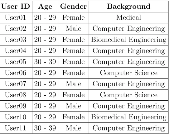
5.2 User Study 01 . . . 73
5.2.1 Subjects . . . 73
5.2.2 Apparatus/Materials . . . 74
5.2.3 Procedure . . . 74
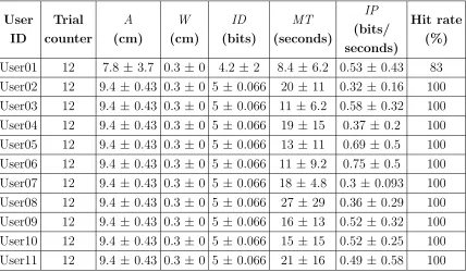
5.2.4 Results . . . 79
5.3 User study 02 . . . 82
5.3.1 Subjects . . . 82
5.3.2 Apparatus/Materials . . . 83
5.3.3 Procedure . . . 83
5.3.4 Results . . . 85
5.4 Discussion . . . 86
6 Investigation of Users’ Depth Perception Based on Structure Through
Motion Visual Cue . . . 89
6.1 Introduction . . . 89
6.2 Methodology . . . 92
6.3 Results . . . 92
6.4 Discussion . . . 95
6.5 Summary and Conclusions . . . 96
7 Conclusion . . . 97
7.1 Thesis summary . . . 97
7.2 Future work . . . 99
References . . . 101
A Appendix A : Research Ethics Approvals . . . 108
B Appendix B : Mental Rotation Test . . . 112
List of Tables
Section Page
2.1 A comparison between AR modalities used in the medical domain. . . 14 2.2 Surgical hierarchal task analysis example. . . 32 4.1 User viewing configuration applied to assign AR simulation scenes and
functionality. . . 56 4.2 Technical specifications of the used environments for the AR simulation
modules. . . 60 5.1 Subjects demographics in the User Study 01. . . 73 5.2 Mean and stand deviation values of subjects’ tasks and its description
throughout the User Study 01 in the first setting. . . 79 5.3 Mean and stand deviation values of subjects’ tasks and its description
throughout the User Study 01 in the second setting. . . 80 5.4 Mean and stand deviation values of subjects’DE,TE, andRE throughout
the User Study 01 in the first setting. . . 81 5.5 Mean and stand deviation values of subjects’DE,TE, andRE throughout
the User Study 01 in the second setting. . . 81 5.6 Subjects demographics in the User Study 02. . . 82 5.7 Mean and stand deviation values of subjects’ tasks and its description
throughout the User Study 02 in the first setting. . . 85 5.8 Mean and stand deviation values of subjects’ tasks and its description
List of Figures
Section Page
1.1 Residents divided their attention between multiple visualization modalities. 5
2.1 Milgram and Kishino’s continuum. . . 11
2.2 Three different visualization modes of real historical scene. . . 11
2.3 AR artefact-based app for medical uses . . . 12
2.4 Three types of AR modalities . . . 13
2.5 MRi and CT scans used for the surgical planning stage. . . 16
2.6 Ventriculostomy procedure workflow. . . 18
2.7 Overview of EVD placement targeting task. . . 19
2.8 Relationship between users’ performance and learning. . . 26
2.9 Overview of common visual depth cues. . . 30
4.1 Typical software development life cycle vs. MDA development vision. . . 48
4.2 A simplified flowchart of MDE-based approach. . . 49
4.3 A surgical scenario of the EVD placement task in Harel statechart notation. 50 4.4 Unity editor and PIM models. . . 52
4.5 Physical version of the two trackable predefined fiducial markers. . . 54
4.6 The registration points in the soft version of the fiducial markers. . . 55
4.7 3D meshes used with the AR simulation modules’ scenes. . . 57
4.8 AR simulation modules’s underlying scenes. . . 59
4.9 AR simulation modules and their environments. . . 61
4.10 AR simulation modules’ use case diagram. . . 62
4.11 AR simulation modules’ class diagram. . . 63
4.12 AR simulation modules’ architecture diagram. . . 64
4.13 Example of users’ speed and accuracy trade-off diagram and its implica-tions on their performance. . . 66
4.14 User‘s trial log file example. . . 67
5.1 Workflow of EVD placement targeting tasks in the user studies . . . 76
5.2 Example of a trial on the AR simulation module. . . 77
5.3 The subject’s stereoscopic view through the running of trial. . . 78
5.4 Workflow of EVD placement targeting tasks in the user studies in the second setting . . . 84
6.1 Users’ performance trade-off through the User Study 01 with targeting the virtual ellipsoids. . . 93
List of Acronyms
2D Two-dimensional
3D Three-dimensional
AR Augmented Reality
CSF Cerebrospinal Fluid
CSV Comma Separated Values
CT Computed Tomography
DOF Degree Of Freedom
EP Entry point
ER Emergence room
EVD External Ventricular Drain
FOV Field Of View
GPS Global Position System
GUI Graphical User Interface
HCI Human-Computer Interaction
HMDs Head-Mounted Displays
HTA Hierarchical Task Analysis
ICU Intensive Care Unit
MDA model-driven Architecture
MR Mixed Reality
MRi Magnetic Resonance Imaging
MRT Mental Rotation Test
OR Operating room
QR Quick Response
PIM Platform-independent Models
PSM Platform-specific Models
SFM Structure From Motion
TP Target point
VR Virtual Reality
List of Symbols
A Amplitude
B Bandwidth
C Human capacity
DE Distance error
ID Index of Difficulty
IP Index of Performance
MT Movement of Time
N Noise power
RE Rotational error
S Single power source
TE Transitional error
List of Appendices
Section Page
Chapter 1
Introduction
In many clinical disciplines, preventable medical errors are considered to be a signifi-cant challenge in clinical settings (e.g., operating rooms (OR), emergency rooms (ER), or intensive care units (ICU)). In the United States of America, a study conducted by the National Practitioner Data Bank stated that at least 4000 patients were injured by preventable surgical errors between 1990 and 2010 [1]. Preventable medical errors are con-sidered as the third leading cause of death, killing 251,000 American citizens. In Canada, the University Health Network’s Annual General meeting stated that preventable medical errors cost Canadian taxpayers over $396 million annually. In 2014 alone, 30,277 Canadi-ans died in acute care due to preventable medical errors, which makes preventable medical errors the main cause of death for Canadians—more than stroke, diabetes, Alzheimer’s disease, and kidney disease combined [2]. These preventable medical errors might be a result of technical errors during complex surgeries with a hierarchy of interconnected surgical tasks. Therefore, mastery of technical surgical skills is becoming increasingly important for novice residents 1 during their training programs.
When Abraham Flexner and William Stuart Halsted proposed the concept of resi-dency (i.e., Halstedian teaching model or apprenticeship model) [3], their training model
became the most used approach for surgical teaching and training. Because the surgical occupation necessitates an education that is as practical as the occupation itself [4], the apprenticeship model has been applied due to its longevity and the adoption of novel sur-gical techniques [5]. Although the apprenticeship model engages novice residents in real surgical scenarios and exposes them to practical tips and feedback from senior residents, the medical community has slowly been moving away from the apprenticeship model due to its disadvantages [5]. The apprenticeship model puts patients’ safety at high risk of medical errors, imposes tremendous operating costs, and lacks flexibility and efficiency. Besides the apprenticeship model, conventional means (e.g., cadavers’ atlases, laboratory animals, and sophisticated mannequins) have been used as supplementary materials to surgical training for the purpose of anatomical knowledge [6]. However, the conventional means also pose many educational limitations. For instance, the conventional means ex-pose medical students to generic and static anatomical materials or different anatomical structures than human beings [7]. Also, the conventional means are unable to on mimic physiological responses that happen in clinical settings [6].
surgical interventions. The interventions include navigation or manipulation of tool tasks, for instance, placement of an external ventricular drain (EVD)’s catheter towards a des-ignated nervous region (e.g., eloquent brain tissues) and targeting a specific fracture of the region. The postoperative stage includes a set of steps for complication management and incision closing [8].
1.1
Motivations and problem statement
Technical and non-technical skills are increasingly valuable for neurosurgeons as part of their training. Technical skills set includes manual dexterity, or specialist knowledge [9], while non-technical surgical skills set includes interpersonal and cognitive skills. Team-work, planning, or resource management are few examples of the interpersonal skills. Situation awareness, decision making, and adaptive strategies are few examples of cog-nitive skills [10]. Mastery of the neurosurgery domain depends on the mixture of both skills set. However, some surgical procedures or tasks might need more emphasis on some skill set more than the others.
Although senior neurosurgeons could proctor a few early ventriculostomy2 training sessions, novice trainees are expected to become proficient very early in their residency programs in such surgery, and they need to cope with expected high volume of operat-ing with their own judgment for new or different neurosurgical cases [11]. Thus, it is improbable that the trainees will benefit from having an access to the same standard of
training. This situation results in a steep learning curve in many parts of the procedure in terms of the whole operation or its interconnected psychomotor tasks, such as the EVD placement task [6].
Trainees suffer from an overwhelming cognitive load while performing EVD place-ment tasks within risky and interesting nervous regions, which is a source of inaccurate intervention planning [12]. Trainees need to mentally synthesize a two-dimensional (2D) version of medical imaging datasets in order to maintain a three-dimensional (3D) view of the inner nervous structure (i.e. 2D-to-3D transformation). Figure 1.1 depicts a typical trainee’s behavior before committing an intervention. The process of receiving a large amount of information from multiple 2D screens at the same time might lead to position and orientation disparity between a working space (i.e., the patient’s organ) and the res-ident’s perception of the working space. This disparity could lead to inaccurate hand-eye coordination for the residents, a contributing factor for ill-posed surgical decisions. This disparity a known issue in 2D user interfaces during studying or consuming two or more imaging datasets [13]. The 3D view is a standard as a guidance base for their current surgical intervention.
Fig. 1.1: Residents divided their attention between multiple visualization modalities to main-tain a mental view and make a decision about the current surgical step [13].
[13]. Access to such training and features would empower trainees to cope with the challenge of anatomical clarity and to shorten their learning curve regarding applying novel surgical techniques [16].
requirements are necessary including diverse technological platforms for deployment and wider user landscapes. Moreover, the influence of human factors, for instance, cogni-tive ergonomics3, plays a critical role when designing neurosurgical simulation tools [12]. Using the same AR technology for EVD placement training and planning aids with the depth perception enhancement remain a largely unexplored area that is ripe for study.
1.2
Research objectives
In this thesis, we focused on the development of an AR simulation module for an EVD placement task (i.e., psycho-motor targeting task) for training and planning aids for novice residents. Our research project had three research aims. The first aim of our study was a description of our approach to accomplish the development of AR simula-tion modules based on model-driven engineering (MDE)4. The outcome of the MDE tech-nique produced two software executables: 1) a stationary AR module, and 2) a portable AR module. Our hypothesis was that the MDE technique is a suitable approach for the development of AR simulation tools with the flexibility to cope with technological issues in terms of patient-specific design requirements, 3D surface rendering, and the fragmentation of multiple computing platforms.
The second aim of our research was to test the feasibility of the developed AR simulation modules. Through two user studies, the verification was intended to detect
3. Cognitive ergonomics is a field concern about mental processes and their interactions among humans and other elements of a system.
quantitative differences in performance between novices with different knowledge back-grounds. The user studies were being conducted through several experimental trials in which participants conducted a variety of targeting tasks into virtual 3D ellipsoids and brain structure meshes. Statistically, discriminating evidence indicates that the AR mod-ule objectively measures the users’ performance between different settings as it is claimed to assess. This data establishes the evidence to strongly advocate for the use of the AR simulation modules as a valuable assessment tool for EVD placement tasks training. Our hypothesis was that whenever users practice the EVD placement tasks with different tri-als, consistent feedback, and cognitive efforts, users’ accuracy to perform the same tasks will improve over time.
1.3
Contributions
Contributions to the AR research and medical communities expected from this research are as follows:
• Provide an empirical evidence that the MDE technique could be used for the devel-opment of psychomotor-task-oriented AR simulation modules for the neurosurgical domain and possible avenues.
• Establish a methodology for quantifying users’ performance using psychomotor-task-oriented AR simulation modules for the EVD placement tasks and demonstrate the developed AR simulation module assessing the skill it intends to assess.
• Understand the effect of users’ physical head motion and perceptual motion cues, as a combined factor, on users’ depth perception while performing EVD placement tasks through the application of the AR simulation modules.
1.4
Thesis organization
Chapter 2
Literature Review
This chapter states basic definitions and background utilized to develop the ideas in this thesis.
2.1
Augmented Reality vs. Virtual Reality
AR is a visualization technology intended to enhance users’ perception of reality through overlaying computer-generated information upon physical objects [16][17]. AR facilitates users’ direct interaction with the physical world while maintaining a perception of the surrounding world and underlying events. VR, another visualization technology, is in-tended to immerse the users in a created virtual world in which the users would gain new insights and live new experiences with configurable settings [18]. VR indulges users in synthetic settings in which users cannot recognize their surroundings [16].
Fig. 2.1: Milgram and Kishino’s continuum and the relationship between AR, VR, and real environment [19].
Fig. 2.2: (Left): the real historical scene without any modifications. (Middle) the real historical scene enhanced with AR information. (Right) the real historical scene represented in VR mode [20].
VR and AR both leverage a mixture of technologies to enable users to navigate and manipulate objects for a particular purpose in the virtual world through their own rules and coordinates system [18]. Both technologies engage users into conventional or new experiences that aren’t commonly expected: a concept that possibly could revolutionize people’s perception and actions in many fields. The primary goal of VR is to completely replace users’ reality with a video-like experience, rather than supply their reality with augmented 3D virtual objects, as in the case of AR technology [14].
codes, barcodes or well-defined images that were scanned by a camera, as the base for the rendering process of 3D virtual information. Geolocated-based AR visualization mode utilizes location sensing and mapping references (e.g., latitude and longitude coordinates) by means of Global Position System (GPS) devices as the base for the rendering process of 3D virtual information [21].
AR technology has been used in many fields, for instance, education and medicine. Figure 2.3 shows an example of a use of an artifact-based AR app for visualization and medical purposes. When users point their AR-supported device at the desired marker, the registered medical information will be aligned with the marker, and their visual representation or feedback will emerge on the device’s screen1 [22].
Fig. 2.3: AR-based app for medical uses (source: http://www.goo.gl/yWbxhk).
coordination or orientation in the physical world. The high AR demand for computing resources requires advanced computing capabilities including high processing and storage, sophisticated graphics cards, and modalities [17]).
AR modalities have been classified into three groups: head-mounted displays (HMDs), handheld displays (e.g., smartphone devices), and spatial displays (e.g., video projectors, holograms), which are depicted in Figure 2.4. HMDs involve semi-transparent mirrors attached to a helmet or goggles to illustrate real scenes and augmented 3D virtual ob-jects over the users’ sight. Handheld displays include smartphone or camera devices with which users see and consume generated or rendered 3D virtual objects upon physical objects. Spatial displays are based on projector devices to cascade graphical information directly onto physical objects in open spaces without the users’ involvement to wear or carry any equipment.
Fig. 2.4: AR displays in three different types: (left) Head-mounted display (HMD), (middle) handheld displays and (right) spatial display [17].
setting in which users are less distracted with multiple displays; they provide researchers with possible accommodation for research prototypes and support a variety of depth perception cues. Also, because HMDs are portable and they do not show physical and virtual object occlusion problems, they allow users a 360-degree view of the surrounding physical space to visualize and interact with 3D virtual images in diverse directions. HMDs, however, have a limited field of view (FOV) and low spatial resolution, and their users might suffer from ergonomic issues due to their weight and fit [23].
Tab. 2.1: A comparison between AR modalities used in the medical domain [13].
appearance [24].
2.2
Neurosurgery
Neurosurgical trainees (i.e., junior residents) spend their residency training for a variety of neurosurgeries2 and building related technical skills (e.g., dexterity) or non-technical skills (e.g., teamwork, communication). The process of any neurosurgery goes through three phases: preoperative (e.g., planning), operative (e.g., intervention), and postoperative (e.g., recovery). The preoperative stage involves creating a blueprint of the neurosurgery’s workflow by the application of health-related records and patient-specific imaging studies (e.g., MRI or CT scan) intended to represent patients’ medical situations as accurate as possible. Figure 2.5(a) shows an example of the two brain imaging studies, and Figure 2.5(b) shows residents using the imaging studies to plan their next surgical steps. The operative stage involves a set of deliberate surgical interventions. The interventions include navigation or manipulation of tool tasks, for instance, placement of a surgical tool towards a designated nervous region (e.g., eloquent brain tissues) and targeting a specific fracture of the region. The postoperative stage includes a set of steps for complications management and incision closing [8].
2.2.1
Preoperative stage
Traditionally, trainees plan their surgical interventions based on a manual review of patient-specific imaging studies as 2D slices or through 2D displays. The review process
(a) (b)
Fig. 2.5: (a) brain imaging studies for CT scan and MRI example (source: http://goo.gl/ 3WvrpJ), and (b) brain imaging studies are used by residents for planning purposes (source: http://goo.gl/6pA9nB).
is necessary for surgeons to build their own 3D cognitive models of the patients’ anatomy and pathology3 [3][7].
According to Navkar et al. [25], the process of the preoperative stage or planning aims to determine two aspects: a point representing an entrance to the patient’s organs and a path towards the nervous region of interest. Eck et al. [26] described the planning process for a neurosurgical procedure as a blend of trajectory preparation and volume exploration processes. Trajectory preparation refers to drawing two 3D points in the Euclidean space within a predefined reference frame. The first position (i.e., target point (TP)) will be located within the region of interest underneath the skin of the head, and the second point (i.e., entry point (EP)) will be located on the outer surface of the skin of the head. The volume exploration process develops the trainee’s ability interactively to 1) visualize patients’ volumetric and geometric information, 2) control the rendered content, and 3) understand spatial relations between nervous structures in the risky and safe regions and the planned trajectories within them.
Trainees usually encounter many challenges in accomplishing accurate planning. For example, preoperative planning intensely consumes a deep level of cognitive capacities (e.g., spatial and perceptual) and sensorimotor skills of the trainees in typical situations. The same task will be more perceptually aggressive for trainees in acute situations of patients at ER or OR [12][27]. This phenomenon occurs because trainees usually perform their planning through a mental 2D-to-3D transformation of patient‘s imaging studies in very limited timespan. The 2D-to-3D transformation is needed to achieve two tasks: 1) to maintain a 3D mental viewpoint of patients‘ circumstances and anatomy during an operation, and 2) to enable the trainees to apply their medical knowledge based on their viewpoint. Without a seamless 2D-to-3D transformation, trainees are prone to cognitive overload, a potential source for inaccurate decision-making in the OR. Moreover, planning will rapidly change due to possible increasing anatomical changeability, therefore the planning process will go through different iterations whenever patients’ circumstances change [3]. Whenever the trajectory changes or is adjusted, by which the TP and EP will change, previous check and recheck step iterations of the risky regions will be invalidated. As a result, the planning process mandates that each trajectory require several iterations of check and recheck steps [26].
2.2.2
Ventriculostomy
a trainee performs a set of interrelated surgical steps creating an intrusion into the ven-tricles. Figure 2.6 depicts an overview of the sequential steps of a ventriculostomy where a resident starts an incision towards a predetermined entry point and inserts a surgical tool (i.e., catheter) to arrive at a designed spot in the patient’s brain.
Annually, trainees perform around 300 ventriculostomies within their training peri-ods [11]. The ventriculostomies are crucial to deescalate a variety of acute neurosurgical situations, for instance, hydrocelphis [15], trauma, Reye Syndrome, or subarachnoid hem-orrhage [28].
Fig. 2.6: Ventriculostomy procedure workflow (source: https://goo.gl/yB6cCn).
2.2.3
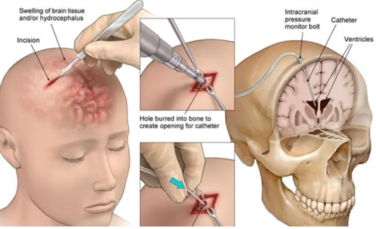
External ventricle drain placement
intersect a lateral ventricle in order to drain cerebrospinal fluid and relieve intracranial pressure.” [5]. Figure 2.7 depicts an overview of EVD placement task.
Annually, Srinivasan et al. [28] estimated that trainees perform 25,000 EVD inser-tions, and it is considered an early and independent task any trainee performs in their academic settings. As a part of their training curriculum, trainees need to develop the targeting skill within the same setting, where they perform it either in intensive care units, OR, or ER.
(a) (b)
Fig. 2.7: (a) A resident arrived at a predefined entry point and penetrated the EVD’s catheter through the dura to extract the cerebrospinal fluid, and (b) the resident passed the catheter which arrived at the bottom of the lateral ventricular [8].
makes multiple hits towards a brain structure by which a set of undesirable trajectories are created [5]. Trainees usually perform EVD placement in the ER with its overarching tasks in sequential stages in active and full systematic analysis capability before acting on patient organs [5].
The deliberate training for the planning process of EVD placement became a ne-cessity. Armstrong et al. [30] stated that EVD placement like neurosurgeries requires trainees to plan a precise trajectory through a designated region on the nervous struc-tures. The trainees will perform their intervention in a relatively narrow fracture within the designated region, which would not be seen by the naked eye. Thus, trainees need to use their cognitive capacities and build a 3D view of where and how to reach to the fracture without damaging other risky regions or eloquent brain areas.
2.3
Surgical Simulators
2.3.1
Types
ensure a safe and free-error training environments. According to Kockro [7], “Training with simulators is of obvious advantage, especially in the eye of the patient: we can learn without causing any harm. In neurosurgery, the potential of simulator technology is still massively underutilized.”
There are several taxonomies mentioned in the literature about surgical simulators and how they were used for different surgical specialties. A simple form of a surgical simulator is an anatomical-related software package that is running on a stationary com-puter with a keyboard or a mouse, where a user interactively engages with the projected virtual objects [3].
According to Halvorsen et al. [31], three types of laparoscopic surgical simulators have been used in different surgical specialties: mechanical, hybrid, and virtual. Mechan-ical simulators are boxes equipped with a digital laparoscope, cameras, screen, surgMechan-ical instruments, and objects. Similar to mechanical simulators, hybrid simulators are boxes that are connected and monitored by a computer system and sensors. Last, virtual simu-lators use a combination of a set of digital technologies aimed to allow trainees to interact with 3D virtual objects in an immersive mode and to get objective feedback about their behavior or performance.
previous two types - are designed to thoroughly engage trainees in a virtual environment by presenting fused surgically-relevant environments in which to practice technical and non-technical-related skills.
In the neurosurgical context, [33] discussed that there are five groups of surgical simulators used in neurosurgical training: computer\VR simulators, cadaveric models, in vivo models, synthetic models, and living patients. Cadaveric and in vivo models include dead animals used for anatomy studies, and synthetic models use sensor-based mannequins that are connected to computer machines [34].
With advanced and low-cost VR\AR technologies, development of simulation-based training gained more attention from medical researchers. With appropriate contextual information throughout a neurosurgical procedure using AR\VR facilities, senior resi-dents and trainees gain a real-time view of specific anatomical segments that were fused upon patient’s organs from different modalities. It is most likely that trainees will be very efficient at their practices with the use of AR\VR visualization modes because they depicted a synthesized version of “anatomic, metabolic, and functional data” from differ-ent sources about a specific patidiffer-ent situation. Also, the visualization mode could also be examined in detail, distributed and discussed with trainees and other peers and research community [3].
2.3.2
Motivation of simulation-based surgical training
surgical teaching method challenges. The apprenticeship model is a classic surgical teach-ing method where trainees attend senior residents’ live surgery sessions to observe and practice on live patients during the surgery.
According to McGaghie et al. [35], it has been shown that training based solely on the apprenticeship model produces inferior results without supplementary simulation techniques. The apprenticeship model is not efficient enough because it consumes a mas-sive amount of hospital resources in terms of money and time. The costs include the costs of actual surgeries from OR operating fees until a complete surgical team [4]. Moreover, trainees usually suffer from lack of real-time feedback during the apprenticeship model, as it would take up to a month for the decision to confirm or reject the surgery performance to appear by the patient’s recovery stage. The apprenticeship model is not flexible; it im-poses a variety of restrictions on senior residents. Senior residents usually have 80 hours of deliberate practice a week to fulfill, but senior residents never exceed 5 hours of delib-erate practice a day with full cognitive capacities [4]. Because the apprenticeship model necessitates the attendance of senior residents to training locations to supervise trainees, trainees would observe only a handful of ventriculostomies to maintain their skills, and the trainees need to use their own judgment for new or rare neurosurgical situations. As a result, trainees might not achieve the expert level within the given residency training timespan [4].
fewer surgical errors, a contributing factor for collateral damage or threat to patients’ safety. Simulation techniques will enable trainees to have adequate surgical practice anywhere and anytime. Also, trainees will receive autonomous surgical training that not only involves a variety of surgical techniques, anatomies, and pathologies but also offers necessary tools to support different surgical specialties and to increase procedures’ success rates [7][12][4].
2.3.3
Simulator validation
Simulator validation is a process in which a particular test or metric measures a specific aspect of a simulator [15]. One of the primary challenges to developing valid surgical simulators is to engage trainees in the complex dynamics of real surgical scenarios in-cluding patients, senior residents, and interconnected surgical steps that occur during the surgical procedure. Many surgical simulators did not last; either they are not sufficient to support enough scenarios in ORs, or they might be seen as a valid option according to subjective evaluation techniques [3].
Psychometric evidence includes face, content, construct, concurrent, and predictive va-lidity tests. A face vava-lidity test is concerned with how a simulator will measure what it is supposed to measure from the perspective of experts. A content validity test evalu-ates the extent a simulator will measure all relevant content or properties of a specific surgical task or procedure based on a checklist or expert opinion. A construct validity test determines the extent a simulator measures a particular trait that the simulator is claimed to measure. Construct validity is most common assessment technique used and is well-documented to quantify the psychometric competency between senior and junior residences in the literature. A concurrent validity test is concerned with how a simulator will comply with well-defined standards for a surgical task or procedure. A predictive validity test concerns how a simulator can predict trainees’ performance in the future through real surgical scenarios.
2.4
User learning process
Learning is a process by which a user acquires a new knowledge, skill or behavior. Prac-ticing a skill for a long time would be necessary to accomplish a competency level in it. For some disciplines, a human could master a particular skill after a long time of intense training [37]. For example, a chess player needs nine years to obtain master status. Even in common folklore from a variety domains such as music, it takes a violinist around 10,000 hours of deliberate practice at the age of 20 to master his or her skills [4].
continuous practice, users learn a variety of everyday tasks until the tasks do not require much cognitive input from them. Then, users would become autonomous, and no per-formance increase is observed. Some professionals reach an advanced level of skill and stop continuing to improve, a situation referred to as arrested development [4].
Fig. 2.8: The relationship between cognitive efforts, experience, and human performance during users’ learning process [38].
In the surgical context, because trainees spent 12 to 13 years in medical schools, it is expected trainees will develop the necessary skills with many hours of deliberate practice on live patients, including supervised and intraoperative training. In reality, trainees might not have cultivated the required mastery of specific skills [4].
making, depth perception) are fragile against how studying images were represented and how trainees will interact with them. The more stimuli4 exist within the OR, the better residents could maintain perception and decisions during their training. Visuospatial reasoning will enable trainees to form a mental perspective about the current nervous structures for further surgical tasks [15]. The immediate and informative feedback could include two types: active and passive. The former type is under computer system con-trol, such as visual or audible notifications, while the latter type emerges as an internal sensation within the user’s body [39].
2.5
Human-computer interaction
Human-computer interaction (HCI) is a technique by which a user accomplishes a task for a specific interface conceptual model and receives a specific feedback [39]. User inter-actions towards a system display take few styles: text entry, menu selection, navigation, and direct manipulation. Direct manipulation is intended to allow users to recognize an object of interest in the user interface and to perform a particular function directly before observing the effect [40].
2.6
Perception depth in surgical training
Among other cognitive processes, the depth perception process includes how trainees observe the effects of their actions with a display screen and reconcile these effects. The
trainees decide to perform a surgical task based on the interaction outcome between their cognitive processes when interacting with a visual stimulus and its background. Inac-curate depth perception is a common problem since low modality resolution and blurry displays cause 3D virtual objects to appear further away from their predetermined loca-tion against the physical objects, a contributing factor to user axis bias and misjudgment of users’ movements. Trainees could perform complex psychomotor tasks or improve their performance by effectively predicting the depth of a target features [15].
A 3D-based user interface enables users to visualize and consume 3D objects in Euclidian dimensions (i.e., X-axis, Y-axis, Z-axis), that includes the depth dimension. For the users to complete their 3D tasks (e.g., navigation or manipulating 3D objects) through 3D-based user interfaces, it is important that users gain an understanding of the dynamic of depth perception of 3D objects and tasks and functionality of the human visual system [41].
Depth perception depends on two factors: distance judgment and visual depth cues. Distance judgment requires the users to maintain a mental interpretation of the physical objects around them and how close or far the objects are. A visual depth cue is a synthesis of an image’s feature, used to convey a sense of depth dimension, and functionality of the human visual system. Figure 2.9 depicts an overview of common visual depth cues.
features (e.g., relative size5, occlusion6, or linear perspective7) to yield depth information [41]; these techniques are depicted in Figure 2.9(a) and Figure 2.9(b), respectively [23].
Oculomotor or observer-centered cues [41] involve muscular tension from the user‘s eye while they are looking at a set of objects from a certain distance. Whenever their eye muscles relax and the eyes‘ lenses become more spherical, then the users see objects as far away. This phenomenon is referred to as accommodation. Another oculomotor cue is referred to as convergence, the rotation of users‘ eyes when the eyes converge and diverge when looking at near or far objects, which are depicted at Figure 2.9(c), respectively, [23].
Motion parallax or dynamic depth cue is a phenomenon that occurs when users look at a set of stationary objects during their physical movement around the objects; the users see the objects moving relatively quickly if they are close and relatively slowly if they are far; these are depicted in Figure 2.9(d) [23].
Binocular disparity and stereopsis refer to the extent a user sees an object using two eyes, by which a difference representation occurs and yields two different images of the same object. The greater the fusion of the two images, the more the users will able to determine how close the object is from his\her eyes Bowman et al. [23].
5. The relative size phenomena is about sorting a set of objects in a way that the user sees the smaller is the object, the more the object appears to be farther away.
6. Occlusion is a phenomenon of sorting a set of objects in a way that the user sees the first object as opaque and occludes another object. Then, the user will see the first object as closer to him\her than the other object
Fig. 2.9: Overview of common visual depth cues [23].
2.7
User performance assessment
2.7.1
User performance
third process involves how the decisions will be carried out.
As interactions between humans and computer systems evolve and the latter be-come increasingly complex, there is a need to analyze human cognitive processes within these interactions [42]. Such analysis is a crucial aspect of measuring users’ performance of tasks accurately. Conventional task analysis approaches do not focus on analyzing and representing the human performance through cognitive processes. Task analysis approaches usually pay more attention to physical or observable aspects of the human performance of tasks.
In the surgical context, a surgical procedure is a combination of generic and ad-vanced surgical tasks that require a certain surgical competency level (i.e., technical, team performance, and communication and decision-making skills). The evaluation of trainees’ performance usually focuses on their surgical competency. Surgical competency is based on a hierarchy of simple psychomotor tasks, for instance, navigation of a tool towards a specific target or manipulation of a physical object [15].
2.7.2
Hierarchical task analysis
A new trend towards a structured assessment technique has been used towards an under-standing of surgical procedures. The technique is referred to as hierarchal task analysis (HTA). HTA technique is a process intended to help analysts to determine the source of errors of a system performance (e.g., human performance) and to propose solutions for the same system [44]. HTA technique is built on the idea of a decomposition of a sys-tem’s goals. Goal decomposition is a process in which an individual goal and its current states are identified through breaking them down to sub-components in a hierarchy [44]. HTA has been used in a variety of studies evaluating surgical procedures or techniques [45][46][47]. Table 2.2 depicts an example of surgical HTA—tabular format—for a task in performing an open inguinal hernia repair [48].
Tab. 2.2: An example of a task in performing an open inguinal hernia repair task with its subtasks and possible recovery steps that lead to the successful completion of a procedure [48].
2.7.3
Fitts‘ law
User performance evaluation is a key aspect of the validity of any solution in the context of surgical simulations. Following the work of information theorist Claude Shannon in evaluating of communication systems’ capacity, Paul Fitts extended Shannon’s Theorem 17 [49] to accommodate measuring simple 2D psychomotor tasks in human behavior to determine the human capacity to fulfill the tasks. Shannon’s Theorem 17 is summarized as equation 2.1:
C = B logS + N
N (2.1)
WhereCis human capacity (in bits\s),B is bandwidth (in 1\s or Hz),Sis a single power source, and N is a noise power [50]. Fitts’ model or Fitts’ Law [51] aims to measure the information capacity of the human motor system through quantitative-based metric referred to as the index of performance (IP). MacKenzie [50] discussed Fitts’ Law as follows:
The realization of movement in Fitts’ model is analogous to the
transmis-sion of information. Movements are assigned indices of difficulty (in units
of bits), and in carrying out a movement task the human motor system is
said to transmit so many bits of information. If the number of bits is divided
by the time to move, then a rate of transmission in bits per second can be
ascertained.
width (W), where the former represents movement distance of the human and the latter represents the width of the region within which a movement terminates. IP is calculated through two approaches. The first approach involves dividing a motor task difficulty or the index of difficulty (ID) by the averaged movement time (M T) over a block of trials to complete a motor task. The second approach involves a regressing line MT on ID, where MT is the dependent variable, and ID is the independent variable. In the first approach, IP is calculated as eq. (2.2)
IP = ID
MT (2.2)
Where ID is calculated as eq. (2.3):
ID = log2 2A
W (2.3)
In the second approach, IP is calculated as the regression line as eq. (2.4):
MT = a + bID (2.4)
Where a and b are regression coefficients. The reciprocal of the slope coefficient, b1 , corresponds to IP. The usual form of Fitts’ Law in the second approach is calculated as following in eq. (2.5):
MT = a + b log2 2A
2.8
Summary and Conclusions
Chapter 3
Related Work
3.1
Introduction
Across surgical disciplines, simulators have been used by novice trainees for image-guidance, deliberate training, and planning aids. In AR based systems, the main moti-vation for surgical simulators is to allow trainees to master an individual task or subtask, the steps of a set of procedures or a wider range of neurosurgeries within predefined cer-tain surgical scenarios, keeping patients safe from any harm from actual training trials [4][52][32][53].
differ-small or almost-no space in the OR, 5) offer a similar experience to the previous systems, 6) support micro and macro views of the workspace, 7) be transparent to the users in terms of errors or loss of tracking, and 8) show a real visual representation of the actual used tools and workspace. This chapter sheds light on related work to the ventricular catheter insertion task (refer to section 2.2.3) for ventriculostomies (refer to section 2.2.2) simulators for deliberate training and planning aid for novice neurosurgery trainees.
3.2
Web-based simulation
3.3
Neuronavigation-system-based simulation
3.4
Mixed-reality-based simulation
trainees performed their tasks with inaccurate depth perception of the virtual catheter, a technical problem that would lead to health complications in a real clinical setting.
different entry points to the interest region with a different hand or direction, and it is most likely re-planning the process of the targeting task would occur if the trainees had uncertainty about the task.
Hooten et al. [61] evaluated a mixed-reality simulator, designed to educate neu-rosurgical trainees to perform complete ventriculostomies. The mixed-reality simulator included 3D-printed concepts of the brain anatomy and a VR system. The system al-lowed for visual and haptic feedback and the trainees’ performance was measured based on a scoring algorithm by combining time and accuracy. Although the simulator was designed for complete ventriculostomies, the simulator could not adapt for possible and rapid scaling up for patient-specific anatomies with a variety of surgical scenarios, for instance, for slim or shifted ventricles.
Lee et al. [62] evaluated a simulator for the training of a catheter insertion task, referred to as Dextroscope, with different entry points and approaches. The authors performed the tasks on only 10 cases with normal-size ventricles for the adult population, and they did not report a comprehensive validation plan for their module regarding usability or user performance.
3.5
3D-printing-based simulation
of training with tactical feedback. On one hand, the trainees were able to assemble the 3D-printed inner parts for the purpose of anatomical knowledge, and the simulators enabled the trainees to adjust the inner parts of the phantom heads to accommodate a set of patients’ cases. Both systems also focused on haptic feedback and brain tissue deformation. On the other hand, both phantom heads did not provide a planning mode to practice or measure the accuracy of catheter insertion or the perception of created trajectories. In addition, the authors did not use objective evaluation metrics to avoid any possible user bias about their performance. Using 3D-printed simulators might be an affordable approach to expand access to basic ventriculostomy training, although, for rare or totally new neurosurgical situations, it might be costly to maintain 3D-print phantom heads for every neurosurgical situation.
3.6
Handheld-modalities-based simulation
3.7
Summary and Conclusions
An ideal AR-based system for ventricular catheter placement simulation for training and planning aids should include a variety of attributes. The system has to engage a user in the 3D environment by offering a 3D user interface. The system needs to cope with the high computation demand of AR information projection, yet the system should not suffer from a long system latency. The system should not limit the input in surgical scenarios regarding lateral ventricular size, entry point position or orientation, or length of the catheter. Also, the system should provide users with visual feedback about their task, and the system should support visual depth cues to lessen the hurdle of inaccurate depth perception. Moreover, the system should enable the dynamic mobility of a trainee’s whole body or some part of it around the patients. Also, the system should measure and record users’ performance according to their accuracy and speed to complete the task without affecting their workflow.
Chapter 4
Design of Augmented Reality Training and
Planning Aid Simulation Modules for EVD
Placement Using Model-driven Engineering
4.1
Introduction
The use of image-guidance systems (e.g., navigation or planning systems) for complex neurosurgical procedures in ORs became popular when they included appropriate con-textual information [59]. These systems have been used: 1) to fulfill surgeons’ need to see
.
• This work has been published as:
– H. Ghandorh, S. De Ribaupierre, R. Eagleson, “Development of Augmented Reality Training Simulator Systems for Neurosurgery Using Model-Driven Software Engi-neering”,IEEE 30th Canadian Conference on Electrical and Computer Engineering (CCECE) [Computer and Software Techniques Track], April 2017, Windsor, Ont, Canada.
real-time patients-specific imaging studies with improved medical diagnoses, and 2) to enhance their cognitive abilities when performing a surgical intervention prior to reducing surgical errors [59][17].
Development of neurosurgical simulation tools for medical education and training purposes is a non-trivial goal. Typical neurosurgical simulation tools use sophisticated equipment and require technical setup and expertise to prepare and operate. Image registration, camera calibration, 3D surface rendering of imaging studies, object tracking and other technical requirements are necessary, including diverse underlying platforms for deployment and wider user landscapes [59]. In addition, the influence of human factors (human perceptual, motor, and cognitive capacities) play a critical role when designing such tools [12]. Moreover, many resources dedicated to simulation systems design and development are wasted in trial-and-error attempts due to inaccurate representations of clinical settings. In addition, a major requirement of such tools is that the tools need to follow a thorough validation process. Therefore, the typical development process will not be able to deliver such tools in low man-hour budgets.
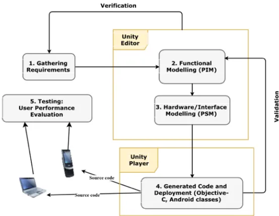
Model-driven engineering (MDE), a software development vision, focuses on mod-eling transformations and technologies to simplify and formalize various software devel-opment activities [66]. MDE promotes the transformations of abstract models as the primary activity in the development life cycle prior to producing executables within a particular domain and set of rules.
Following the MDE vision, developers will be able to deliver executables while hiding complex platform details, reducing manual coding errors, and allowing the sharing of experts’ knowledge in a specific domain [67]. As a result, high-quality executables could be delivered more frequently in shorter timespans.
This chapter fulfills the first aim of our research study, and it describes the extent to which MDE is a suitable development approach to generate two AR simulation modules1: 1) stationary module (i.e., desktop version) , and 2) portable module (i.e., smartphone app). The purpose of the locally developed AR simulation modules is to enable users: 1) to visualize AR information needed as training and planning aids for an EVD placement or insertion task (refer to Section 2.2.3), 2) to enable users to practice the EVD placement with predefined surgical scenarios, and 3) to enable domain experts or evaluators with a mode of assessment to measure novices‘ performance as a trade-off between speed and accuracy.
4.2
Methodology
4.2.1
Our MDE-based approach
We adopted the MDA technique, a well-documented and most common technique for MDE-based development. Figure 4.1 presents a comparison between a typical software development life cycle and MDA development vision.
MDA entails three main milestones: 1) crafting of platform-independent models (PIMs), 2) crafting of platform-specific models (PSMs), and 3) building of code
Fig. 4.1: Typical software development life cycle vs. MDA development vision [68].
tors. PIMs are domain-related specifications that should be modeled in a formal modeling language (e.g., United Modeling Language (UML) profile). PSMs, which are derived from PIMs, are platform-related specifications. Both PIMs and PSMs are created by modeling tools, and PSM will be used as the input for a code generator, a component intended to source code production that complies with a particular set of domain rules [67].
Fig. 4.2: A simplified flowchart of MDE-based approach.
First, we identified a set of requirement specifications (Figure 4.2(1)). A neurosur-gical scenario, the input for AR simulation modules, includes a description of an EVD placement trial. Figure 4.3 depicts an example of a neurosurgical scenario for EVD place-ment within a ventriculostomy procedure (refer to Section 2.2.2), represented in Harel statechart notation. The neurosurgical scenario is represented as surgical HTA (refer to Section 2.7.2) in a bullet-point format. A portion of the neurosurgical scenario is presented as follows:
• Phase 0: Review imaging studies (CT scans or MRI).
• Phase 1: Localize the closest entry point in the right frontal bone in the skull to the lateral ventricles.
• Phase 2: Visualize the shape and location of lateral ventricles in your mind.
• Phase 4: Advance the EVD towards the lateral ventricles until you hit the frontal horn :
– Penetrate the EVD’s catheter through the inner table of the skull bone per-pendicularly to the brain surface slowly to<= 6 cm depth.
– The stylet then is removed and the catheter is kept in the burr hole.
– A slight pop in resistance emerged (i.e., increase resistance than a loss of resistance).
– When the frontal horn is cannulated, CSF drainage should emerge through the catheter.
We focused on the Phase 4.1 step from the scenario that fulfills the required EVD placement targeting task. Listing 4.1 depicts an example of textual description of the EVD placement task in JavaScript Object Notation (JSON) tags format.
Fig. 4.3: A surgical scenario of the EVD placement task in Harel statechart notation.
{
"id": "413a41e3-ce7d-4d18-904c-411f53ccca0e",
"name": "Demo Targeting 1",
"assets":
[{
"tag": "Target 1",
"position": { "x": -6.45441866, "y": 0.5490153, "z": 1},
"rotation": { "x": 0, "y": 0,"z": 0},
"scale": {"x": 1.23174512, "y": 1.23174512,"z": 1},
"assetId": "00000000-0000-0000-0000-000000000000"
}]
}
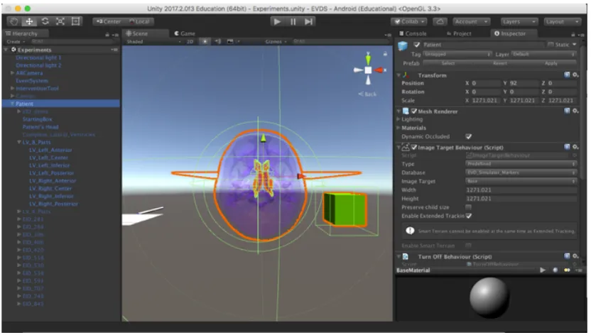
The neurosurgical scenario was maintained and came from a third-party system (i.e., ScenarioSim) that facilitated the transformation of the textual description of the oral description of the neurosurgical scenario from a domain expert (i.e., senior neurosurgeon). Then, we maintained visual representations (i.e., PIM) of the AR scene and verified them with the domain expert during the designing of both systems. The visual representations were then designed through the editor of Unity (Unity engine 2017.2.0 Educational, Unity Technologies, San Francisco, California, USA) (Figure 4.2(2)). The Unity editor is a game engine editor intended to create and model game scenes including 3D interactive objects, which is depicted in Figure 4.4.
platform-Fig. 4.4: Unity editor and PIM models.
specific visual representations (i.e., PSM) (Figure 4.2(3)). Then, we utilized the Unity Player (i.e., code generator) in order to complete the virtual scene settings, a necessary step to generate and deploy an artifact towards the desired platform (Figure 4.2(4)). Af-terwards, the Unity Player converted the PSM and transformed it into the source code, and deployed it towards a single platform based on the virtual scene settings through command line programs (e.g., Shell).
4.3
Simulation modules description
4.3.1
Design considerations
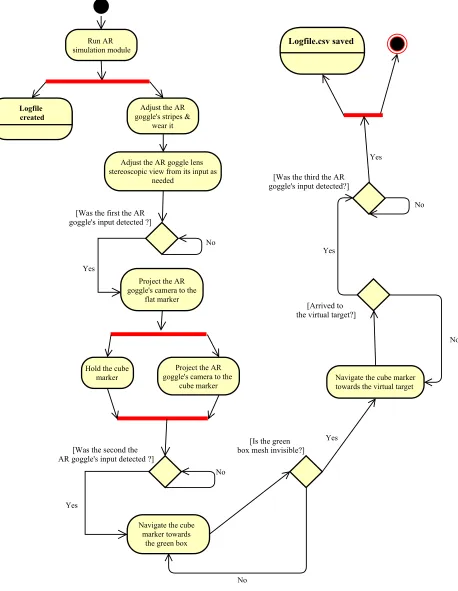
We received well-defined requirements from the domain expert that include a few spec-ifications. The simulation modules need to build such that they will allow novices to apply the EVD placement task complying with an initial planning stage. The planning stage enables a series of surgical scenarios, including trial sets and entry points’ position and rotation coordinates. The simulation modules need to allow for not only interaction with patient-specific 3D meshes but also for the performance of surface rendering with possible visual depth cues at running time. Moreover, the simulation modules would be deployed in stationary and portable environments to demonstrate extendibility with the potential for cross-platform deployment. During the trials, the simulation modules will record the novices’ trajectory and time taken in the virtual space. The simulation modules also will offer a stereoscopic vision mode and record the novices’ head and hand tracking (i.e. head and hand physical movements) prior to producing readable log files prior to users’ performance analysis.
4.3.2
Simulation modules setup
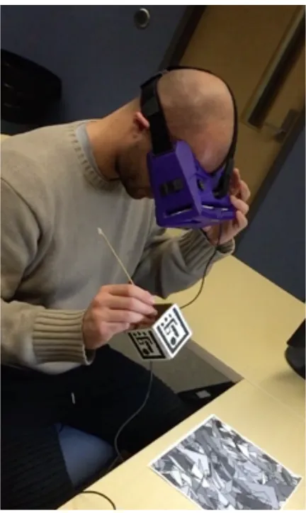
cardboard papers and a wood stick for stability and mimicking the catheter’s geometrical shape and weight.
Fig. 4.5: Physical version of the two trackable predefined fiducial markers.
The functionality of AR simulation modules depends on the integration of off-the-shelf software packages (i.e. Unity editor and libraries, and Vuforia). Unity is a sophisticated game engine that aims to create game scenes, and it allows for a mode to adjust 3D meshes-related configuration in terms of location, rotation, size, surface-based texture, and functionality within a game scene. Vuforia (Vuforia SDK version 5.5, Parametric Technology Corporation, Needham, Massachusetts, United States) is an image tracking and detection framework that provides device or objects detection and tracking functionality, stereo rendering, and reconstructing VR\AR scenes among the real-world space.
the fiducial markers. Vuforia target manager examines the validity of fiducial markers and ranks their tractability based on a local scale (i.e., augmentation level) by identifying image features and sharp edges. The more the fiducial markers are sharp with spiked fea-tures, carved texture, rich details, and good contrast, the higher will be the augmentation level, which increases the tracking robustness. Vuforia target manager is responsible to also to generate a trackable fiducial marker dataset (i.e. VuforiaDataset.unitypackage) as an input for the Unity Editor. Figure 4.6 indicates the fiducial markers after application of image processing and determining their detection landmark.
Fig. 4.6: The registration points in the soft version of the fiducial markers.
scene, if needed. Whenever any adjustments are performed or needed, the Unity Editor gives a visual feedback of the current state of these components, accordingly.
For camera calibration purposes, Vuforia SDK performs dynamic camera calibra-tion by creating calibracalibra-tion profiles. The calibracalibra-tion profiles are created based on a model similar to the four-step internal camera model proposed by Heikkila and Silven [69]. The calibration profiles assumed that the intrinsic camera parameters are pre-calibrated, where it utilized the pre-defined markers in a real scene.
For the viewing mode, Unity and Vuforia integration allowed for a mode to select the simulation scene to be seen through mono or binocular views. Table 4.1 indicates the applied configurations to accomplish a stereoscopic view for the AR simulation modules: Tab. 4.1: User viewing configuration applied to assign AR simulation scenes and functionality.
Attribute Value
Eyewear type Video see-through
Camera distortion mode Dual texture for Android devices Camera distortion coefficients K1: 0.07. K2: 0.03
Field of view of the
camera in degrees 95
Camera offset 0.09
Camera projection type Orthographic The clipping plane
distance for the camera Near: 0.05, Far: 5000 The viewing volume (depth) of an
orthographic camera 700 Max simultaneous tracked
objects\images 5
The aspect ratio (width divided by height). Automatic Frame per second 30 - 45
(a) Original skull meshes.
(b) The skull mesh in a transparent shedder.
(c) Ellipsoid (i.e. target). (d) Brain structures (Lateral ventricle).
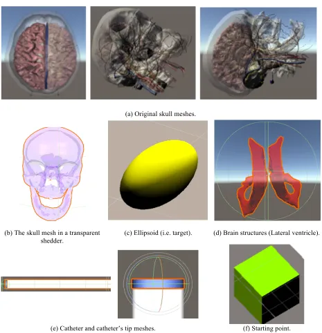
(e) Catheter and catheter’s tip meshes. (f) Starting point. {fig:Ch04-ThreeD-meshes-within-AR-scenes}: 3D meshes used with the AR simulation modules’ scenes
rendering mode. We integrated a set of 3D meshes into the Unity editor as the main assets of the game scene, where the Unity editor was used as a canvas to host the 3D meshes. The skull and brain structures meshes were designed through a sequential series of 362 high-resolution of a female patient. The images were converted into grayscale format prior to being inserted into Amira 5.6 (FEI Visualization Services Group, Hillsboro, OR) for manual segmentation purposes. The segmented objects (i.e., meshes) went through a multiple step process: 1) editing process of meshes through MeshLab (MeshLab 1.2.1., ISTI-CNR, Pisa, IT), and 2) refining process of missing surfaces or components though Blender 3D (Blender 3D 2.76, Blender Foundation, Amsterdam, Netherlands). More details about the meshes designing are available in [70]. Other meshes, for instance, ellipsoid, catheter, or catheter’s tip, were designed from available geometrical shapes from the Unity library.
In each generated artifact, the AR simulation modules’ screen includes two game scenes, which are depicted in Figure 4.8. The UI scene includes an eye chart image intended to allow users to adjust or calibrate the AR goggle’s view with suitable distance according to their comfort. The AR scene includes a virtual camera, Vuforia dataset, 3D meshes (skull, brain structures, ellipsoids, catheter, catheter’s tip, and the starting box). Other components were used, for instance, lights and audio sound effects, to give the AR simulation modules’ scenes the best intuitive user experience.
Fig. 4.8: The AR simulation modules’ screen and underlying game scenes.
4.3.3
Hardware description
A live demo of portable AR simulation module is available at https://youtu.be/
fDpfzJb9ixw. The first and the second generated artifact were deployed in the
follow-ing environments, described in Table 4.2 and Figure 4.9. Both generated executables were deployed and running through a preliminary validation stage to ensure their usability for future assessment.
Tab. 4.2: Technical specifications of the used environments for the AR simulation modules.
Environment Technical specifications
Stationery module
3200
screen RCA TV, 1366x768 resolution, 16:9 widescreen. External
camera Logitech HD Webcam C270. MacBook
Pro laptop machine
OS X El Capitan 10.11 operating system, 2.66 GHz Intel Core 2 Duo processor, and 16GB 1067MHz DDR3 Ram.
Portable module
AR goggle
Merge VR DSCVR headset with two 34 mm biconvex lenses, fits for 5.9600, screen phones, and purple color.
LG Nexus 5
smart-phone
4.9500 screen, 1080x1920 resolution, 137.9x69.2x8.6
mm, Android v6.0 (Marshmallow) operating system, Quad-core 2.3 GHz Krait 400 processor, camera 8 MP.
Fiducial markers
Flat marker
Single image with multiple triangles shapes that uses Vuforia’s Image target object.
Cube marker
Four images with multiple shapes that use Vuforia’s Image target object.
LED desk lamp OttLite LED white lamp, 20.5
00 height, different brightness
settings (maximum of 345 lumens), and rubberized flexible neck. Cardboard cube Diameter x height (7x7)(cm).
![Fig. 2.9: Overview of common visual depth cues [23].](https://thumb-us.123doks.com/thumbv2/123dok_us/1923070.1252441/44.612.99.542.65.392/fig-overview-common-visual-depth-cues.webp)