Copyright © 2003, American Society for Microbiology. All Rights Reserved.
Immunoglobulin G Subisotype Responses of Pneumonic and Healthy,
Exposed Foals and Adult Horses to
Rhodococcus equi
Virulence-Associated Proteins
Kathleen E. Hooper-McGrevy,† Bruce N. Wilkie, and John F. Prescott*
Department of Pathobiology, University of Guelph, Guelph, Ontario N1G 2W1, Canada Received 23 October 2002/Returned for modification 27 January 2003/Accepted 26 February 2003
Rhodococcus equicauses severe pyogranulomatous pneumonia in foals and in immunocompromised humans. Replication of virulent isolates within macrophages correlates with the presence of a large plasmid which encodes a family of seven virulence-associated proteins (VapA and VapC to VapH), whose functions are unknown. Although cell-mediated immunity is thought to be crucial in eliminatingR. equiinfection, antibody partially protects foals. The antibody response to both VapA and VapC was similar in six adult horses and six naturally exposed but healthy foals, as well as in eight foals withR. equipneumonia. The immunoglobulin G (IgG) subisotype response of pneumonic foals to Vap proteins was significantly IgGb biased and also had a trend toward higher IgGT association compared to the isotype association of antibody in adult horses and healthy exposed foals. This suggests that in horses, IgGb and IgGT are Th2 isotypes and IgGa is a Th1 isotype. Furthermore, it suggests that foals which developR. equipneumonia have a Th2-biased, ineffective immune response whereas foals which become immune develop a Th1-biased immune response. Pneumonic foals had significantly more antibody to VapD and VapE than did healthy exposed foals. This may indicate a difference in the expression of these two Vap proteins during persistent infection. Alternatively, in pneumonic foals the deviation of the immune response toward VapD and VapE may reflect a bias unfavorable toR. equiresistance. These data indicate possible age-related differences in the equine immune response affecting Th1-Th2 bias as well as antibody specificity bias, which together favor the susceptibility of foals toR. equipneumonia.
Rhodococcus equi causes pyogranulomatous bronchopneu-monia in foals younger than 4 months and induces significant economic losses on endemically infected horse-breeding farms (29). This gram-positive, facultatively intracellular bacterium is an opportunistic pathogen in immunocompromised humans, such as those infected with human immunodeficiency virus (2, 9, 13). Foal-virulent R. equistrains possess an 81-kb plasmid and express VapA, a plasmid-encoded, surface-expressed lipo-protein (38, 40–42). VapA is a member of a family of seven Vap proteins (VapA and VapC to VapH), which have homol-ogy in their C-terminal halves and are encoded within a 27-kb pathogenicity island on the large plasmid (37). The function of these proteins is not known.
Protection of foals againstR. equiappears to rely on coop-eration and interdependency between the antibody-mediated and cell-mediated immune response. Antibody appears to con-tribute to protection since the period of maximum susceptibil-ity coincides with declining levels of maternally derived anti-body (29). In addition, antianti-body opsonizesR. equifor uptake and killing by macrophages and neutrophils in vitro (45, 47, 48) and may facilitate the killing of R. equi in vivo. Vap-specific antibodies protect immunosuppressed mice since purified im-munoglobulin G (IgG) from APTX (a VapA-enriched anti-gen)-vaccinated horses protected against intraperitoneal
chal-lenge withR. equiwhereas nonimmune equine IgG failed to protect (10).
Pulmonary clearance of virulent R. equi in mice requires functional T lymphocytes (3, 46). Both CD4⫹ and CD8⫹T
cells apparently contribute to protection (26, 33), and CD4⫹
cells are necessary for complete pulmonary clearance ofR. equi
in mice (16). Mice in which a Th2 cytokine response was in-duced by administration of monoclonal antibodies against gamma interferon (IFN-␥) prior to experimental infection with virulent R. equi failed to clear the bacteria and developed pulmonary granulomas (17). In contrast, immunocompetent BALB/c mice developed a Th1 cytokine response and cleared the infection (17). Adoptive transfer ofR. equi-specific Th1 or Th2 cell lines in mice supported the conclusion that a Th2 response is detrimental whereas a Th1 response is beneficial in clearance ofR. equi (18). VirulentR. equi can modulate the cytokine response in foals, down-regulating IFN-␥mRNA ex-pression in CD4⫹T cells and up-regulating lung interleukin-10
expression (12). These cytokines may influence the Th1-Th2 balance of the immune response in foals.
Although antibody provides partial protection against R. equi pneumonia (14), the role of the Ig isotype has not been described. Prescott et al. determined that the antibody re-sponse of young foals to the APTX antigen with aluminium hydroxide adjuvant induced a more IgGb- and IgGT-biased subisotype response than did natural infection, which induced an IgGa-dominant response (30). Vaccination with this antigen and adjuvant exacerbated disease in the foals after natural challenge with R. equi (30). An aluminium hydroxide-based influenza vaccine also induced an IgGT-biased response in horses, with some evidence of an IgGc response. These
vacci-* Corresponding author. Mailing address: Department of Pathobi-ology, University of Guelph, Guelph, Ontario N1G 2W1, Canada. Phone: (519) 824-4120, ext. 4716. Fax: (519) 767-0809. E-mail: prescott @uoguelph.ca.
† Present address: Canadian Food Inspection Agency, National Cen-tre for Foreign Animal Disease, Winnipeg, Manitoba R3E 3M4, Can-ada.
nated horses were not resistant to infection even though they had an anamnestic IgGT response (25). In contrast, natural infection with influenza virus induced virus-specific IgGa and IgGb (25).
In mice and humans, the antibody isotype reflects the Th1-Th2 bias of the immune response. A similar bias may occur in horses. The IgG subisotype profile of the antibody response associated with protective immunity toR. equiinfection has not been described. The study reported here addresses the hypoth-esis that rhypoth-esistance or susceptibility to R. equi pneumonia in foals is associated with distinct IgG subisotype-related anti-body responses to the seven virulence-related Vap proteins and that in pneumonic foals the profile reflects a Th2-biased response whereas in healthy foals and adults the profile reflects a Th1-biased response.
MATERIALS AND METHODS
Experimental design.Serum was collected biweekly from clinically normal pony foals (n⫽6) kept on pasture at a University of Guelph research farm that had a history ofR. equiinfections in foals. The serum sample in which the peak antibody response to VapA was observed over the first 6 months of a foal’s life was used to determine the isotype profile of the anti-Vap response compared to sera obtained from a group of clinically normal, unrelated adult horses (n⫽6) at the same farm and from a third group of foals with clinicalR. equipneumonia (n⫽8). The clinical case samples were obtained from client animals at the University of Florida and the Ontario Veterinary College. The horses were categorized according to defined resistance or susceptibility status to provide a framework in which to make correlations based on specificity and isotype relat-edness of serum anti-Vap antibody. The three groups represent (i) a clinically normal, healthyR. equi-exposed foal group; (ii) a clinically normal and healthy adult group (and since adult horses do not become sick withR. equipneumonia, presumably an immune group); and (iii) a susceptible group of foals that had developedR. equipneumonia and were therefore assumed to be nonimmune. The use of the horses for this research complied with all relevant national guidelines and University of Guelph Animal Care Committee policies.
Vap protein purification.Vap proteins were obtained as glutathioneS -trans-ferase fusion proteins. Eachvapgene was amplified from theR. equistrain 103⫹
plasmid by PCR using primers described in Table 1. The primers contained 5⬘
extensions encoding restriction enzyme recognition sites forBamHI orEcoRI. Amplified products were digested withBamHI andEcoRI, and the digestion products were ligated to a similarly digested plasmid vector (pGEX-2T; Amer-sham Pharmacia Biotech, Baie d’Urfe´, Quebec, Canada). The resulting plasmids were transformed intoEscherichia coliXL1Blue (Stratagene, La Jolla, Calif.). These plasmids allowed in vitro production of recombinant Vap proteins fused to glutathioneS-transferase. Fusion proteins were purified, using glutathione-Sepharose 4B beads (Amersham Pharmacia Biotech), and Vap proteins were cleaved from glutathioneS-transferase using thrombin (Amersham Pharmacia Biotech). Protein production was assessed by sodium dodecyl sulfate-polyacryl-amide gel electrophoresis, Western blot analysis, and protein assay (Pierce, Rockford, Ill.).
Although cloning and protein production were successful for each Vap pro-tein, purification using the glutathione-Sepharose 4B beads was not always achieved because of the inability of the fusion protein to bind to the beads. In these cases, a protein agglutination protocol was employed to concentrate the Vap protein. Briefly, a 100-ml overnight culture, which had reached an optical
density at 600 nm (OD600) of 0.6, was stimulated with 5 mM isopropyl--D
-1-thiogalactopyranoside (IPTG) for 3 h. The culture was then centrifuged at 1,500⫻gfor 15 min at 4°C, and the supernatant was decanted. The pellet was resuspended in 4 ml of 25% sucrose–50 mM Tris (pH 8.0) and frozen at⫺70°C. The suspension was then thawed quickly in a 37°C water bath, and 1 ml of a lysozyme solution (10 mg of lysozyme [Sigma-Aldrich, Oakville, Ontario, Cana-da] per ml of 250 mM Tris [pH 8.0] was added. This mixture was incubated on ice for 15 min, and 30 ml of a 2⫻RIPA-TET detergent mixture at a 5:4 ratio (2⫻
RIPA is 20 mMTris-HCl [pH 8.0], 300 mM NaCl and 2% sodium deoxycholate; TET is 100 mM Tris-HCl[pH 8.0], 50 mM EDTA [pH 8.0], and 2% Triton X-100) was added. The mixture was left on ice for a further 5 min and then sonicated on ice three times for 30 s. The sonicate was then centrifuged at 15,000
⫻g for 15 min at 4°C, and the supernatant was decanted. The pellet was resuspended in 500l of sterile water and analyzed by sodium dodecyl sulfate-polyacrylamide gel electrophoresis, Western blot analysis, and protein assay.
Serological testing.Equine sera were absorbed forE. coli-specific antibodies because Vap proteins produced by agglutination contained trace amounts of
E. coliantigens. Briefly,E. coliXL1Blue cells were grown on tryptic soy agar plates at 37°C for 24 h. The bacteria were washed from the plates with 5 ml of phosphate-buffered saline, using a glass rod. The OD of the bacterial suspension was measured and adjusted with phosphate-buffered saline to an OD600of 2.0.
The suspension was subjected to three rounds of 3 min of sonication and freeze-thawing and cultured to confirm killing. The suspension was then added in a 1:1 ratio to each serum sample and incubated at 4°C for 18 h with gentle rocking (Lab-line 3-D bidirectional rotator; Fisher Scientific, Nepean, Ontario, Canada). The sera were then centrifuged (14,000⫻gfor 30 min) to remove debris.
Titers of serum antibodies to each of the Vap proteins were determined by enzyme-linked immunosorbent assay (ELISA). The natural log of the dilution which produced an OD double that of the negative control was considered the titer of that sample. Each sample was run in duplicate, and the average titer of the group (adults, clinically ill foals, and clinically normal foals) was used in a comparison of the total antibody response (expressed as a ratio over the total antibody response to VapA) and the IgG subisotype response to each of the Vap proteins. A ratio was used for the total antibody comparison to control for possible variation associated with differences in antibody avidity. It was not used for the comparison of isotype response because this would have removed most of the data. Ninety-six-well polystyrene plates (Immunolon 1; Dynatech Laborato-ries Inc., Chantilly, Va.) were coated overnight at 4°C with each Vap protein at 2g/ml in carbonate-bicarbonate coating buffer (1.59 g of Na2CO3per liter,
2.93 g of NaHCO3per liter [pH 9.6]), washed three times using 0.05% PBS–
Tween 20, and blocked for 1 h at room temperature using 0.05% Tween 20 and 0.5% gelatin in PBS (PBS-TG). To measure antibody associated with all Ig isotypes, peroxidase-conjugated goat anti-horse Ig (Jackson ImmunoResearch Laboratories, Inc., West Grove, Pa.) was used at 1:10,000 in PBS-TG and incu-bated for 1 h at 37°C. Substrate was then applied (ABTS; Roche Diagnostics, Mannheim, Germany), the plate was incubated for 1 h in the dark at room temperature, and the OD was read at 405/630 nm (BioTek EL311; Biotek Instruments, Fisher Scientific). To determine isotype-specific response, peroxi-dase-conjugated anti-IgGa, anti-IgGb, anti-IgGc, and anti-IgGT antibodies (Se-rotec Inc., Oxford, England) were used at 1:100,000 for anti-IgGa, anti-IgGb, and anti-IgGc and 1:10,000 for anti-IgGT. Positive and negative controls were ob-tained from a knownR. equi-exposed mare and a foal immediately following birth, respectively.
Analysis.Two-tailed Student’st tests and LSD random permutation tests (analysis of variance) in SAS (SAS/STAT user’s guide version 6, 1990; SAS Institute Inc., Cary, N.C.) were used to analyze the data and to determine the significance of differences between groups(Pⱕ0.05). The first test was used to test for significant differences in the actual titers of each group, and the second test was used to test for significant differences between the groups for each
TABLE 1. Primers used for PCR of each of the seven members of the Vap family
Vap Primer 1 Primer 2
VapA 5⬘-GCCGGATCCACTAATGCGACCGTTCTT-3⬘ 5⬘-CATGAATTCCTAGGCGTTGTGCCA-3⬘ VapC 5⬘-GCCGGATCCGCCAATGTAGTCGCTCCGTC-3⬘ 5⬘-CATGAATTCGCGAGCGTTTACCTTCCGAC-3⬘ VapD 5⬘-GCCGGATCCACGGCGAGAAGACGAAAGTGT-3⬘ 5⬘-CATGAATTCTTGTTCCTCACGCAGCCCAC-3⬘ VapE 5⬘-GCCGGATCCATAGCATTGGTTCTAATCGCA-3⬘ 5⬘-CATGAATTCTACTGCACTGCAGCCGGACT-3⬘ VapF 5⬘-CACGGATCCAACAGATGTTGTGGCGATTGC-3⬘ 5⬘-CCTGAATTCTTCGTCCGCTTCTGCCAATC-3⬘ VapG 5⬘-GCGAATTCCAGAAGAGGTACGTGGCATTG-3⬘ 5⬘-CTGGGATCCATGGTATCCACTACAGCAGC-3⬘ VapH 5⬘-CATGGATCCGTGTACCAACGGCTGAAGCTC-3⬘ 5⬘-CCTGAATTCCACGTGATTCTTCGCTTGGAC-3⬘
antibody subisotype against each Vap protein in terms of there being detectable antibody or no antibody.
RESULTS
Antibody response to each of the Vap proteins.Antibody to VapA and VapC increased following natural exposure to R. equi(Fig. 1). In contrast, antibody to VapF, VapG, and VapH did not change throughout the 6-month observation period and antibody to VapE was not detected. The level of anti-VapD antibody rose slightly in some foals a few weeks after the peak of the response to VapA and VapC. Figure 1 data from one foal are representative of the pattern of the response observed in six foals kept at pasture, which remained appar-ently healthy, although the response pattern shifted along the
xaxis between foals, so that the day on which the anti-VapA response peaked differed between foals. For this reason, data for foals were not combined.
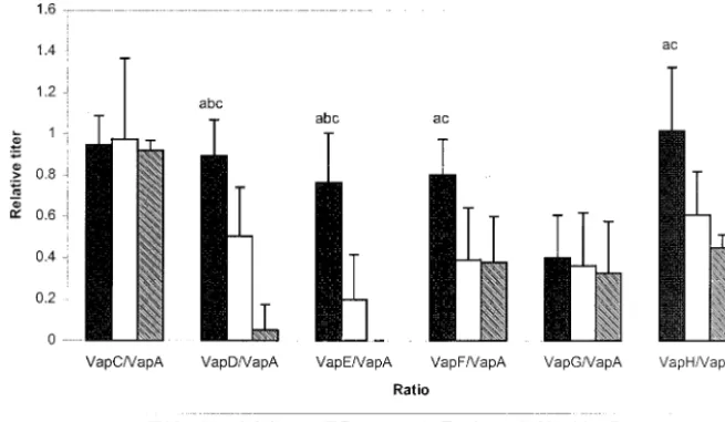
There was a marked antibody response to VapA and VapC by all groups in the study. The antibody response relative to anti-VapA was significantly lower in both foal groups than in the immune adult horses for VapD, VapE, anti-VapF, and anti-VapH (Fig. 2). The pneumonic foals had sig-nificantly more antibody to VapD and VapE than did the normal foals, which as a group had no antibody response to VapE and only a low response to VapD by one foal.
Subisotype of the IgG response.The antibody response to each of the proteins by the three groups of animals (pneumo-nic foals, cli(pneumo-nically normal foals, and immune adults) was fur-ther studied by investigating the subisotype (IgGT, IgGb, IgGc, and IgGa) of IgG and the differences were analyzed by Stu-dent’sttest. With the exception of three pneumonic and two clinically normal foals responding to VapA, an IgGc subisotype
response was not detected to any of the Vap proteins. The IgGT response of pneumonic foals was significantly greater than that of the immune adult group to VapA, VapC, and VapG (Fig. 3). The adults produced an IgGa- versus an IgGT-or IgGb-biased response against VapA, VapC, VapF, and VapH (Fig. 3). Although the pneumonic foals also produced IgGa against VapA and VapC, this group had an IgGb-dom-inant response to all the Vap family members (Fig. 3). The IgGb response of ill foals was significantly greater(Pⱕ0.05 by Student’sttest) than that of immune adults for VapA, VapC, VapD, VapE, and VapG and significantly greater than the normal foal group for VapC, VapE, and VapG (Fig. 3).
The mean IgGa-to-IgGb and IgGa-to-IgGT ratios against all of the Vap family members were consistently higher in the adults than in the pneumonic foals (Table 2) and were usually higher in adults than in healthy foals. When the immune adult group was compared to the pneumonic-foal group, the IgGa-to-IgGb ratio was as much as three times higher and the ratio difference was significant for VapA, VapC, and VapF. The IgGa-to-IgGb ratios in the healthy foals tended to also be higher than in the pneumonic foals, as did the IgGa-to-IgGT ratio. When the overall IgGb response to all the Vap proteins was compared between the horse groups by analysis of vari-ance, the IgGb response was significantly(Pⱕ0.05) greater in the pneumonic-foal group than in both the clinically normal group and the adult group.
DISCUSSION
Antibody function differs by immunoglobulin subisotype (7, 22). The Ig subisotype bias of the response to infectious or-ganisms reflects a finely regulated multifactorial system that
steers the response to provide an environment appropriate for control of the particular organism (15). In effect, the isotype profile of the immune response reflects the overall type of response (Th1 or Th2) induced by the pathogen or vaccine (32). This division of the CD4⫹T helper cell population into
two complementary groups has been extensively investigated by using humans and mice (32). The relationship of the bias of the response (Th1/Th2) to the degree of resistance to specific pathogens or to the response to vaccination has be-come an important area of research since it may be possible to increase resistance to disease by steering the immune response (11).
Assuming that the Th1 bias is protective in equine infections by the intracellular pathogenR. equias it is in mice, the results reported here support the conclusion that IgGT reflects a Th2-like response since a significantly greater IgGT response toR. equiantigens VapA, VapC and VapG was observed in foals that had developed R. equipneumonia than in the im-mune adult group. However, this difference was not noted between the two foal groups. The significantly greater overall amount of IgGb anti-R. equiantibody in the sera of pneumonic foals compared to adult and healthy foals suggests that the ratio of IgGa to IgGb is important in determining resistance, since the amounts of IgGa were similar in clinically normal foals, pneumonic foals, and adults (Fig. 3). These results sug-gest that a higher IgGa-to-IgGb anti-R. equi ratio (Table 2) reflects a Th1-biased immune response and greater protection from R. equiinfection. This is an important finding, since a fundamental question regardingR. equipneumonia in foals is why foals are particularly susceptible to disease since this infection is almost unique to foals (as well as occurring in immunodeficient individuals of other species). The present results support the hypothesis that foals developR. equi pneu-monia because an inappropriate Th2-dominant response to infection develops whereas foals and adults which develop a Th1 response become immune (and remain so as adults). It has been suggested that the neonatal period in general is marked
by a high susceptibility to infections and that although neonatal T cells are immunocompetent, their differentiation is biased toward a Th2 profile under neutral conditions (19). Work with rodents and humans indicates that this susceptibility may be a result of a combination of factors including a greater costimu-latory requirement of neonatal T cells than of adult T cells (1), differences in antigen handling by neonatal B cells and low major histocompatibility complex-peptide density which favors priming of Th2-type CD4 cells (23), the lack of anatomical structures (germinal centers) required for lymphoid cell mat-uration (8, 24), and an immature phenotype of neonatal B cells in comparison to adult B cells (21). In addition to these pos-sible mechanisms which may or may not operate to produce a relative Th2 bias in foals, theR. equi virulence plasmid may drive a Th2-biased immune response. For example, Gigue`re et al. (12) reported that foals with severe pneumonia caused by virulentR. equistrains differed significantly from foals infected with avirulentR. equistrains in that the former had reduced amounts of IFN-␥mRNA in bronchial lymph node CD4⫹T
cells as well as enhanced quantities of interleukin-10 mRNA in the lungs. Since the presence of the virulence plasmid carrying
R. equiVap genes was the only difference between the virulent and avirulent R. equi strains used in the experimental infec-tions, this suggests that an important function of the virulence plasmid involves driving an ineffective Th2 immune response so that foals develop pneumonic disease rather than clearing infection. One factor determining whether foals become im-mune (Th1 response) or develop disease (Th2 response) in response to virulentR. equimay be the dose of virulent bac-teria initiating the infection. By analogy, the dose of Mycobac-terium tuberculosis BCG has been shown experimentally to determine whether a Th1 or a Th2 response developed, with relatively low doses leading to an almost exclusively cell-medi-ated, Th1 response (28). It may also be predicted that, as in mycobacterial infections, individuals vary in their ability to mount Th1 and Th2 immune responses (36, 44).
Aluminum hydroxide induces a Th2-biased response in
FIG. 2. Mean antibody response of the adult horse group, the pneumonic foal group, and the clinically normal foal group to each of the six Vap proteins (VapC to VapH) expressed as a ratio to the VapA titer. Titers of antibody to each protein were determined by ELISA. Significant differences(Pⱕ0.05 by Student’sttest) between the adult and pneumonic groups are indicated by a, those between the pneumonic and clinically normal foal groups are indicated by b, and those between the adult and clinically normal group are indicated by c.
mice, humans, and other species, which is reflected in the Ig subisotype of the antibodies produced (5, 6, 43). It was previ-ously demonstrated that vaccination of foals with APTX anti-gen in aluminum hydroxide was associated with exacerbation
ofR. equipneumonia and induced a greater IgGb and IgGT response than did natural infection, which induced an IgGa-biased response (30). Serum antibody responses following vac-cination of horses with an aluminum hydroxide-based
enza vaccine which failed to produce protection were restricted to IgGT, whereas protective immunity produced by natural infection resulted in an IgGa and IgGb serum antibody re-sponse. An IgGa isotype bias was also observed in horses naturally infected withStreptococcus equi(35), in contrast to vaccinated animals, which produced only small amounts of serum IgGa. Horses infected with intestinal nematodes such as
Anoplacephala perfoliata and Strongylus vulgaris produced a strong IgGT response (27, 31). These studies and the data presented here suggest that in horses the IgGT isotype reflects a Th2-like response and the IgGa isotype reflects a Th1-like response. The role of IgGb (the predominant isotype in serum, comprising 77% of total serum Ig) (34) is still unclear. How-ever, the present data indicate that they reflect susceptibility in foals to R. equi pneumonia and therefore that they reflect a Th2 bias in the immune response. Interestingly, Lopez et al. (20) recently observed a greater than fourfold increase in VapA-specific IgGa and IgGb antibody levels following intra-bronchial challenge of adult horses with live R. equiwhereas the IgGT antibody levels increased only 2.6-fold. IgGa and IgGb opsonize microbes and fix complement, whereas IgGT not only does not fix complement but also may inhibit com-plement fixation by IgGa and IgGb (4, 22). A recent study of foals naturally exposed toR. equiinfection also identified the dominance of IgGa and IgGb in these foals but did not relate this to the development of pneumonia in any of the foals (39). The course of the antibody response to each of the seven members of theR. equiVap protein family in clinically normal foals suggests either that there is a differential pattern of an-tigen expression and/or that VapA and VapC are more immu-nogenic than the other Vap proteins, as well as possibly being cross-reactive. VapE appears not to be expressed early in in-fection or has low immunogenicity, since antibody to this pro-tein was undetectable or present only in small amounts in all six pneumonic foals. The relative antibody response to each of the Vap proteins suggests that horses eventually develop anti-bodies to VapE. Anti-VapE antibody was detected in the pneumonic foal group, whereas the clinically normal foals had no response to this protein. The antibody response to VapD was also significantly greater in the pneumonic foals than in the clinically normal group. It may be possible to use a lack of
antibody response to VapE in parallel with an antibody re-sponse to VapA to identify pneumonic foals or foals at risk of disease.
In conclusion, pneumonic foals had significantly more anti-body to VapD and VapE than did healthy exposed foals, which may indicate a difference in the expression of these two pro-teins during persistent infection. Alternatively, it may reflect a skewing of the immune response toward VapD and VapE, which may influence anti-R. equi resistance. In addition, this study suggests that in horses the Ig isotypes IgGT and IgGb are Th2 related and that antibodies associated with them are prob-ably ineffective in clearance ofR. equiand allow for coloniza-tion and disease in foals. The IgGa isotype, in contrast, reflects a Th1 response in foals which do not develop pneumonia. The relative IgGa-to-IgGb and IgGa-to-IgGT ratios appear to in-fluence the outcome of infection withR. equi(Table 2). The results of the comparison of immune adults and healthy foals to pneumonic foals suggest that the higher the IgGa-to-IgGb and IgGa-to-IgGT ratios, the better the animals are protected againstR. equi. This study also suggests a possible age-related difference in the equine immune response affecting the Th1-Th2 bias, which, together with the antibody specificity bias toward VapD and VapE, may favor a susceptibility of foals to
R. equipneumonia.
ACKNOWLEDGMENTS
This work was supported by the Grayson-Jockey Club Equine Re-search Fund, the Natural Sciences and Engineering ReRe-search Council of Canada, and the Ontario Ministry of Agriculture and Food.
We thank Steeve Gigue`re for pneumonic foal sera, Vivian Nicholson for technical assistance, and William Matthes-Sears for assistance with statistical analysis.
REFERENCES
1. Adkins, B.1999. T-cell function in newborn mice and humans. Immunol. Today20:330–335.
2. Arlotti, M., G. Boboli, G.L. Moscatelli, G. Magnati, R. Masterati, V. Borghi, M. Andreoni, M. Libanore, L. Bonazzi, A. Piscina, and R. Ciammarughi.
1996.Rhodococcus equiinfection in HIV-positive subjects: a retrospective analysis of 24 cases. Scand. J. Infect. Dis.28:463–467.
3. Balson, G. A., J. A. Yager, and B. A. Croy.1992. SCID/beige mice in the study of immunity toRhodococcus equi, p. 49–53.InP. D. Rossdale and J. F. Wade (ed.), Equine infectious diseases, 6th ed. R&W Publications, Newmarket, United Kingdom.
TABLE 2. IgGa-to-IgGb and IgGa-to-IgGT ratios for mean IgGa-, IgGb-, and IgGT-associated antibody to each of theR. equiVap proteins for each horse group
Group and ratio Value of ratio for:
VapA VapC VapD VapE VapF VapG VapH
IgGa/IgGb
Healthy adults 1.89⫾0.61 1.76⫾0.94 —a — 1.27⫾0.45 — 1.04⫾0.91
Pneumonic foals 1.01⫾1.75 0.50⫾1.66 0.37⫾0.85 0.53⫾1.31 0.35⫾0.91 0.35⫾0.94 0.50⫾1.24 Healthy foals 1.30⫾0.94 1.56⫾0.94 0.00⫾0.00 — 0.62⫾0.91 0.67⫾0.94 0.47⫾1.36
Significanceb ac ab — — a b
IgGa/IgGT
Healthy adults 2.57⫾0.88 — 0.56⫾0.77 — 3.75⫾0.44 — 2.62⫾0.44
Pneumonic foals 0.95⫾1.07 1.10⫾0.72 — — — 0.62⫾0.78 —
Healthy foals 1.35⫾0.50 5.49⫾0.98 — — — 1.25⫾0.79 4.90⫾1.64
Significanceb ac b
aDashes indicate an inability to calculate the ratio due to the absence of a response and hence a zero denominator.
bSignificant differences (Pⱕ0.05 by Student’sttest) between the adult and pneumonic foal group are indicated by a, those between the pneumonic and healthy foal
groups are indicated by b, and those between the adult and healthy foal group are indicated by c.
4. Banks, K. L., and T. C. McGuire.1975. Surface receptors on neutrophils and monocytes from immunodeficient and normal horses. Immunology28:581– 588.
5. Bomford, R.1998. Will adjuvants be needed for vaccines of the future? Dev. Biol. Stand.92:13–17.
6. Brewer, J. M., M. Conacher, C. A. Hunter, M. Mohrs, F. Brombacher, and J. Alexander.1999. Aluminium hydroxide adjuvant initiates strong antigen-specific responses in the absence of IL-4 or IL-13-mediated signaling. J. Im-munol.163:6448–6454.
7. Burton, D. R., and J. M. Woof.1992. Human antibody effector function. Adv. Immunol.51:1–84.
8. Dijkstra, C., and E. Dopp.1983. Ontogenetic development of T- and B-lymphocytes and non-lymphoid cells in the white pulp of the rat spleen. Cell Tissue Res.229:351–363.
9. Donisi, A., M. G. Suardi, S. Casari, M. Longo, G. P. Cadeo, and G. Carosi.
1996.Rhodococcus equiinfection in HIV-infected patients. AIDS10:359– 362.
10. Fernandez, A. S., J. F. Prescott, and V. M. Nicholson.1997. Protective effect againstRhodococcus equiinfection in mice of IgG purified from horses vaccinated with virulence associated protein (VapA)-enriched antigen. Vet. Microbiol.56:187–192.
11. Furesz, S. E., B. N. Wilkie, B. A. Mallard, S. Rosendal, and J. I. MacInnes.
1998. Anti-haemolysin IgG1 to IgG2 ratios correlate with haemolysin neu-tralization titers and lung lesion scores inActinobacillus pleuropneumonia
infected pigs. Vaccine16:1971–1975.
12. Gigue`re, S., B. N. Wilkie, and J. F. Prescott.1999. Modulation of cytokine response of pneumonic folas by virulentRhodococcus equi. Infect. Immun.
67:5041–5047.
13. Harvey, R. L., and J. C. Sunstrum.1991.Rhodococcus equiinfections in patients with and without human immunodeficiency virus infection. Rev. Infect. Dis.13:139–145.
14. Hooper-McGrevy, K. E., S. Giguere, B. N. Wilkie, and J. F. Prescott.2000. Evaluation of equine immunoglobulin specific forRhodococcus equi viru-lence-associated proteins A and C for use in protecting foals against Rhodo-coccus equi-induced pneumonia. Am. J. Vet. Res.62:1307–1313. 15. Jefferis, R., and D. S. Kumararatne.1990. Selective IgG deficiency:
quanti-fication and clinical relevance. Clin. Exp. Immunol.81:357–367.
16. Kanaly, S. T., S. A. Hines, and G. H. Palmer.1993. Failure of pulmonary clearance ofRhodococcus equiinfection in CD4⫹T-lymphocyte-deficient
transgenic mice. Infect. Immun.61:4929–4932.
17. Kanaly, S. T., S. A. Hines, and G. H. Palmer.1995. Cytokine modulation alters pulmonary clearance ofRhodococcus equiand development of gran-ulomatous pneumonia. Infect. Immun.63:3037–3041.
18. Kanaly, S. T., S. A. Hines, and G. H. Palmer.1996. Transfer of CD4⫹Th1
cell line to nude mice effects clearance ofRhodococcus equifrom the lung. Infect. Immun.64:1126–1132.
19. Kovarik, J., and C.-A. Siegrist.1998. Immunity in early life. Immunol. Today
19:150–152.
20. Lopez, A. M., M. T. Hines, G. H. Palmer, D. C. Alperin, and S. A. Hines.
2002. Identification of pulmonary T-lymphocyte and serum antibody isotype responses associated with protection againstRhodococcus equi. Clin. Diagn. Lab. Immunol.9:1270–1276.
21. Marshall-Clark, S., D. Reen, L. Tasker, and J. Hassan.2000. Neonatal immunity: how well has it grown up? Immunol. Today21:35–41. 22. McGuire, T. C., T. B. Crawford, and J. B. Henson.1972. The isolation,
characterization and functional properties of equine immunoglobulin classes and subclasses, p. 364–381.InJ. T. Bryans and H. Gerber (ed.), Proceedings of the 3rd International Conference on Equine Infectious Diseases. S. Karger, Basel, Switzerland.
23. Murry, J.1998. How the MHC selects Th1/Th2 immunity. Immunol. Today
19:157–163.
24. Namikawa, R., T. Mizuno, H. Matsuoka, H. Fukumi, R. Ueda, and G. Itoh.
1983. Ontogenic development of T and B cells and non-lymphoid cells in the white pulp of human spleen. Immunology57:61–69.
25. Nelson, K. M., B. R. Schram, M. W. McGregor, A. S. Sheoran, C. W. Olsen, and D. P. Lunn.1998. Local and systemic isotype-specific antibody responses to equine influenza virus infection versus conventional vaccination. Vaccine
16:1306–1313.
26. Nordmann, P., E. Ronco, and C. Nauciel.1992. Role of T-lymphocyte sub-sets inRhodococcus equiinfection. Infect. Immun.60:2748–2752. 27. Patton, S., R. E. Mock, J. H. Drudge, and D. Morgan.1978. Increase in
immunoglobulin T concentration in ponies as a response to experimental infection with the nematodeStrongylus vulgaris. Am. J. Vet. Res.39:19–24. 28. Power, C. A., G. Wei, and P. A. Bretscher.1998. Mycobacterial dose defines the Th1/Th2 nature of the immune response independently of whether immunization is administered by the intravenous, subcutaneous, or intrade-mal route. Infect. Immun.66:5743–5750.
29. Prescott, J. F.1991.Rhodococcus equi: an animal and human pathogen. Clin. Microbiol. Rev.4:20–34.
30. Prescott, J. F., V. M. Nicholson, M. C. Patterson, M. C. Zandona Meleiro, A. Caterino de Araujo, J. A. Yager, and M. A. Holmes.1997. Use of Rhodo-coccus equivirulence-associated protein for immunization of foals against
Rhodococcus equipneumonia. Am. J. Vet. Res.58:356–359.
31. Proudman, C. J., and A. J. Trees.1996. Correlation of antigen specific IgG and IgGT responses withAnoplocephala perfoliatainfection intensity in the horse. Parasite Immunol.18:499–506.
32. Romangnani, S.1997. The Th1/Th2 paradigm. Immunol. Today18:263–266. 33. Ross, T. L., G. A. Balson, J. S. Miners, G. D. Smith, P. E. Shewen, J. F. Prescott, and J. A. Yager.1997. Role of CD4⫹, CD8⫹and double negative T-cells in the protection of SCID/beige mice against respiratory challenge withRhodococcus equi. Can. J. Vet. Res.60:186–192.
34. Sheoran, A. S., and M. A. Holmes.1996. Separation of equine IgG subclasses (IgGa, IgGb and IgGT) using their differential binding characteristics for staphylococcus protein A and streptococcal protein G. Vet. Immunol. Im-munopathol.55:33–43.
35. Sheoran, A. S., B. T. Sponseller, M. A. Holmes, and J. F. Timoney.1997. Serum and mucosal antibody isotype responses to M-like protein (SeM) of
Streptococcus equiin convalescent and vaccinated horses. Vet. Immunol. Immunopathol.59:239–251.
36. Surcel, H. M., M. Troye-Bloomberg, S. Paulie, G. Anderson, C. Moreno, G. Pasvol, and J. Ivanyi.1994. Th1/Th2 profiles in tuberculosis, based on the proliferation and cytokine response of blood lymphocytes to mycobacterial antigens. Immunology81:171–176.
37. Takai, S., S. Hines, T. Sekizaki, V. M. Nicholson, D. A. Alperin, M. Osaki, D. Takamatsu, M. Nakamura, K. Suzuki, N. Ogino, T. Kakuda, H. Dan, and J. F. Prescott.2000. DNA sequence and comparison of virulence plasmids fromRhodococcus equiATCC 33701 and 103. Infect. Immun.68:6840–6847. 38. Takai, S., K. Koike, S. Ohbushi, C. Izumi, and S. Tsubaki.1991. Identifica-tion of 15- to 17-kilodalton antigens associated with virulentRhodococcus equi. J. Clin. Microbiol.29:439–443.
39. Takai, S., I. Nakata, N. Fujui, Y. Kimura, Y. Sasaki, T. Kakuda, S. Tsubaki, T. Kondo, and T. Sugiura.2002. Isotype-specific antibody responses to
Rhodococcus equiin foals on a horse-breeding farm with a persistent inci-dence ofR. equiinfection. J. Equine Sci.13:63–70.
40. Takai, S., T. Sekizaki, T. Ohbushi, C. Izumi, T. Sungawara, Y. Watanabe, and S. Tsubaki.1991. Association between a large plasmid and 15- to 17-kilodalton antigens in virulentRhodococcus equi. Infect. Immun.59:4056– 4060.
41. Tan, C., J. F. Prescott, M. C. Patterson, and V. Nicholson.1995. Molecular characterization of a lipid-modified virulence-associated protein of Rhodo-coccus equiand its potential in protective immunity. Can. J. Vet. Res.59:
51–59.
42. Tkachuk-Saad, O., and J. F. Prescott.1991.Rhodococcus equiplasmid: isolation and partial characterization. J. Clin. Microbiol.29:2696–2700. 43. Victoratos, P., M. Yiangou, N. Avramidis, and L. Hadjipetrou.1997.
Regu-lation of cytokine gene expression by adjuvantsin vivo. Clin. Exp. Immunol.
109:569–578.
44. Wilsher, M. L., C. Hagan, R. Prestidge, A. U. Wells, and G. Murison.1999. Human in vitro responses toMycobacterium tuberculosis.79:371–377. 45. Yager, J. A., C. K. Duder, J. F. Prescott, and M. C. Zink.1987. The
inter-action ofRhodococcus equiand foal neutrophilsin vitro. Vet. Microbiol.
14:287–294.
46. Yager, J. A., C. A. Prescott, D. P. Kramer, H. Honnah, G. A. Balson, and B. A. Croy.1991. The effect of experimental infection withRhodococcus equi
on immunodeficient mice. Vet. Microbiol.28:363–376.
47. Zink, M. C., J. A. Johnson, J. F. Prescott, and P. J. Pascoe.1982. The interaction ofCorynebacterium equiand equine alveolar macrophages in vitro. J. Reprod. Fertil. Suppl.32:491–496.