Scholarship@Western
Scholarship@Western
Electronic Thesis and Dissertation Repository
3-26-2018 9:00 AM
Validation of an Algorithm Allowing Identification of Primary
Validation of an Algorithm Allowing Identification of Primary
Spontaneous Pneumothorax Cases from Administrative
Spontaneous Pneumothorax Cases from Administrative
Databases
Databases
Eric FrechetteThe University of Western Ontario
Supervisor
Malthaner, Richard
The University of Western Ontario Welk, Blayne
The University of Western Ontario Choi, Yun-Hee
The University of Western Ontario
Graduate Program in Epidemiology and Biostatistics
A thesis submitted in partial fulfillment of the requirements for the degree in Master of Science © Eric Frechette 2018
Follow this and additional works at: https://ir.lib.uwo.ca/etd Part of the Pulmonology Commons
Recommended Citation Recommended Citation
Frechette, Eric, "Validation of an Algorithm Allowing Identification of Primary Spontaneous Pneumothorax Cases from Administrative Databases" (2018). Electronic Thesis and Dissertation Repository. 5324. https://ir.lib.uwo.ca/etd/5324
This Dissertation/Thesis is brought to you for free and open access by Scholarship@Western. It has been accepted for inclusion in Electronic Thesis and Dissertation Repository by an authorized administrator of
Introduction: There is no differentiation between primary spontaneous pneumothorax (PSP)
and secondary pneumothorax (SP) in the current version of the International Classification of
Diseases (ICD-10). Objective: To validate the accuracy of an algorithm that identifies cases
of PSP from administrative databases. Methods: The charts of 150 patients who attended the
emergency room (ER) with a recorded main diagnosis of pneumothorax were reviewed. The
corresponding hospital administrative data collected during previous hospitalizations and ER
visits were processed through the proposed algorithm. The results were compared over two
different age groups. Results: The results obtained from the PSP algorithm demonstrated a
significantly higher sensitivity (97% vs. 81%, p=0.038) and positive predictive value (87%
vs. 46%, p<0.001) in patients under 40 years of age compared to older patients. Conclusions:
The proposed algorithm is adequate to identify cases of PSP from administrative databases in
the age group classically associated to the disease.
Keywords
Epidemiology; Pneumothorax; Pleural Diseases; International Classification of Diseases;
ii
Co-Authorship Statement
The validation study and the study protocol presented here were designed and undertaken by
Eric Fréchette, whose tasks include but were not limited to the studies design, data collection,
data analysis and manuscript production. This was made possible with the support and
regular feedback from the supervisory committee and from the co-authors whose names
appear below.
Dr. Richard A Malthaner was the primary supervisor and was involved in different aspects of
this work.
The support and review of the co-authors of the publication below allowed to optimize the
study protocol in order to obtain appropriate funding and finalize the manuscript which was
accepted for publication in the Canadian Respiratory Journal on May 12, 2016.
Eric Fréchette, Keegan Guidolin, Ayman Seyam, Yun-Hee Choi, Sarah Jones, J. Andrew McClure, Jennifer Winick-Ng, Blayne Welk and Richard A. Malthaner, “Identifying Primary Spontaneous Pneumothorax from Administrative Databases: A Validation Study,” Canadian Respiratory Journal, vol. 2016, Article ID 1690482, 6 pages, 2016.
iii
Acknowledgments
Supervisory committee: I am thankful for the support provided by my supervisor Dr Richard
A Malthaner throughout the steps of this work. He has been very supportive and encouraging
and has been an excellent source of knowledge and inspiration. I want also to thank Dr
Blayne Welk for providing an excellent support related to population-based studies, and Dr
Yun-Hee Choi for her help regarding data analysis.
Surgical Investigators Group at ICES Western: I would like to thank the members of the
group including Chris Bailey, Muriel Brackstone, Sumit Dave, Luc Dubois, Steve
Latosinsky, Danielle MacNeil, JacobMcGee, Dave Nagpal, Stephen E. Pautler, and Kelly
Vogt who served as scientific and clinical advisors for this work and who have facilitated the
work with their support.
Colleagues from the Department of Surgery: I want to acknowledge the support of
colleagues involved in research in the Department of Surgery of London Health Sciences
Centre, more specifically Dr Keegan Guidolin, Dr Ayman Seyam, Mr J. Andrew McClure,
and Mrs JenniferWinick-Ng, for their involvement in data collection and their help with data
analysis.
Funding support: This work has been made possible with the financial support of the
Department of Surgery, (Schulich School of Medicine & Dentistry, Western University); I
am deeply grateful for their provision of an Internal Research Fund Grant.
Division of Thoracic Surgery: This work and my recent epidemiology training would not
have been possible without the everyday support from my colleagues and the staff of the
Division, who were always available and helpful, providing a great working environment
allowing time and resources to be used for education and research. To Dr Dalilah Fortin, Dr
Richard Malthaner and Dr Richard Inculet: thank you! To Deb, Khrystyn, Linda H., Linda
M, Raquel, Danielle, Maria, Nancy and Kendra: Thank you!
Family: For their constant understanding, trust and love, my wife Katharina and all my
family members need to share a great part of the acknowledgments, always being present and
iv
Table of Contents
.
Table of Contents.
Abstract………..ii
Co-Authorship Statement……….iii
Acknowledgments………iv
Table of Contents………...v
List of Tables………..…viii
List of Figures………...………...ix
List of Appendices………...…….xi
Chapter 1………1
1 Introduction……….…………2
1.1 Background and Overview………..2
Chapter 2………4
2 Literature review……….5
2.1 Definition and incidence of primary spontaneous pneumothorax………...5
2.2 Clinical presentation and management………5
2.3 Reported PSP recurrence rates………...………11
2.4 Risk factors for pneumothorax………...……11
2.5 Risk factors for PSP recurrence………..………...…13
v
Chapter 3………..………....…18
3 Rational, research question and hypothesis.….………19
3.1 Rational….……….………19
3.2 Research question and hypothesis……….………19
Chapter 4 ……….20
4 Methods………...………..21
4.1 Study design………...21
4.2 Creation of the algorithm………...21
4.3Validation study cohort creation………24
4.4Reference standard……….25
4.5Statistical methods……….25
4.6Sample size calculation………..26
Chapter 5………..29
5 Results………...30
5.1 Manual chart review………..30
5.2 ICD-10-CA codes used as inclusion criteria………..30
5.3 Results from the algorithm……….30
5.4 Age dichotomization cutting point……….32
Chapter 6………..…34
vi
6.1 Importance of differentiating PSP from SP………...………...35
6.2 Need for epidemiological studies on PSP………..35
6.3 Limitations of the ICD-10-CA classification………...………..36
6.4 Validation of administrative data………...36
6.5 Study strengths and limitations………..37
6.6 Future direction………..38
6.7 Conclusions………38
References………40
Appendices……….……..45
vii
List of Tables
Chapter 4. Methods
Table 1. Chronic conditions identified as potential cause of secondary pneumothorax with
corresponding ICD-10-CA codes.………....23
Table 2. Acute conditions identified as potential cause of secondary pneumothorax with
corresponding ICD-10-CA codes………..……...24
Chapter 5. Results
Table 3. Distribution of patients with a main diagnosis code of pneumothorax according to age
group..………..………....31
Table 4. Diagnostic performance of the administrative data algorithm for the identification of
viii
List of Figures
Chapter 2. Literature review
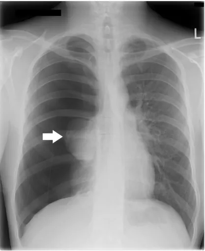
Figure 1. Postero-anterior chest radiograph documenting a complete right-sided pneumothorax
with shifting of the mediastinum towards the left chest. The right lung is completely collapsed
(arrow) creating a large intrapleural space filled with air in which the normal aspect of lung
parenchyma and vasculature is no longer visible………...……….……6
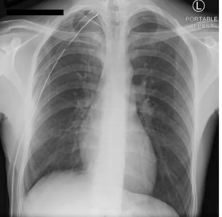
Figure 2. An incomplete re-expansion of the right lung can be seen on this chest radiograph
following the insertion of a drainage catheter for a spontaneous pneumothorax (white arrow).
A discrete amount of subcutaneous emphysema is also visible in the subcutaneous tissues
around the site of chest tube insertion (black arrow)………….………...……..…….8
Figure 3. Following surgery for primary spontaneous pneumothorax with persistent air leak,
the expanded right lung occupies the totality of the ipsilateral pleural space. This chest
radiograph documents the presence of a chest tube and close to its tip postoperative changes
of the apex of the lung related to bullectomy……...………….………...………….10
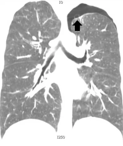
Figure 4. Coronal view of the chest computed tomodensitometry performed in a patient
presenting a left PSP recurrence 18 month following ipsilateral axillary thoracotomy for
bullectomy with vibramycin pleurodesis. A residual bulla can be seen next to the stapler line
(arrow). In this case, the absence of adhesion between the lung and the chest wall was suspected
to be secondary to the weak efficacy of the antibiotic as agent for pleurodesis……….14
Chapter 4. Methods
Figure 5. Saw-tooth appearance of the graph reporting the calculated power of an exact test for
binomial proportions in a non-inferiority study attempting to document a minimal acceptable
ix
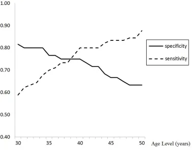
Chapter 5. Results
Figure 6. Sensitivity and specificity of the algorithm’s modifications applied to the 150 patients
and restricting the diagnosis of primary spontaneous pneumothorax to patients below the age
x
List of Appendices
Appendix A: Proposed ICES study abstract………...……….45
Appendix B: Health Science Research Ethic Board approval……….50
Appendix C: Lawson Health Research Institute approval………..….51
Appendix D: Schulich Surgery Internal Research Fund grant……….52
1
Introduction
1.1 Background and Overview
Primary spontaneous pneumothorax (PSP) is one of the most common lung diseases
treated by thoracic surgeons. It is a medical condition affecting generally young and
healthy individuals who are presenting with sudden onset shortness of breath and
shoulder pain. It results from the rupture of a lung bulla, a pocket of air located at the
superior portion of the lung, leading to the accumulation of intrathoracic atmospheric air
and collapsing the lung at some degree. Spontaneous pneumothorax can also occur in
patients presenting different types of lung diseases such as lung emphysema or cystic
fibrosis, which will impact the long-term management. While PSP is most commonly a
benign condition that can sometimes simply be observed, it has the potential, if
unrecognized, to progress to a catastrophic situation in which the heart and other
intra-thoracic organs are compressed by a high pressure tension pneumothorax potentially
creating a hemodynamic instability, which if untreated can be fatal.
Depending on the size of the pneumothorax and its associated clinical presentation, the
initial management may vary. It may include simple observation, needle aspiration, or
chest drainage allowing an evacuation of the intrathoracic trapped air until the lung
breach has healed. But sometimes, a persistent air leak from the lung originating from a
“broncho-pleural fistula” will need to be addressed surgically. This is one of the
indications to perform a surgical intervention for PSP in which the bullae of the lung are
removed and the lung defect is being closed. It is normally associated to another
procedure, named pleurodesis, that can be either mechanical or chemical and that aims to
induce the formation of scar tissue within the chest cavity to permanently maintain the
lung surface adherent against the inner portion of the chest wall, which will decrease the
chance of another episode.
However, the main indication to perform surgery for PSP is to prevent recurrence. Many
episode of PSP, but the most important is the number of prior PSP episodes. Most
patients will be offered surgery after a second documented episode.
Unfortunately, the exact risk of recurrence following an episode of PSP is hard to predict,
in part because only few studies with a limited number of patients have been conducted
specifically on the subject. Some population-based studies are available but include both
primary and secondary spontaneous pneumothorax which are often pooled under the
same classification codes in hospital discharge summaries. This is problematic, as the
recurrence risk, and the factors influencing recurrence, may be different among
secondary pneumothorax. Large administrative data studies specifically on PSP are
lacking.
Administrative hospital data are potentially powerful data sources, allowing
epidemiological studies of diseases, but the accuracy of the data must be confirmed. The
tenth edition of the International Classification of Diseases (ICD-10-CA) was introduced
in Canada in 2000, and it includes codes allowing the identification of patients seeking
medical treatment for pneumothorax. A subdivision of the code J93 further differentiates
between spontaneous tension pneumothorax, other spontaneous pneumothorax, other
pneumothorax, and pneumothorax (unspecified). Unfortunately, the classification does
not clearly allow the identification of PSP, which would be necessary for the conduct of
large population-based studies on PSP using administrative data from emergency room
visits and hospital discharge summaries.
The clinical significance of this lack of knowledge on PSP is that it might be difficult for
a physician to predict the risk of recurrence of a particular patient and therefore decide
which treatment would be the most appropriate.
The objective of this work is to validate the accuracy of an administrative data algorithm
designed to identify PSP cases from the recorded emergency room and hospital discharge
2. Literature review
2.1 Definition and incidence of primary spontaneous
pneumothorax
The accumulation of air into the pleural cavity, or pneumothorax, can happen suddenly in
previously asymptomatic individuals. The term secondary pneumothorax (SP) has been
used to define this condition when it is caused by an underlying medical condition, such
as emphysema or trauma. It is different than primary spontaneous pneumothorax, which
is occurring mainly in previously healthy patients.1 It is the result of the rupture of blebs
or bullae located at the apex of the lung, or by the presence of pleural porosities in this
area.2 Melton et al. reported 77 cases of PSP occurring over a 24-year period in Olmstead county, Minnesota and calculated an age-adjusted incidence rate of 7.4/100 000 persons
per year in males and 1.2/100 000 persons per year in females (adjusted for the USA
white population age distribution from 1960). The phenomenon was described as being
6.2 times more frequent in males than in females.3 Others have reported incidence rates varying between 18-28 cases/100 000 persons per year for males and between 1.2-6
cases/100 000 persons per year for females.4 The peak of incidence is believed to be between the age of 20 and 30 years-old.5 More recently, an analysis of a French database identified over a 4-years period, 42595 patients requiring hospitalization for either
primary and secondary pneumothorax and calculated an annual incidence rate of 22.7
cases/100 000 persons.6
2.2 Clinical presentation and management
Patients presenting an acute episode of pneumothorax will complain most commonly of
dyspnea and severe chest pain irradiating to the shoulder. The physical examination will
reveal decreased breath sounds on auscultation and hyperresonance at chest percussion of
the involved side. A postero-anterior chest radiography will confirm the diagnosis and
Figure 1.Postero-anterior chest radiograph documenting a complete right-sided
pneumothorax with shifting of the mediastinum towards the left chest. The right
lung is completely collapsed (arrow) creating a large intrapleural space filled with
air in which the normal aspect of lung parenchyma and vasculature is no longer
Depending on the severity of the clinical symptoms and the size of the pneumothorax,
most patients with will be treated by simple observation, needle aspiration or chest-tube
insertion. Although the clinical course of most patients will be benign, PSP might have
occasionally serious complications. In some cases a tension pneumothorax might
develop. This is a life-threatening condition caused by the accumulation of air under
pressure within the chest, from a one-way valve phenomenon at the site of the air leak.7 Increased thoracic pressure, shifted mediastinal structured and a compromised blood
return to the right atrium may lead to hypoxia, hypotension and cardiovascular collapse.8 Another reported complication from PSP is persistent air leakage from a broncho-pleural
fistula, which may result in a prolonged hospital stay. In these cases the site of rupture at
the apex of the lung will not heal for many days and the patient will require chest tube
insertion to evacuate the air from the pleural cavity, a procedure usually associated with
minimal complications, however pain or discomfort might be significant. Sometime, a
larger broncho-pleural fistula will lead to air accumulating within the chest despite
previous drainage and additional chest tube insertion, or suction chest drainage is
necessary.9 (Figure 2.) A prolonged air leak will be an indication to address PSP by surgery. Although there is no consensus on the most appropriate length of time to wait
until the leak stops before proceeding with a surgical intervention, most experts will
recommend a waiting period of 5 days.4
Surgical procedures for PSP are some of the most commonly performed interventions in
thoracic surgery and are usually definitive. Through an open axillary approach or with the
use of thoracoscopy, the procedure includes the removal of the apical blebs and bullae
which are thought to be causal factors. In addition, in order to improve the surgical
results and minimize the potential for recurrence, some type of pleurodesis will be
performed to promote scarring at the apex of the lung, which will adhere to the inner
surface of the thoracic cavity. Mechanical pleurodesis includes pleural abrasion or
pleurectomy and can only be performed at the time of surgery. A chemical pleurodesis
might be performed at the time of surgery, but also in the endoscopy unit during medical
thoracoscopy, or even at the patient`s bedside by the injection of sclerosing agents
approach, but can be a valid option in frail patients who aren`t candidates for a surgical
intervention.11
Figure 2.An incomplete re-expansion of the right lung can be seen on this chest
radiograph following the insertion of a drainage catheter for a spontaneous
pneumothorax (white arrow). A discrete amount of subcutaneous emphysema is
also visible in the subcutaneous tissues around the site of chest tube insertion (black
Several products have been suggested as potential sclerosing agents to use in cases of
PSP, the most effective being talc, which can be directly insufflated within the chest or
injected as a suspension into the chest tube. Chemical pleurodesis is rarely used alone in
patients with PSP in Canada and in the United States, but many European authors have
suggested that this could represent an adequate alternative to surgery and that the
resection of the causative ruptured blebs or bullae could be optional.12 Pain and talc toxicity (including acute pneumonitis, acute renal failure and myocardial infarction) have
been described as potential adverse events of talc pleurodesis.13
As previously mentioned, surgical intervention might be necessary in cases of prolonged
air-leak. In those cases, the procedure will allow to stop the air-leakage and withdraw the
drainage catheter, to eventually discharge the patient from the hospital. (Figure 3.) In
those cases, the necessity of the procedure is obvious. Similarly, an intervention to
prevent PSP recurrence is recommended in certain hazardous situations, such as when a
patient is living far away from accessible medical care, working in pressurized
environment (e.g. airplane pilot, divers) and when a pneumothorax recurrence can
become a catastrophic situation.14
Outside of those obvious conditions, surgery for pneumothorax is most commonly aimed
at recurrence prevention. The problem is that this risk of recurrence in a specific patient is
difficult to define. As detailed by Massard et al., the risk of recurrence increases after
each new episode of pneumothorax, and patients presenting with recurring PSP, either
ipsilateral or contralateral, should be offered surgery.8 This has become a generally well accepted concept, even considering the paucity of information on this subject in the
medical literature. In a Delphi consensus statement on the management of pneumothorax
by the American College of Chest Physicians, 85% of the expert panel members
recommended surgery after the first recurrence of PSP. The number of previous episodes
Figure 3.Following surgery for primary spontaneous pneumothorax with persistent
air leak, the expanded right lung occupies the totality of the ipsilateral pleural
space. This chest radiograph documents the presence of a chest tube and close to its
2.3 Reported PSP recurrence rates
Because of the fact that recurrences seem to guide the treatment offered to patients
suffering from PSP, one would expect to find in the medical literature a thorough
description of this risk and of the different factors modelling it. But this is not the case: a
wide variation in the recurrence rates have been described, ranging from 10 to 21% after
a first episode, and rising as high as 50% to almost 80% after a second and third episode.8 The time interval during which the recurrences were captured varies from one study to
the other, offering generally a short period of follow up, and reporting a wide variety of
incidence of this late complication. In their article published in 1991, Lippert et al. were
the first to report appropriately the results on a time-related basis.16 Their 5-year
cumulative survival without pneumothorax in patients without lung disease was estimated
to be 77% (95% confidence interval: 73-81%). Many authors cited the information
published by Cran and Rumball reporting data collected on spontaneous pneumothorax in
the British Royal Air Force between 1941 and 1962, on 994 men aged between 16 and 50
years-old.17 A global recurrence rate of 18.7% was described, with most recurrences
occurring within one year (59.5%) or two years (81.0%) after the first episode. The
proportion of spontaneous pneumothorax recurrences occurring after five years was
minimal (3.2%). In a recent study from Korea, 232 patients were treated without surgery
and underwent telephone follow up for 4 to 8 years following their episode of PSP. The
number of PSP episodes was not included in the analysis, but a recurrence rate of 29%
was noted in patients treated with observation. The patients who required chest drainage
had a 54% recurrence rate, which is higher than what has been reported before.18
2.4 Risk factors for pneumothorax
Many clinical elements have been described as potential risk factors for the development
of a first episode of PSP. It is important to understand those characteristics as they could
also cause recurrence. This was suggested in a review of 1491 patients who presented
in clinical factors could be identified between initial and recurrent cases.19 The most
commonly cited patient-related risk factor is gender. Males are known to be more than
three times more at risk than females to develop a pneumothorax, and this risk rises up to
six times when the analysis is restricted to PSP.3,6 Age is also related to the risk of developing PSP. In a review from the General Practice Research Database (GPRD)
including 5.6% of the patients treated in England and Wales, Gupta et al. described a
biphasic distribution of age in patients consulting for any pneumothorax. The first peak of
incidence was thought to be mainly due to PSP and was corresponding to the 20-24y.o.
age group in male and 30-34y.o. age group in female.5 Another risk factor for a spontaneous pneumothorax is smoking. In a population-based study in the county of
Stockholm, Sweden, Bense et al. reported in 1987 a 9-fold and 22-fold risk increase of
developing pneumothorax with tobacco smoking in women and men, respectively.20 Marijuana smoking has also been identified as detrimental for respiratory functions and it
is suspected to increase even more the risk of developing bullous lung diseases and
causing pneumothorax in individuals who are most commonly already smokers.21 Tall and thin individuals are known to be at higher risk for PSP. The effect of the
height-weight ratio on the risk of recurrence has been confirmed by Nakamura to be independent
from the effect of the level of tobacco consumption.22 Patients height seems to be a more
important factor than weight for the occurrence of PSP, taller individuals being at higher
risk.23 Rapid changes in the configuration of the upper chest occurring during
adolescence was found to have an impact on the risk of developing PSP in a case-control
study on 404 patients treated in Taiwan.24 The intensity of physical activity was initially suspected as being a significant etiological factor for PSP, but this has been refuted.4 Some psychological or emotional factors were once suspected to cause PSP. In an
interesting cooperative work involving departments of psychiatry and emergency
medicine, no clear relation could be identified between episodes of PSP and patients’
anger, anxiety or depression levels.25 Certain environmental factors, such as weather conditions, atmospheric pressure, level of air pollution, have been suggested to promote
be identified between the occurring of pneumothorax and atmospheric pressure, relative
humidity, rainfall, wind speed or temperature.30
2.5 Risk factors for PSP recurrence
It is unusual for patients to develop PSP recurrence following surgical treatment, unless
an incomplete bullae resection is performed with an inadequate pleurodesis. (Figure 4.)
Many reports in literature have defined the factors related to those unsuccessful
interventions, however it was decided for this work not to include them and to limit the
review to the natural history of PSP prior to any definitive treatment.
The presence of air trapping on tomodensitometry (CT-Scan), has been described as
being a risk factor for PSP.31 In a study on 176 patients treated without surgery and followed for at least 12 months after an episode of PSP, the risk of recurrence increased
from 6% to 68% in patients found to have blebs or bullae on high-resolution CT-scan.32 Similarly, in a retrospective study of 114 patients treated non-surgically for PSP, Young
Choi et al. noted after a mean follow up of 43 months a recurrence rate higher (60% vs
31%) in patients documented to have blebs or bullae identified on high-resolution
CT-scan. In a retrospective review of 153 patients treated for PSP, Sadikot et al. reported
over a four-year period a 54.2% rate of recurrence. Gender, height and smoking status
were the only three independent factors for recurrence that could be found as
significant.33 Similarly, in a study of 122 subjects treated for a first episode of spontaneous pneumothorax, Lippert et al. documented a higher risk of recurrence in
patients with lower height-weight ratio, in smokers, in patients over 60 years of age and
in patients presenting pulmonary fibrosis on chest X-Ray. This study however included
patients who underwent surgical treatment for pneumothorax and possibly also cases of
secondary pneumothorax, making the results from their analysis not applicable to
Figure 4.Coronal view of the chest computed tomodensitometry performed in a
patient presenting a left PSP recurrence 18 month following ipsilateral axillary
thoracotomy for bullectomy with vibramycin pleurodesis. A residual bulla can be
seen next to the stapler line (arrow). In this case, the absence of adhesion between
the lung and the chest wall was suspected to be secondary to the weak efficacy of the
The patient BMI seems to be related to the risk of recurrence. In a retrospective analysis
of a cohort of 273 patients aged less than 30 years and treated for a first episode of PSP,
Tan et al. recorded 81 recurrences (30%). Sixty-four percent of their patients underwent
surgery. They performed a Cox regression analysis and identified 3 factors associated to
recurrence: a low body-mass index (BMI) (less than 18.5 kg/m2), the absence of surgical treatment, and a large-size pneumothorax (more than 50% of the lung volume).34 In a similar study design including 553 patients treated surgically for PSP and analyzing the
risk factor for contralateral recurrence, Chen et al. identified both low BMI and visible
blebs/bullae on HDCT to be related to an additional episode of PSP.35 Another group from China (Chiu et al.), analyzing the results of 128 patients treated medically for a first
episode of pneumothorax, identified the size of the pneumothorax to be associated to
further episodes.36
Other factors related to the type of treatment offered to patients with PSP have been
related to the risk of recurrence. The decision to treat the patient initially with chest air
aspiration through thoracentesis instead of chest tube insertion was found to have a
higher incidence of recurrence during the first week of follow-up in two studies published
in the mid-1990’s.37 This finding is however not surprising because multiple
thoracentesis sessions may be necessary when this procedure, most commonly performed
in Europe, is used when dealing with PSP.38 In a meta-analysis of randomized trials
comparing the results of thoracentesis and chest drainage in the treatment of PSP, no
significant difference could be identified in recurrence rates at one year.39 The performance of chemical pleurodesis through the chest-tube at the moment of initial
treatment for PSP has been shown to be an effective way to reduce recurrences, and
reports have detailed the benefits of using tetracycline, minocycline or talc suspension to
obtain an effective sclerotherapy of the pleural space.40,41
2.6 Pneumothorax in administrative data studies
Administrative hospital data are potentially powerful data sources, allowing
tenth edition of the International Classification of Diseases (ICD-10-CA) was introduced
in Canada in 2000, and implemented in Ontario in 2002. It includes codes allowing the
identification of patients seeking medical treatment for pneumothorax.42 A subdivision of the code J93 further differentiates between spontaneous tension pneumothorax, other
spontaneous pneumothorax, other pneumothorax, and pneumothorax (unspecified).43 Unfortunately, the classification does not clearly allow the identification of PSP, which is
necessary to conduct large population-based studies on the subject using administrative
data from emergency room visits and hospital discharge summaries.
Gupta et al. published in 2000 their work on the epidemiology of pneumothorax in
England. They identified from the General Practice Research Database and the Hospital
Episode Statistics obtained from the Office for National Statistics over 20,000
consultations for pneumothorax between the years 1991 and 1995. The authors could not
specifically target the patients presenting for PSP and included in their study patients
treated for secondary pneumothorax and traumatic or iatrogenic pneumothorax. They
were however able to demonstrate the impact of age on the incidence of pneumothorax,
as both in males and females a biphasic distribution of cases of pneumothorax. The first
peak of incidental cases occurred between ages 20-24 in males and between the ages
30-34 in female. It was suggested that these early age pneumothorax were representing
mostly PSP cases.
In another population-based study, Bobbio et al, reported from the French database
“Programme de Médicalisation des Systèmes d’Information” between 2008 and 2011, 59
637 hospitalizations of 42 595 patients for pneumothorax. Patients treated as outpatients
were not included in the study. The authors calculated an annual incidence of 22.7 cases
per 100,000 population for France. The mean hospital stay was 7 days, 24% of patients
underwent surgery with thoracoscopic resection of blebs (76% or intervention) associated
to mechanical pleurodesis in 52% and talc pleurodesis in 24%. The authors excluded
traumatic pneumothorax and differentiated between secondary pneumothorax and what
was termed “idiopathic pneumothorax”. The authors used the ICD classification for the
diagnosis of pneumothorax and it is unclear how they differentiated between idiopathic
study, however, a high proportion of the cases represented a re-hospitalization, (48% of
the cases in females and 49% of the cases in males), and in those cases the patients were
found to be significantly younger (35 vs 40 years when no previous pneumothorax could
be documented), and underwent more often a surgical procedure. There was in this study
a high proportion of idiopathic pneumothorax (85% of patients) even in the group of
patients above 50 years old (65% of cases in males and 77% in females). This raises the
question whether or not the idiopathic pneumothorax cases in this study represent cases
of PSP.
There is to our knowledge no large population-based epidemiological study specifically
3 Rationale, research question and hypothesis
3.1 Rationale
PSP is one of the most common chest pathologies treated by chest physicians. The main
indication for surgery is the prevention of recurrence in high risk patients. There have
been multiple publications reporting the experience of different medical centers, some of
them having a significant experience. However, there are only a few rare
population-based studies on the subject, and none adequately or specifically examine the
epidemiology, risk factors and recurrence rates for PSP. This is in part a consequence of
the absence of specific codes differentiating primary and secondary spontaneous
pneumothorax in the International Codification of Disease. The consequence is a
variability between the recommendations of the different thoracic societies, and an
important non-adherence to national guidelines of treatments.4,15,44 We conducted this work with the hope to provide researchers with a tool to facilitate large population-based
studies on PSP.
3.2 Research question and hypothesis
The objective of this study was to create and validate an administrative data algorithm
which would allow the identification of PSP cases from the recorded emergency room
and hospital administrative data. We hypothesized that we could build an algorithm
identifying PSP cases with a sensitivity and positive predictive value of at least 80%, to
4 Methods
4.1 Study design
We performed a retrospective validation study to assess the accuracy of an algorithm
designed to identify cases of PSP amongst patients presenting to the emergency room
with a main diagnosis of pneumothorax. The results obtained from the proposed
algorithm were compared to the reference standard obtained from manual chart review.
The research protocol was reviewed and approved by the Health Sciences Research
Ethics Board at the University of Western Ontario (00000940-105409).
4.2 Creation of the algorithm
The development of the algorithm was based on the same concepts used clinically to
differentiate between PSP and other types of pneumothorax. It was derived from clinical
reasoning and designed following discussions amongst the research collaborators after a
complete review of the ICD-10-CA codes to identify the different clinical entities that are
recognized to be a cause of SP. Four main diagnostic codes of pneumothorax were
chosen as algorithm inclusion criteria because they were potentially associated with
patients presenting PSP. These codes were included the following:
- J93.0 Spontaneous tension pneumothorax
- J93.1 Other spontaneous pneumothorax
- J93.8 Other pneumothorax
- J93.9 Pneumothorax, unspecified
We decided not to include some additional diagnosis of pneumothorax found in the
ICD-10-CA in our algorithm, as they likely represented cases of secondary, iatrogenic or
traumatic pneumothorax. Those unused codes were:
- postprocedural pneumothorax (J95.811)
- traumatic pneumothorax (S27.0)
- tuberculous pneumothorax (A15)
- pyopneumothorax (J86)
The next step in the algorithm creation was the exclusion of all cases not believed to be
related to PSP and representing potentially either an iatrogenic pneumothorax, a
secondary pneumothorax consecutive to a chronic condition, or a pneumothorax related
to an acute illness or event such as trauma, pneumonia, status asthmaticus, etc. Below are
the details of the three steps of algorithmic exclusion:
1. Patients with a hospital admission record in Discharge Abstract Database (DAD)
in the previous 30 days were excluded unless their main diagnosis was pneumothorax, as
they could represent cases of iatrogenic pneumothorax from a procedure or an
intervention having occurred during this hospitalization.
2. Patients with a chronic condition associated with SP where excluded. We
identified these patients based on all diagnosis in DAD and National Ambulatory Care
Reporting System (NACRS) databases recorded for 14 conditions present at the index
Table 1. Chronic conditions identified as potential cause of secondary
pneumothorax with corresponding ICD-10-CA codes.
3. We also excluded the patients presenting with an acute condition potentially
causing SP in the 30-days prior to the first visit for PSP. (Table 2.)
For each patient all information from index date and previous visits DAD and NACRS
recorded locally were obtained. The information collected for the analysis included the
admission and discharge dates and all ICD-10-CA diagnostic codes. The data from the
different visits and databases were pooled for each individual and submitted to the
algorithm.
Chronic conditions
Chronic Obstructive Pulmonary disease J40, J410, J411, J418, J42, J431,432, J438-J441, J961, J982 Thoracic endometriosis N808
Pneumocystis B59
Sarcoidosis D860-J863, D868,D869 Tuberous sclerosis Q851
Rheumatoid arthritis M051-M053, M058-M060, M062-M064, M068, M069, M080 Ankylosing spondylitis M081, M45
Scleroderma L940-L941
Ehler-Danlos syndrome Q796
Marfan syndrome Q874
Langerhan's disease D760, D763
Cystic fibrosis E840, D841, D848, D849 Interstitial lung disease J841, J848, J849
Table 2. Acute conditions identified as potential cause of secondary pneumothorax
with corresponding ICD-10-CA codes.
4.3 Validation study cohort creation
The charts of the first 150 consecutive patients who presented at the LHSC Victoria
Hospital from January 2003 to March 2010 with a main ICD-10-CA diagnosis of
pneumothorax were considered for inclusion in the study. The sample size estimation is
detailed at chapter 4.7. The patient`s charts where identified from the NACRS database
information stored locally at the hospital’s Medical Records service. The NACRS
database contains data for hospital-based ambulatory care and is mandatory in Ontario for
all day surgery and emergency department visits. It includes the recording of the main
diagnosis along with up to 25 additional ICD-10-CA diagnostic codes collected from
trained abstractors. In this study, patient’s recruitment was limited to individuals aged at
the time of consultation between 18 and 65 years, representing an age distribution that
Acute conditions
Chest trauma S202-S204, S207, S208, S2100, S2101, S2110, S2111, S2120, S2121, S2170, S2171, S2180, S2181, S2190, S2191, S22000, S22001, S22010, S22011, S22090, S22091, S22100, S22101, S22200, S22201, S22300, S22301, S22400, S22401, S22410, S22411, S22490, S22491, S22500, S22501, S22800, S22900, S230-S235, S240, S2410-S2413, S2418-S2420, S2428, S2438, S2440, S2448, S2458, S2468,
S250-S255, S257-S259, S26000, S26001, S26800, S26801, S26810, S26811, S26880, S26881, S26890, S26891, S27000, S27001, S27100, S27101, S27200, S27201, S27300, S27301, S27310, S27311, S27380, S27381, S27390, S27391, S27400, S27410, S27480, S27490, S27500, S27510, S27511, S27580, S27590, S27600, S27601, S27610, S27611, S27680, S27690, S27700, S27701, S27710, S27711, S27780, S27790, S27791, S27800, S27801, S27810, S27811, S27840, S27841, S27850, S27851, S27860, S27861, S27890, S27891, S27900, S27901, S27980, S27981, S280, S281, S2900, S2908, S297-S299, T001, T008-T011, T0180, T0181, T0190, T0191, T0210, T0211, T0270, T0271, T0280, T0281, T0290, T0291, T031, T039, T041, T047, T048, T049, T058-T065, T068, T07, T080, T081, T090, T091, T095, T098, T099, T140-T149, T792, T797, T798, T799
Foreign bodies T173-T175, T178, T179, T181, T188, T189 Status asthmaticus J4501, J4511, J4581, J4591
Pneumonia J120-J123, J128, J129, J13, J14, J150-J160, J168, J180-J182, J188, J189
would allow the recruitment of a sufficient number of both primary and secondary
pneumothoraxes while excluding the pediatric population. The cases of pneumothorax
were identified from one of the NACRS main diagnosis codes: J930 (spontaneous tension
pneumothorax), J931 (other spontaneous pneumothorax), J938 (other pneumothorax), or
J939 (pneumothorax, unspecified), correlating to the inclusion criteria of the proposed
algorithm. These codes included the complete range of disease severity.
For this study, it was decided to perform the validation from a group of patients having
one of the ICD-10-CA codes of pneumothorax. It would have been difficult to recruit
enough patients having a PSP from a wider inclusion characteristic such as the presence
of shortness of breath or pleuritic chest pain at presentation. But because the goal of the
algorithm was to differentiate PSP cases from other types of pneumothorax, and because
of the high accuracy of the J93 code, the cohort was built from patients diagnosed at the
emergency with a diagnosis of pneumothorax.
4.4 Reference standard
To define the validity of the propose algorithm, the results needed to be compared to a
reference standard obtained by manual chart review. Patient charts were abstracted by
two independent physicians, blinded to the recorded administrative data coding (KG,
AS). The reviewers confirmed or rejected the diagnosis of pneumothorax from the
documents contained in the charts, including the radiology reports and images if needed.
All cases of pneumothorax were defined as PSP or SP. Where there was disagreement
between reviewers, the diagnosis was obtained following a review by a third physician
(EF).
4.5 Statistical methods
The results obtained from the algorithm and from the chart review were compared. The
years or older, this threshold representing the midpoint of the usual distribution peaks for
PSP and SP.
The chart review results were considered as the gold standard reference for the validation
of the proposed algorithm. Comparing the results from the chart review and
administrative data algorithm, sensitivity, specificity, positive predictive value (PPV),
and negative predictive value (NPV) were calculated, including the 95% confidence
interval. SAS software version 9.3 was used. Fisher’s exact test was used to compare
categorical data of the two patients groups including sensitivity, specificity, PPV and
NPV. The kappa statistic was calculated as a measure of agreement between reviewers’
results. Additional calculations of sensitivity and specificity were performed to evaluate
the ability of the algorithm to define PSP when limiting the diagnosis to patients below
different age thresholds set between 30 and 50 years of age.
In this study, sensitivity was defined as the proportion of PSP patients (identified from
the reference standard) adequately having a positive result from the algorithm. Specificity
as the proportion of patient without PSP from the reference standard and negative from
the algorithm. PPV was the proportion of algorithm positive patients truly having PSP
from the reference standard, and NPV was the proportion of algorithm negative patients
not having a PSP from the reference standard.
4.6 Sample size calculation
To evaluate the diagnostic performance of the proposed algorithm to diagnose primary
pneumothoraxes, sensitivity and specificity was obtained from a comparison to the
reference standard, taken from manual chart review. The necessary sample size has been
calculated following the binomial exact approach for a dichotomous diagnostic test using
the SAS 9.3 software (SAS Institute Inc., Cary (NC) USA). A significance level of 0.05
has been chosen to calculate the total number of patients necessary to reach a power of
90% in a non-inferiority trial, comparing the proposed algorithm binomial results to a
standard reference obtained from manual chart review, using a one-sided exact test with
specificity of 80%. As shown in Figure 5, because of the nature of the binomial
distribution, a “saw-toothed” curve was acquired when power was expressed for different
values of sample size. This phenomenon was expected as previously described.45 From a conservative analysis of this graph, it is noted that the power of the study to reject the null
hypothesis was superior to 90% for any value of sample size superior to 130. Considering
possible incomplete data and other challenges that might present during chart reviews, an
additional 15% cases have been added to the number in order to reach a total sample size
Figure 5. Saw-tooth appearance of the graph reporting the calculated power of an
exact test for binomial proportions in a non-inferiority study attempting to
document a minimal acceptable sensitivity or specificity of 80% when a result of
5 Results
5.1 Manual chart review
Of the 150 patients included in this study, 95 were under the age of 40 years and 55 were
aged 40 years and over. The chart review could not identify any evidence of
pneumothorax in the charts of 6 patients, while 96% had a pneumothorax corresponding
appropriately to the main diagnosis. These included 90 cases of PSP, representing 60%
of patients. Eighty-two percent of these (74 cases) were found in patients under 40
years-old, while 18% were identified in older patients.
There was an agreement of 88% between the two reviewers; corresponding to a kappa
statistics of 0.76 and a third review was necessary in 18 cases.
5.2 ICD-10-CA codes used as inclusion criteria
The review of the administrative data revealed that the code J939 (pneumothorax,
unspecified) was the most commonly used to describe the main diagnosis, present in 85
of the 150 patients. However in younger patients, the code J931 (other spontaneous
pneumothorax) was also commonly used and was identified in 48% of the 95 patients.
The codes J930 (spontaneous tension pneumothorax) and J938 (other pneumothorax)
were retrieved in only 4 patients for each of them.
5.3 Results from the algorithm
The reason to suspect a SP from the administrative data was most commonly related to an
associated chronic condition which could be identify in 23 of the 39 patients not
considered to have a PSP from the results of the algorithm. In some patients, more than
one potential cause of secondary pneumothorax was identified. These results are detailed
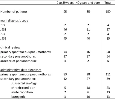
Table 3. Distribution of patients with a main diagnosis code of pneumothorax
according to age group.
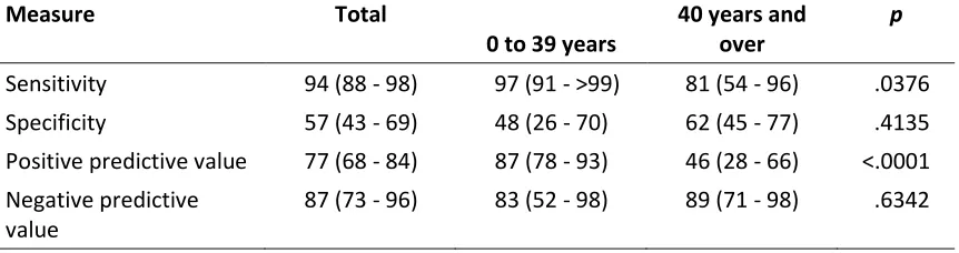
The ability of the proposed algorithm to identify cases of PSP is detailed in Table 4,
which summarizes diagnostic accuracy. Overall, sensitivity of the algorithm for PSP was
94%, being higher in younger patients (97%) than in older patients (81%), p=0.0376.
Similarly, a higher PPV was obtained when the algorithm was applied to the younger
patients population, 87%, compared to 46% for the patients ages 40 years or more
(p<0.0001). The overall specificity of the algorithm was 57%, and no significant
difference was identified between the younger and older patient groups. Similarly, there
was no significant difference between the two groups NPVs, the overall value estimated
to 87%.
0 to 39 years 40 years and over Total
Number of patients 95 55 150
main diagnosis code
J930 2 2 4
J931 46 11 57
J938 2 2 4
J939 45 40 85
clinical review
primary spontaneous pneumothorax 74 16 90
secondary pneumothorax 17 37 54
absence of pneumothorax 4 2 6
administrative data algorithm
primary spontaneous pneumothorax 83 28 111
secondary pneumothorax 12 27 39
suspected etiology:
chronic condition 5 18 23
acute condition 7 6 13
Table 4. Diagnostic performance of the administrative data algorithm for the
identification of primary spontaneous pneumothorax cases (estimate and 95%
confidence interval).
Measure Total
0 to 39 years
40 years and over
p
Sensitivity 94 (88 - 98) 97 (91 - >99) 81 (54 - 96) .0376 Specificity 57 (43 - 69) 48 (26 - 70) 62 (45 - 77) .4135 Positive predictive value 77 (68 - 84) 87 (78 - 93) 46 (28 - 66) <.0001 Negative predictive
value
87 (73 - 96) 83 (52 - 98) 89 (71 - 98) .6342
5.4 Age dichotomization cutting point
In order to evaluate the appropriateness of the 40-years threshold used in the study, we
performed additional modifications on the algorithm for different age levels varying
between 30 and 50 years. We calculated the sensitivity and specificity of the algorithm
applied to the 150 patients when limiting the diagnosis of PSP to patients younger than
each age point. Between the levels of 30 to 50 years, the sensitivity varied from 59% to
6 Discussion
6.1 Importance of differentiating PSP from SP
PSP is by definition a spontaneous and unprovoked event usually occurring in young and
previously healthy individuals. It differs from SP, which represents a proportion of about
half of all pneumothorax.3,46-48 All pneumothorax will require similar initial management; however, once urgent treatment is completed, the care given to patients presenting with
either PSP or SP will differ.49,50 Treatment of the underlying cause of SP will be
necessary and could include chest wall stabilization, optimization of the COPD or asthma
medication, and antimicrobial treatment of pneumonia or lung abscess. For PSP, aside
from smoking cessation and recommendation to avoid certain activities, most individuals
will require no further treatment, but surgery could be offered to patients who are at a
higher risk of recurrence or complications.4,8,15
6.2 Need for epidemiological studies on PSP
There is a need for large epidemiological studies on the subject of PSP to help define
predictors of recurrence and lead to treatment recommendation tailored to this otherwise
healthy and potentially productive population. Population-based studies from United
Kingdom and France have been published in the last few years and have produced
significant knowledge on the subject; however, these included both primary and SP.
Hallifax and Rahman noted an unexpectedly low proportion of SP (14%) in a report from
the French national healthcare database, which has not been fully explained and suggests
6.3 Limitations of the ICD-10-CA classification
The 10th version of the ICD, used in Canada as in many other jurisdictions, does not
include a sub-classification allowing the identification of the different types of
spontaneous pneumothorax. This situation is problematic and limits the ability of
researchers to target PSP specifically. In the United States, a different version of the ICD
has been implemented in October 2015, ICD-10-CM (Clinical Modification), which
includes a subdivision of the J931 code corresponding to PSP: J9311.52 However, it is
difficult to anticipate the effect of this sub-classification on eventual epidemiological
studies because of the presence of three other J93 codes which could be used for PSP. In
our study, only 38% of patients were coded J931, the majority (57%) of the charts were
labeled with the code J939 (pneumothorax, unspecified).
6.4 Validation of administrative data
Code-validation is necessary before conducting population-based studies to ensure
adequacy of the patient cohorts.53 In Canada, the Institute for Clinical Evaluative Sciences (ICES) reported in 2006 that the pneumothorax code J93 is amongst the most
accurately used (95%) of the main diagnosis codes, and this correlates to the results of
this review which identified a miscode rate of only 4%.42 However, to our knowledge,
this study represents the first attempt to differentiate between PSP and SP using hospital
administrative data. Defining PSP by only the absence of associated codes for underlying
lung disease was not considered sufficient in the design of this study and three exclusion
criteria were created with the goal of obtaining a better case definition. Based on the
results of the administrative data algorithm, these exclusion criteria identified a
significant proportion of patients experiencing SP, particularly in younger patients for
which the presence of a chronic condition was responsible for only 42% of cases defined
6.5 Study strengths and limitations
PSP is known to occur in a younger population than SP, and multiple studies have
suggested that peak incidence occurs between 20 and 30 years of age. When creating the
inclusion criteria for a population-based study oriented towards this age group, an option
would be to use the patient`s age to minimize the number of misdiagnoses that would be
associated with inclusion of older patients. This study’s dichotomization of younger
versus older patients allowed observation of the effect of this strategy. The ability of the
proposed algorithm to accurately identify cases of PSP cases was significantly better
when limited to the younger group of patients. However, using this method would
exclude cases of true PSP occurring in older patients (16 individuals in this study) and
limit the participation to about 82% of potential patients. However, these individuals
might also have a non-diagnosed underlying lung condition.
The proposed algorithm demonstrated a good diagnostic accuracy in defining PSP cases
amongst patients under 40 years of age. The 97% sensitivity rate found in this age group
suggests that almost all PSP cases can be identified with the use of administrative data.
And, as demonstrated by the 87% PPV, the identification of a PSP case from the
algorithm will be correct in a very high proportion of patients. These findings also
suggest that limiting the diagnosis of PSP to the absence of recorded concurrent lung
conditions might be insufficient and could be improved by the use of more restrictive
exclusion criteria.
The limitations of the proposed algorithm are related to its lower than expected
specificity. This is a direct consequence of a high number of patients falsely considered
to have a PSP, particularly in the older population. This suggests either that the strict
criteria used in this study for PSP definition might not be sufficient or more likely, that
the recording of secondary diagnosis into NACRS and DAD databases is occasionally
incomplete. Thus, it would be prudent to limit the use of the algorithm to the suggested
target population of PSP, as its accuracy might be insufficient when attempting to build a
Choosing an appropriate reference standard is an important part of a validation study. The
“golden” standard used in this study was based on the review of patients charts by two
independent reviewers. Even if the patients were not directly evaluated by the reviewing
physicians, the ability to proceed with a third chart review in cases of non-agreement is
thought to have provided this study with a solid reference standard.
6.6 Future direction
The findings of this study support the use of the proposed algorithm as an appropriate
method of PSP identification for the conduction of population-based epidemiological
studies based on administrative data using ICD-10 codes. As a result, an ICES study
proposal abstract has been written. It proposes the use of this study’s algorithm to
evaluate in the population of Ontario the epidemiology of PSP and its recurrence factors
(Appendix A). Because of the wide acceptance of the ICD-10 codification system, it is
possible that the proposed algorithm could be exported to other provincial and national
databases even if discharged summaries slightly different than DAD or NACRS could
exist in other jurisdictions. Eventually, a clinical algorithm for the management of PSP
might be created. In the development of such prognostic models, the process of
derivation is different than the validation: it will allow the identification of prognostic
factor according to a subset of patient from regression analysis. The prognostic model
obtained needs then to be validated to ensure its external validity, often using a second
subset of the studied population. Such clinical algorithms may have a significant impact
on the clinical practice.
6.7 Conclusions
PSP is a clinical entity distinct from SP for which there is a paucity of epidemiological
studies. The ICD-10 codes used in most countries do not allow its direct identification.
An algorithm was created, which uses the information available in NACRS and DAD to
overcome this deficiency. Of 150 patients presenting with a diagnosis of pneumothorax,
we validated this algorithm to have a sensitivity of 97% and a PPV of 87% when used in
a population of young patients known to be at higher risk of developing a PSP. These
give researchers the tools required to conduct epidemiological studies on large
population-based data. Such studies are expected to improve the understanding of the
References
1. Onuki T, Ueda S, Yamaoka M, et al. Primary and Secondary Spontaneous Pneumothorax: Prevalence, Clinical Features, and In-Hospital Mortality. Can Respir J. 2017;2017:6014967.
2. Noppen M. Spontaneous pneumothorax: epidemiology, pathophysiology and cause. European respiratory review : an official journal of the European Respiratory Society. 2010;19(117):217-219.
3. Melton LJ, 3rd, Hepper NG, Offord KP. Incidence of spontaneous pneumothorax in Olmsted County, Minnesota: 1950 to 1974. The American Review of
Respiratory Disease. 1979;120(6):1379-1382.
4. MacDuff A, Arnold A, Harvey J, Group BTSPDG. Management of spontaneous pneumothorax: British Thoracic Society Pleural Disease Guideline 2010. Thorax. 2010;65 Suppl 2:ii18-31.
5. Gupta D, Hansell A, Nichols T, Duong T, Ayres JG, Strachan D. Epidemiology of pneumothorax in England. Thorax. 2000;55(8):666-671.
6. Bobbio A, Dechartres A, Bouam S, et al. Epidemiology of spontaneous pneumothorax: gender-related differences. Thorax. 2015;70(7):653-658.
7. Simpson G, Vincent S, Ferns J. Spontaneous tension pneumothorax: what is it and does it exist? Internal Medicine Journal. 2012;42(10):1157-1160.
8. Massard G, Thomas P, Wihlm JM. Minimally invasive management for first and recurrent pneumothorax. The Annals of Thoracic Surgery. 1998;66(2):592-599.
9. Baumann MH. Pneumothorax. Seminars in respiratory and critical care medicine. 2001;22(6):647-656.
10. Bintcliffe O, Maskell N. Spontaneous pneumothorax. Bmj. 2014;348:g2928.
11. Ashby M, Haug G, Mulcahy P, Ogden KJ, Jensen O, Walters JA. Conservative versus interventional management for primary spontaneous pneumothorax in adults. Cochrane Database Syst Rev. 2014(12):Cd010565.
13. Gonzalez AV, Bezwada V, Beamis JF, Jr., Villanueva AG. Lung injury following thoracoscopic talc insufflation: experience of a single North American center. Chest. 2010;137(6):1375-1381.
14. Baumann MH. Pneumothorax and air travel: lessons learned from a bag of chips. Chest. 2009;136(3):655-656.
15. Baumann MH, Strange C, Heffner JE, et al. Management of spontaneous pneumothorax: an American College of Chest Physicians Delphi consensus statement. Chest. 2001;119(2):590-602.
16. Lippert HL, Lund O, Blegvad S, Larsen HV. Independent risk factors for cumulative recurrence rate after first spontaneous pneumothorax. The European respiratory journal : official journal of the European Society for Clinical Respiratory Physiology. 1991;4(3):324-331.
17. Cran IR, Rumball CA. Survey of spontaneous pneumothoraces in the Royal Air Force. Thorax. 1967;22(5):462-465.
18. Noh D, Lee S, Haam SJ, Paik HC, Lee DY. Recurrence of primary spontaneous pneumothorax in young adults and children. Interact Cardiovasc Thorac Surg. 2015;21(2):195-199.
19. Kepka S, Dalphin JC, Parmentier AL, et al. Primary Spontaneous Pneumothorax Admitted in Emergency Unit: Does First Episode Differ from Recurrence? A Cross-Sectional Study. Can Respir J. 2017;2017:2729548.
20. Bense L, Eklund G, Wiman LG. Smoking and the increased risk of contracting spontaneous pneumothorax. Chest. 1987;92(6):1009-1012.
21. Martinasek MP, McGrogan JB, Maysonet A. A Systematic Review of the Respiratory Effects of Inhalational Marijuana. Respir Care. 2016;61(11):1543-1551.
22. Nakamura H, Izuchi R, Hagiwara T, et al. Physical constitution and smoking habits of patients with idiopathic spontaneous pneumothorax. Japanese journal of medicine. 1983;22(1):2-8.
23. Melton LJ, 3rd, Hepper NG, Offord KP. Influence of height on the risk of spontaneous pneumothorax. Mayo Clinic proceedingsMayo Clinic. 1981;56(11):678-682.
25. Eryigit H, Ozkorumak E, Unaldi M, Ozdemir A, Cardak ME, Ozer KB. Are there any psychological factors in male patients with primary spontaneous
pneumothorax? Int J Clin Exp Med. 2014;7(4):1105-1109.
26. Haga T, Kurihara M, Kataoka H, Ebana H. Influence of Weather Conditions on the Onset of Primary Spontaneous Pneumothorax: Positive Association with Decreased Atmospheric Pressure. Annals of Thoracic and Cardiovascular Surgery : Official Journal of the Association of Thoracic and Cardiovascular Surgeons of Asia. 2012.
27. Araz O, Ucar EY, Yalcin A, et al. Do Atmospheric Changes and the Synodic Lunar Cycle Affect the Development of Spontaneous Pneumothorax? Acta Chir Belg. 2015;115(4):284-287.
28. Bense L. Spontaneous pneumothorax related to falls in atmospheric pressure. European journal of respiratory diseases. 1984;65(7):544-546.
29. Bertolaccini L, Cassardo C, Viti A, Terzi A. The relationship between
meteorological variations and the onset of spontaneous pneumothorax. Surgery today. 2013;43(3):345-346.
30. Heyndrickx M, Le Rochais JP, Icard P, Cantat O, Zalcman G. Do atmospheric conditions influence the first episode of primary spontaneous pneumothorax? Interact Cardiovasc Thorac Surg. 2015;21(3):296-300.
31. Smit HJ, Golding RP, Schramel FM, Deville WL, Manoliu RA, Postmus PE. Lung density measurements in spontaneous pneumothorax demonstrate airtrapping. Chest. 2004;125(6):2083-2090.
32. Casali C, Stefani A, Ligabue G, et al. Role of blebs and bullae detected by high-resolution computed tomography and recurrent spontaneous pneumothorax. The Annals of Thoracic Surgery. 2013;95(1):249-255.
33. Sadikot RT, Greene T, Meadows K, Arnold AG. Recurrence of primary spontaneous pneumothorax. Thorax. 1997;52(9):805-809.
34. Tan J, Yang Y, Zhong J, et al. Association Between BMI and Recurrence of Primary Spontaneous Pneumothorax. World J Surg. 2017;41(5):1274-1280.
35. Chen YY, Huang HK, Chang H, Lee SC, Huang TW. Postoperative predictors of ipsilateral and contralateral recurrence in patients with primary spontaneous pneumothorax. J Thorac Dis. 2016;8(11):3217-3224.