SHRESTHA, NAVIN. Mapping Quantitative Trait Loci (QTL) Associated with Flavonoids in Tomato (Under the direction of Dr. Dilip R. Panthee).
The flavonoids are a remarkable group of plant metabolites. These are the only secondary metabolites which have been credited with so many diverse essential functions in growth and development of the plant. Anthocyanins are one of the significant components of flavonoid in a plant system. They play a significant role in plant resistances to biotic and abiotic stress. Their bioavailability and biological activities in humans appear to be strongly influenced by their chemical nature. A variety of biological activities possessed by flavonoids include antiallergic, antiinflammatory, antiviral, antiproliferative, and anticarcinogenic activities. Anthocyanins are getting more attention because of their strong antioxidant activity.
Cultivated tomatoes (Solanum lycopersicum L.) do not produce anthocyanin in the fruit, and this trait has been introgressed from wild varieties of tomato. Fruit with the genes
different groups; low, medium and high. Some progenies fall outside the range of parents, suggesting that there was transgressive segregation, which is one of the characteristics of a quantitative trait. For QTL analysis, we adopted QTL-seq approach and identified four QTL on chromosome 1, 2 and 4.
In order to assess the role of anthocyanins against early blight (EB) in tomato, 32 genotypes of three varying degree of anthocyanin content; 12, 12 and 8 genotypes from high, medium and low anthocyanin level group, incorporating NC1CELBR as negative control and NC84173 as positive control was examined in CRD in laboratory environment. Similarly, 250 BC1S1 genotypes including those 32 genotypes and control lines were tested in MRS, Waynesville, NC in Randomized Complete Block Design (RCBD). Spores of Alternaria linariae in the concentration of ~105 spores/ml was inoculated in experimental genoytpes both in the field and laboratory. The disease was scored using Horsfall-Barratt score. Anthocyanins were found to be insignificant against early blight in tomato suggesting other phenolics like flavonols to be included for carrying out further similar experiments.
by Navin Shrestha
A thesis submitted to the Graduate Faculty of North Carolina State University
in partial fulfillment of the requirements for the degree of
Master of Science
Horticultural Science
Raleigh, North Carolina 2019
APPROVED BY:
_____________________________ Dr. Dilip R. Panthee
Committee Chair
DEDICATION
BIOGRAPHY
Navin Shrestha was born in Gaindakot, -2, Nawalpur (then Nawalparasi), Nepal. He received his primary and secondary education from Continental English Secondary Boarding School (CESBS), Gaindakot, Nawalpur. After completion of School Leaving Certificate (SLC) from CESBS, he accomplished his high school from Apex Academy, Narayanghat, Chitwan. Then he taught Mathematics and Science in primary level at CESBS for few years. He joined the Institute of Agriculture and Animal Science (IAAS), Tribuvan University (TU), Nepal for a Bachelor of Science in Agriculture degree in 2007., with Plant Breeding as an elective in 2011. After completion of his undergrad, he started working as District Agriculture Officer (DAO) for Suahaara (Good Nutrition) project-an USAID funded project to improve the nutritional status of women and children under 2 years. He started his Master of Science degree with Department of Natural Resources and Environmental Sciences, Alabama Agricultural and Mechanical
ACKNOWLEDGMENTS
First and foremost, I would like to thank Dr. Dilip Raj Panthee, whose tireless effort and support to seeing this project succeed made all of this work possible. His encouragement and regular suggestion have allowed me to take this work beyond anything. I express my sincere gratitude to him for all of his efforts to secure the success of my work.
I would have not been able to complete this project without that constant inspiration and encouragement from my mother, Bishnu Kumari Shrestha, despite her unwellness when there was no one to take care of her at that very critical situation. My wife, Shradda Shrestha-whom I left in other part of the world after being together for just 17 days, her moral support towards me played a bigger part in making this research possible. I also express my sincere gratitude to my sister, Anita Shrestha Giri, for her motivation.
I really appreciate Dr. Penelope Perkins-Veazie and Dr. Counselo Arellano for their constructive criticism in bringing this thesis in this shape. I am grateful to Dr. Anthony LeBude for providing me his greenhouse to conduct heat stress experiment. Without his support this experiment would have not been possible to complete. Special thanks are due to Dr. Tom Kon, who graciously allowed me to use his laboratory space for conducting early blight experiment despite the peak season of grading, analysing and performing his own experiment on apples in the same laboratory.
I thank Rachel Kries for allowing me to use the tools and equipment, and space of her pathology laboratory to conduct my experiment. Similarly, I really appreciate Nathan Lynch for his promptness and efforts to help me whenever I come across with the difficulties in the
I extend hearty thanks to Jianbo Zhang. He must be mentioned for his expertise in bioinformatics and assisting me in the analysis of mapping quantitative trait loci (QTL). I am grateful to Pragya Adhikari for her guidance, encouragement, and pushing me to take my research to next level, Xingue Gong was generous in helping me in the field and greenhouse. I would be remiss to overlook her of being so helpful and available every time whenever I need her.
This project had the assistance of many other people. I am indebted to Biplav Acharya for assisting me in statistical analysis of the experiments. To Ann Piotrowski I express my sincere appreciation for her time and expertise taking care of experimental plants in greenhouse and field. I am also thankful to Bryan Konsler for being available any time in solving any hardware and software technical issues.
TABLE OF CONTENTS
LIST OF TABLES ... ix
LIST OF FIGURES ... x
Chapter 1: Review of Literature ... 1
Background ... 1
Chemistry of Flavonoids ... 1
Flavones ... 3
Flavonols ... 3
Flavanones ... 3
Flavanols ... 4
Iso-flavones ... 4
Anthocyanidins ... 4
History of Flavonoids ... 4
Hypothesis for Evolution of Flavonoids ... 7
Anthocyanins in Tomato ... 8
Biosynthetic Pathway of Anthocyanin ... 10
Induction of Anthocyanin Biosynthesis ... 16
Photoinduction ... 16
Cold Temperature Induction ... 16
Osmotic Induction ... 16
Genes Governing the Expression of Anthocyanin in Tomato ... 17
Functions of Flavonoids ... 21
Flavonoids as an Antioxidant ... 21
Flavonoids as Defensive Compounds ... 22
Preformed Flavonoids and Herbivory ... 22
Antifungal activity of Preformed Flavonoids ... 22
Induced Defensive Flavonoids (Biotic Stress) ... 24
Induced Defensive Flavonoids (Abiotic Stress) ... 25
Flavonoids in Clinical Effects ... 26
Anti-inflammatory Effects ... 26
Anti-tumor Effects ... 27
Anti-thrombogenic Effects ... 27
Anti-osteoporotic Effects ... 27
Anti-viral Effects ... 27
Flavonoids as UV-filters ... 28
Flavonoids as a Signal Molecule for Symbiotic Interactions ... 29
Flavonoids as Attractants for Pollination and Seed Dispersal ... 30
Flavonoids as Phytoalexins ... 31
Role of Anthocyanin in the Shelf life of Tomato ... 33
Research Objectives ... 34
References ... 36
Chapter 2: Mapping Quantitative Trait Loci (QTL) associated with Flavonoid in Tomato ... 53
Introduction ... 54
Materials and Methods ... 58
Plant Materials and Plant Growth ... 58
Anthocyanin Quantification ... 58
DNA Analysis ... 59
Whole-genome Sequence of Bulked DNA ... 60
Alignment of Short Reads to the Reference Sequence and Sliding Window Analysis... 60
Confidence Interval ... 62
Phenotyping and Heritability Analysis ... 65
Results ... 66
Distribution Pattern of Anthocyanins ... 66
Heritability and Descriptive Statistics Analysis ... 68
QTLs for Anthocyanin Biosynthesis ... 69
Discussion ... 72
References ... 74
Chapter 3: Role of Flavonoids in Tomato against Early Blight (Alternaria linariae) ... 79
Abstract ... 79
Introduction ... 80
Materials and Methods ... 82
Plant Materials (BC1 Population) ... 82
Anthocyanin content and BC1S1 population ... 83
Alternaria linariae isolates ... 84
Culture and Sub-culture of A. linariae ... 84
Spore Collection ... 85
Plant Materials in Laboratory ... 79
Plant Materials in the Field ... 85
Phenotyping of the Population in the Field ... 86
Disease Evaluation ... 87
Statistical Analyses ... 89
Results ... 90
Discussion ... 92
References ... 96
Chapter 4: Role of Flavonoids in Tomato against Heat Stress ... 99
Abstract ... 99
Introduction ... 99
Materials and Methods ... 102
Plant Materials (BC1 Population) ... 102
Anthocyanin Content and BC2S1 Population ... 102
Plant Materials in the Greenhouse ... 103
Phenotyping of the Plant Material ... 104
Statistical Analyses ... 106
Results……….106
Distribution of Fruit Numbers ... 109
Fruit Set Index ... 109
Distribution of Style Length ... 111
Distribution of Style Protrusion ... 111
Distribution of Anther Length ... 112
Distribution of Fruit Size ... 113
Distribution of Fruit Color ... 113
Discussion ... 115
References ... 117
APPENDIX ... 121
LIST OF TABLES
Table 1.1 Total Anthocyanin Content in Some Common Fruits and Vegetables ... 10 Table 1.2 Flavonoids in the testa layer of barley genotypes and resistance to Fusarium ... 23 Table 2.1 Descriptive statistics for the different traits related to anthocyanins in BC1 and BC1S1 generations in 2017 and 2018 and the heritability (h2) of those traits ... 68 Table 2.2 QTL for anthocyanin synthesis ... 69 Table 3.1 Pearson’s correlation coefficient (r) among different traits studied about early blight disease in different tomato genotypes ... 91 Table 3.2 Comparison of the effect of different level of anthocyanin in disease score of tomato genotypes (LSD) ... 91 Table 4.1 Mean squares for various characters related to heat stress conducted in green
house environment ... 107 Table 4.2 Pearson’s correlation coefficient (r) among different traits related to heat stress
tolerance evaluated under greenhouse conditions ... 107 Table 4.3 Comparison of different genotypes of tomato for various traits related to heat stress tolerance evaluated under greenhouse conditions ... 108 Table 4.4 General statistics for the different attributes observed for heat stress experiment
LIST OF FIGURES
Figure 1.1 Basic carbon skeleton of flavonoids ... 2
Figure 1.2 Classification of Polyphenols ... 3
Figure 1.3 Structure of major classes of flavonoids ... 4
Figure 1.4 Schematic representation of oxidative pressure hypothesis illustrating the evolutionary concept of flavonoid distribution and abundance. Symbols indicate positive (+) or negative (-) effects ... 8
Figure 1.5 Anthocyanin Biosynthesis Pathway ... 14
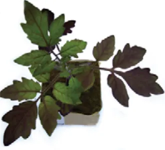
Figure 1.6 Solanum lycopersicum fruit with Aft gene ... 18
Figure 1.7 Solanum lycopersicum plant with atv gene ... 19
Figure 1.8 Solanum lycopersicum fruit with both Aft and atv gene ... 20
Figure 1.9 Total flavonoids (TF) content in healthy and infected tomato fruits by Alternaria linariae ... 25
Figure 1.10 Links between the working mechanisms of flavonoids and their effects on disease ... 28
Figure 1.11 Schematic model of flavonoid secretion from soybean roots and the inter- recognition between legume and rhizobium ... 30
Figure 1.12 Example of Stress-Induced Polyphenols ... 32
Figure 1.13 Model for the mechanism of shelf-life extension in anthocyanin rich tomatoes ... 34
Figure 2.1 Flow chart of QTL-seq ... 63
Figure 2.2 A simplified scheme of QTL-seq applied in tomato ... 64
Figure 2.3 Distribution of anthocyanins in BC1 population ... 67
Figure 2.4 Distribution of anthocyanins in BC1S1 population ... 67
Figure 2.5 QTL Analysis Plot ... 70
Figure 3.2 Field in Mountain Research Station, Waynesville, NC ... 87 Figure 3.3 Control lines NC1CELBR (left) and NC84173 (right) used for field trial and lab trial in field ... 88 Figure 3.4 EB disease scored in tomato field at MRS, Waynesville, NC ... 90 Figure 3.5 Comparison of tomato genotypes with various levels of anthocyanins for early
blight resistance under field and laboratory ... 92 Figure 4.1 Heat Stress Experiment conducted in the Greenhouse at MHCREC, Mills River, NC ... 104 Figure 4.2 Flower showing protruded style ... 105 Figure 4.3 Style length measurement ... 105 Figure 4.4 Figure showing different shapes and color of tomato genotypes experimented for heat stress tolerance in greenhouse ... 105 Figure 4.5 Distribution of flowers in heat stress experiment conducted in greenhouse
environment ... 109 Figure 4.6 Distribution of fruits in heat stress experiment conducted in greenhouse
environment ... 110 Figure 4.7 Fruit set index of tomato genotypes experimented for heat stress tolerance in
greenhouse environment ... 110 Figure 4.8 Distribution of style length in heat stress experiment conducted in greenhouse
environment ... 111 Figure 4.9 Distribution of style protrusion in heat stress experiment conducted in
greenhouse environment ... 112 Figure 4.10 Distribution of anther length in heat stress experiment conducted in greenhouse
environment ... 113 Figure 4.11 Distribution of fruit size in heat stress experiment conducted in greenhouse
environment ... 114 Figure 4.12 Distribution of fruit color in heat stress experiment conducted in greenhouse
CHAPTER 1: REVIEW OF LITERATURE BACKGROUND
Flavonoids are low molecular weight, polyphenolic secondary metabolic compounds, found in almost all fruits and vegetables (Samanta et al., 2011). Flavonoids include soluble pigments found in the vacuoles of plant cells (Samuel, 2018). They are called secondary
metabolites because they are not regarded as an essential element for the plant’s survival as they don’t have a direct role in the normal growth, development, and reproduction of the plants but the plant does suffer in their absence (James, 2017; Samuel, 2018). Secondary plant metabolites are useful in the long run, such as defense against biotic and abiotic stress (Treutter, 2005).
Flavonoids are beneficial for plants in several ways: they are the essential pigments for producing colors needed to attract pollinating insects, required for UV filtration, nitrogen fixation and act as a chemical messenger. These compounds can be important for human health. Their bioavailability and biological activities in humans appear to be strongly influenced by their chemical nature (Higdon, 2005). A variety of biological activities possessed by flavonoids include antiallergic, anti-inflammatory, antiviral, antiproliferative, and anticarcinogenic activities (Yao et al., 2004). There has been a growing interest in dietary flavonoids because of their beneficial effects as antioxidants in the prevention of human diseases such as cancer and cardiovascular diseases, and some pathological disorders of gastric and duodenal ulcers, allergies, vascular fragility, and viral and bacterial infections. (Zand et al., 2002).
CHEMISTRY OF FLAVONOIDS
Szostak-Wegierek, 2014). These naturally-occurring compounds are the most wide-spread, due to the wide range of chemical structures arising from their biosynthetic pathway (Holton and Cornish, 1995; Gould et al., 2008).
Flavonoids are defined by their basic skeleton, the flavan-nucleus, which consist of two aromatic rings with six carbon atoms (ring A and B) interconnected by a hetero cycle including three carbon atoms (ring C) as shown in Figure 1.1. In other words, flavan-nucleus consists of 15 carbon atoms arranged in three rings (C6-C3-C6). Based on the modification of central C ring (chromane ring or pyrane ring) they are divided into different structural classes: flavones, flavonols, flavanones, flavanols, isoflavones and anthocyanidins, while individual compounds within a group vary in the substitution pattern of the A and B rings. Alternatively, dietary flavonoids differ in the arrangements of hydroxyl, methoxy, and glycosidic side groups, and in the conjugation between the A- and B-rings (Heim et al., 2002). During metabolism, hydroxyl groups are added, methylated, sulfated or glucoronidated (Heim et al., 2002). It can also be said that diversity in flavonoid structures is due to modifications of the basic skeleton by enzymes such as glycosyl transferases, methyl transferases and acyl transferases (Bovy et al, 2007).
Based on the chemical structure, flavonoids are calssified into 12 major groups.
and anthocyanidins are the major six classes of flavonoids (Higdon, 2005), which is shown in Figure 1.2 along with the classification of polyphenols.
*Anthocyanidins with one or more sugar moieties (anthocyanidin glycosides) are called
anthocyanins
General description of the structure of six major classes is given below:
Flavones: In flavones, ring C contains a double bond between positions 2 and 3, and a ketone on position 4 (Figure 1.3). Most flavones of fruits and vegetables hold a hydroxyl group on position 5 of ring A. Apigenin and luteolin are the most widespread flavone aglycones Flavonols: The only difference between flavones and flavonols is the hydroxyl group on position 3 of the latter (Figure 1.3). This group is perhaps the most common in fruits and
vegetable. The most common flavonol aglycones are quercetin and kaempferol.
Flavanones: They are also known as dihydroflavones. The structural features of flavanones are the same as those of flavones, except that flavanones lack the double bond between positions 2 and 3 (Figure 1.3). Two representative aglycones of this group are naringenin and hesperetin.
Flavanols: They are also called flavan-3-ols, as the hydroxyl group is almost always attached to the position 3 carbon of ring C (Figure 1.3). Flavanols are also interchangeable with the term catechins. Flavanols or catechins are often found in the skins of fruits and certain vegetables.
Iso-flavones: In contrast to other dietary flavonoids, where ring B is attached to the C2 position of ring C, in isoflavonoids ring B is attached to the C3 position of ring C (Figure 1.3). Genistein and daidzein are the most common isoflavones found in soybean sprouts (Lee et al., 2007).
Anthocyanidins: Chemically anthocyanidins are flavylium cations (Figure 1.3) and
generally exist in the form of chloride salts (Yahia et al, 2010). Anthocyanidins give plants distinctive colors. The color of anthocyanidins can be affected by acylation or methylation at the hydroxyl groups of ring A and B (Yahia et al, 2010). For more clarity, anthocyanins are
glycosides of anthocyanidins, with the sugar group mostly attached to the C3 position of ring C.
HISTORY OF FLAVONOIDS
Flavonoids in tomato were first studied by the team of Euler in 1933. They found yellow flavonoid dye in the tomato cv. Golden Queen which was found to be soluble in alkali and insoluble in carbon disulfide (Euler et al., 1933). This study then never gained any momentum. However, rutin, which gives tomato peel its typical yellow color, was isolated from leaves of Figure 1.3: Structure of major classes of flavonoids
tomato plants and later from tomato stem epidermis (Charaux, 1924 and Fontaine at al., 1947). In 1958, two flavonoids were isolated for the first time from fruit epidermis of three tomato
cultivars-Ponderosa, Rutgers and Sunny Ray (Wu and Burrell, 1958). Those two flavonoids were naringenin and quercetin (= quercetin 3-rhamnoside). In addition, two of the cultivars-Ponderosa and Sunny Ray exhibited these flavonoids in leaves also, whereas Rutgers contained quercetin only. Flavonoids were not detected in the flesh of the fruit of all of these three cultivars.
The occurrence of rutin (quercetin 3-O-rutinoside) from the tomato pastes made from cv. VF-145 was identified in 1968 (Rivas and Luh, 1968). This compound was found to be one of the major flavonoids in ripened tomatoes (Rivas and Luh, 1968). Rutin was the main flavonoid found both in stalk and fruit of cv Marmande (Tronchet, 1969). Similarly, other flavonoids; quercetin 3-rhamnosyl-diglucoside and quercetin triglycoside were also reported from cv. Marmande. However, their concentration was lower than that of rutin (Tronchet, 1969). In addition to this, trace amount of an uncharacterized quercetin derivative along with four kaempferol derivative was detected (Tronchet, 1969).
1980, Hunt and Baker identified chalconaringenin, naringenin and naringenin 7-glucoside in the fruit epidermis of cv. Ailsa Craig, Grower’s Pride and Alicante.
With the advent of modern HPLC technology in the1980s, there has been an increase in the detection of flavonoid compounds. Moreover, in association with the liquid chromatography-mass spectrometry (LC-MS), the number of flavonoids from tomatoes has increased
tremendously since the beginning of millennium (Moco et al., 2007, Lijima et al., 2008 and Mintz-Oron et al., 2008). However, these new compounds are partially described, and identities of sugar moieties and their positions on the flavonoid aglycones have not been identified. A p-coumaric acid was found in low levels in fruit and this was the first report on an acylated flavonoid from tomatoes (Senter et al., 1988).
During the last decade, the number of identified flavonoids in tomatoes have increased visibly and this is possible due to the adoption of advanced technology like high resolution LC-MS coupled with molecular calculation tools. Different flavonoids
compounds which were unidentified in yesteryears were identified with this technology. 12 compounds were identified from different ripening stages of the cvs. Aromata, Conchita,
Campri, Favorita, Macarena, Celine, and Cedrico (Moco et al., 2007). Similarly, in tomato fruits cv. Ailsa Craig, two derivates of kaempferol, six quercetins, four naringenins, two eriodictyols, six chalconaringenins and four of either naringenin or chalconaringenin (24 in total) were
identified by the use of ultra-performance liquid chromatography combined with a time-of-flight mass spectrometry (TOF-MS) (Mintz-Oron et al., 2008). Except for rutin, naringenin and
chalcoeriodictyl (2), naringenin (7), eriodictyl (6), kaempferol (13), and quercetin (13) (Lijima et al., 2008).
So, with the advancement of technology, new flavonoid compounds are being identified and its number is expected to increase in upcoming days.
HYPOTHESIS FOR EVOLUTION OF FLAVONOID
Close and McArthur (2002) have formulated the ‘Oxidative pressure hypothesis’ to explain the evolutionary concept of flavonoid distribution and abundance.
Oxidative stress is an imbalance between reactive oxygen species and the antioxidant defense system, the latter of which relies heavily on nutrients. There has been some debate about the reasons why flavonoids have been developed during evolution. ‘Oxidative pressure
hypothesis’ states that the distribution and abundance of many phenolics including flavonoids is a response by plants to prevent or minimize photodamage, but not as a trade-off in resource allocation in resource-limited environments or a response of herbivory (Treutter, 2006). Close and McArthur (2002) suggest that the level of many, if not most, phenolics vary in plants under different light and nutrient conditions, for the same reason that levels of other, well established antioxidants vary. That is, plants may increase phenolic production directly in response to the oxidative pressure produced from excess light energy (Figure 1.4), and as a physiological response to quench reactive chemical species. The authors suggested further that the observed ecological pattern in levels of most phenolics is more consistent with the concept that
photodamage is the main elicitor. At the evolutionary level, patterns of phenolic levels between species may reflect different selective pressure from the potential risk of photodamage.
for only some phenolics. According to the hypothesis, abiotic factors, which decrease the efficiency of photosynthesis, are the strongest selective agents or elicitors of phenolics. In this concept, herbivory affects oxidative pressure less than the abiotic factors and mainly when severe enough to cause nutrient stress at the whole plant level.
ANTHOCYANINS IN TOMATO
Anthocyanins are water-soluble pigments, which are derived from flavonoids and are found in all plant tissues throughout the plant kingdom. Anthocyanins are either developmentally transient appearing in juvenile or senescing tissues, or may be permanent (Chalker-Scott, 1989). Similarly, anthocyanin may be environmentally transient, appearing and disappearing with changes in photoperiod, temperature or other signals (Chalker-Scott, 1989).
Anthocyanins are the most common class of purple, red, and blue plant pigments. Anthocyanins are getting more concern because of their strong antioxidant activity as measured by oxygen radical absorbing capacity (ORAC) assay (Jones et. al., 2003). Dietary phytonutrients and antioxidants obtained from the fruits and vegetables are of primary importance in the
prevention and disease and aging. Anthocyanins and related polyphenolic and flavonoid compounds are considered important phytonutrient contributors (Proteggente et al. 2002).
Table 1.1: Total Anthocyanin Content in Some Common Fruits and Vegetables. Fruits and vegetables Total anthocyanins
Blueberry 22-495
Blackberry 83-326
Raspberry
black 214-428
red 20-60
Sweet cherry 350-450
Cranberry 78
juice 18-87
Strawberry 7-30
juice 21-333
Red grapes 30-750
Red wine 100-1000
Apple (Scugog) 10
Red onion 9-21
All values are expressed on a fresh weight basis (mg/100g) except for juice and wine, expressed in mg/L. (Reproduced from Wang et al. (1997))
BIOSYNTHETIC PATHWAY OF ANTHOCYANIN
Two categories of genes are involved in the synthesis of anthocyanin-structural genes and regulatory genes. Structural genes encode anthocyanin biosynthesis enzymes (chalcone synthase, chalcone isomerase, flavanone 3-hydroxylase, flavanoid 3’ hydroxylase, flavonoid 3’ 5’
Anthocyanin biosynthesis is a process that is interlinked with various vital cellular process such as Calvin cycle which is a part of photosynthesis, the pentose phosphate pathway which is involved in the production of NADPH, and pentoses that are also part of other vital processes such as pyruvate decarboxylation which in turn is connected to the Krebs cycle (Routray and Orsat, 2011). There are 3 main pathways which lead to the synthesis of
anthocyanin; they are the shikimate pathway, the phenyl propanoid pathway, and the flavonoid pathway (Routray and Orsat, 2011).
The shikimate pathway consists of combining phosphoenol pyruvate with erythrose-4-phosphate resulting in the formation of chorismate, which is a precursor of many aromatic compounds including amino acid phenylalanine and sometimes this pathway ultimately leads to the formation of phenylalanine (Routray and Orsat, 2011).
The phenylpropanoid pathway is the first step in the polyphenol biosynthesis. This pathway uses amino acid phenylalanine from the shikimate pathway as the initial substrate. The phenylalanine is converted to an activated form of cinnamic acid, that is, coumaryl CoA
(Stafford, 1990).
these flavanones to dihydroflavonol dihydrokaempferol (DHK), as a result of the action of the flavonoid 3-hydroxylase (Bovy et. al., 2007, Routray and Orsat, 2011, and Marti et.al., 2016). Dihydrokaempferol can be further hydroxylated, either at the 3’ or both 3’ and 5’ position of B ring forming dihydroquercetin (DHQ) and dihydromyricetin (DHM) respectively. The former hydroxylation is catalyzed by flavonoid 3’-hydroxylase (F3’H) and the latter by flavonoid 3’, 5’-hydroxylase (F3’5’H).
Kaempferol, quercetin and myricetin are major flavonols in tomato (Marti et al., 2016). Dihydrokaempferol directly gets converted into flavonol kaempferol when catalyzed by flavonol synthase (FLS). Dihydroquercetin and dihydromyricetin are the substrates for enzyme FLS, leading to the formation of flavonols quercetin and myricetin respectively (Bovy et. al., 2007, and Marti et.al., 2016). Quercetin is a major flavonoid in tomato peel, in addition to naringenin chalcone (Bovy et. al., 2007 and Marti et.al., 2016). Quercetin is important, as it is the source of rutin in tomato (Marti et. al., 2016).
into peonidin-3- glucoside and petunidn- or malvidin-3-glucoside, respectively through
glycosylation (Routray and Orsat, 2011; Petrussa et. al., 2013). Delphinidin-type anthocyanins are usually found only in vegetative tissues of tomato (Bovy et al., 2007). In a research
conducted by Jones et. al. (2003), they identified petunidin, followed by malvidin and delphinidine as the principle anthocyanidins in tomato fruit.
Flavonoids that are tangible in the peel during fruit ripening are naringenin chalcone, quercetin-glycosides, and kaempferol-glycosides (Krause and Galensa, 1992; Muir et al., 2001). In fruit pericarp, enzymes chalcone synthase (CHS), chalcone isomerase (CHI) and flavonol synthase (FLS) are below detection level whereas in the peel CHS, flavonoid-3 hydroxylase (F3H), and FLS are expressed in abundance (Verhoeyen et al., 2002).
Figure 1.5: Anthocyanin Biosynthesis Pathway Adapted from:
• Blueberries and Their Anthocyanins: Factors Affecting Biosynthesis and Properties, Winny Routray and Valerie Orsat, 2011
• Molecular Biology of Chilli Pepeer Anthocyanin Biosynthesis, Aza-Gonzalez et al., 2012 • Tomato as a source of carotenoids and polyphenols targeted to cancer prevention, Marti
INDUCTION OF ANTHOCYANIN BIOSYNTHESIS
Photoinduction: Anthocyanins are photoinduced by wavelengths in the UV, visible and far-red regions. Induction of anthocyanins by light is supported by its inhibition in the dark (Chalker-Scott, 1999). However, too much radiation in the UVB region inhibits anthocyanin synthesis. (Buchholz et. al., 1995). Various hypotheses have been postulated regarding anthocyanin synthesis, with some studies say that synthesis is induced by only the UVB
photoreceptor while others say that synthesis is induced through some combination of the UVB photoreceptor, phytochrome and cryptochrome. However, many types of research have shown that UVB is the only photoinducer of anthocyanins, while their relative amounts are modulated by phytochrome (Reddy et. al., 1994).
Cold temperature induction: Even though the role of cold temperature is less talked in the induction of anthocyanin, this process has evidence in deciduous plants every fall (Chalker-Scott, 1999). Christie et al. (1994) consider the anthocyanin biosynthesis pathway to involve cor (cold-regulation) genes but observe that very cold temperatures destroy the biosynthetic
capability. However, low temperatures in the absence of either visible light or UVB prevent anthocyanin biosynthesis (reviewed by Chalker-Scott, 1999). McKown et al. (1996) also stated that there is a relationship between anthocyanin biosynthesis and freezing tolerance, in either the synthetic or regulatory pathways leading to both.
grown in the presence of various sugars. Anthocyanin accumulation induced by saline conditions was seen in Zea mays roots, Morus alba leaves and the lower stems of Casuarine equisetifolia seedlings (reviewed by Chalker-Scott, 1999).
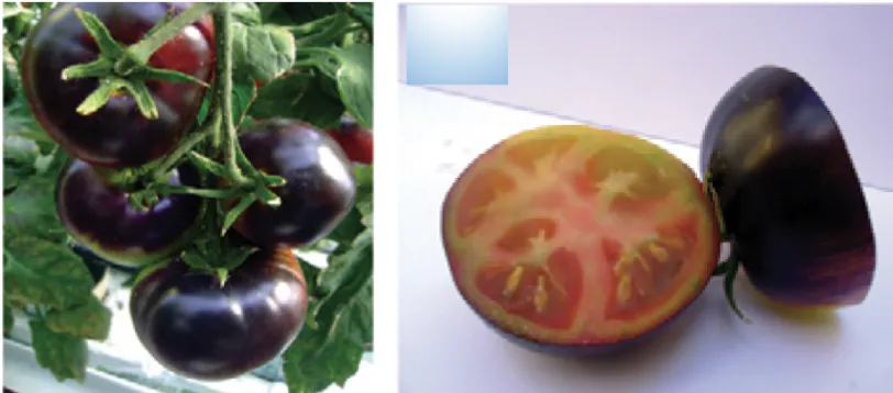
GENES GOVERNING THE EXPRESSION OF ANTHOCYANIN IN TOMATO Till date, there are three reported genes that are responsible for expression of anthocyanin in fruit. They are Anthocyanin fruit (Aft) from Solanum chilense, Aubergine (Abg) from Solanum lycopersicoides, and atroviolacium (atv) from Solanum cheesmanii. (Georgiev, 1972 and Peter et al., 2008)
Anthocyanin fruit was derived from a Solanum esculentum X Solanum chilense cross (Georgiev, 1972). Anthocyanin fruit gene was first acronymed as Af when described by
Georgiev, but later on renamed as Aft by Jones to prevent confusion with anthocyanin free gene af which was already in use for an independent gene, the recessive anthocyanin free, which lacks anthocyanin in vegetative tissues (reviewed by Jones et al., 2003). DeJong et al (2004) suggested that Aft is an allele of the AN2 locus on chromosome 10. Aft is responsible for initiating the expression of anthocyanin in immature green fruit with gradual accumulation throughout development (Mes et. al., 2008). Cold temperature and particularly high light intensity enhance this gene expression (Mes, 2004). The pigment is first expressed in epidermal cells located outside the placental septa which later on spreads uniformly across the fruit where it is exposed to light (Mes et. al., 2008).
Rick et al. (1994) described a second gene - Aubergine (Abg), responsible for anthocyanin production in tomato fruit. Abg was obtained from the intergeneric cross S. lycopersicoides X S. lycopersicum and relies on chromosome 10 (Rick et. al. 1994). Abg is phenotypically similar to Aft in the sense that it also requires high light intensity to induce
anthocyanin expression, however, unlike Aft, Abg has a variegated expression (Mes et. al., 2008). This gene is also found to be located on chromosome 10 in the same arm where Aft resides, but it has not been confirmed whether it lies at the same locus as Aft (Mes et. al., 2008). S.
lycopersicoides shows a paracentric inversion for this arm (lower arm) of chromosome 10, which has prevented stable introgression and study of the trait in S. lycopersicum in this cross (S.
lycopersicum X S. lycopersicoides) (Canady et. al., 2006 and Boches and Myers, 2007). So, stable homozygotes of Abg have not been obtained (Jones et al., 2003). However, chromosomal inversion has not been known in S. chilense, the donor genome for Aft in relation to S.
lycopersicum (Jones et. al., 2003).
The other gene, atroviolaceum (atv) is responsible for anthocyanin accumulation in the entire plant. This gene was first found in Solanum cheesmaniae and is located on chromosome 7 of tomato (Rick et. al.,1968). The atv was found to be weakly expressed as purple fruit
Figure 1.6: Solanum lycopersicum fruit with Aft gene
phenotype when transferred into a cultivated tomato (reviewed by Mes et al., 2008). Expression of atv is more intense in vegetative tissue than in fruit tissue.
In fruit tissue, atv expression appears to be expressed in different cell layers which are not visually detectable (reviewed by Mes et al., 2008). Accumulation of atv-induced anthocyanin is enhanced by cool temperatures (Mes, 2004). When atv is present in the homozygous recessive condition, the phytochrome signaling pathway perceives a high irradiance response, resulting in an elevated anthocyanin response throughout the plant. This anthocyanin response is twice as strong in red light compared to blue light (Kendrick et al., 1997). An example of this is shown in Figure 1.7.
A study conducted by Mes et. al. (2008) revealed a progression of pigment intensity as Aft Aft atv atv > Abg » Aft > atv. Pigment intensity expressed by the combination Abg- Aft- is slightly higher than the gene expressed singly. On the other hand, the combination Abg- atv atv is similar to the combination Aft- atv atv in terms of pigment intensity. Phenotypically, double mutants of atv with either Aft or Abg is distinguishable by the presence of purple stems, leaves,
Figure 1.7: Solanum lycopersicum plant with atv gene.
and intensely pigmented fruit. Solanum lycopersicum fruit with both Aft and atv genes is shown in Figure 1.8 (Gonzali et al., 2009)
Since Aft and Abg have not been characterized yet, the pathway in which they may interact is unknown. However, they may act within the same pathway but are tissue specific. Aft and Abg do not produce an elevated anthocyanin in vegetative plant tissues, as atv does, but they all produce the same anthocyanidin in their specific target tissue. Aft and Abg are responsible for an anthocyanin response in the cell layer directly beneath the epidermis (Mes et. al., 2008). The intensity and timing of the response vary among the two genes, even though they share the property that no other tissue layers are affected (Kendrick et al., 1994). Jones (2000) reported that Aft does have characteristics similar to that of high-photoperiod response mutant along with elevated carotenoid content, which suggests that it may regulate the phytochrome pathway. The atv gene regulates the phytochrome response (Kerchoffs et al., 1997).
Anthocyanin content can be increased through the combination of the anthocyanin-affecting genes with selection for smaller fruit size (Jones et al., 2003).
Figure 1.8: Solanum lycopersicum fruit with both Aft and atv genes
FUNCTIONS OF FLAVONOIDS
In plants, flavonoids play major roles in many biological processes such as in
providing pigmentation to flowers, fruits, and seeds to attract pollinators and seed dispersers, in plant defense against pathogenic micro-organisms, in plant fertility and germination of pollen, in protection against ultraviolet and in acting as signal molecules in plant-microbe interactions. (Koes et al., 1994; Harborne & Williams, 2000). Flavonoid is also the most important source of non-green coloration. It is important in yellow, blue, purple, white and black colors.
I. Flavonoids as an antioxidant:
Davies (1995) stated that oxidation is the transfer of electrons from one atom to another and represents an essential part of aerobic life and our metabolism since oxygen is the ultimate electron acceptor in the electron flow system that produces energy in the form of ATP (reviewed by Pietta, 2000). However, problems may arise when the electron flow becomes uncoupled (transfer of unpaired single electrons), generating free radicals. Free radicals known as reactive oxygen species (ROS) include superoxide (O2-), peroxyl (ROO), alkoxyl (RO), hydroxyl (OH-), and nitric oxide (NO). ROS continuously affect the body cells and tissues causing injury to the endothelial wall resulting in atherosclerotic changes (Nijveldt et. al., 2001). Various studies have shown the positive effect of flavonoid against heart disease like coronary heart disease.
II. Flavonoids as Defensive Compounds:
Flavonoids can be divided into two groups based on its defense mechanism- “preformed” and “induced”. Plants synthesize the “induced” compounds in response to physical injury, infection, or stress. The “preformed” flavonoids are natural compounds that are synthesized during the normal development of plant tissue. These preformed compounds are generally stored at sites where they may play a signaling and a direct role in defense (Truetter, 2006).
Preformed flavonoids and herbivory:
F
avonoids do play a role in plant-insect interaction (Treutter, 2006). Among plant secondary metabolites, flavonoids are reported to have significant role against herbivores (Feeny, 1976). In contradiction to this, Simmonds (2003), states that it is not clear whether insects or plants get the benefit. Insects might sequester the flavonoid in their cuticle to get prevented from the predators or in their wings to attract mates. On the other hand, flavonoids may behave as antifeedants, as toxins, and as digestibility reducers so that insects are deterred (reviewed by Treutter, 2006). Flavonols quercetin and its glycoside rutin cause larval mortality of the tobacco armyworm (Spodoptera liture) (Mallikarjuna et al., 2004). Similarly, the biochemical basis for nematode resistance of banana is because of flavan-3,4-diols andcondensed tannins (Collingbom et. al., 2000). Laitinen et. al. (2004) reported that the flavonoids in birch bark tree are responsible for resistance to mammalian herbivores, such as hares.
Antifungal activity of preformed flavonoids: The antifungal activity is based on the inhibition of spore development and mycelium hyphae elongation (Blount et al.,1992). The strongest antifungal activity is demonstrated by unsubstituted flavones and unsubstituted
flavanones. Hydroxyl groups in these compounds reduce antifungal properties against Alternaria tenuissima, Cladosporium cladosporioides, Spicellum roseum and Trichoderma hamatum
methyl groups reduce antifungal properties. Flavonoids like isoflavones, flavanes and flavanones inhibit root pathogens belonging to fungi such as Fusarium oxysporum f.sp. Phaseoli (Makoi and Ndakidemi, 2007). Preformed flavanols in apple leaves are said to play a defense role against the fungus Venturia inaequalis (reviewed by Treutter, 2006).
A study conducted by Skadhauge et. al., (1997) in barley suggested that
proanthocyanidins and even small amounts of dihydroquercetin are involved in the defense against Fusarium species (F. poae, F. culmorum, and F. graminearum) Table 1.2 summarizes this research.
Table 1.2: Flavonoids in the testa layer of barley genotypes and resistance to Fusarium. (Adapted and modified after Skadhauge et. al. (1997) and Treutter (2006))
Genotypes Flavonols in the testa layer Inhibition of
Fusarium infection
Triumph (WT) anth., cat., pa + +
Alf (WT) anth., cat., pa + +
Grit (WT) anth., cat., pa + +
ant 13-152 - -
ant 17-148 (FHT-mutant) homoeriodoctyol, chrysoeriol -
ant 19-159 (DFR-mutant) dihydroquercetin +++
ant 19-109 anth., cat., procyanidin B3 + ant 22-1508 homoeriodoctyol, chrysoeriol -
ant 25-264 anth. -
ant 26-485 anth., cat. +
ant 27-489 anth., cat. +
ant 28-484 anth. +
ant 29-2110 anth., pa +
Note:
FHT = flavanone 3 -hydroxylase
DFR = dihydroflavonol-4-reductase anth. = anthocyanins
cat. = catechins
pa = proanthocyanidins - no inhibition
Induced defensive flavonoids (Biotic Stress): In plants, flavonoids are seen to rise in their concentration, or their formation are induced after attack or injury by pathogens or pests preventing the hosts from further loss (Barry et. al., 2002; Gallet et. al., 2004). Production of epidermal anthocyanin in cotton leaves as resistance to bacterial blight (Xanthomonas campestris pv. malvacearum) is a good example of induced flavonoids as defense (Kangatharalingam et. al., 2002). Flavanols get accumulated in the lesion margins of Eucalyptus globulus when infected by fungi Cytonaema sp. (Eyles et al., 2003). Similarly, Pearce et al. (1998) reported that the
synthesis of phenolic compounds was induced in tomato leaf tissues when wounded. The
amounts of two phenolic compounds, E-feruloyltyramine and E-p-coumaroyltyramine, increased 10-fold in tomato leaves in response to mechanical wounding.
The involvement of flavonoids in plant defense depends on the species. Species having low flavonoids are less significant in plant defence (Logemann and Hahlbrock 2002). In bilberry (European blueberry), infection by a fungal endophyte (Paraphaeosphaeria sp.) and a fungal pathogen (Botrytis cinerea) lead to increased biosynthesis of phenolic compounds (Koskimaki et al., 2009). Accumulation of phenolic compounds was specific for each infection. Infection by the pathogen promoted specifically accumulation of epigallocatechin, quercetin-3-glucoside,
quercetin-3-O-α-rhamnoside, quercetin-3-O-(4’’-HMG)-R-rhamnoside, chlorogenic acid and coumaroyl quinic acid. The endophyte infected plants had a higher content of quercetin-3-glucuronide and coumaroyl iridoid (Koskimaki et al., 2009). The uninfected samples contained high quantities of insoluble proanthocyanidins, whereas the infected samples had an abundance of less polymerized, oligomeric proanthocyanidins (Koskimaki et al., 2009).
Induced defensive flavonoids (Abiotic Stress):
F
lavonoids along with other phenolic compounds are sometimes attributed to playing role in frost hardiness (Chalker –Scott & Krahmer, 1989) and drought resistance (Pizzi & Cameron, 1986) in the cell wall and atmembranes. Furthermore, Tattini et al (2004) also believe that flavonoids have a protective role Figure 1.9: Total flavonoids (TF) content in healthy and infected tomato fruits by
Alternaria linariae
during drought stress. One of the significant roles of flavonoids is photoprotection which is contributed by high quercetin: kaempferol ratio, which is recorded in Petunia leaves (Ryan et al., 2002).
Role of temperature in the synthesis of phenolic compounds in tomatoes has not been properly assessed (Dumas et al. 2003). In Arabidopsis thaliana, the lower temperature between 5 and 100C was seen to induce flavonol accumulation (reviewed by Slimestead and Verheul, 2009). Differences between day and night temperatures have a marked effect on the
accumulation of anthocyanin pigments in apple and berry skin (Tomas-Barberan and Espin, 2001).
Depending on the plant and parts of the plant, high temperature either decreases or increases the synthesis of anthocyanins. High temperature decreases anthocyanin synthesis in reproductive parts of red apples, chrysanthemums and asters whereas high temperature stress increases the anthocyanin accumulation in vegetative tissue of rose and sugarcane leaves (reviewed by Wahid et al., 2007). Low concentration of anthocyanin even at a high temperature may be due to its decreased rate of synthesis and stability (Shaked-Sachray et al. 2002).
Flavonoids are also believed to play a role in response to heat stress. Teklemaraim and Blake (2003) suggest that the antioxidant activity of flavonoids may increase heat tolerance, which held for cucumber seedlings (Cucumis sativus L.). The UV-induced flavonols reduced the oxidative degradation of membrane lipids.
III. Flavonoids in clinical effects
cycloxygenase and lipoxygenase pathways (Moroney et. al., 2001). Cycloxygenases and
lipoxygenase play an essential role as inflammatory mediators. They are involved in the release of arachidonic acid, which is a starting point for a general inflammatory response (Nijveldt et. al., 2001).
Antitumour Effects: ROS can damage DNA, and the division of cells with unrepaired or misrepaired damage leads to mutations, increasing the exposure of DNA to mutagens (Nijveldt et al., 2001). Flavonoids as an antioxidant have also shown its ability to inhibit carcinogenesis (Stefani et al. 1999). The inverse association between flavonoid intake and the subsequent incidence of lung cancer has been suggested by the study conducted by Knekt et al. in 1997.
Antithrombogenic Effects: Aggregation of platelet is responsible for the development of atherosclerosis and acute platelet thrombus formation, leading to embolization of stenosed arteries (Nijveldt et al., 2001). These platelets adhering to vascular endothelium generate lipid peroxides and oxygen free radicals, which inhibit the endothelium formation of prostacyclin and nitrous oxide (Nijveldt et al., 2001). Flavonols are supposed to maintain proper concentrations of endothelial prostacyclin and nitrous oxide by directly scavenging free radicals (Gryglewski et al., 1987).
Antiosteoporotic Effects: The flavonoids present in tea is found to be responsible for the prevention of osteoporosis (Hegarty et al., 2000).
An overview of the links between the working mechanisms and clinical effects of flavonoids is graphically presented in Figure 1.10.
IV. Flavonoids as UV-filters:
Flavonoids protect the plant against UV damage as they act as screen absorbing UV radiation. Since, flavonoids are accumulated mostly in the epidermis and hypodermis of leaves and stems, apical meristem and pollen, this reduces the chances of UV light penetrating to vulnerable tissues or organs (Mierziak et. al., 2014). The spectrum of UV radiation reaching the earth’s surface has been divided into lower energy UV-A (320-400nm), higher energy UV-B (280-320 nm) and UV-C (254-280 nm). The damage caused by UV-B is severe as it may affect vital processes in plants like photosynthesis, transpiration and pollination (Tevini and
Teramura,1989; Lindroth, 2000). Besides, it also causes DNA and cellular damage, induces plant Figure 1.10: Links between the working mechanisms of flavonoids
and their effects on disease.
foliar chemistry, including a decrease in carbohydrate concentrations (Tevini and
Teramura,1989; Lindroth et al., 2000). UV-B radiation is also reported to weaken the defense mechanism of plants promoting the pathogenicity of the pathogens like fungus (Sharma, 2001). Flavonoids generally absorb UV-B radiation and act as a filter protecting the tissues from damage (reviewed by Samanta et la., 2011). It does so by reducing epidermal penetration of UV-B radiation selectively protecting internal tissues without interfering photosynthesis (Caldwell et al., 1983).
V. Flavonoids as signal molecule for symbiotic interactions:
During normal growth and development of plants, a wide variety of flavonoids are
synthesized in both roots and shoots. Flavonoids are involved in the nodulation process (Ferreyra et. al., 2012). This is further supported by Wassen et al. (2006); flavonoid deficient roots of transgenic plants produced by RNA interference of chalcone synthase were unable to produce nodules. Legume-rhizobial nodulation starts with a signal exchange between the symbiotic partners and this process commences with the production of host-specific signal molecules flavonoid and isoflavonoid through roots. Charactersistics flavonoid is produced by legume which acts as signals for various microbes, including symbionts and pathogens (Straney et al., 2002). The release of flavonoids is strongest at root tips and the emerging root hairs, which are the target sites for Rhizobium infections (Abdel-Lateif et al., 2012). Flavonoids act as
chemoattractants for rhizobia and as specific inducers of rhizobial nodulation genes (nod-genes), which control root nodule formation in the nitrogen-fixing bacteria (Tsai et al., 1991). However, flavonoids can influence the expression level of bacterial nod genes both positively and
the other hand, naringenin stimulates nod formation in Rhizobium leguminosarum, but quercetin represses its expression (Mierziak et al., 2014). Mechanism of nodule formation initiates through binding of flavonoids to bacterial NodD proteins (Mierziak et al., 2014). Figure 1.11 (Samanta et al., 2011) shows a schematic model of flavonoid secretion from soybean roots and the inter-recognition between legume and Rhizobium.
Flavonoids also play a role in the formation of mycorrhiza, which is a symbiotic
relationship between a plant and soil-borne fungi that colonize the cortical tissue roots (Mierziak et al., 2014). During mycorrhiza development in white clover, melon roots, and Medicago
truncatula, it was found that the level of flavonoid was raised (Ponce et al., 2004; Akiyama et al., 2002; Schliemann et al., 2008). Similarly, flavonoids such as quercetin, quercetin galactoside, and kaempferol, have a positive effect on the growth of hyphae and spore germination of mycorrhizal fungi (reviewed by Mierziak, 2014).
VI. Flavonoids as attractants for pollination and seed dispersal
Insect pollinated plants (e.g. Petunia) generally have flowers with large, brightly colored petals, while most of the wind pollinated (e.g. corn) have flowers which are often small, dull and
Figure 1.11: Schematic model of flavonoid secretion from soybean roots and the inter-recognition between legume and rhizobium
apetalous (Koes et. al., 1994). Pigmentation acts as a signal to attract pollinating insects or birds. Anthocyanins are the major flavonoid compounds that contribute to the pigmentation of flowers (Kong et al., 2003). In addition to this, various flavonols and flavones act as co-pigments with anthocyanins that help to intensify flower color (Forkmann and Martens, 2001). Even colorless flavonoids contribute to flower color giving creamy or ivory appearance in petals which
otherwise would be translucent (Forkmann, 1991).
Flavonoids (anthocyanins. flavonols, chalcones) often accumulate in both the male and female sex organs in flower, including the pollen grains (Farrant, 2000). The role of flavonoid in pollen fertility was first established in maize, with its well characterized anthocyanin mutants (Mo et al., 1992). Pollak et al. (1993) reported that flavonoid deficient mutants are lacking chalcone synthase were generated in maize and petunia to elucidate the roles of flavonoids, and they were male sterile as no functional pollen tube was produced. However, this deficiency could be reversed by the addition of flavonol kaempferol at pollination (Mo et al., 1992).
Flavonoids also determine which animal vector is attracted to accomplish pollination. In Petunia integrifolia, flowers are red and bees pollinate them, whereas in Petunia axillaris, flowers are white and moths pollinate them (Wijsman, 1983).
Flavonoids present in fruit are thought to attract frugivorous consumers which assist in seed dispersal. However, this is applicable for larger plants such as trees, for which the seeds need to carry away from their parent territory to ensure germination (Anderson and Markham, 2005)
VII. Flavonoids as Phytoalexins
antimicrobial compounds synthesized in response to pathogen attack. Their level increases around the site of infection to concentrations toxic to pathogens in in vitro bioassays. (Dixon and Paiva, 1995). Phenylpropanoid compounds are also induced in response to wounding or feeding by herbivores. Increased levels of coumestrol and coumarin are toxic to potential herbivores, causing estrogenic and anticoagulant effects (reviewed by Dixon and Paiva, 1995). Wound induced chlorogenic acid, alkyl ferulate esters, and cell wall-bound phenolic esters may act directly as defense compounds or may serve as precursors for the synthesis of lignin, suberin, and other wound-induced polyphenolic barriers (Bernards and Lewis, 1992).
Induction of phenylpropanoids by other stresses have been less well studied. Though Christie et. al. (1994) found that cold stress increases the levels of anthocyanin, but the reasons for this increment are not clear. The concentration of phenylpropanoids increased in roots or root exudates due to various nutritional disorder. For example, low nitrogen induces flavonoid and isoflavonoid nod gene inducers and chemoattractants for nitrogen-fixing symbionts (Wojtaszek
Figure 1.12: Example of Stress-Induced Phenylpropanoids
et al., 1993). Example of stress-induced phenylpropanoids is represented in Figure 1.12 (Dixon and Paiva, 1995)
VIII. Role of Anthocyanin in the shelf life of tomato:
Anthocyanins do play a role in extending the shelf life of tomato by delaying
overripening and reducing susceptibility to gray mold (Zhang et. al., 2013; Bassolino et. al., 2013). Shelf life is one of the most important horticultural traits of tomato which is determined by two major components; fruit softening during over ripening and susceptibility to fungi Botrytis cinerea, an important economic postharvest pathogen (Dean et al., 2012). Bassolino et. al., (2013) reports that the softening of fruit is delayed in Aft/Aft atv/atv tomatoes when
compared to wild type (WT) Ailsa Craig. Aft/Aft atv/atv fruits and Ailsa Craig fruit when stored at 170C, 100% of the fruit softened after 70 days and 42 days respectively and the proportion of fresh weight loss was higher in Ailsa Craig than in Aft/Aft atv/atv fruits. Zhang et. al., (2013) also reported the similar finding of anthocyanin extending the self-life of tomato by 2-fold. The susceptibility anthocyanin rich fruit (Aft/Aft atv/atv) to an important necrotrophic pathogen Botrytis cinerea is reduced when compared to WT (Ailsa Craig). This reduced susceptibility could be due to their antioxidant activity which counterbalances the oxidative burst induced by B. cinerea (Zhang et. al., 2013). The oxidative burst is thought to heighten the infection by this necrotrophic fungus that feeds on dead tissue. Anthocyanin alters the mechanism of ROS burst generated by B. cinerea which in turn reduces the host cell death creating an unfavorable condition for the growth of necrotroph (Zhang et. al., 2013).
Thus, anthocyanins are found to have beneficial functions protecting plants against various biotic and abiotic factor. Besides their protective roles in plants, interest on anthocyanins is increasing because of their health properties in the human diet. So, the tomato is an excellent candidate for the study of a protective role against early blight and heat stress, since it is most important, commonly consumed vegetables in human diets worldwide (Bovy et al., 2002; Willits et al., 2005). Furthermore, tomato fruits are naturally devoid of anthocyanins (Torres et al., 2005, Mes et al., 2008. Gonzali et al., 2009)
RESEARCH OBJECTIVES
While three genes associated with flavonoid levels have been reported, there may be minor genes controlling the flavonoid levels in tomato. We hypothesize that genotypes with an elevated level of flavonoid will have improved level of tolerance to biotic and abiotic stress tolerance. Hence the proposed thesis research has the following research objectives:
Figure 1.13: Model for the mechanism of shelf life extension in anthocyanin rich tomatoes
1. Mapping QTL associated with flavonoid content in tomato
2. Investigate the response of tomato genotypes with different levels of flavonoids to early blight, and
REFERENCES
Abdel-Lateif, K., Bogusz, D., & Hocher, V. (2012). The role of flavonoids in the establishment of plant roots endosymbioses with arbuscular mycorrhiza fungi, rhizobia, and frankia bacteria. Plant Signaling & Behavior, 7(6), 636-641.
Akiyama, K., Matsuoka, H., & Hayashi, H. (2002). Isolation and identification of a phosphate deficiency-induced C-glycosylflavonoid that stimulates arbuscular mycorrhiza formation in melon roots. Molecular Plant-Microbe Interactions, 15(4), 334-340.
Al-sane, KO (2011). Physiological and genetic control of anthocyanin pigmentation in different species. Doctoral dissertation. VU-University, Amsterdam, p. 10.
Andersen, O. M., & Markham, K. R. (2005). Flavonoids: Chemistry, biochemistry, and applications CRC press. P. 413.
Aza-González, C., Núñez-Palenius, H. G., & Ochoa-Alejo, N. (2012). Molecular biology of chili pepper anthocyanin biosynthesis. Journal of the Mexican Chemical Society, 56(1), 93-98. Barry, K., Davies, N., & Mohammed, C. (2002). Effect of season and different fungi on
phenolics in response to xylem wounding and inoculation in eucalyptus nitens. Forest Pathology, 32(3), 163-178.
Bassolino, L., Zhang, Y., Schoonbeek, H., Kiferle, C., Perata, P., & Martin, C. (2013).
Accumulation of anthocyanins in tomato skin extends shelf life. New Phytologist, 200(3), 650-655.
Beckman, C. H. (2000). Phenolic-storing cells: Keys to programmed cell death and periderm formation in wilt disease resistance and in general defence responses in
Beggs, C. J., Kuhn, K., Böcker, R., & Wellmann, E. (1987). Phytochrome-induced flavonoid biosynthesis in mustard (sinapis alba L.) cotyledons. enzymic control and differential regulation of anthocyanin and quercetin formation. Planta, 172(1), 121-126.
Bernards, M. A., & Lewis, N. G. (1992). Alkyl ferulates in wound healing potato tubers. Phytochemistry, 31(10), 3409-3412.
Blount, J. W., Dixon, R. A., & Paiva, N. L. (1992). Stress responses in alfalfa (medicago sativa L.) XVI. antifungal activity of medicarpin and its biosynthetic precursors; implications for the genetic manipulation of stress metabolites. Physiological and Molecular Plant
Pathology, 41(5), 333-349.
Boches, P., & Myers, J. (2007). The anthocyanin fruit tomato gene (aft) is associated with a DNA polymorphism in a MYB transcription factor. Paper presented at the Hortscience, 42. (4) pp. 856-856.
Boss, P. K., Davies, C., & Robinson, S. P. (1996). Expression of anthocyanin biosynthesis pathway genes in red and white grapes. Plant Molecular Biology, 32(3), 565-569. Bovy, A., Schijlen, E., & Hall, R. D. (2007). Metabolic engineering of flavonoids in tomato Bovy, A., Schijlen, E., & Hall, R. D. (2007). Metabolic engineering of flavonoids in tomato
(solanum lycopersicum): The potential for metabolomics. Metabolomics, 3(3), 399. Bovy, A., de Vos, R., Kemper, M., Schijlen, E., Almenar Pertejo, M., Muir, S., et al. (2002).
High-flavonol tomatoes resulting from the heterologous expression of the maize transcription factor genes LC and C1. The Plant Cell, 14(10), 2509-2526.
Caldwell, M. M., Robberecht, R., & Flint, S. D. (1983). Internal filters: Prospects for UV‐ acclimation in higher plants. Physiologia Plantarum, 58(3), 445-450.
Canady, M. A., Ji, Y., & Chetelat, R. T. (2006). Homeologous recombination in solanum Chalker‐Scott, L. (1999). Environmental significance of anthocyanins in plant stress
responses. Photochemistry and Photobiology, 70(1), 1-9.
Chalker-Scott, L., & Krahmer, R. L. (1989). Microscopic studies of tannin formation and
distribution in plant tissues. Chemistry and significance of condensed tannins (pp. 345-368) Charaux, C. (1924). Biochemical hydrolysis of rutin. Derivation of a New Sugar,
Rutinose.Compt. Rend, 178, 1312-1314.
Christie, P. J., Alfenito, M. R., & Walbot, V. (1994). Impact of low-temperature stress on general phenylpropanoid and anthocyanin pathways: Enhancement of transcript abundance and anthocyanin pigmentation in maize seedlings. Planta, 194(4), 541-549.
Close, D. C., & McArthur, C. (2002). Rethinking the role of many plant phenolics–protection from photodamage not herbivores? Oikos, 99(1), 166-172.
Collingborn, F. M., Gowen, S. R., & Mueller-Harvey, I. (2000). Investigations into the biochemical basis for nematode resistance in roots of three musa cultivars in response to radopholus s imilis infection. Journal of Agricultural and Food Chemistry, 48(11), 5297-5301.
Cooper, J. E. (2004). Multiple responses of rhizobia to flavonoids during legume root infection. Advances in botanical research, Elsevier, 41. pp. 1-62.
interaction betweenXanthomonas campestrispv. malvacearumand cotton. Physiological and Molecular Plant Pathology, 49(5), 285-306.
Davies, K. J. (1995). Oxidative stress: The paradox of aerobic life. Biochemical Society Symposia, 61. pp. 1-32.
De Jong, W., Eannetta, N., De Jong, D., & Bodis, M. (2004). Candidate gene analysis of
anthocyanin pigmentation loci in the Solanaceae. Theoretical and Applied Genetics, 108(3), 423-432.
De Stefani, E., Boffetta, P., Deneo-Pellegrini, H., Mendilaharsu, M., Carzoglio, J. C., Ronco, A., et al. (1999). Dietary antioxidants and lung cancer risk: A case-control study in
uruguay. Nutrition and Cancer, 34(1), 100-110.
De Stefani, E., Boffetta, P., Deneo-Pellegrini, H., Mendilaharsu, M., Carzoglio, J. C., Ronco, A., et al. (1999). Dietary antioxidants and lung cancer risk: A case-control study in
uruguay. Nutrition and Cancer, 34(1), 100-110.
Dean, R., Van Kan, J. A., Pretorius, Z. A., Hammond‐Kosack, K. E., Di Pietro, A., Spanu, P. D., et al. (2012). The top 10 fungal pathogens in molecular plant pathology. Molecular Plant Pathology, 13(4), 414-430.
Dixon, R. A., Achnine, L., Kota, P., Liu, C., Reddy, M., & Wang, L. (2002). The
phenylpropanoid pathway and plant defence—a genomics perspective. Molecular Plant Pathology, 3(5), 371-390.
Dumas, Y., Dadomo, M., Di Lucca, G., & Grolier, P. (2003). Effects of environmental factors and agricultural techniques on antioxidantcontent of tomatoes. Journal of the Science of Food and Agriculture, 83(5), 369-382.
Euler, H. V., Karrer, P., Krauss, E. V., and Walker, O. (1931). Biochemistry of the pigments of the tomato. Helvetica Chimica Acta, 14, 154-162.
Eyles, A., Davies, N., Yuan, Z., & Mohammed, C. (2003). Host responses to natural infection by cytonaema sp. in the aerial bark of eucalyptus globulus. Forest Pathology, 33(5), 317-331. Falcone Ferreyra, M. L., Rius, S., & Casati, P. (2012). Flavonoids: Biosynthesis, biological
functions, and biotechnological applications. Frontiers in Plant Science, 3, 222. Farrant, J. M. (2000). A comparison of mechanisms of desiccation tolerance among three
angiosperm resurrection plant species. Plant Ecology, 151(1), 29-39.
Feeny, P. (1976). Plant apparency and chemical defense. Biochemical interaction between plants and insects (pp. 1-40) Springer.
Fontaine, T. D., & MA, R. (1947). Isolation of rutin from tomatin concentrates. Archives of Biochemistry, 15(1), 89-93.
Forkmann, G. (1991). Flavonoids as flower pigments: The formation of the natural spectrum and its extension by genetic engineering. Plant Breeding, 106(1), 1-26.
Forkmann, G., & Martens, S. (2001). Metabolic engineering and applications of flavonoids. Current Opinion in Biotechnology, 12(2), 155-160.
Galensa, R., & Herrmann, K. (1980). Analysis of flavonoids by high-performance liquid chromatography. Journal of Chromatography A, 189(2), 217-224.
Georgiev, C. (1972). Anthocyanin fruit (af). Tomato Genet.Coop. Rpt, 22(10)
Gonzali, S., Mazzucato, A., & Perata, P. (2009). Purple as a tomato: Towards high anthocyanin tomatoes. Trends in Plant Science, 14(5), 237-241.
Gould, K., Davies, K. M., & Winefield, C. (2008). Anthocyanins: Biosynthesis, functions, and applications Springer Science & Business Media, p.398.
Gryglewski, R. J., Korbut, R., Robak, J., & Świȩs, J. (1987). On the mechanism of antithrombotic action of flavonoids. Biochemical Pharmacology, 36(3), 317-322. Harborne, J. B., & Williams, C. A. (2000). Advances in flavonoid research since
1992. Phytochemistry, 55(6), 481-504.
Hegarty, V. M., May, H. M., & Khaw, K. (2000). Tea drinking and bone mineral density in older women–. The American Journal of Clinical Nutrition, 71(4), 1003-1007.
Higdon, J. (2005). Flavonoids. Linus Pauling Institute, Oregon State University. Available online:
http://lpi.oregonstate.edu/mic/dietary-factors/phytochemicals/flavonoids#sources. Retrieved on June 19, 2018
Heim, K. E., Tagliaferro, A. R., & Bobilya, D. J. (2002). Flavonoid antioxidants: Chemistry, metabolism and structure-activity relationships. The Journal of Nutritional
Biochemistry, 13(10), 572-584.
Herrmann, K. (1979). Übersich über die inhaltsstoffe der TomatenConstituents of
tomatoes. Zeitschrift Für Lebensmittel-Untersuchung Und Forschung, 169(3), 179-200. Iijima, Y., Nakamura, Y., Ogata, Y., Tanaka, K., Sakurai, N., Suda, K., et al. (2008). Metabolite
James, K. (2017). Animal metabolites: From amphibians, reptiles, Aves/Birds, and invertebrates. Pharmacognosy (pp. 401-411) Elsevier.
Jones, C. M. (2000). Evaluation of Carotenoids and Anthocyanins in High Pigment, Processing, Heirloom and Anthocyanin Fruit Tomatoes, Doctoral Dissertation, Oregon State University. Jones, C., Mes, P., & Myers, J. (2003). Characterization and inheritance of the anthocyanin fruit
(aft) tomato. Journal of Heredity, 94(6), 449-456.
Kangatharalingam, N., Pierce, M. L., Bayles, M. B., & Essenberg, M. (2002). Epidermal
anthocyanin production as an indicator of bacterial blight resistance in cotton. Physiological and Molecular Plant Pathology, 61(3), 189-195.
Kendrick, R., Kerckhoffs, L., Tuinen, A. V., and Koornneef, M. (1997). Photomorphogenic mutants of tomato. Plant, Cell & Environment, 20(6), 746-751.
Kendrick, R. E., Kerckhoffs, L., Pundsnes, A., Van Tuinen, A., Koorneef, M., Nagatani, A., et al. (1994). Photomorphogenic mutants of tomato. Euphytica, 79(3), 227-234.
Kerckhoffs, L., De Groot, N., Van Tuinen, A., Schreuder, M., Nagatani, A., Koornneef, M., et al. (1997). Physiological characterization of exaggerated-photoresponse mutants of
tomato. Journal of Plant Physiology, 150(5), 578-587.
Knekt, P., Järvinen, R., Seppänen, R., Heliövaara, M., Teppo, L., Pukkala, E., et al. (1997). Dietary flavonoids and the risk of lung cancer and other malignant neoplasms. American Journal of Epidemiology, 146(3), 223-230.
Kong, J., Chia, L., Goh, N., Chia, T., & Brouillard, R. (2003). Analysis and biological activities of anthocyanins. Phytochemistry, 64(5), 923-933.