HIGHLIGHTED ARTICLE
| WORMBOOK
EVOLUTION AND ECOLOGY
From
“
the Worm
”
to
“
the Worms
”
and Back Again:
The Evolutionary Developmental Biology
of Nematodes
Eric S. Haag,*,1David H. A. Fitch,†and Marie Delattre‡
*Department of Biology, University of Maryland, College Park, Maryland 20742,†Department of Biology, New York University, New York 10003, and‡Laboratoire de Biologie Moléculaire de la Cellule, CNRS, INSERM, Ecole Normale Supérieure de Lyon, 69007, France ORCID IDs: 0000-0002-6102-3498 (E.S.H.); 0000-0001-6940-7544 (D.H.A.F.); 0000-0003-1640-0300 (M.D.)
ABSTRACTSince the earliest days of research on nematodes, scientists have noted the developmental and morphological variation that exists within and between species. As various cellular and developmental processes were revealed through intense focus on Caenorhabditis elegans, these comparative studies have expanded. Within the genusCaenorhabditis, they include characterization of intraspecific polymorphisms and comparisons of distinct species, all generally amenable to the same laboratory culture methods and supported by robust genomic and experimental tools. TheC. elegansparadigm has also motivated studies with more distantly related nematodes and animals. Combined with improved phylogenies, this work has led to important insights about the evolution of nematode development. First, while many aspects ofC. elegansdevelopment are representative ofCaenorhabditis, and of terrestrial nematodes more generally, others vary in ways both obvious and cryptic. Second, the system has revealed several clear examples of developmentalflexibility in achieving a particular trait. This includes developmental system drift, in which the developmental control of homologous traits has diverged in different lineages, and cases of convergent evolution. Overall, the wealth of information and experimental techniques developed inC. elegansis being leveraged to make nematodes a powerful system for evolutionary cellular and developmental biology.
KEYWORDSC. elegans; connectome; developmental systems drift; embryo; evolution; gene regulatory network; sex determination; sperm; vulva; WormBook
TABLE OF CONTENTS
Abstract 397
Unique Attributes of the Caenorhabditis System 398
Findings 401
Zygotic mitosis 401
Postzygotic cell lineage and founder cell specification 403
Developmental regulation of gene expression 404
Neuronal development 405
Sex determination 405
Germ cell proliferation 408
Spermatogenesis 408
Continued
Copyright © 2018 by the Genetics Society of America doi:https://doi.org/10.1534/genetics.118.300243
Manuscript received August 31, 2017; accepted for publication August 3, 2018 Available freely online through the author-supported open access option.
CONTENTS,continued
Vulva development 410
Variation in the position of the vulva 411
Variation in the number and fate of VPCs 411
Variation in the mechanisms of VPC induction 413
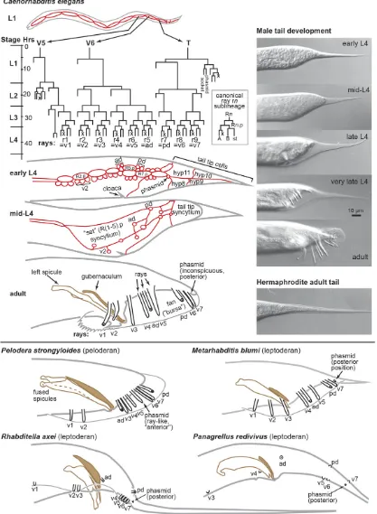
Male tail 414
Evolution of ray pattern 417
Evolution of phasmid position 418
Male-specific tail tip morphogenesis and its evolution 418
Dauer formation and phenotypic plasticity 419
Discussion 419
An accurate, well-resolved phylogeny is essential for EDB 419
Many genes, pathways, and cells are highly conserved 420
Early events are surprisingly evolvable 420
Gene duplication allows partition and gain of gene functions 421 Genetic network architecture may influence evolutionary trajectories 421
Pervasive DSD and its implications for research 421
Microevolution is reflected in macroevolution 421
Repeated evolution involves both reproducible co-options and idiosyncratic components 422
T
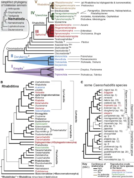
HE small, laboratory-friendly nematodes of the genus Caenorhabditiswerefirst developed as a system for genetic analysis of animal development by a few early champions. One of thefirst experimental studies onC. eleganswas performed by Japanese American Hikokuro Honda, who found that sperm determine the sex of progeny, and discovered that oocyte mei-osis is not completed until after fertilization (Honda 1925). Two decades later, the French biologist Victor Nigon and his American colleague Ellsworth Dougherty greatly extended this work (Nigon 1943; Dougherty and Nigon 1949; Ferris and Hieb 2015; Nigon and Félix 2017), aided by improvements in culture methodology by Briggs (1946). These workers set the stage for Sydney Brenner’s breakthroughs with C. elegans (Brenner 1974, 2009). Along with French biologist Emile Maupas, who first describedC. elegans(Maupas 1900), all of these early researchers were struck by the fact that, within a stereotypical body form, evolutionary variation in habitat choice, feeding strategy, reproductive mode, behavior, and anatom-ical details are rampant. Thus, research focusing on C. elegans was always complemented by the work of other nematologists working in other groups, such as other nema-todes in the order Rhabditida (Figure 1) (Sudhaus 1976). It can therefore be fairly said that questions of biodiversity, the evolution of developmental processes, and their connections to ecology were very much lingering over thefield even in the earliest days. The authors of this review represent examples of contemporary biologists who share their predecessors’ fasci-nation with the evolution of nematode development. Trained in theC. elegansparadigm, we and others take particular delight in gazing outward across the phylogeny, always on the lookout for new phenomena and explanations for how they evolved.Unique Attributes of the Caenorhabditis System
implicated in chemosensation, such as rhodopsin-related G protein-coupled receptors, have been amplified and diversified (Bargmann 2006).
A striking variable distinguishing some nematodes, such as Caenorhabditis,Pristionchus, and some other clade V taxa re-lates to sexual mode. Although the ancestral gonochoristic (ma-le-female), obligately outcrossing mode is retained by most species, several have evolved a self-fertile hermaphrodite (Kiontkeet al.2011) (Figure 1). Males persist at greatly reduced frequencies, creating an androdioecious mating system. Andro-dioecy is rare in both animals and plants (Pannell 2002; Weeks et al.2006), but because it makes genetic manipulations simpler and faster, selfing species likeC. elegansandC. briggsae(and peas!) have always been favored by experimental biologists. A major area of research reviewed below involves comparisons between close relatives with different sexual modes.
Despite its fame for exhibiting “invariant” development, C. elegans also offers one of the best-characterized examples of an adaptive phenotypic plasticity: the formation of the dauer larva. This resistant variant of the third larval stage is triggered by crowding or starvation in the previous stage (Albertet al. 1981), which, in turn, alters pheromones and nutritional status. These cues are then translated into differential states of signal-ing pathways and circulatsignal-ing hormones (Fielenbach and Antebi 2008). Because the dauer larva appears to be a universal dis-persal form for both free-living and parasitic terrestrial nema-todes (Crook 2014), the cues that induce its development and the attributes it possesses are likely to vary with ecological niche. Somefirst examples of this variation are reviewed below. C. eleganshas also enjoyed early and intense attention to the characterization of its genome and its relation to various processes. It was thefirst animal species to have a complete sequence assembly (C. elegansSequencing Consortium 1998), and this quickly became a handmaiden to gene-focused EDB (e.g., Kuwabara and Shah 1994; Haag and Kimble 2000). In-terest in examining interspecies variation led to a collection of genome assemblies from otherCaenorhabditisspecies (Stein et al., 2003; Hillieret al.2007; Rosset al.2011; Fierstet al.
2015; see also http://www.nematodes.org/nematodege-nomes/index.php/Main_Page). This work is ongoing on an ever-larger scale, driven by both the discovery of many new species (Kiontkeet al.2011; Barrière and Félix 2014; Huang et al.2014; Ferrariet al.2017; Sloset al.2017) and advances in sequencing technology (seecaenorhabditis.org). Note that genome sequencing and annotation have been completed, or are in progress, for all of theCaenorhabditisspecies shown in Figure 1C except forC. sonorae, which has been refractory to reisolation. Within species, the genomes of many genetically distinct isolates from around the world are also being charac-terized (Cutteret al.2006; Rockman and Kruglyak 2009; Dey et al.2012; Thomaset al.2015; Cooket al.2016). This presents a rich resource with which to examine standing variation in molecules and processes (e.g., Cooket al.2017).
Perhaps not surprisingly, evolutionary studies ofCaenorhabditis grew as comparative offshoots of the major topics ofC. elegans research. Essentially, once an aspect of the development of C. elegans came to be understood in some detail, several obvious questions followed quickly: Is that general? If it is general, can it help us understand natural variation in form? If it is not general, how did it evolve? Sometimes the reverse line of questioning, starting with an appreciation of variation in a particular feature, has also sparked more in-depth work inC. elegansitself. In this fashion, EDB using nematodes has focused on these topics:
zygotic mitosis and founder cell specification. embryonic cell lineage.
developmental regulation of gene expression. neuroanatomy.
sex determination. germ cell development.
spermatogenesis and sexual behavior. vulva and somatic gonad development.
nongonadal somatic sexual dimorphism and male develop-ment,e.g., the tail.
dauer formation.
Below, we summarize keyfindings fromC. elegansresearch on the above developmental processes, and discuss the evo-lutionary studies that they have enabled. While the latter would, in principle, include many studies of deeply diverged nematodes and other phyla, we emphasize here the more recent evolution revealed by comparisons within Rhabditida (Clade V, Figure 1). Finally, we attempt to distill the major insights that have emerged from nematode EDB.
Findings
Zygotic mitosis
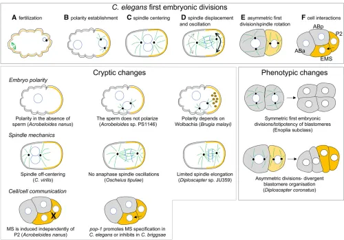
Thefirst embryonic divisions of theC. elegansembryo have been extensively described (Rose and Gönczy 2014) (Figure 2). Briefly, oocytes are blocked in prophase of meiosis I and un-polarized. At fertilization, the sperm brings in the paternal DNA and a pair of centrioles. These centrioles rapidly recruit pericentriolar material, which locally destabilizes the corti-cal actomyosin contractility, leading to the asymmetric re-partition of the PAR polarity proteins. At fertilization, the anteroposterior (AP) axis of the cell is thus established, and the sperm entry site defines the posterior side of the cell. In response to the PAR polarity, cytoplasmic proteins localize asymmetrically in the cell, and the mitotic spindle that is initially centrally located becomes posteriorly positioned along the AP axis during anaphase (Figure 2). This asym-metric displacement comes with impressive transverse os-cillations of the spindle—the manifestation of excess forces pulling on posterior astral microtubules. Because the cell cleavage plane is perpendicular to the spindle, two daughter cells of unequal size and asymmetric fate are formed, the posterior cell P1 being smaller than the anterior cell AB. This stereotyped asymmetric division has become a model to study oriented cell division because of the exquisite spa-tiotemporal resolution of events during thisfirst cell cycle and because of the strong conservation of molecules in-volved across phyla (Neumuller and Knoblich 2009). At each subsequent division, a similar asymmetric cell division is reproduced in the P lineage, ultimately giving birth to the founder cell of the germline, the P4 cell.
At the second cell cycle, while the founder cell AB divides symmetrically to generate ABa and ABp, P1 gives rise to the small P2 cell and EMS. During this division, the mitotic spindle of P1 rotates along the AP axis of the embryo and becomes perpendicular to the spindle of AB. This leads to a rhomboid organization of the fourfirst blastomeres, which is essential for the subsequent cellular interactions (Figure 2). Indeed, at the four-cell stage, P2 sends a Wnt signal to EMS, which then divides asymmetrically to give rise to the founder cell of the intestine (the E cell) and the founder cell of the me-soderm (MS). In the absence of P2 or Wnt signaling, EMS gives rise to two MS cells. Through Notch/Delta signaling, P2 also induces different fate acquisition in ABp compared to ABa. P2 next divides to give the founder cell C and P3, which divides again to give the founder cells D and P4.
These cell divisions thus rapidly produce the six key founder cells ofC. elegansembryos (Sulstonet al.1983).
The embryos of most nematodes, in particular free-living forms, can easily develop ex-utero. Thefirst cell divisions are easy to monitor under slide and coverslip because cells are large and transparent and the pace of cell divisions is relatively fast. These properties allowed the analysis of the early steps of embryogenesis in very diverse nematode species, starting with the founding work of T. Boveri onAscaris megalocephala (=Parascaris equorum) (Maderspacher 2008) and followed by Nigon and others in the early twentieth century (Nigon and Félix 2017).
Among the long list of free-living and parasitic species that have been observed since then, only species from Enoplia (Clade I, Figure 1A) undergo a series of symmetric embryonic
embryos always faces the vulva, suggesting that the orientation of the oocytes within the gonadal tract provides a polarity cue. In contrast, there is no correlation between embryo orien-tation within the uterus and the position of the posterior pole inDiploscapter, suggesting that polarity in these spe-cies is established randomly. Because polarization relies on the local destablilization of the actomyosin network in C. elegans, one could imagine that spontaneous self-organization of the actomyosin cortex triggers symmetry breaking to define the anterior–posterior axis of the em-bryo inDiploscapter.
Early embryo polarization is also observed in the parasitic nematodeBrugia malayi(Spirurina) (Landmannet al.2014). In this species, a microtubule-organizing center is found in oocytes prior to fertilization at the future posterior side of the cell, opposite to the location of the female meiotic spindle,
suggesting a microtubule-based mechanism of polarization from a maternal origin.Wolbachiaendosymbionts are found enriched at the posterior side of the one-cell embryo and in the P1 cell after thefirst division in this species; their removal leads to polarity defects in two-cell embryos (Landmannet al. 2014). WhetherWolbachiaare required for the initiation of polarity or its maintenance remains to be determined, but this example nicely illustrates the diversity of mechanisms that exist to establish the first embryonic polarity axis of nematode embryos during thefirst cell cycle.
stereotyped. In one study, thefirst two embryonic divisions of 34 rhabditids were scored, uncovering a large degree of variability in these subcellular phenomena (Brauchle et al. 2009). Farhadifaret al.(2015) analyzed thefirst embryonic cell division of 42 different rhabditid species and of natural isolates and mutation accumulation lines ofC. elegans. Spin-dle length appears to be constrained by stabilizing selection on cell and embryo size, with the two linked inC. elegansby a linear scaling relationship. However, the observed variations in spindle movements could not be explained by evolutionary changes in cell size between species (Valfort et al. 2018). Moreover, traits associated with spindle movements com-bined in ways contrasting with the expectation based onC. elegansstudies, suggesting that mechanical optimization of the mitotic spindle differs between species despite a con-served output phenotype: the asymmetry of division.
The origin of differences in spindle positioning between C. elegans and its congenerC. briggsaehave been explored (Richeet al.2013). InC. briggsae, at the onset of mitosis, the spindle is anteriorly shifted in contrast to a central position found inC. elegans. During anaphase, the spindle is pulled posteriorly in both species. However, this movement is ac-companied by much-reduced transverse spindle oscillations inC. briggsaecompared toC. elegans. These phenotypes were attributable to the GPR-1/2 proteins—components of the
cortical force generator complex. While two recently dupli-cated genes gpr-1 and gpr-2 are found in the genome of C. elegans,C. briggsaehas only onegpr-2gene. This difference in gene copy number correlated with a lower expression level inC. briggsaecompared toC. elegansbut also with a different spatio-temporal regulation.. Thus, the processes that produce a conserved and essential cellular feature, asymmetric spindle position, are distinct. This represents a case of what has been dubbed developmental system drift (DSD; True and Haag 2001) or phenogenetic drift (Weiss and Fullerton 2000) at the earliest stages of embryonic development.
Postzygotic cell lineage and founder cell specification
Although descriptions of early embryogenesis in Enoplia and Dorylaimia (Clades I and II; Figure 1) remain scarce because species of these clades are difficult to maintain in laboratory conditions (Schulze and Schierenberg 2011), what is known suggests that a striking diversity of mechanisms for early-development evolved early in the phylum. InEnoplus brevis (Enoplia, Clade I) thefirst embryonic divisions are symmetric and body axes are not specified during thefirst cell divisions. Moreover, except for the endoderm (E) lineage, no founder cells are identified and cells become determined later “en bloc”(Schulze and Schierenberg 2011). In Pontonema vul-gare, the spatial arrangements of the blast cells producing specific lineages can also vary substantially among embryos (Malakhov 1994; Voronov 1999), also suggestive of “regula-tive” development. In another representative of Enoplia, Tobrilus, a blastocoel is even observed with a canonical gastrulation—a feature that was unexpected in this phylum
of pseudocoelomate worms. However, anteroposterior polar-ity is established at the four-cell stage, and three founder cells for the germline, the pharynx, and the intestine are found (Schierenberg 2005; Schulze and Schierenberg 2011). Re-sults obtained in Prionchulus punctatus (Mononchida) are contradictory. On the one hand, laser ablation of half of the embryo does not prevent the development of a normal fertile adult (Borgonieet al.2000). On the other hand, there arefive founder cells (E, pharynx, D, C, and P), suggesting an early specification of cellular identities (Schulze and Schierenberg 2011). WhileRomanomermis culicivorax(Dorylaimia, a.k.a. Clade II) has six founder cells like C. elegans, tissues are formed by rings of cells, reminiscent of a segmentation pro-cess (Schulze and Schierenberg 2008, 2009). These species present extremely divergent early embryonic development, making it difficult to infer the ancestral pattern of develop-ment in nematodes. Nevertheless, because it is shared with outgroup phyla, the absence of deterministic lineage was most likely an ancestral character associated with slow embryogenesis.
divides first (Brauchleet al.2009). Moreover, species have either a rhomboid organization of blastomeres as in C. ele-gans, or a linear arrangement at the four-cell stage when both AB and P1 spindles rotate to align along the AP axis. Such linear organization is found in Diploscapter and some “Protorhabditis”species (Dolinskiet al.2001; Brauchleet al. 2009; Lahlet al.2009; Fradinet al.2017) or inMeloidogyne (Dolinskiet al.2001; Calderón-Urreaet al.2016) (Figure 2). In species with linear arrangement of the early blasto-meres, the question of lineage specification remains open. InC. elegans, ABp fate is induced by P2 via Notch signaling (Melloet al.1994; Mickeyet al.1996). The linear arrange-ment in the four-cell embryo means that this signaling must occur in a different way, if it occurs at all (Brauchle et al. 2009). Also, inDiploscapter coronatus, and some other spe-cies of the“Protorhabditis”group, P2 has already divided into C and P3 at the time of EMS and ABp division (Lahlet al. 2009; Fradinet al.2017). Moreover, the orientation of C and P3 is random, at least inDiploscapter coronatus. Thus, in only 50% of embryos does ABp contact C while EMS contacts P3. Despite these random contacts, ABp and ABa have a distinct lineage, suggesting that ABp specification is independent of an induction by either C or P3. Whether EMS requires an inductive signal by a neighboring cell or is cell-autonomous remains to be determined. Importantly, removal of EMS leads to an absence of intestinal cells, demonstrating an absence of multipotency, as inC. elegans(Lahlet al.2009). In striking contrast, inAcrobeloides nanus, where cellular contacts at the four-cell stage are similar toC. elegans, the absence of P2 does not prevent gut specification (Figure 2). Rather, any cell at the three-cell stage can give rise to intestinal cells after abla-tion of the others. Similarly, if AB is ablated, EMS takes over and C becomes EMS. Thus, in this species, multipotency and hierarchy of transformations is observed, despite an early segregation of the lineage in wild-type embryos (Wiegner and Schierenberg 1998, 1999). Unexpectedly, in the distantly related Plectus, the situation resembles C. elegans, since an induction of EMS by P2 is necessary to specify the intestine (Schulze et al. 2012). Therefore, many different solutions and reversals are found over the course of nematode evolu-tion to specify cellular identities during early embryogenesis. Interestingly, even within Caenorhabditis, differences in gut specification have been revealed at the molecular level, despite conservation of cellular interactions and blastomere specification (Coroianet al.2006; Linet al.2009). Upon Wnt signaling by P2, the transcription factorsSKN-1andPOP-1
act to specify E and MS identity. WhilePOP-1has a positive contribution to MS specification inC. elegans, it represses the MS fate in C. briggsae. In an interesting twist to the story,
MED-1,2, two GATA transcription factors that act down-stream of SKN-1, evolved in the lineage to C. elegans and are not present inC. briggsae. One model for the co-option of these new factors is via a transitional feed-forward archi-tecture in whichSKN-1acts both through and independently of MED-1,2 (Maduro 2009). Given the highly conserved cell lineages in the two species (Zhaoet al.2008), such an
opposite role for a key signaling pathway is an unexpected case of DSD.
The above results demonstrate that—despite a very strained body plan—early steps of embryogenesis vary con-siderably between nematodes. The molecular signature of such diversity in the early steps of embryogenesis was ex-plored infive different species withinCaenorhabditis(Levin et al.2012). Embryos from 10 different morphological stages were collected, from four-cell stage embryos to L1 larvae, and their transcriptomes were analyzed. Despite species-specific developmental timing, embryos from specific stages showed a similar pattern of gene expression across species, suggest-ing the existence of conserved“milestones”in development. Importantly, at midembryogenesis, corresponding to ventral enclosure, transcriptomes from different species were the least divergent. Moreover, genes that were activated at this stage showed enrichment in crucial functions such as pattern-ing by Hox genes or locomotion. These results led to the proposition (Levin et al.2012) that for nematodes, ventral enclosure represents a key, body plan-defining point in devel-opment, the so-called phylotypic stage (Slack et al.1993; Richardsonet al.1998). Transcriptome profiles throughout embryonic development were also performed in 20 mutation accumulation lines ofC. elegans, in which the effect of selec-tion is largely abolished. For all developmental stages, except ventral enclosure, variation in gene expression was much higher in the MA lines. This result strongly suggests that gene expression during ventral enclosure is highly con-served because of stabilizing selection (Zalts and Yanai 2017). Regardless of whether or not there is a phylotypic stage, these results do support an hourglass model (Raff 1996), in which nematode development shows the greatest diversity prior to or after a conserved point midway through embryogenesis.
Developmental regulation of gene expression
expression and anterior excretory duct cell location. Forcing expression ofLIN-48in theC. briggsaeexcretory duct cell is sufficient for anterior location. Thus, the gain of a novel reg-ulatory linkage during evolution altered bothlin-48 expres-sion and morphology. In addition, enhancers that mediate the conserved hindgut expression oflin-48, which are bound by
EGL-38, have diverged betweenC. elegans andC. briggsae (Wanget al.2004).
Gene regulatory evolution has also been examined in sub-sets of homologous neurons conserved acrossCaenorhabditis. Barrièreet al.(2012) focused on the GABAergic cell marker
unc-47. Though expressed in identical ways inC. elegansand C. briggsae, cross-species reporter transgenes produced addi-tional, ectopic sites of expression. Further experiments revealed that coordinated evolution betweencisandtrans fac-tors has occurred in each lineage. A subsequent study (Barrière and Ruvinsky 2014) expanded the neuronal genes analyzed to seven (unc-25,unc-46,unc-47,oig-1,acr-14,gpa-5, andmod-5) and the species to five (C. elegans,C. briggsae,C. remanei, C. brenneri, andC. japonica). Again, while regulatory regions from non-elegans species generally drive expression in the expected C. elegans cells, ectopic expression and/or cell-specific lack of expression is seen in nearly all cases. Interest-ingly, ectopic expression of cross-species transgenes is much more common, suggesting that the repressive mode of regu-lation evolves faster than the activating mode. Similar reporters based on homologs from the much more distantly related par-asitesMeloidogyne,Brugia, andTrichinella(Figure 1) showed that conserved patterns of expression can be driven by se-quences that are essentially unalignable (Gordonet al.2015).
The above studies show that changes incis-regulatory se-quences evolve rapidly. They can sometimes have develop-mental effects, but more often remain phenotypically cryptic. This is likely due to the action of stabilizing selection, which mandates an outcome, but not a mechanism. This allows compensatory evolution (or apparently compensatory, see Haag 2007) to proceed unchecked, accelerated by directional selection on other loci that share trans-regulators (Johnson and Porter 2007). Over time complex dependencies between distinct promoter regions form (Ludwiget al.2000).
Neuronal development
Of the 957 somatic cells of the C. elegans hermaphrodite, 302 are neurons, with another 56 providing support (Chalfie and White 1988). Males have over 100 additional neurons and glia, mostly with mating-related roles. Pioneer-ing work of John White and his colleagues determined the full connectome of the hermaphrodite (Whiteet al.1986), and 25 years later a full description of the male posterior nervous system completed the picture (Jarrellet al.2012). A large body of literature has described normal and per-turbed nervous system development in C. elegans as well (Hobert 2010; Cherra and Jin 2015; Shaham 2015). Such a wealth of information about this one species, as with other topics explored here, begs the question of conservation. Are all Caenorhabditis nematodes put together this way? How
about more distantly related nematodes? The earliest com-parisons were with the larger, distantly related parasite, Ascaris(e.g., Sulstonet al.1975; Walrondet al.1985; Niebur and Erdos 1993; Holden-Dye and Walker 1994), and revealed a surprisingfine-scale congruence of neurons over a large evo-lutionary distance (Schafer 2016).
Perhaps not surprising given their overtly similar anatomy, homologous neurons are produced inC. briggsaefrom a con-gruent embryonic cell lineage (Zhaoet al.2008). The more distantly relatedP. pacificusshares all 20 of the pharyngeal neurons, despite substantial divergence in feeding strategies (Bumbarger et al. 2013). Interestingly, however, these ho-mologous pharyngeal neurons are connected in substantially different ways. The cell lineages producing them have yet to be determined in P. pacificus, but even if they differ some-what, the nervous system appears to evolve novel connec-tions far faster than novel neurons. That finding presents an interesting parallel to work on the evolution of gene reg-ulatory networks (GRNs; Peter and Davidson 2011). In both cases, homologous components (either neurons or genes) evolve distinct regulatory connections to other components. How neural development is modified to produce novel con-nections is an important area for future research.
Sex determination
Sex determination was one of thefirst aspects ofC. elegans development to be tackled using forward genetic approaches (Hodgkin and Brenner 1977; Hodgkin 2002). X chromosome dosage had long been known to be the ultimate regulator of sexual fate (Nigon 1951). The discovery of a genetic pathway linking X dosage to cell fate (Hodgkin 1986) was subse-quently confirmed by molecular cloning of the genes (reviewed by Zarkower 2006). It soon became apparent, however, that this pathway did not resemble those that link chromosomes to sexual fate inDrosophilaor mammals (Cline and Meyer 1996; Eggerset al.2014). The cloning ofC. ele-gansmab-3revealed thefirst widely conserved sex-specifier, the DM family of transcription factors (Raymondet al.1998; Zarkower 2001). Thus, the disparity in sex determination mechanisms among different phyla is not due to wholly in-dependent origins of sexual dimorphism, but rather to rapid divergence of sex determination pathways, most likely up-stream and downup-stream of conserved DM factors (Haag and Doty 2005; Kopp 2012). This realization provided fur-ther motivation to examine the evolution of sex determina-tion over shorter time scales.
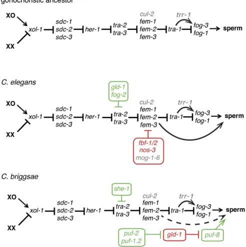
The first comparisons of sex determination genes within Caenorhabditisfocused on the“core pathway”that regulates dimorphism body-wide (Figure 3), starting with the identifi -cation ofC. briggsaehomologs of the genestra-2(Kuwabara and Shah 1994; Kuwabara 1996) and tra-1(de Bono and Hodgkin 1996). These early studies revealed rapid sequence evolution but conserved functions in the promotion of female somatic development. Similar results were subsequently re-ported for the male-promotingxol-1,her-1,fem-2, andfem-3
2002; Luzet al.2003) and the male-promotingtra-3(Kelleher et al. 2008).XOL-1 and FEM-3 are particularly divergent, with only 22 and 38% amino acid sequence identity be-tween their C. elegans and C. briggsae orthologs, respec-tively. ForFEM-3, its overall rapid divergence is mirrored by the region of the C-terminal domain ofTRA-2with which it interacts (Haag and Kimble 2000). In three species tested, the interaction between conspecificTRA-2andFEM-3 part-ners was conserved, but interspecies pairings invariably failed (Haaget al.2002). Less complete interspecies incom-patibility was observed for the FEM-2-FEM-3 interaction (Stothard and Pilgrim 2006). Another interaction, between a C-terminal domain ofTRA-2and TRA-1, has been docu-mented in bothC. elegansandC. briggsae(Lumet al.2000; Wang and Kimble 2001). These results indicate that, contrary to the conventional wisdom of molecular biology, even pro-tein domains of critical importance can evolve rapidly. This may be especially true if the only role of a sequence is to interact with one other partner (i.e., there is no pleiotropy at the molecular level). Abundant polymorphisms that do not disrupt interaction are observed in C. remanei TRA-2 and
FEM-3(Haag and Ackerman 2005). A population model sug-gests such variants can allow rapid coevolution by reducing the deleterious effects of other changes that would reduce
fitness on their own (Haag and Molla 2005).
In addition to rapid ortholog sequence evolution,C. brigg-saeis apparently lacking a clear ortholog ofsea-1, an autoso-mal regulator ofxol-1, the upstream-most“master regulator” of sexual fate (E.S.H., unpublished data). Thus, over the roughly 20 MY since C. elegans and C. briggsae diverged (Cutter 2008), their global sex determination pathways have undergone rapid sequence evolution and coevolution of con-served genes, and have begun to exhibit gene-level pathway incongruence. The existence of a highly diverged tra-1 ho-molog in the more distantly relatedP. pacificus(Pires-daSilva and Sommer 2004) suggests that key aspects of the core sex determination pathway nevertheless remain after substan-tially longer periods of divergence.
Self-fertile hermaphrodites have evolved at least three times withinCaenorhabditis(Kiontkeet al.2004, 2011) (Fig-ure 1 and Fig(Fig-ure 3). This novel strategy is enabled by pro-duction of sperm in the XX ovary, making germline sex determination an obvious topic of interest for EDB. Before examining that, however, it is worth noting that, unlike Drosophila and mammals, the somatic niches for germline stem cells are very similar (if not identical) in male and fe-male Caenorhabditis (Kimble and Hirsh 1979; Kimble and White 1981; Millozet al.2008), and a male somatic gonad is not required to support the differentiation of spermatocytes (Graham and Kimble 1993; Grahamet al.1993). Further, the C. eleganshermaphrodite does not expressHER-1, a secreted protein that specifies male fate in XO animals, even in the L4 stage when sperm are produced (Trentet al.1991; Perryet al. 1993). Self-fertility thus represents a cell-autonomous change in sexual fate. Extensive mutagenesis screens for XX ani-mals with germline-specific sexual transformations (e.g., the
masculinization of germline, or Mog, and feminization of germline, or Fog phenotypes) have identified cis-regulatory elements in core sex-determination gene mRNAs that are sites of negative regulation by germline RNA-binding proteins [RBPs, reviewed by Zanetti and Puoti (2013)]. The reconfi gu-ration of RBP-target mRNA networks thus appears to be the key to XX spermatogenesis, distinguishing it from other phe-notypic novelties that are rooted in changes in transcription factors and their target genes (Carroll 2008).
What were the changes that allowed XX spermatogenesis to evolve, and how similar are they in selfing species that evolved convergently? Examination of conserved global sex-determiners in the hermaphroditic C. briggsae and the outcrossing C. remanei revealed identical roles for the fe-male-promotingtra-1,tra-2, andtra-3(de Bono and Hodgkin 1996; Kuwabara 1996; Haag and Kimble 2000; Kelleheret al. 2008), and the male-promotingher-1(Streitet al.1999). In contrast, while RNAi knockdown ofCbr-fem-2andCbr-fem-3
function could feminize the germ cells ofC. briggsaemales, it had no effect on hermaphrodites (Haaget al.2002; Stothard et al.2002). The dispensability of theC. briggsae femgenes for hermaphrodite spermatogenesis was subsequently
con-firmed by deletion mutations and exhaustivetra-2(ts) sup-pressor screens (Hill et al. 2006). These results suggested that regulatory mechanisms that allowC. briggsae spermato-genesis act downstream of thefemgenes.Cbr-fem-3;Cbr-tra-1
double mutants have the perfect male soma characteristic of Cbr-tra-1 mutants, but a well-regulated hermaphrodite germline, as inCbr-fem-3mutants (Hill and Haag 2009). This indicates that, as inC. elegans(Hodgkin 1986), thefem mu-tations are epistatic totra-1in the germ line. Interestingly, in both speciesfog-3 expression, which is controlled bytra-1, and thus by the femgenes, remains high in tra-1mutants even when the germline is feminized by simultaneous loss of one or morefemgenes (Chen and Ellis 2000; Hill and Haag 2009). This indicates that thefemgenes act in multiple places near the terminus of the germline sex determination path-way. The degree of identity of these sites of control be-tween the two selfing species, and the extent to which they were present in their gonochoristic ancestors, remains to be determined.
C. briggsae. This may have occurred because of its simple RNA target motif (Ryderet al.2004) and conserved expres-sion in early meiotic germ cells (Joneset al.1996; Nayak et al.2005).FOG-2, an F-box protein cofactor forGLD-1in C. elegansthat is essential for XX (but not male) spermato-genesis (Schedl and Kimble 1988; Cliffordet al.2000) is a recent gene duplicate that is found only in this species (Nayaket al.2005).
Thegld-1mRNA is itself subject to translational repression via its own 39UTR by the PUF family RBP FBF (Crittenden et al. 2006). The PUF family is somewhat dynamic in Caenorhabditis, such that inC. briggsaethere are not strict
orthologs of FBF. However, both biochemical and genetic studies indicate that the three paralogs of the PUF-2 subfam-ily (Cbr-puf-2, Cbr-puf-1.1, and Cbr-puf-1.2) represent the C. briggsaeequivalents (Liuet al.2012). Given the opposite roles ofC. briggsaeandC. elegansgld-1in sex determination, it is not surprising that simultaneous RNAi knockdown of
Cbr-puf-2andCbr-puf-1.2function feminizes the germ line, rather than masculinizes as does C. elegans fbf(RNAi). Sur-prisingly, however, complete elimination ofCbr-puf-2activity alone (via a deletion mutation) leads to a fully penetrant larval arrest. Subsequent studies revealed this was due to a defect in pharyngeal development, apparently related to the brief expression ofCbr-puf-2in three pharyngeal muscle cells (Liu and Haag 2014). This suggests that the PUF protein family may spin off paralogs as they acquire novel roles out-side of the germ line, an example of the neofunctionalization process thought to favor retention of otherwise redundant gene copies (Lynchet al.2001).
The above studies revealed evolutionary variation through reverse-genetic targeting of conserved genes. Another fruitful approach has been to conduct unbiased forward screens for germline-specific feminizers inC. briggsae(reviewed by Ellis 2017). For example, alleles ofCbr-gld-1emerged from screens forMoghermaphrodites (Beadellet al.2011). Similarly, screens forfog-2-like mutations conferring hermaphrodite-specific germline feminization led to the discovery ofshe-1
(Guoet al.2009). LikeFOG-2,SHE-1is an F-box protein that depends upontra-2for its function. However, there is no in-dication that it directly regulates tra-2, nor that it interacts with GLD-1. Its exact role in enabling XX spermatogenesis thus remains a subject for future work.
Another novel factor required for sperm development of both sexes ofC. briggsaeis encoded bytrr-1(Guoet al.2013). This component of the Tip60 histone acetyl transferase com-plex is conserved acrossCaenorhabditis, but loss oftrr-1alone is incapable of causing similar feminization of theC. elegans germ line. Cbr-trr-1mutations enhance the incomplete so-matic masculinization ofCbr-tra-2, and, in the germ line, help activate fog-3 expression, suggesting that TRR-1 promotes male development. However, the effect onfog-3is dependent upon the presence oftra-1. This suggests that, as for Gli and its other hedgehog pathway transcription factor homologs,
TRA-1 has both activating and repressing effects on target genes, withTRR-1being important for the former. A previ-ously unknown role of theC. eleganstrr-1ortholog in promot-ing male development can be revealed through enhancement of weakfemalleles (Guoet al.2013). These results are consis-tent with existence of separate and conservedtra-2/fem (repres-sor) andtra-1/trr-1(activator) branches of the sex determination pathway. Though apparently conserved in both C. elegansand C. briggsae, their relative importance is reversed. The case of
trr-1also shows how use of a second“satellite model”organism can shed important light on cryptic evolution underlying con-served phenotypes of the more widely studied species.
The impact of trr-1described above, as well as related work in C. elegans(Grote and Conradt 2006) suggest that Figure 3 Convergent evolution of self-fertility via distinct changes
chromatin regulators may be frequent contributors to sexual regulation via modulation of TRA-1 function. Chen et al. (2014) thus pursued possible roles for the nucleosome remod-eling factor (NURF) complex inC. briggsae. Using the TALEN-based genome editing methods they had developed (Weiet al. 2014a), they discovered that, while complete loss ofCbr-isw-1
and Cbr-nurf-1 were sterile, hypomorphic mutations were sometimesFog, and RNAi knockdown of either gene increased the penetrance of this. Surprisingly, however, the feminizing impact is not observed in C. elegans, or in the outcrossing C. nigoniandC. remanei. The NURF complex thus appears to be uniquely important inC. briggsae, and likely represents an-other component of the species-specific regulation that each hermaphrodite evolved to produce sperm transiently.
Germ cell proliferation
The proliferation of germ cells at the distal tip of theC. elegans gonad is directed by a somatic niche, comprised of the many
finger-like projections of a single distal tip cell (DTC, Hall et al. 1999; Byrd et al.2014). The DTC stimulates mitotic proliferation of germline stem cells via Notch signaling (Kimble and Hirsh 1979; Kimble and White 1981; Austin and Kimble 1987; Cinquin et al. 2010). As proliferation pushes stem cells out of the DTC niche, they undergo afinal mitosis and then enter meiosis. No further mitoses are nor-mally observed in either sex, and there is no evidence for a mostly quiescent, or“label-retaining”subpopulation of stem cells (Crittendenet al.2006). In addition, for allCaenorhabditis species that are self-fertile, spermatocytes are found only during the L4 larval stage and (depending on species) the first few hours of adulthood as defined by thefinal molt. Thus, sperm are of a finite number established prior to ovulation, and when sperm are exhausted reproduction ceases unless mat-ing with a male occurs. Recent studies in other nematode groups have revealed significant deviations from these aspects ofCaenorhabditisgerm cell proliferation.
The recently described genus Auanema (Kanzaki et al. 2017) has presented several unexpected aspects of germline development. Though similar to Caenorhabditis in overall form and habitat, and within the same family,“Rhabditidae” (Kiontke and Fitch 2005), at least three Auanema species (A. rhodensis,A. freiburgenesis, andA. viguieri) exhibit a re-productive polyphenism in the development of XX individu-als, such that those that develop directly via a normal L3 larva mature into females, while those produced from dauer larvae (L3d) develop as selfing hermaphrodites (Félix 2004; Kanzaki et al.2017). This presents another convergently evolved self-fertile taxon, which has now been examined in some detail. Among their unexpected features, hermaphrodite spermatocytes are not specified briefly in the L4 stage, as in Caenorhabditis, but instead are continuously replenished via coherent popu-lations of spermatagonia (McCaig et al.2017). These form elongated cysts that proliferate mitotically far from the distal stem cell niche, and undergo meiosis and spermatogenesis adjacent to oocytes. Other surprising features of Auanema germline biology are described below.
More distant relatives ofCaenorhabditisare the mamma-lian filarial parasites (onchocercids, Spiruromorpha, Figure 1), such asBrugia malayi, the causative agent of humanfi l-ariasis. These parasites have a radically different life history from the bacteriovores in“Rhabditidae”discussed thus far. A femaleBrugiaadult can lay over 1000 embryos per day, and sustain this rate for over 5 years (Tayloret al.2010)—a re-productive output three orders of magnitude greater than that ofC. elegans. In addition, they and many of their relatives have harboredWolbachiabacteria as obligatory symbionts for millions of years (McLaren et al. 1975; Bandi et al. 1998; Tayloret al.1999). Importantly, curing these nematodes of Wolbachia with antibiotics adversely affects them without harming their mammalian host (Bosshardt et al.1993). In Onchocerca ochengi, a parasite of livestock, tetracycline treat-ment kills adults (Langworthyet al.2000). In curedBrugia malayi and B. pahangi, females produce inviable embryos that die via extensive apoptosis, while males retain normal fertility (Bandiet al.1999; Landmannet al.2011). This in-viability is likely caused by the requirement forWolbachiain proper polarization of thefirst zygotic cell division, as noted earlier (see section Zygotic mitosis). A subsequent study (Foray et al. 2018) revealed that the Wolbachia symbiont and theBrugiafemale have coevolved to jointly support oo-cyte proliferation. The dynamics of this proliferation differ markedly from that ofCaenorhabditis, in that it occurs predom-inantly in a zone proximal to the distal stem cell niche, with the most distal cells represent a quiescent population (Forayet al. 2018). Loss ofWolbachiastimulates ectopic proliferation in the distal zone, with the effect of exhausting the quiescent pool. It thus appears thatWolbachiahas become such an integral part of the female germline development that the nematodes can no longer prosper without it. What, if anything, the nematode hosts derive from the symbiosis is another mystery.
In addition to the presence of theWolbachiasymbiont, the somatic niche for germline stem cells differs between Caenorhabditis andBrugia (Foray et al.2018). Ablation of the DTC inBrugiais not sufficient to eliminate germline pro-liferation, as it is inCaenorhabditis. Nevertheless, broad treat-ment with inhibitors of Notch signaling reduce proliferation. These results suggest that the somatic niche inBrugiais sim-ilar to that ofCaenorhabditis, but on a larger scale. Thisfi nd-ing is consistent with the ongond-ing anatomical (Rundell and Leander 2010) and genomic (Aboobaker and Blaxter 2003) miniaturization of nematodes that accompanied their invasion of tiny meiofaunal habitats.
Spermatogenesis
Just withinCaenorhabditis, sperm can differ in volume as much as 50-fold between species (Vielleet al.2016). Sperm size is correlated with competitive ability within species (LaMunyon and Ward 1998). In selfing species, male sperm are consistently larger than those of hermaphrodites, in part because of somatic gonad effects (Baldi et al.2011). How-ever, male sperm of outcrossing species are generally larger than those of males from selfing species (LaMunyon and Ward 1999; Hill and L’Hernault 2001). Further, conditions that select for the most competitive sperm also increase sperm size (LaMunyon and Ward 2002). These correlations indicate that postcopulatory sexual selection and its relaxation in selfing species is a major force that shapes sperm develop-ment. They also suggest a simple effect of sperm size on com-petitive ability, yet interspecies matings reveal a more complex relationship. Males of outcrossing species frequently suppress self-fertility in hermaphrodites, and tend to have larger sperm. However, across a matrix of many pair-wise crosses, the extent of this effect is not correlated with difference in the sperm size of the two species (Tinget al.2014). This suggests that other factors contribute to competitiveness. A likely candidate is the sperm proteome, which can be much larger in outcrossing species (Thomaset al.2012b; Yinet al.2018).
Several lines of evidence have revealed that male-expressed genes are disproportionately lost as part of wide-spread genome shrinkage in self-fertile lineages (Thomas et al. 2012b; Fierst et al. 2015). One case that has been investigated functionally is that of the MSS family of sperm surface glycoproteins. Yinet al.(2018) found thatmssgenes are found in nearly all outcrossingCaenorhabditis, but are missing in all self-fertile species. MSS proteins are both nec-essary (inC. remanei) and sufficient (when restored to C. briggsae) for optimal sperm competition. The increased suc-cess in siring cross-progeny that anmss+transgene confers toC. briggsaemales (Yinet al.2018) may provide an impor-tant clue about its independent loss. With greater suppres-sion of selfing comes a greater fraction of male progeny. The reproductive assurance and lack of inbreeding depression of selfing species (Dolgin et al. 2008), combined with the small, transient habitats they favor likely create conditions that select for lower male frequency via interdemic selec-tion. This is reminiscent of the local mate competition sce-nario of Hamilton (1967). Loss ofmssmay provide a way to reduce male frequency without complete loss of outcross-ing, which is likely needed at some level (Morran et al. 2009a,b).
Beyond competiveness, the sperm of some nematodes exhibit oddities that lead to unexpected sex ratios, as in the heterogonic sheep parasiteStrongyloides papillosus. Adults in a host are always parthenogenic females. Many of their XX progeny develop directly into infective larvae, creating a sim-ple asexual life cycle. However, females can also produce sexual XX female and XO male progeny, which mate outside the host and produce outcrossed infective larvae. How does a parthenogenic XX female produce a male without mating? Albertsonet al.(1979) had suggested that one X chromosome
(present as part of an X-autosome fusion in this species) may be lost in some diploid oocytes via chromosomal diminution. Using molecular markers and heroic crosses through sheep, Nemetschkeet al.(2010) found clear support for this hypothesis. Which of the two X chromosomes is lost appears to be random, but some mechanism must prevent both from being lost.
Auanema rhodensis presents another interesting sperm-mediated sex ratio anomaly. Though males are XO and fe-males XX, cross progeny are,2% male (Félix 2004). Exam-ination of male spermatogenesis provided an explanation (Shakeset al.2011). As spermatocytes proceed through mei-osis I, the two X chromosomes are not paired, as inC. elegans. This produces secondary spermatocytes with one X chroma-tid. When these divide, the spermatid possessing the X at-tracts nearly all of the organelles required for sperm function (mitochondria, membranous organelles, and MSP), while the nullo-X chromosome set ends up in a residual body incapable of supporting spermiogenesis. As a result, nearly all spermato-zoa capable of fertilizating an oocyte are X-bearing, which, in turn, produces extremely female-biased broods. Interestingly, matings between male and free-living female Strongyloides papilillosus also produce all-female broods, but it is not yet known whether the mechanism is the same as that described forA. rhodensis(Streitet al.1999).A. rhodensishermaphrodite morphs employ yet another non-Mendelian mechanism of X chromosome segregation during spermatogenesis, as the func-tional self-sperm contain two X chromosomes (Tandonnet et al.2018). This is coupled with loss of both oocyte X chro-mosomes to thefirst polar body. As a result, self-progeny are always XX, but crosses between XX hermaphrodites and males yield exclusively male progeny. These dynamics are another strong indication that selection on sex ratio can push the evo-lution of sperm attributes, in this case via unexpected meiotic novelties.
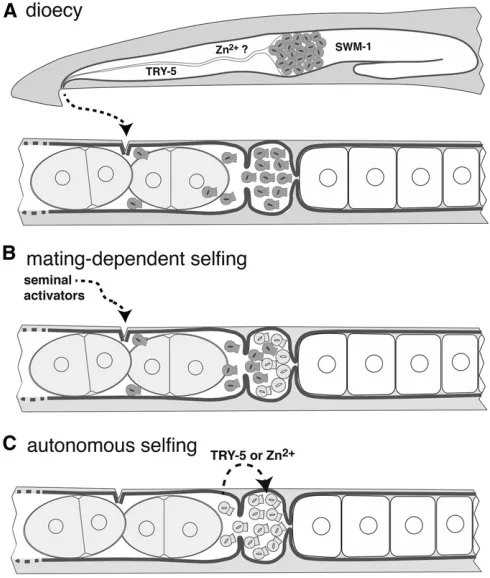
Though it is obvious that self-fertility depends upon XX spermatogenesis, the final step of sperm development— spermiogenesis or activation—plays another important role in its evolution. In male nematodes, spermatids are stored in an inactive state in the seminal vesicle, and are not activated to become motile spermatozoa until exposure to factors dur-ing their passage through the vas deferens activates two par-allel pathways (Ellis and Stanfield 2014). One of these pathways is composed of SPE-8and associated sperm pro-teins, which responds to a signal from the vas deferens (Nishimura and L’Hernault 2010) that may be zinc cations (Liuet al.2013). The other is mediated by the seminal pro-tease TRY-5 and its inhibitor,SWM-1(Stanfield and Ville-neuve 2006; Smith and Stanfield 2011). The requirement for activators expressed in the male somatic gonad presents a problem for would-be selfing hermaphrodites, which must evolve male-independent sperm auto-activation.
C. elegans spe-8 group mutants exhibit hermaphrodite-specific activation defects, suggesting that only the spe-8
used the identical means to achieve sperm auto-activation. Weiet al.(2014b) found that genes of both male sperm ac-tivation pathways are conserved across the genus. Knockout mutants in multiplespe-8group genes cause self-sterility in C. briggsaehermaphrodites (but not males), suggesting par-allel co-options of the same sperm activation pathway. How-ever, loss ofspe-8group homologs had no effect onC. tropicalis hermaphrodites, buttry-5mutant hermaphrodites were self-sterile. This indicates that, in the C. tropicalislineage, the alternative pathway evolved to enable auto-activation (i.e., convergence). Surprisingly,C. tropicalismales are also ren-dered sterile upon loss of onlytry-5, indicating that the two pathways are no longer redundant in this species.
Given the need for both XX spermatogenesis and sperm auto-activation, how did self-fertility ever evolve? In an elegant experiment, Baldiet al.(2009) simulated this trans-formation using the gonochoristicC. remanei. Partial loss of
Cre-tra-2function with RNA interference creates XX pseudo-hermaphrodites that produce sperm (Haag and Kimble 2000), but these sperm are not active and the animals are not self-fertile. However, mating with males is sufficient to activate these sperm and allow production of selfed progeny. Moreover, whenCre-swm-1 is also knocked down, pseudo-hermaphrodite self sperm spontaneously activate and sire self-progeny (Baldiet al.2009). Because there is no known role for TRY-5protease in females, this ability ofCre-swm-1
(RNAi) to activate these XX sperm is surprising. Examination of sex-specific transcriptome data (Thomaset al.2012a) reveals thatC. remaneiswm-1is abundantly and comparably expressed in both females and males, whiletry-5is highly male-biased. One possibility is that the low level ofTRY-5expression inC. remanei females is sufficient to activate self sperm whenSWM-1is elim-inated. Alternatively, knockdown of tra-2 may elevate TRY-5
levels to a point that potentiates loss ofswm-1. In either case, simultaneous modification of sex determination and sperm acti-vation factors is sufficient to allow rudimentary selfing.
The above experiments suggest a two-step model for the evolution of self-fertility (Figure 4). In thefirst phase, a germ-line-specific change in the regulation of the sex determina-tion (discussed above) could have produced a small population of XX spermatids. By virtue of developing in a female body, these were initially inactive, and also smaller than male sperm (Baldi et al.2011). However, theirtrans -activation could be achieved via seminalfluid from conspe-cific (or closely related) males, as shown by Baldi et al. (2009). Evolving in a population of gonochoristic conspe-cifics, such mates would likely be readily available to the in-cipient selfer. This would produce a mixed-paternity brood with an XX-skewed sex ratio, which has two potential conse-quences. First, selfed progeny would retain the maternal ge-notype that promotes XX spermatogenesis, which is likely a recessive trait (Woodruffet al.2010). Second, the XX-biased broods may be adaptive if population sizes shrink and local mate competition conditions set in (see above). Such partial selfing would have the further benefit of allowing recessive deleterious mutations to be gradually purged (Garcia-Dorado
2012). Eventually, a greater degree of selfing would become well tolerated. In thefinal phase, hermaphrodites could have evolved sperm auto-activation by upregulating expression of one of the spermiogenesis signals in their otherwise female reproductive tract. Baldiet al.(2009) also suggested that the capacity for XX sperm autoactivation may have evolved as a neutral polymorphismfirst, allowing subsequent changes in sex determination that enable spermatogenesis to achieve great impact without mating. In either case, an autonomous selfer would enjoy reproductive assurance at low density with-out accompanying inbreeding depression, allowing them access to habitats that would be marginal for obligate outcrossers.
Vulva development
The vulva is an opening in the center of theC. elegans her-maphrodite (but can be in different positions in other taxa) that serves for copulation and egg laying through its direct connection with the uterus. Because of extensive work on the vulva inC. elegans, it has also become an important evo-devo model and is a primary exemplar of DSD. The vulva is a simple organ that originates from a handful of ventral epider-mal cells during the larval stages (Figure 5). Cellular division and organogenesis can be tracked by differential interference contrast (DIC) microscopy. Moreover,C. elegansmutants with abnormal vulvae remain fertile, which has allowed vulva de-velopment to be explored in exquisite detail (Sternberg 2005; Guptaet al.2012). While the morphology of the adult vulva can be a slit or a round pore depending on the species (Kiontkeet al.2007), the fate patterns of the vulva precursor cells (VPCs) are quite conserved between species. Are the VPCs specified similarly in all species? Where does the induc-tion signal come from, and is it always the same molecular signal? Studies that have posed these questions have revealed an impressive diversity of cryptic changes (i.e., changes in mech-anism without changes in phenotype), between both closely and distantly related species, and even strains of the same spe-cies. The field has also benefited from the establishment of other selfing species,P. pacificusandOscheius tipulae, as genet-ically tractable systems to explore changes in vulval develop-ment over large evolutionary distances (Félix 2006; Sommer 2006). Cryptic genetic changes have been deciphered further by exploring different species of the same genus and even dif-ferent natural isolates of the same species in the three genera, Caenorhabditis,OscheiusandPristionchus.
to the cuticle to anchor the vulva). P3.p, P4.p and P8.p daughter cells adopt a 3°(nonvulval) fate, because they do not form the vulva in wild type animals. All of these P(5–7).p cells form a vulval competence group, as any of these cells can replace an ablated cell and become vulval. All the other Pn.p cells are incapable of forming the vulva, even upon ab-lation of P3.p to P8.p (Sternberg and Horvitz 1986). This vulval competence group is defined by expression of two Hox proteins, withLIN-39in central cells promoting compe-tence and MAB-5repressing it in more posterior Pn.p cells (Clandininet al.1997). The AC sends aLIN-3/EGF signal that acts as a morphogen on the VPCs (Hill and Sternberg 1992). Closest proximity to the signal determines the 1°fate (gen-erally P6.p); P5.p and P7.p receive a lower dose and adopt the 2°fate. Activation of the EGF/Ras signaling pathway in P6.p activates the Notch/Delta pathway (Sternberg 1988).
This leads to the inhibition of the 1°fate in P5.p and P7.p through lateral inhibition and to the activation of the 2°fate in these same cells. The Wnt pathway is also involved in vulval specification, as loss of negative regulators of Wnt causes mote than three VPCs to be induced (Gleasonet al. 2002). Conversely, VPCs adopt a 3° fate or fuse with the hypodermis in the absence of positive regulators of the Wnt pathway (Eisenmann and Kim 2000).
Variation in the position of the vulva: C. eleganshas two gonadal arms extending from the center of the animal, with a central uterus and vulva derived from the central epidermal Pn.p cells. Some species have a single gonadal arm (mono-delphy), which extends anteriorly. The evolution of monodel-phyper sewill not be covered here (but see Félix 1999). In most cases, monodelphy is accompanied by a posterior shift of the uterus and the vulva. In the monodelphic species P. redivivus, the vulva forms at 60% of body length because of a posterior displacement of the central Pn.p cells and because the vulva is centered in between P6.p and P7.p (Sternberg and Horvitz 1982). Within“Rhabditidae,”monodelphy and a posterior vulva are derived and evolved several times (Kiontkeet al.2007). In the three posterior-vulva species of Cruznema,Mesorhabditis, andTeratorhabditisthat have been analyzed, again only the central Pn.p cells (P5.p to P7.p) are competent, and they migrate posteriorly (Sommer and Sternberg 1994). However, mechanisms of vulva induction differ between species. The developing gonad induces the VPCs inCruznema, but is not required to induce the VPCs in Mesorhabditis and Teratorhabditis. Establishment of the competence group by LIN-39 is conserved in P. pacificus (Eizinger and Sommer 1997) andO. tipulae(Louvet-Vallee et al.2003). HOX specification represents a constraint on specifying which cells can form the vulva; to make a pos-terior vulva, this constraint has been overcome in at least four independent lineages in Rhabditida by a similar mechanism, i.e., posterior migration of the vulva cells (Kiontkeet al.2007).
Variation in the number and fate of VPCs:Large variations are found in the size of the competence group, in the number of divisions of competent cells, as well as in the fate of the noncompetent Pn.p cells (for an evolutionary synthesis of most of these differences among species in Rhabditida, see Kiontkeet al. 2007). While the competence group includes P3.p up to P10.p inP. redivivus(Sternberg and Horvitz 1982), much larger than the number of cells that are induced, in RhabditophanesandStrongyloides rattithe competence group is restricted to the cells that form the vulva (Félix et al. 2000a). The size of the competence group can also vary be-tween closely related species. For instance, P3.p is competent inC. elegansbut not inC. briggsaeor in some otherCaenorhabditis species (Pénigault and Félix 2011). Overexpression of Wnt in C. briggsaeis sufficient to induce the division of P3.p, while downregulation in C. elegansprevents the division of P3.p (Pénigault and Félix 2011). Because several Wnt ligands Figure 4 Scenario for the evolution of self-fertility inCaenorhabditis. (A) In
are expressed in a gradient from posterior to anterior in the C. elegansbody (Gleasonet al.2006), one possible explana-tion is that the lack of competency of P3.p inC. briggsaecould be due to a shorter Wnt gradient inC. briggsaecompared to C. elegans (Figure 5; Pénigault and Félix 2011). It is also possible that, in C. briggsae, P3.p is less sensitive to Wnt signals. Within Pristionchus, P8.p is partially competent in P. pacificus(Sommer 1997), but is a true VPC inP. lheritieri (Srinivasanet al.2001).
Within the competence group, the pattern of cell division is also variable. The numbers of cells that form the vulva vary
between 16 cells in O. tipulae, to 34 cells in Rhabditoides regina(Sommer and Sternberg 1995). WithinCaenorhabditis, 22 cells form the vulva in all species that have been observed (Félix 2007). However, the division pattern of the competent cell P3.p is highly variable, between and withinCaenorhabditis species (Delattre and Félix 2001; Félix 2007; Pénigault and Félix 2011). Similarly, the pattern of P4.p and P8.p division shows a high degree of intraspecies and interspecies varia-tion inOscheius. The fate of the cells that are not competent to form the vulva also vary. Pn.p cells that do not express
(Louvet-Vallee et al. 2003), while the expression of LIN-39
prevents cell death in P. pacificus (Eizinger and Sommer 1997). The restriction of the competence group by cell death is a derived character that is observed several times in the phylogeny, yet reversals of this restriction are very rare, constituting an interesting evolutionary bias (Sommer and Sternberg 1996; Kiontkeet al.2007). InTurbatrix aceti, as in three other Panagrolaimomorpha species, the survival of the VPCs depends on a signal emanating from the gonad dur-ing the L2 stage, in contrast to P. pacificus (Sternberg and Horvitz 1982; Félix and Sternberg 1997, 1998).
Variation in the mechanisms of VPC induction:Laser ab-lation of the AC or the gonad has been performed in a wide range of species. These experiments have revealed an impres-sive diversity of induction mechanisms, apparently evolved from an ancestral two-step induction signal from the gonad (Kiontkeet al.2007). Most surprisingly, systematic character-ization ofCaenorhabditisandPristionchushas uncovered cryp-tic genecryp-tic changes (e.g., changes in the contributions of different signaling mechanisms, in competence level and even genetic variation affecting the requirement for induction) be-tween closely related species and even bebe-tween strains of the same species (Srinivasan et al.2001; Félix 2007; Zauner and Sommer 2007; Millozet al.2008; Kienle and Sommer 2013).
In Clades IV and V (Figure 1), the VPCs can be induced independently of the gonad, as inBrevibucca, or require con-tinuous or possibly consecutive signals from the gonad, as in Halicephalobus sp. (Félix et al. 2000a). As shown above, Mesorhabditis and Teratorhabditis also do not rely on the gonad for induction (Sommer and Sternberg 1994), while two consecutive signals from the AC are required inO. tipulae andRhabditella axei(Félix and Sternberg 1997). InO. tipu-lae, the early and late induction signals depend on the activity of MEK kinase—a component of the Ras pathway involved in C. eleganslate-only induction (Dichtel-Danjoy and Félix 2004b). One possible evolutionary scenario is that a hetero-chronic shift occurred in the Caenorhabditis lineage with regard to both the requirement for, and expression of, the homologous induction event (Kiontkeet al.2007).
InP. pacificus, a continuous 10-hr induction from several cells of the somatic gonad is required to induce the VPCs (Sigrist and Sommer 1999), seemingly comparable to the two-step induction ofO. tipulae(Kiontkeet al.2007). As in C. elegans(Gleasonet al.2006), simultaneous inactivation of several Wnt ligands and receptors leads to Vulvaless pheno-types inP. pacificus(Zhenget al.2005; Tianet al.2008; Wang and Sommer 2011). However, after inactivation of Ppa-bar-1/b-catenin (the Wnt signal transducer), VPCs do not die of
apoptosis but adopt a 3° fate, similar to the phenotype obtained after gonad ablation (Tian et al. 2008). Further, Wnt ligands MOM-2 and LIN-44 are expressed in the AC before the division of VPCs and in the central cells of the somatic gonad, respectively (Tianet al.2008). This suggests that Wnt signals comprise the gonadal signal that induces formation of the vulva inP. pacificus(Figure 5), whereas they
are primarily involved in establishing VPC competence to re-spond to that signal in Caenorhabditis. The involvement of
LIN-3 and its downstream cascade has not been demon-strated inP. pacificusvulval induction, raising the possibility that a secondary, largely redundant Wnt pathway in one species (C. elegans) could be central to the homologous process in another (P. pacificus). Interestingly, P8.p and the mesoblast M cell are both responsible for the lateral inhibi-tion that prevents too many VPCs from adopting a 1° fate (Jungblut and Sommer 2000). Thus, P8.p inhibits the induc-tion of VPCs even though it is not a VPC itself. Of note, Wnt signaling is also used inP. pacificusto shape the distinct “pret-zel” morphology of the somatic gonad (Rudelet al. 2008). Selection on either gonad shape or vulva induction would thus target a pleiotropic Wnt module, with potential conse-quences (overt or cryptic) for the other trait.
Variation of the induction mechanism of VPCs is also found between species belonging to the same genus or even between strains of the same species. For instance, a single late induction from the AC is required for VPC divisions in Panagrolaimussp. PS1579 as inC. elegans, while early and continuous (or possibly two consecutive) signals from the gonad are necessary in anotherPanagrolaimusspecies,P. sp. PS1732 (Félixet al.2000). WithinPristionchus, the system of induction found in the laboratory strainP. pacificusPS312is not widely conserved. For instance, inP. lheritieriandP. mau-pasiand even different strains ofP. pacificus, some VPCs are induced even when the gonad is ablated just after hatching, and the extent of the lateral inhibition exerted by P8.p on the VPCs can vary (Srinivasanet al.2001; Zauner and Sommer 2007). Mapping of the quantitative trait locus (QTL) respon-sible for the differences in the gonad-independent induction of VPCs between P. pacificusstrains revealed a new role for the Notch ligandapx-1/Delta(Kienle and Sommer 2013). In many wild strains, absence of a binding site for the HAIRY transcription factor in thecis-regulatory region ofapx-1 leads to its expression in P6.p and confers a gonad-independent induction of this cell, while in the laboratory strainPS312,
apx-1is not expressed in the VPCs, which thus require the gonad for induction (Kienle and Sommer 2013).
While the pattern of division of the VPCs is very conserved amongCaenorhabditisspecies, cryptic changes in the mech-anism of induction were revealed by ablation of the AC or overexpression of the LIN-3/EGF inductive signal (Félix 2007). Early ablation of the AC leads to adoption of the 3° fate for all VPCs in all species. However, ablation of the AC during patterning,i.e., mid-L3 stage, has different outcomes depending on the species or strain within a species (Félix 2007; Milloz et al. 2008). For instance, inC. remanei, the VPCs adopt a 2°3°2°pattern, suggesting that, in contrast to C. elegans, a low level of induction from the AC is sufficient for P6.p to induce its neighboring cells, but not enough for its own fate acquisition. In C. briggsae, the same experiment leads to a 2°2°2°pattern. Moreover, mild overexpression of
ofLIN-3(Katzet al.1995). Thus, lateral inhibition from P6.p on adjacent Pn.p cells can be overcome easily inC. briggsae and less so in C. elegans. Nevertheless, the LIN-12/Notch pathway is still involved in lateral inhibition in C. briggsae andLIN-3/EGF acts in a dose-dependent manner on VPC fate specification (Félix 2007). However, inactivation of genes of the vulva specification pathways inC. elegansandC. briggsae often leads to different phenotypes, revealing a difference in the respective contributions of these pathways (Rudel and Kimble 2001; Sharanya et al. 2012, 2015; Mahalak et al. 2017). Thus, evolutionary changes in the patterning of the vulva are not necessarily due to rewiring of the signaling pathways, but can also be attributed to quantitative changes of the same network of signaling pathways (Haag and True 2007). Modeling of the vulva induction pathways confirms that quantitative tuning of the same network parameters could account for the different vulva patterning obtained experimentally amongCaenorhabditisspecies (Hoyos et al. 2011).
Other experiments provided indirect evidence of cryptic genetic changes between species or between strains of the same species. One approach is mutagenesis screens for vulva defects. These yielded a different spectrum of mutations depending on the species. In O. tipulae, although 50,000 gametes were mutagenized, only a handful of hypo- and hyper-induction mutants were isolated. The far larger cate-gory was represented by mutations that affect the number of cell division of VPCs, but not their specification, while this category of mutants was very rarely found inC. elegans. These results may indicate a rewiring of the system, depend-ing on the species. Alternatively, higher pleiotropy of the genes involved in vulval patterning inO. tipulaecould lead to embryonic death or sterility, thus preventing the detection of specific classes of mutants (Dichtel et al. 2001; Louvet-Vallee et al. 2003; Dichtel-Danjoy and Félix 2004a). Simi-larly, screens in C. briggsae (Sharanya et al. 2012, 2015) failed to identify mutants lacking VPC induction, and map-ping of mutants indicates there are novel players relative to theC. elegansparadigm.
An alternative approach is to characterize the spectrum of vulval defects in mutation accumulation lines. Phenotypes observed in C. elegans are quantitatively different from those obtained in C. briggsae (Braendle et al. 2010). Al-though the division of the VPCs is highly reproducible within a strain, developmental errors arise at low frequency (1%; Braendle and Félix 2008). Interestingly, the frequency and type of errors differ between closely related species or strains of the same species (Zauner and Sommer 2007; Braendle and Félix 2008). Introgression of a mutant allele in different strains ofC. elegansalso revealed intraspecific cryptic changes. For instance, the impact of different alleles of the Ras pathway on vulva induction vary, depending on the genetic background (Milloz et al. 2008). The back-ground factors that distinguish C. elegans natural isolates were next explored by introgressing an allele of the EGF receptorlet-23and performing QTL mapping. This revealed
thatC. elegansN2harbors a mutation in the conserved ace-tyltransferase NATH-10 that is mainly responsible for the difference in expressivity of the let-23allele. Because this
nath-10 allele also confers high fitness on the laboratory strain N2 compared to others, this experiment demon-strated that cryptic genetic changes can accumulate in the genomes by indirect selection and pleiotropic effects (Duveau and Félix 2012). Even seemingly constant features of the signaling network are subject to DSD at the molecular level. For example, while expression oflin-3/EGF remained constant betweenCaenorhabditisspecies andO. tipulae, the cis-regulatory elements that underlie it have been substan-tially reconfigured, with elements required for expression in one species completely missing in the other (Barkoulas et al.2016).
Last, but not least, the development of the vulva has been shown to vary between female and hermaphrodite morphs of A. rhodensis (Félix 2004; Kanzaki et al.2017). While both females and hermaphrodites have a competence group formed by P(4-8).p, P8.p divides in females but not in her-maphrodites. Most strikingly, while three successive gonadal inductions are necessary to form a vulva in hermaphrodites, two rounds of induction are sufficient in females. Although the molecular basis of such a switch remains unknown, this example illustrates that vulva induction can go through dif-ferent routes even for animals from the same genotype.
The comparative work on vulva development reviewed above has initiated a virtuous cycle, in which interesting differences between C. elegans and its relatives have been appreciated directly, and also motivated further research in C. elegans. For example, the observation of an intrinsic differ-ence among Pn.p cells inMesorhabditis(Sommer and Sternberg 1994) led to re-evaluation of the differential competence of cells of the“equivalence”group inC. elegans(Clandininet al.1997). Similarly,C. briggsaepry-1mutants are multi-vulva (like theirC. eleganscounterparts), but also frequently show a failure of P7.p induction. This led to the discovery of a similar, albeit weakly penetrant, defect in C. elegans pry-1 mutants (Seetharaman et al.2010).
Male tail