HIGHLIGHTED ARTICLE
GENETICS | INVESTIGATION
Avoiding the Ends: Internal Epitope Tagging of
Proteins Using Transposon Tn7
Rebecca E. Zordan,*,1Brian J. Beliveau,* Jonathan A. Trow,* Nancy L. Craig,†and Brendan P. Cormack*,2 *Department of Molecular Biology and Genetics, Johns Hopkins University School of Medicine, Baltimore, Maryland 21205-2185,
and†Department of Molecular Biology and Genetics, The Howard Hughes Medical Institute, Johns Hopkins University School of
Medicine, Baltimore, Maryland 21205-2185
ABSTRACTPeptide tags fused to proteins are used in a variety of applications, including as affinity tags for purification, epitope tags for immunodetection, orfluorescent protein tags for visualization. However, the peptide tags can disrupt the target protein function. When function is disrupted by fusing a peptide to either the N or C terminus of the protein of interest, identifying alternative ways to create functional tagged fusion proteins can be difficult. Here, we describe a method to introduce protein tags internal to the coding sequence of a target protein. The method employsin vitroTn7-transposon mutagenesis of plasmids for random introduction of the tag, followed by subsequent Gateway cloning steps to isolate alleles with mutations in the coding sequence of the target gene. The Tn7-epitope cassette is designed such that essentially all of the transposon is removed through restriction enzyme digestion, leaving only the protein tag at diverse sites internal to the ORF. We describe the use of this system to generate a panel of internally epitope-tagged versions of the Saccha-romyces cerevisiae GPI-linked membrane proteinDcw1 and theCandida glabrata transcriptional regulator Sir3. This internal protein tagging system is, in principle, adaptable to tag proteins in any organism for which Gateway-adapted expression vectors exist.
KEYWORDSSaccharomyces; epitope tag; protein tagging; transposition
R
ECOMBINANT fusion proteins are essential tools in molec-ular studies across all organisms. Fusingfluorescent pro-teins to a protein of interest (POI) allows for direct visualization in situ. Fusing peptide epitopes, for which antibodies have been developed, to the POI is fundamental to many techniques, includ-ing Western blots, immunofluorescence, co-immunoprecipitation, chromatin immunoprecipitation, and purification (Arnauet al. 2006; Younget al.2012; Bellet al.2013).We describe an approach to add a protein or peptide epitope to a POI. One challenge to epitope tagging is choosing the location to attach the epitope to the POI. The standard
design appends the epitope to the N- or C-terminus of the POI. However, in some instances, proteins cannot be tagged at the N or C terminus because a tag interferes with function, disrupting, for example, trafficking or post-translational mod-ification. Tags can also interfere with protein folding or structure and disrupt protein–protein interactions.
Our efforts to study the localization and function ofDcw1 inSaccharomyces cerevisiaehave been hampered because we cannot introduce a tag at either terminus. Dcw1 and its paralogDfg5are important in cell-wall structure and integrity in S. cerevisiaeand other fungi (Kitagaki et al.2002, 2004; Gonzalezet al.2010; Maddiet al.2012). Traditional methods of N- or C-terminally taggingDcw1are expected to fail due to interference with protein localization becauseDcw1localization at the cell membrane requires an N-terminal secretory signal sequence and a C-terminal signal sequence for addition of a glycosylphosphatidylinositol (GPI) anchor. Modeled after pub-lished constructs (Kitagakiet al.2002), we initially engineered an HA-tag located at residue 26 inDCW1(Dcw1-HA26), just downstream of the N-terminal signal sequence, but found that this tagged protein, while viable, is only partially functional, conferring a temperature-sensitive phenotype. This motivated Copyright © 2015 by the Genetics Society of America
doi: 10.1534/genetics.114.169482
Manuscript received August 8, 2014; accepted for publication February 23, 2015; published Early Online March 5, 2015.
Supporting information is available online athttp://www.genetics.org/lookup/suppl/ doi:10.1534/genetics.114.169482/-/DC1.
Sequence data from this article have been deposited with the GenBank Data Library under accession no. KP698385 (pRZ49 = Tn7-mEOS2); no. KP698386 (pRZ98 = biotin); no. KP698387 (pRZ99 = 6His); no. KJ939358 (pRZ101 = Tn7-FLAG); no. KP698388 (pRA102 = Tn7-HA); no. KP698389 (pRZ103 = Tn7-myc); and no. KP698390 (pRZ106 = Tn7-GFP).
1Present address: Department of Biology, Notre Dame of Maryland University, Baltimore, MD 21210.
2Corresponding author: Johns Hopkins University School of Medicine, PCTB 522, 725 N. Wolfe St., Baltimore, MD 21205-2185. E-mail: [email protected]
the development of a transposon-based system to introduce epitope tags throughoutDCW1; the resulting library of epi-tope (33FLAG)-tagged alleles ofDCW1was then screened for function, permitting the isolation of functional internally tagged alleles ofDCW1. To demonstrate the generality of the method, we mutagenized a second gene,SIR3fromCandida glabrata. SIR3is not an essential gene but is absolutely re-quired for subtelomeric transcriptional silencing inC. glabrata (De Las Penas et al. 2003). We used the transposon-based method to internally tag CgSIR3with four different protein tags, including thefluorescent proteins GFP and mEOS2.
Transposons have been used as tools to introduce epitope tags andfluorescent proteins randomly into targets (Merkulov and Boeke 1998; Ross-Macdonald et al. 1999; Manoil and Traxler 2000; Kumaret al.2002; Sheridanet al.2002; Kumar et al. 2004; Osawa and Erickson 2005). In some cases, the transposons are used to identify regions of the target POI into which a tag may be inserted without disrupting target func-tion, requiring later cloning steps to insert the epitope tag at permissive sites (Spreghiniet al.2003). In a recently described system, TAGIT, Tn5 transposition is used to introduce cas-settes containing epitope tags internally into target genes (Gregory et al. 2010). Resulting insertions are screened to identify in-frame fusions in the target gene. Excision of the bulk of the transposon is donein vivo, using Cre recombinase. We describe a method, related to TAGIT, using transposon Tn7 andin vitromutagenesis to introduce epitopes into two different open reading frames (ORFs), generating functional internally tagged alleles. Tn7 mini transposons have been shown to have very little sequence bias, making them ideal tools for random mutagenesis (Bieryet al.2000; Seringhaus et al. 2006; Greenet al.2012). Our plasmid-based epitope-tagging system generates large libraries of internally tagged ORFs, which can be screened for function to identify useful fusion proteins.
Materials and Methods
Plasmids used in this study are listed inSupporting Information,
Table S1. DNA primers are listed inTable S2.
Media
Escherichia coliwas routinely grown at 37°in LB media con-taining appropriate antibiotics for selection. For any media including trimethoprim, Oxoid Isosensitest media was used instead of LB. Carbenicillin, not ampicillin, was used to select for plasmids marked with the ampicillin-resistance gene. Anti-biotics were added at the following final concentrations: carbenicillin (Car; 100mg/ml), kanamycin (Kan; 30mg/ml), and trimethoprim (Tmp; 10 mg/ml). Solid media forE. coli growth was supplemented with 1.5% agar.
S. cerevisiaeandC. glabratastrains were typically grown at 30°on YPD media (10 g/liter yeast extract, 20 g/liter peptone, 2% dextrose). All solid yeast media contained 2% agar. To maintain His- and Ura-marked plasmids, SD-His (1.7 g/liter yeast nitrogen base without amino acids or ammonium sulfate,
5 g/liter ammonium sulfate, 1.92 g/liter SC-His amino acid mixture, 2% dextrose) or SD-Ura media (1.7 g/liter yeast nitrogen base without amino acids or ammonium sulfate, 5 g/liter ammonium sulfate, 6 g/liter casamino acids, 2% dex-trose) were used. To maintain plasmids with a nourseothricin (NAT) marker, YPD was supplemented with 50mg/ml NAT in liquid media or 100mg/ml in solid media. To select against Ura-marked plasmids, 5-FOA media (1.7 g/liter yeast nitrogen base without amino acids or ammonium sulfate, 5 g/liter ammonium sulfate, 6 g/liter casamino acids, 25 mg/liter uracil, 1 g/liter 5-FOA, 2% dextrose) was used.
Strains and transformation
AllS. cerevisiaestrains used in this study are listed inTable S3; all C. glabratastrains are listed inTable S4. Standard lithium acetate transformation protocols were used (Hillet al.1991). DH10 E. coli cells were used for routine cloning. Strain BW23473 was used to maintain the Tn7 donor plasmids, which have a R6Kgorigin (Metcalfet al.1996). DB3.1 (Life Technol-ogies) was used to propagate Gateway destination vectors con-taining theccdBcassette. Highly competent MegaX DH10B T1R cells (Life Technologies) were used to maintain large libraries of isolates throughout the mutagenesis procedure.
BY240 strain construction
DCW1 was deleted in a clean two-step loopout from the dfg5DG strain from the yeast knockout collection (Winzeler et al.1999). BecauseDCW1 andDFG5are synthetically le-thal (Kitagaki et al. 2002), the dfg5DG yeast strain was transformed with a His-marked plasmid carrying a wild-type copy of DCW1, prior to deletion of DCW1. The intergenic regions immediately flanking DCW1 were amplified from genomic DNA using primers 1975–1978. The resulting frag-ments were cloned into YIPlac211 (URA3-marked). YIPlac211-DCW1was linearized with aKpnI digest (separating the 59and 39flanking regions) and integrated at theDCW1genomic locus. YIPlac211-DCW1was replaced with a clean deletion construct, created by amplifying theflanking regions from YIPlac211 with primers 1975 and 1978. Clean deletions were identified by counterselection against theURA3marker in YIPlac211-DCW1. This completely removes theDCW1ORF from the genomic locus and leaves a KpnI scar in its place. A plasmid shuffle replaces theDCW1(His) plasmid with pCU-DCW1, creating BY240.
Tn7-FLAG donor plasmid construction
The transposon FLAG donor plasmid is pRZ101, which carries Tn7-FLAG and is based on the suicide plasmid backbone pJP5603 (Penfold and Pemberton 1992), which contains the R6Kg origin of replication (ORI). We modified pJP5603 by removing theXbaI site from the polylinker by treatment with Klenow and religation. Overall, the transposon was assembled modularly in other vector backbones and then subcloned into the modified pJP5603 backbone. Tn7L was amplified as a 236-bp fragment from pIC6 (Castanoet al.2003) using primers 3201 and 3202. This PCR product introduces anFseI site at the distal
end of Tn7L and was cloned as aBamHI-AscI fragment. The FLAG epitope is a 33FLAG tagflanked byflexible linkers. It was synthesized by DNA2.0 and is flanked byAscI andXbaI restriction sites, allowing the FLAG tag to be easily subcloned into the Tn7 donor backbone. The dhfr gene was amplified from pAT-2 (Devine and Boeke 1994) cloned as anXbaI-PstI fragment. The Tn7R end is derived from Tn7R1-70*in pMCB64 (Biery et al. 2000) (which includes a PmeI site at the distal Tn7R end) and was synthesized as a PstI-EcoRI fragment (DNA2.0).
Other versions of the Tn7-tag donor vector were also constructed.The HA, biotinylation target sequence (bio), 63His, and 33myc epitope tags were all synthesized by DNA2.0. The mEOS2 epitope tag was PCR-amplified from pET28-ftsZ-mEOS (a gift of Jie Xiao) using primers 4724 and 4725. The GFP tag was PCR-amplified from pGRB2.3 (Zordanet al.2013) using oligos 5300 and 5301 and then subcloned into the DNA2.0 backbone to position the GFP tag betweenAscI andXbaI sites in the backbone. All tags were subcloned into the Tn7-tag do-nor backbone (as described for pRZ101) with theAscI andXbaI sitesflanking the epitope tag.
The following sequences for the Tn7-tag donor vectors are available from GenBank: pRZ49 = Tn7-mEOS2 (accession no. KP698385), pRZ98 = Tn7-biotin (accession no. KP698386), pRZ99 = 6His (accession no. KP698387), pRZ101 = Tn7-FLAG (accession no. KJ939358), pRA102 = Tn7-HA (accession no. KP698388), pRZ103 = Tn7-myc (accession no. KP698389), and pRZ106 = Tn7-GFP (accession no. KP698390).
DCW1 plasmid construction
pCU-DCW1: This plasmid is used in BY240 to cover the synthetic lethality betweenDCW1andDFG5gene deletions. It is derived from p416GPD (Mumberget al.1995), containing theTDH3(GPD) promoter,CYC1transcription terminator, and CEN/ARS andURA3markers for maintenance and selection in S. cerevisiae. TheDCW1ORF was PCR-amplified fromS. cerevisiae genomic DNA using primers 1629 and 1630 and subcloned into p416GPD usingBamHI andEcoRI.
DCW1-DONR vector (target):TheDCW1gene was amplified from genomic DNA using oligos that appended standard attB1 (59-GGGGACAAGTTTGTACAAAAAAGCAGGCTA-39) and attB2 (59-GGGGACCACTTTGTACAAGAAAGCTGGGTA-39) sites onto the forward and reverse oligos, respectively. The product was recombined into a Gateway entry vector pDONR201 using a standard Gateway BP clonase II reaction. The sequence of the plasmid was confirmed.
DCW1-destination vector: The entire intergenic region up-stream of the DCW1 ORF was PCR-amplified using primers 6333 and 6334 and cloned into a ccdB-containing backbone using SacI and XbaI restriction sites present in the primers. A SacI-XhoI digest was used to subclone the promoter and ccdB region into the p413GPD backbone (Mumberget al. 1995), which contains theCYC1transcriptional terminator and aHIS1auxotrophic marker for selection in yeast.
Wild-type DCW1 expression vector: Gateway LR clonase II (Invitrogen protocols) was used to recombine theDCW1-DONR vector andDCW1-destination vector, creating a wild-type, un-tagged version of theDCW1expression vector pRZ160.
Dcw1-HA26vector:This vector is a derivative of pRZ160 with an HA tag inserted at amino acid position 26.
Verify DCW1 expression vector functions
Before beginning mutagenesis, it was important to test whether the destination vector and target gene would function in our system. We sequence-verified the wild-typeDCW1 expression vector pRZ160 and then transformed it into BY240 and selected for growth on SC-His media (selecting for pRZ160). The result-ing strain was grown on 5-FOA to verify that the strain could lose the original pCU-DCW1plasmid and survive with the new
DCW1(His) plasmid, pRZ160. This confirms that the DCW1
promoter andDCW1ORF functioned inS. cerevisiaeand would be suitable substrates for mutagenesis.
Transposition reaction and processing pool
A detailed transposition and processing protocol is inFile S1. In short, TnsA, TnsB, and TnsCA225V enzymes (purified as de-scribed in Gamas and Craig 1992 and Choiet al. 2013) were used in anin vitroreaction, mobilizing the Tn7-tag cassette from a Tn7 donor vector into a target vector containing the DCW1 ORF or the CgSIR3ORF. The mutagenized plasmids were re-covered by transforming intoE. coliMegaX cells and selecting for appropriate drug resistance. TheDCW1ORF was mobilized using Gateway recombination enzymes in a dedicated DCW1 destination vector, creating a mutagenized DCW1 expression pool; likewise, following mutagenesis, theCgSIR3ORF was mo-bilized into a correspondingSIR3destination vector. These ex-pression pools were recovered inE. coliMegaX cells and selected in sequential rounds of Car and Tmp selection. The left end of Tn7 was removed byFseI digestion; the right end of Tn7 and the TmpRmarker were removed byPmeI digestion.
Screening and analysis of Dcw1-FLAG alleles in S. cerevisiae
The finalDCW1-FLAG plasmid pools were transformed into
S. cerevisiaeand grown on SD-His plates at 30°for 2 days. Transformants on the SD-His plates were replica-plated to 5-FOA plates and grown at 37°for 1 day and then re-replica-plated to 5-FOA plates and grown for 1 day at 37°. Only cells with functionalDCW1-FLAG alleles will grow; those with nonfunc-tionalDCW1-FLAG alleles will die on 5-FOA because of coun-terselection against the pCU-DCW1plasmid.
We performed colony PCR on functional FOARtransformants to qualitatively determine where the FLAG was inserted within theDCW1ORF. The PCR to determine FLAG insertion position used primers 2766 and 6244 or primers 5160 and M13F. We also performed PCR with 2766 and a primer (5626) that reads out of Tn7L; any isolates that gave a PCR product were elim-inated from further study. A subset of isolates predicted to have different sites of insertion (based on PCR product size) were
sequenced with primer 6244 to identify the exact placement of the FLAG tag. We screened 276 isolates by colony PCR, sent 48 isolates for sequencing, and found 10 unique DCW-FLAG alleles with internal insertions.
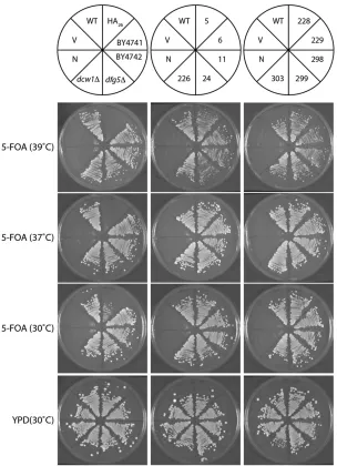
For the 10 unique isolates, functionalDCW1-FLAG plasmids were isolated fromS. cerevisiaeand recovered by transforma-tion intoE. coli(Hoffman 2001).The plasmids were individu-ally retransformed into BY240, and their growth phenotype was confirmed. These 10 clean functional strains were used for subsequent growth analysis and for Western blots. The 10 clean DCW1-FLAG strains, the DCW1-HA strain, and control strains were streaked onto YPD, and complementation was tested again on 5-FOA (at 39°, 37°, and 30°), Ura, SC-His, and YPD plates at 30°. After growth for 2 days, pictures were taken on an Alpha Imager and growth was compared (Figure 4).
Construction of C. glabrata SIR3 Gateway vectors
The C. glabrata SIR3 destination vector was constructed by PCR-amplifying the regionsflanking theSIR3ORF from strain CBS138 (Dujonet al.2004). Primers 3220 and 3221 were used to amplify the 59flanking region; primers 3222 and 3223 were used to amplify the 39 flanking region. These PCR products were subcloned into a Gateway backbone on either side of accdBcassette, using the restriction sites present in the prim-ers. This Gateway backbone contains replication origins and and an AmpRcassette for selection inE. coli, as well as a CEN/ ARS and NatR marker for maintenance and selection in
C. glabrata. The SIR3 entry vector was created by PCR-amplifying the SIR3 ORF fromC. glabratastrain BG2 (Cormack and Falkow 1999) using primers 4470 and 4484 and subse-quently introducing the ORF into the Gateway backbone pDONR201 using a Gateway BP reaction (Life Technologies). We note that there are three polymorphisms in theSIR3ORF, relative to the CBS138 sequence, but these do not affect func-tion, as a wild-typeSIR3expression vector generated from this ORF allele (pRZ47) complements asir3Ddefect inC. glabrata. Schematic drawings of the SIR3 entry vector and destination vector are shown inFigure S2.
Microscopy of Sir3-GFP strains
Eight C. glabratastrains carrying various Sir3-GFP alleles, as well as a negative control strain carrying an untagged Sir3 vector, were grown to stationary phase in liquid YPD+Nat. Cells were washed in PBS and resuspended in PBS, and 5ml of the cells was mounted on a slide. Images were taken using a Zeiss Axioskop microscope with a 1003objective. The cap-tured images are automatically displayed with optimized brightness and contrast settings in Image J; these maximum and minimum values for the entire captured image are listed in Figure 8. Image J software was used to adjust the constrast to the same settings for all images, thereby allowing more direct comparison of GFP brightness across all images. The adjusted contrast images were converted to 8-bit images, cropped, and resized using Adobe Photoshop and Adobe Illustrator.
S. cerevisiae protein preparation and Westerns
S. cerevisiaestrains carrying variousDCW1-FLAG plasmids, or a wild-typeDCW1untagged plasmid (negative control), were grown in SD-His media to mid-log phase (OD600 between 0.1325 and 0.2325). Cells were pelleted by centrifugation and stored at280°. Cell pellets were weighed on a microscale; these weights were used later to normalize loading between strains. Lysates were prepared similar to the method of Frieman and Cormack (2004).To prepare lysates, resuspend cell pel-lets in 50 mM Tris (pH 8.0) supplemented with protease inhib-itors (Roche, 04693132001) and lyse with glass beads using a FastPrep (Bio101 Thermo Savant) (three times: 45 sec, 6.5 speed, ice 1 min between beatings). Supernatants were trans-ferred to clean tubes, and beads were washed in 400 ml Tris (pH 8.0) + protease inhibitors and combined with an earlier fraction (800ml lysate total). Lysates were clarified by centri-fugation at 13,0003g, 10 min, 4°in a microfuge. Superna-tant was saved as the “cytoplasmic”fraction. The pellet was resuspended in 1 ml 50 mM Tris (pH 8.0) + 2% SDS and boiled for 20 min, vortexing to mix every 10 min. After cen-trifugation (13,000 3 g, 10 min, 4°), the supernatant was saved as the“plasma membrane”fraction. The remaining pellet material was resuspended, washed in Tris+SDS, boiled, and centrifuged (as above) three additional times; supernatants were discarded. The pellet was washed four times (twice in 1 ml, twice in 500 ml) in 50 mM Tris + protease inhibitors and spun as before. The pellet was washed once in 500 ml 33 mM potassium phosphate and then resuspended in 100ml 33 mM potassium phosphate + 60 mMb-mercaptoethanol; this is the“cell-wall”fraction.
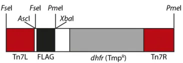
Material from each fraction was loaded onto an SDS-PAGE gel; volumes were normalized across strains using pellet weights. Proteins were transferred to Immobilon-P membrane and immunoblotted using antibodies as indicated. Mouse monoclonala-FLAG (F1804, Sigma) was used at 1:2000 di-luted in TBS+3% milk; a-Pgk1 (A6457, Molecular Probes) was used at 1:1000 in TBS+3% milk. Both of these were used in conjunction witha-mouse HRP-linked (Cell Signaling) sec-ondary antibody at a 1:2000 dilution. Thea-Dcw1 polyclonal antibody was raised against the peptide VELDLDNYESLQ, rep-resenting amino acids 22–33 in the Dcw1protein (Covance, Princeton, NJ). ForDcw1detection, thea-Dcw1 primary an-tibody was diluted at 1:1000 in TBS+3% milk, and the sec-ondary antibody was a 1:5000 dilution ofa-rabbit HRP-linked antibody (Cell Signaling). Amersham’s ECL kit (RPN2132) was Figure 1 Schematic of the Tn7-FLAG construct. Areas indicated by red are Tn7 left and right ends. The 33FLAG epitope tag is indicated by black.
The gray area is thedhfrgene, which confers resistance to trimethoprim
(TmpR). Restriction sites of interest are indicated above the schematic.
used for chemiluminescent detection; thea-FLAG anda-Pgk1 blots were exposed tofilm for 5–10 min; thea-Dcw1 blots were exposed for 45 min.
C. glabrata protein preparation and Westerns
C. glabrata strains carrying different Sir3-GFP plasmids or a wild-type SIR3 untagged plasmid (negative control) were grown in YPD+Nat media to log phase (OD600between 0.5 and 0.635). Cells were pelleted by centrifugation and stored at280°. Lysates were prepared in a urea lysis buffer (Ubersaxet al.2003), supplemented with protease inhibitors (Roche, 11836170001) . Proteins were separated on a 3– 8% Tris–acetate SDS-PAGE gel (Nupage) and transferred to an Immobilon-P PVDF membrane. A polyclonal a-GFP primary antibody (Abcam, ab290) was used at 1:5000 dilu-tion in PBST+5% milk; the a-rabbit HRP-linked secondary antibody (GE Healthcare, NA934V) was used at a 1:5000 di-lution. Chemiluminescent detection was performed using the Amersham ECL kit (RPN2132).
Viability selection and sequence analysis of functional tagged SIR3 alleles in C. glabrata
SIR3was mutagenized four separate times, using the Tn7-bio, Tn7-myc, Tn7-mEOS2, or Tn7-GFP donor vectors. After process-ing the mutagenized plasmid pools to remove leftover Tn7 sequence, the final bio, myc, mEOS2, and Sir3-GFP pools were transformed into C. glabratastrain CGM293, and transformants were selected by growth on YPD+Nat plates for 2 days at 30°. Strain CGM293 carriesURA3integrated into a subtelomeric location, where it is subjected to Sir3-dependent transcriptional silencing. Only cells that carry a functional tagged version of Sir3 have intact subtelomeric silencing, thus silencing the URA3 gene at the telomere and permitting growth on plates containing 5-FOA. To select functional clones of Sir3, the YPD+Nat plates were replica-plated onto 5-FOA plates and grown for 2 days at 30°. For the Sir3-myc trans-formation the pool was outgrown in liquid YPD+Nat prior to plating. This pool showed skewed representation of particular insertion sites. Subsequently, for the other three pools, the transformations were plated directly onto YPD+Nat plates, and these pools show a more even representation of different functional alleles. As with theDCW1screening, we performed colony PCR to qualitatively determine where the tag had inserted into SIR3. Judging from these PCR product sizes, plasmids carrying SIR3 with a range of insertion sites were sent for sequencing to determine the exact position of the tag withinSIR3(Table S6andTable S7).
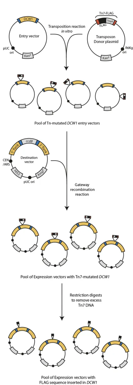
Figure 2 Overview of mutagenesis workflow. Anin vitromutagenesis reaction introduces the Tn7-FLAG transposon randomly throughout the
target entry vector (here,DCW1). MutagenizedDCW1plasmids are
se-lected on the basis of TmpRand KanR. To isolate only those versions that
have a Tn7-FLAG inserted inDCW1, and not in the backbone, a Gateway
LR reaction is used to mobilize theDCW1gene into aDCW1destination
vector. After transformation into MegaXE. colicells and selection with
Tmp and Car, only expression vectors that have a mutagenizedDCW1
gene should remain. Restriction digests are used to remove the bulk of
the transposon: FseI removes the Tn7L end; PmeI removes the TmpR
cassette and Tn7R. After this series of digestions, the pool will consist
ofDCW1expression vectors with a 33FLAG epitope inserted randomly,
in either direction, in theDCW1gene.
Results
Transposon design
The Tn7-FLAG transposon (Figure 1) is based off the miniTn7 design (Bieryet al.2000), using truncated Tn7 left and right (Tn7L and Tn7R) ends. The Tn7L region is 206 bp long, whereas the Tn7R region is 71 bp long. Previous research has shown the Tn7 ends can accommodatePmeI sites and still mobilize during in vitro transposition reactions (Bieryet al. 2000). Here, we engineeredFseI andPmeI sites at the distal Tn7 transposon ends to facilitate removal of the bulk of the transposon sequence using restriction digests after the muta-genesis occurs (see later description). These restriction enzymes have 8-bp restriction sites, which occur rarely in the DNA genome sequence. The Tn7-FLAG construct contains two FseI sites, which flank the Tn7L end; two PmeI sites flank a trimethoprim resistance (TmpR) cassette and the Tn7R end.
PmFseI digestion, therefore, removes the left end of the trans-poson, while PmeI digestion removes the right portion of the transposon. The transposon contains a 33FLAG epitope tag, located between the internalFseI andPmeI sites. The entire Tn7-FLAG construct is carried on the plasmid pRZ101, which serves as a Tn7 donor plasmid. The pRZ101 backbone includes a kanamycin resistance marker (KanR) and replicates using a R6KgORI. This ORI functions only inE. colicells containing the P protein, encoded by the pir gene (Kolter et al. 1978; Haldimannet al.1996; Metcalfet al.1996).
During Tn7 mutagenesis, the Tn7 transposase, containing TnsA and TnsB subunits, creates double-strand breaks at the Tn7 ends, releasing the Tn7-FLAG cassette from pRZ101. TnsCA225Vmediates insertion of the Tn7-FLAG cassette into a va-riety of targets with essentially no sequence specificity (Green et al.2012). During host-mediated repair of the insertion prod-uct, a 5-bp duplication is introduced at the insertion site. The epitope tag is positioned within Tn7-FLAG so that it will be translated correctly in only one reading frame after subcloning to excise transposon sequences. All other reading frames contain STOP codons. Thus,five of six transposon mutants are expected to be nonfunctional as a consequence of the epitope tag being
out of frame and generating an internal stop codon. After all mutagenesis and processing of the transposon mutants, the Tn7-FLAG cassette introduces 159 bp into the target (including the 5-bp duplication), resulting in a 53-amino-acid insertion internally fused into the target protein.
Tn7-FLAG can be easily adapted to introduce other epitope tags. UniqueAscI andXbaI sitesflank the FLAG cassette, and these sites can be used to change the epitope tag to any other tag of interest. In our own work, we generated 6His, Tn7-myc, Tn7-GFP, Tn7-mEOS2, Tn7-biotin, and Tn7-33HA donor plasmids, all of which have been confirmed to mobilize during in vitrotransposition reactions (not shown).
Methodology overview
Thein vitromutagenesis method to introduce internal epitope tags is outlined schematically in Figure 2. To illustrate the utility of this system, we chose to internally tag the protein Dcw1, a GPI-anchored protein, which cannot be functionally tagged at either the N- or C-terminus. The method consists of three separable steps, all of which are carried outin vitro, with re-covery of reaction products by transformation intoE. coli. First, transposition is used to generate a pool of Tn7 insertions throughout the plasmid carrying the gene to be tagged. Second, Tn7 insertions within the ORF are isolated by a Gateway-mediated recombination step. Third, this pool of Tn7-mutated ORFs is treated with restriction enzymes to remove essentially all transposon sequences, leaving just the introduced protein tag.
Transposition: Using a target plasmid containing theDCW1 ORF cloned into a Gateway entry vector, we performed an in vitromutagenesis reaction with recombinant Tn7 transposase proteins. The reaction mobilizes the Tn7-FLAG cassette from the Tn7 donor plasmid into the DCW1target plasmid. This creates a pool of mutagenizedDCW1entry vectors. This pool is transformed into highly competent E. coli DH10 cells and selected for both TmpRand KanRin liquid culture. The use of DH10 cells selects against the Tn7-FLAG donor plasmid, as this plasmid’s R6KgORI will not replicate in DH10 cells. Dou-ble drug selection selects against unmutagenizedDCW1entry Table 1 Sizes of mutagenizedDCW1pools
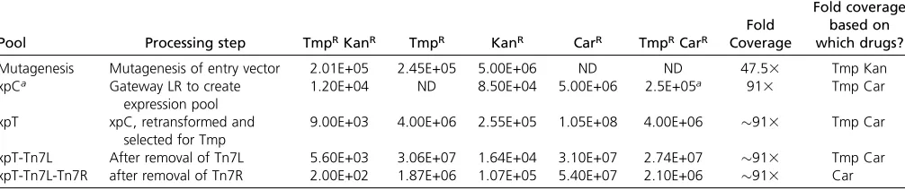
No. of colonies (calculated to full reaction size)
Pool Processing step TmpRKanR TmpR KanR CarR TmpRCarR
Fold Coverage
Fold coverage based on which drugs?
Mutagenesis Mutagenesis of entry vector 2.01E+05 2.45E+05 5.00E+06 ND ND 47.53 Tmp Kan
xpCa Gateway LR to create
expression pool
1.20E+04 ND 8.50E+04 5.00E+06 2.5E+05a 913 Tmp Car
xpT xpC, retransformed and
selected for Tmp
9.00E+03 4.00E+06 2.55E+05 1.05E+08 4.00E+06 913 Tmp Car
xpT-Tn7L After removal of Tn7L 5.60E+03 3.06E+07 1.64E+04 3.10E+07 2.74E+07 913 Tmp Car
xpT-Tn7L-Tn7R after removal of Tn7R 2.00E+02 1.87E+06 1.07E+05 5.40E+07 2.10E+06 913 Car
Colony counts, as plated after recovery during transformation, prior to drug selection in pool. Cells were diluted appropriately to prevent a lawn of growth and plated onto media as indicated. Numbers in the table are calculated to represent the number of colonies in the full transformation resistant to the given drug. ND, not determined. axpC colony counts were revised; seeFile S3andTable S5. Fold coverage was calculated by comparing the number of colonies from the indicated drug selection to the size of
the DNA available for mutagenesis (File S2).
vectors. Thus, after growth overnight in Tmp and Kan, only cells carrying mutagenizedDCW1entry vectors should survive.
Isolation of mutagenized ORFs by Gateway recombina-tion: The pool consists of plasmids that have a transposon inserted in theDCW1ORF as well as plasmids where the trans-poson is inserted in the plasmid backbone (and which have an unmutagenized ORF). To isolate only those ORFs that have been insertionally mutagenized, we used Gateway recombina-tion to mobilize the DCW1 ORF from the entry vector into aDCW1destination vector (Figure 2) to generate what we refer to as the expression pool. The DCW1 destination vector con-tainsDCW1promoter and terminator regions,flanking a ccdB cassette; it also contains replication origins and markers for propagation and selection in both E. coli and yeast. This DCW1 expression pool was recovered by transformation into E. coliDH10 cells. Expression pool plasmids, with the transposon-containingDCW1ORF cassette, confer resistance to both Tmp and Car. We found that this double selection was maximally effective if we performed the drug selections sequentially (data not shown); accordingly, the transformed cells were initially se-lected only for carbenicillin resistance (CarR), which selects for all destination vectors carrying a mutagenized or unmutagenized DCW1ORF. Entry vectors and destination vectors that did not recombine are selected against based on the CarR andccdB counterselectable cassette, respectively. After selecting for CarR, plasmid DNA is recovered from the expression vector pool and transformed again intoE. coliDH10 cells, this time selecting for TmpR. After growth in Tmp, only cells carryingDCW1 expres-sion vectors with a Tn7-FLAG cassette remain. Note that the Tn7-FLAG may be inserted in any reading frame at this point, as the pool of mutants has not been screened for function in yeast yet.
Removal of transposon sequences: Removal of the bulk of the transposon, leaving only the epitope tag, requires two restriction digests and ligation steps. These cloning steps are performed on the pooled DNA, allowing for easy processing of the entire insertion library. Digestion withFseI releases the Tn7L end from each plasmid, which is then recircularized by ligation. This“–Tn7L”pool of plasmid DNA is transformed intoE. coli, and the liquid culture is treated with Tmp and Kan simulta-neously. This second round of Tmp selection is possible since the Tn7R end anddhfrgene remain in the construct and selects against any residual unmutagenizedDCW1expression vectors that have escaped the previous selection step. Next, aPmeI di-gest is used to remove thedhfr(TmpR) gene and the Tn7R end (Figure 1). After intramolecular ligation, the plasmid pool is transformed intoE. coliand selected for CarR. The DNA isolated from this pool contains mutagenizedDCW1expression vectors with the 59-aa FLAG epitope inserted in any of the six reading frames.
Monitoring pool sizes and complexity
Transposition efficiency was monitored by plating on various selective media. Transformation of our mutagenesis reaction
resulted in 5 3106KanRcolonies, representing all plasmids whether mutagenized or not. Of these, 23105(4%) contained transposon insertions (TmpRKanR) (Table 1). This represents a 47-fold coverage of all possible insertion sites for the plasmid (File S2).
The efficiency of Gateway LR mobilization, as well as the fraction of ORFs carrying the Tn7-flag, was estimated by scoring for Car and Tmp resistance (File S3). We determined that the initial expression pool (xpC) contained 2.5 3 105 TmpRCarRindependent recombinants (Table 1). Thus, the ma-jority of the pool complexity is maintained in the expression pool. For subsequent steps, FseI digestion andPmeI digestion, we recovered at least 4 3106transformants, again ensuring that pool complexity was maintained through different cloning steps.
Complexity of the pool was qualitatively assessed using restriction digests of pool DNA at each step of the processing (File S4). Additionally, the plasmids from a subset of the final mutagenized expression pool were isolated and se-quenced with primer 6244 to assess the distribution of Tn7 insertion sites in the plasmid. From 48 sequenced iso-lates, we found 29 had FLAG inserted in the reverse orien-tation (representing 18 unique sites) and 19 had FLAG inserted in the forward direction (representing 18 unique sites). For two nucleotide positions, we isolated FLAG inser-tions in both the“forward”and the“reverse”orientations in different isolates.
Selection for functional clones in yeast
The final DCW1-FLAG pool was transformed in batch intoS.
cerevisiae to screen for functional clones. Mutations in DCW1 and the related geneDFG5are synthetically lethal inS. cerevisiae (Kitagakiet al. 2002). We use the strain BY240, which is adcw1D dfg5D strain carrying a wild-type copy ofDCW1on
aURA3-marked plasmid (pCU-URA3). The plasmid backbone
of theDCW1-FLAG expression pool contains a yeast CEN/ARS sequence and aHIS3marker for selection. After transforming
theDCW1-FLAG pool into BY240, we selected against the
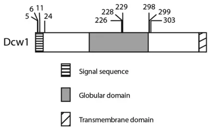
orig-inal wild-type pCU-URA3 plasmid by growth in the presence of Figure 3 Location of FLAG insertions in functionalDCW1-FLAG alleles. Domain structure of Dcw1 is based on predictions from ELM. The
loca-tions of FLAG inserloca-tions within the functionalDCW1-FLAG alleles used in
subsequent experiments are indicated by hash marks above the diagram. Dcw1 is 499 amino acid residues in length.
5- 5-FOA. Only strains transformed with a functional DCW1-FLAG allele will grow on 5-FOA plates. We performed the 5-FOA selection at 37°; the elevated temperature is a mild cell-wall stress and ensures that any cells that grow have a fully functionalDCW1-FLAG allele. We screened 100,000 trans-formants, of which 5% had functional versions of DCW1-FLAG (data not shown).
Transformants werefirst screened for presence of a FLAG insertion using whole-cell PCR (data not shown). Products had a range of sizes, representing FLAG insertions across the DCW1 ORF. A subset of these was chosen for further char-acterization. Plasmids carrying functional DCW1-FLAG alleles were isolated from individualS. cerevisiaetransformants and recovered in E. coli. The location of the FLAG insertion within the DCW1 ORF was determined by sequencing. A number of unique insertion sites were identified, as illus-trated on the schematic shown in Figure 3. The majority of insertions were, as expected, in the ORF. In addition, several isolates were identified in which the FLAG was inserted just
upstream or downstream of the DCW1ORF in the sequence between the Gateway recombination sites and the ORF in the expression vectors (data not shown). We found that the Gate-way attL1 and attL2 recognition sequences, required for mobili-zation of theDCW1ORF into the destination vector, are partially permissive such that certain Tn7-disrupted attL1 and attL2 sequences still, surprisingly, function in the Gateway reaction. These few insertions, therefore, have the epitope outside of the ORF sequence and represent contamination in the overall library.
Unique DCW1-FLAG plasmids were retransformed into S. cerevisiae strain BY240, and function was confirmed by testing for growth on 5-FOA plates (Figure 4). All internally taggedDCW1-FLAG isolates tested grew robustly on 5-FOA at temperatures up to 39°, confirming that theDCW1-FLAG alleles are fully functional. The DCW1-HA26allele showed reduced growth on 5-FOA at 30°and no growth at 37°and 39°, indicating that it is a partially functional hypomorphic allele.
Figure 4 SelectDCW1-FLAG isolates rescue synthetic lethal-ity ofdcw1Ddfg5Dmutants. Each yeast strain was grown on 5-FOA at three temperatures to test for Dcw1 function. Strains were also grown on YPD to verify overall viability. The posi-tions of each strain are shown in the schematic at the top. Three control strains, labeled here as N, C, and WT, were streaked onto each experimental plate. Strain“N”is a nega-tive control: the parental strain BY240, adcw1Ddfg5D
pCU-DCW1 strain derived from BY4742. Strain“V”is another
negative control: BY240 transformed with an empty (His) vec-tor, pRZ159. The “WT” strain is a positive control: strain
BY240 transformed with a wild-type DCW1(His) plasmid.
BY4741 and BY4742 areDCW1 DFG1 ura3Dstrains used
to derive other strains. dfg5D and dcw1D are deletion
mutants derived from BY4741 from the yeast knockout col-lection (Winzeleret al.1999). The strain BY240 transformed
with a DCW1-HA26(His) plasmid is in the position labeled
“HA26.”The remaining strains are BY240 transformed with variousDCW1-FLAG (His) plasmids and labeled to indicate the amino acid position of the FLAG insertion.
Detecting FLAG by Western blot
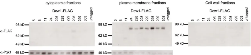
To verify that the FLAG epitope introduced into Dcw1 is detectable by Western analysis, we monitored Dcw1-FLAG protein levels in our strains carrying functionalDCW1-FLAG clones. Cytoplasm, plasma membrane, and cell-wall fractions were isolated from log-phase cultures of each strain.Dcw1is a membrane-bound GPI protein (Kitagaki et al. 2002), and abundant FLAG signal is easily detectable in the cell membrane fraction in 8 of the 10 strains expressing Dcw1-FLAG alleles, consistent with the fact that these are fully functional Dcw1-FLAG alleles (Figure 5). UntaggedDcw1, which would not be visible on thea-FLAG immunoblot, is predicted to have a mo-lecular weight of 49.5 kDa. Dcw1-FLAG fusion proteins are expected to have a molecular weight of 55 kDa. However, ma-ture Dcw1 is glycosylated, and previous studies have shown that Dcw1-HA has an apparent molecular weight of 80 kDa (Kitagakiet al.2002), similar to what we observe in Figure 5. For three alleles, the FLAG insertion (at residue 5, 6, or 11) is predicted to disrupt the signal sequence (Figure 3). For two of these three, Dcw1-FLAG5and Dcw1-FLAG6, the immunoblot showed no signal, and for one, Dcw1-FLAG11, the protein was detectable in the membrane fraction, but its molecular weight is lower than expected (60 kDa, instead of80 kDa). To confirm that the localization observed of the Dcw1-FLAG fusion proteins represents true localization of untaggedDcw1, we also performed immunoblots using these same cellular frac-tions with ana-Dcw1 antibody raised against a peptide portion of native Dcw1(Figure S1). The signal is much fainter than what we observed using a-FLAG antibodies; importantly, the Dcw1-FLAG fusion proteins show similar size distributions using both the anti-Dcw1 and anti-FLAG antibodies.
Mutagenesis of a second target gene, C. glabrata SIR3
The demonstrate the generality of the tagging methodology, we used the same approach to mutagenize theSIR3gene of Candida glabrata, which is required for silencing of sub-telomeric regions of theC. glabratagenome (De Las Penaset al.2003). Schematic diagrams of the Sir3 Gateway constructs are shown in Figure S2. We used four different Tn7 derivatives (Tn7-myc, Tn7-GFP, Tn7-mEOS2, Tn7-biotin) to generate
li-braries of Tn7 insertions with between 3 and 63105 inde-pendent insertions in each pool. Functional tagged alleles were identified by complementation of a sir3D strain. We sequenced the insertion sites for fully functional alleles, iden-tifying tag insertion sites distributed across the ORF (Figure 6,
Table S6, and Table S7). We chose to further characterize
eight GFP-taggedSIR3alleles, which complement for the telo-meric silencing defect (Figure S3), representing insertions sites distributed across the ORF. By Western analysis, all eight were expressed at approximately similar levels (Figure 7). For all eight alleles, Sir3-GFP was localized in puncta, as has been reported previously for wild-type Sir3 protein in S. cerevisiae (Cockellet al.1995) (Figure 8).
Discussion
Here we describe a robust system for creating a library of internally tagged target protein constructs. This mutagenesis is carried outin vitro, and all subsequent steps of selecting for insertional mutants and removing excess Tn7 sequence are carried out on the bulk pool in vitro. This batch processing greatly reduces the effort in maintaining and screening the thousands of possible mutants.
As proof of principle, we mutagenized a target gene,DCW1, to saturation, introducing a 33FLAG epitope throughout the ORF. Screening in yeast identified fully functional tagged DCW1-FLAG alleles, of which we chose 10 for further analysis. All 10DCW1-FLAG alleles that we created proved to have wild-type levels of function when assayed for growth at elevated temperatures.
Monitoring the distribution of Dcw1 in fractionated cell lysates by Western blot illustrated that the available FLAG anti-bodies were more sensitive than antianti-bodies directly raised against a peptide in Dcw1. The majority of alleles had a sub-cellular distribution similar to the wild-type protein—primarily in the membrane fraction, consistent with its proposed function (Kitagaki et al. 2002). Two alleles, Dcw1-FLAG5 and Dcw1-FLAG6, were not detectable in any cell fraction in thea-FLAG Western blots. We suspect that the FLAG tag, located in the signal sequence in these two alleles, may have been cleaved off Figure 5 Western blot analysis of Dcw1-FLAG expression inS. cerevisiae. (Top) Anti-FLAG immunoblots ofS. cerevisiaestrains expressing Dcw1-FLAG alleles, representing the cytoplasmic, plasma membrane, and cell-wall fractions from each strain. Material from 7.53105cells is loaded for each strain in each fraction. Wells are labeled with the amino acid site of the FLAG tag within Dcw1. (Bottom) Anti-Pgk1 immunoblot of the cytoplasmic and plasma
membrane fractions from strains carrying Dcw1-FLAG alleles. Cytoplasmic material from 3.753105cells is loaded for each strain. Membrane material
from 3.53105cells was loaded for each strain
during normal protein trafficking. In the case of Dcw1-FLAG11, the protein was detected in Western blots, but appeared smaller than the expected size (Figure 5). This apparent size difference could be due to truncation or complete post-translational glycosylation. We note that in-troduction of internal epitopes may still alter protein function. However, if multiple functional tagged alleles have the same phenotype and show the same subcellular distribu-tion—as we observed for 7 of 10 alleles—this increases con-fidence that they reflect wild-type function.
We also mutagenized the C. glabrata SIR3 gene and iso-lated 126 functional, unique internally tagged alleles (four different tags). By Western analysis, GFP internally tagged proteins were expressed at similar levels, and all showed a punctate distribution similar to that documented for Sir3 inS. cerevisiae. Since the tranposon insertion is random and generates many nonfunctional insertions, the utility of the method depends on a robust screen for functional alleles. This can be an assay for viability (as forDCW1). For Sir3, the assay for function was not viability but growth in the presence of 5-FOA as a measure of transcriptional silencing of a subtelomeric URA3gene. ForDCW1andSIR3,5% of insertions were func-tional, suggesting that, for some proteins at least, functional alleles could also be identified by manual screening of a rela-tively small number of insertions.
For DCW1, the functional alleles had FLAG insertions
clustered in a few regions of the gene, including in the central globular glycosyl hydrolase domain (identified by protein BLAST and ELM motif finding) (Kitagaki et al. 2002). ForSIR3as well, functional insertions for all four tags were found across the ORF, including insertions in N- and C-terminal predicted globular domains. It would have been difficult to predict that these regions would tolerate insertions
solely based on bioinformatic modeling of the protein struc-ture. Unlike other epitope-tagging systems (Khmelinskiiet al. 2011; Ramsdenet al.2011), this Tn7-based system requires no prior knowledge of permissive sites to introduce tags in the POI. The efficientin vitromutagenesis and cloning steps allow researchers to create and recover an unbiased set of tagged versions of a target gene, which can then be screened for function.
The system of Tn7-based epitope tagging that we describe should be broadly useful. We have described the application to epitope tagging of an essential yeast gene. However, several aspects of the methodology make it suitable to tagging of proteins in virtually any system. First, once Gateway vectors are constructed, mutagenesis, cloning, and screen-ing (in yeast) takes 2–3 weeks total. Second, the mutagenesis is carried outin vitro, and Tn7 has minimal insertion sequence bias. Third, the insertions in the ORF are isolated without regard to function of those insertions. Fourth, the library of tagged proteins is generated infinal form inE. coli, obviating the need to remove transposon sequences in the cell type where functional screening is done. Rather, thefinal library can simply be introduced into the appropriate cell type and screened directly.
Since a major use of transposon epitope-tagging methods is to generate fully functional tagged versions of the POI, it is imperative that the screening be done only on mutagenized ORFs since any untagged wild-type alleles in the pool would pass the functional screen. In the highly useful Tn5-based system, TAGIT, the selection for tagged alleles is by presence of the marker gene carried in the transposon. This allows introduction of the mutagenized library into the cell where screening is carried out; at this stage the transposon sequences are removed by expression in the cell of Cre recombinase. Our Figure 6 Distribution of functional tagged alleles ofC. glabrata
SIR3. (Top) Linear representation of theC. glabrataSir3
pro-tein (1088 residues). The gray areas are predicted globular domains (by ELM), which have been found to have roles in associating with histones (N-terminal domain) and Sir3 di-merization (C-terminal domain). In the schematics below, a black line is located at the position for each unique in-sertion site identified from among the functional Sir3-bio, Sir3-myc, Sir3-mEOS2, and Sir3-GFP pools. (Bottom) Sche-matic overlays all sites at which a functional tagged Sir3 was
identified. The color of the line indicates the number of
mutagenesis pools in which the particular insertion site was identified; light blue indicates that the site was
identi-fied in only one pool; black indicates that it was found in all four mutagenesis pools.
method provides an alternative to this approach. Insertions in the ORF are efficiently isolated from nonmutagenized ORFs by Gateway-mediated recombination. This step is highly efficient and can be carried out in batches, permitting construction of a library of tagged ORFs comprising tens or hundreds of thousands of independent insertion events. Subsequent efficient restriction digestions of the pool to remove the left and right ends of the transposon result in a complex pool of tagged ORFs in an appropriate expression vector. This library can then be directly screened for function as dictated by function of the POI, without the need to express recombinase enzymes in the target cell. As a caveat, we note that we recovered a small number of functional expression plasmids where the epitope had inserted between the Gateway recombination sites and the ORF (data not shown); it appears that the attL1 and attL2 sites are somewhat permissive for insertions. This leaves the target ORF unmu-tagenized, and our current plasmid architecture makes it impossible to eliminate these extraneous plasmids from the pool.
An advantage of this Tn7-based method over other transposon systems is the very limited amount of transposon-derived sequence left in the insertion site. In our method, the FseI andPmeI sites are engineered to be present within three bases of the transposon end, and following excision, there are only 22 nucleotides (essentially theFseI andPmeI sites) in addition to the epitope tag present at the insertion site. This increases, we would argue, the likelihood of identifying functional epitope insertions since it limits the amount of extraneous sequence shared by all insertions to a few amino acids.
Finally, the method in principle could be used for analysis of proteins in many different organisms. The mutagenesis of the cloned gene is done in vitro, and mobilization into the expression vector exploits Gateway recombination. This can be adapted most easily to any system for which Gate-way-modified expression vectors exist. We suggest that the method will be useful in a range of systems, including model eukaryotic systems like S. cerevisiaeas well as mammalian systems.
Acknowledgments
We thank Matthew Frieman for creating the dcw1Ddfg5D pCU-DCW1 strain BY240. We thank Irene Castaño for strain CGM293, Yuxia Ren for her assistance cloning the Tn7 donor vectors, and Jie Xiao for the gift of pET28-ftsZ-mEOS. R.E.Z. is supported by an American Cancer Society Postdoctoral Fellowship (PF-09-194-01-MPC). This work was supported by National Institutes of Health grants R01A1046223 (to B.P.C.) and RO1GM076425 (to N.L.C.). N.L.C. is an Investigator of the Howard Hughes Medical Institute.
Figure 7 Western blot analysis of Sir3-GFP inC. glabrata. An
anti-GFP immunoblot of C. glabrata strains carrying different Sir3-GFP
alleles. Wells are labeled with the amino acid site of the GFP insertion
within Sir3;“x”indicates an empty lane. The antibody cross-reacted
withC. glabratawhole-cell lysates, as shown by the banding see in
the untagged strain. The band representing Sir3-GFP is marked with an arrow.
Figure 8 Imaging of live C. glabratastrains carrying various Sir3-GFP alleles. Eight strains carrying different versions of Sir3-GFP, as well as an untagged Sir3 control strain, were grown to stationary phase in liquid culture. Live cells were imaged using differential interference contrast
bright-field andfluorescence microscopy. GFPfluorescence is shown with
both the contrast settings as captured (automated contrast) and adjusted so each strain has the same contrast levels (adjusted contrast). The max-imum and minmax-imum contrast settings for each strain are listed on the automated contrast panel. The adjusted contrast panels all have a minimum
of 202 and a maximum of 727. Bar, 5mm.
Literature Cited
Arnau, J., C. Lauritzen, G. E. Petersen, and J. Pedersen, 2006 Current strategies for the use of affinity tags and tag removal for the purification of recombinant proteins. Protein Expr. Purif. 48: 1–13.
Bell, M. R., M. J. Engleka, A. Malik, and J. E. Strickler, 2013 To fuse or not to fuse: What is your purpose? Protein Sci. 22: 1466–1477. Biery, M. C., F. J. Stewart, A. E. Stellwagen, E. A. Raleigh, and N. L. Craig, 2000 A simple in vitro Tn7-based transposition system with low target site selectivity for genome and gene analysis. Nucleic Acids Res. 28: 1067–1077.
Castano, I., R. Kaur, S. Pan, R. Cregg, L. Penas Ade et al., 2003 Tn7-based genome-wide random insertional mutagene-sis of Candida glabrata. Genome Res. 13: 905–915.
Choi, K. Y., Y. Li, R. Sarnovsky, and N. L. Craig, 2013 Direct in-teraction between the TnsA and TnsB subunits controls the het-eromeric Tn7 transposase. Proc. Natl. Acad. Sci. USA 110: E2038–E2045.
Cockell, M., F. Palladino, T. Laroche, G. Kyrion, C. Liu et al., 1995 The carboxy termini of Sir4 and Rap1 affect Sir3 local-ization: evidence for a multicomponent complex required for yeast telomeric silencing. J. Cell Biol. 129: 909–924.
Cormack, B. P., and S. Falkow, 1999 Efficient homologous and illegitimate recombination in the opportunistic yeast pathogen Candida glabrata. Genetics 151: 979–987.
De Las Penas, A., S. J. Pan, I. Castano, J. Alder, R. Cregget al., 2003 Virulence-related surface glycoproteins in the yeast pathogen Candida glabrata are encoded in subtelomeric clusters and subject to RAP1- and SIR-dependent transcriptional silenc-ing. Genes Dev. 17: 2245–2258.
Devine, S. E., and J. D. Boeke, 1994 Efficient integration of arti-ficial transposons into plasmid targets in vitro: a useful tool for DNA mapping, sequencing and genetic analysis. Nucleic Acids Res. 22: 3765–3772.
Dujon, B., D. Sherman, G. Fischer, P. Durrens, S. Casaregolaet al., 2004 Genome evolution in yeasts. Nature 430: 35–44. Frieman, M. B., and B. P. Cormack, 2004 Multiple sequence
sig-nals determine the distribution of glycosylphosphatidylinositol proteins between the plasma membrane and cell wall in Sac-charomyces cerevisiae. Microbiology 150: 3105–3114. Gamas, P., and N. L. Craig, 1992 Purification and characterization
of TnsC, a Tn7 transposition protein that binds ATP and DNA. Nucleic Acids Res. 20: 2525–2532.
Gonzalez, M., N. Goddard, C. Hicks, R. Ovalle, J. M. Rauceoet al., 2010 A screen for deficiencies in GPI-anchorage of wall glyco-proteins in yeast. Yeast 27: 583–596.
Green, B., C. Bouchier, C. Fairhead, N. L. Craig, and B. P. Cormack, 2012 Insertion site preference of Mu, Tn5, and Tn7 transpo-sons. Mob. DNA 3: 3.
Gregory, J. A., E. C. Becker, J. Jung, I. Tuwatananurak, and K. Pogliano, 2010 Transposon assisted gene insertion technology (TAGIT): a tool for generatingfluorescent fusion proteins. PLoS ONE 5: e8731. Haldimann, A., M. K. Prahalad, S. L. Fisher, S. K. Kim, C. T. Walsh et al., 1996 Altered recognition mutants of the response regu-lator PhoB: a new genetic strategy for studying protein-protein interactions. Proc. Natl. Acad. Sci. USA 93: 14361–14366. Hill, J., K. A. Donald, and D. E. Griffiths, 1991 DMSO-enhanced
whole cell yeast transformation. Nucleic Acids Res. 19: 5791. Hoffman, C. S., 2001 Preparation of yeast DNA. Curr. Protoc. Mol.
Biol. Chapter 13: Unit13 (13.11-1-13.11.4).
Khmelinskii, A., M. Meurer, N. Duishoev, N. Delhomme, and M. Knop, 2011 Seamless gene tagging by endonuclease-driven homologous recombination. PLoS ONE 6: e23794.
Kitagaki, H., H. Wu, H. Shimoi, and K. Ito, 2002 Two homologous genes, DCW1 (YKL046c) and DFG5, are essential for cell growth and encode glycosylphosphatidylinositol (GPI)-anchored
mem-brane proteins required for cell wall biogenesis in Saccharomy-ces cerevisiae. Mol. Microbiol. 46: 1011–1022.
Kitagaki, H., K. Ito, and H. Shimoi, 2004 A temperature-sensitive dcw1 mutant of Saccharomyces cerevisiae is cell cycle arrested with small buds which have aberrant cell walls. Eukaryot. Cell 3: 1297–1306.
Kolter, R., M. Inuzuka, and D. R. Helinski, 1978 Trans-complementation-dependent replication of a low molecular weight origin fragment from plasmid R6K. Cell 15: 1199–1208.
Kumar, A., S. Agarwal, J. A. Heyman, S. Matson, M. Heidtmanet al., 2002 Subcellular localization of the yeast proteome. Genes Dev. 16: 707–719.
Kumar, A., M. Seringhaus, M. C. Biery, R. J. Sarnovsky, L. Umanskyet al., 2004 Large-scale mutagenesis of the yeast genome using a Tn7-derived multipurpose transposon. Genome Res. 14: 1975–1986. Maddi, A., C. Fu, and S. J. Free, 2012 The Neurospora crassa dfg5 and
dcw1 genes encode alpha-1,6-mannanases that function in the in-corporation of glycoproteins into the cell wall. PLoS ONE 7: e38872. Manoil, C., and B. Traxler, 2000 Insertion of in-frame sequence
tags into proteins using transposons. Methods 20: 55–61. Merkulov, G. V., and J. D. Boeke, 1998 Libraries of greenfl
uores-cent protein fusions generated by transposition in vitro. Gene 222: 213–222.
Metcalf, W. W., W. Jiang, L. L. Daniels, S. K. Kim, A. Haldimann et al., 1996 Conditionally replicative and conjugative plasmids carrying lacZ alpha for cloning, mutagenesis, and allele replace-ment in bacteria. Plasmid 35: 1–13.
Mumberg, D., R. Muller, and M. Funk, 1995 Yeast vectors for the controlled expression of heterologous proteins in different ge-netic backgrounds. Gene 156: 119–122.
Osawa, M., and H. P. Erickson, 2005 Probing the domain struc-ture of FtsZ by random truncation and insertion of GFP. Micro-biology 151: 4033–4043.
Penfold, R. J., and J. M. Pemberton, 1992 An improved suicide vector for construction of chromosomal insertion mutations in bacteria. Gene 118: 145–146.
Ramsden, R., L. Arms, T. N. Davis, and E. G. Muller, 2011 An intein with genetically selectable markers provides a new approach to internally label proteins with GFP. BMC Biotechnol. 11: 71. Ross-Macdonald, P., P. S. Coelho, T. Roemer, S. Agarwal, A. Kumar
et al., 1999 Large-scale analysis of the yeast genome by trans-poson tagging and gene disruption. Nature 402: 413–418. Seringhaus, M., A. Kumar, J. Hartigan, M. Snyder, and M. Gerstein,
2006 Genomic analysis of insertion behavior and target spec-ificity of mini-Tn7 and Tn3 transposons in Saccharomyces cer-evisiae. Nucleic Acids Res. 34: e57.
Sheridan, D. L., C. H. Berlot, A. Robert, F. M. Inglis, K. B. Jakobsdottir et al., 2002 A new way to rapidly create functional,fluorescent fusion proteins: random insertion of GFP with an in vitro trans-position reaction. BMC Neurosci. 3: 7.
Spreghini, E., D. A. Davis, R. Subaran, M. Kim, and A. P. Mitchell, 2003 Roles of Candida albicans Dfg5p and Dcw1p cell surface proteins in growth and hypha formation. Eukaryot. Cell 2: 746–755. Ubersax, J. A., E. L. Woodbury, P. N. Quang, M. Paraz, J. D. Blethrow et al., 2003 Targets of the cyclin-dependent kinase Cdk1. Nature 425: 859–864.
Winzeler, E. A., D. D. Shoemaker, A. Astromoff, H. Liang, K. Anderson et al., 1999 Functional characterization of the S. cerevisiae ge-nome by gene deletion and parallel analysis. Science 285: 901–906. Young, C. L., Z. T. Britton, and A. S. Robinson, 2012 Recombinant protein expression and purification: a comprehensive review of affinity tags and microbial applications. Biotechnol. J. 7: 620–634. Zordan, R. E., Y. Ren, S. J. Pan, G. Rotondo, A. De Las Penaset al., 2013 Expression plasmids for use in Candida glabrata. G3 (Bethesda) 3: 1675–1686.
Communicating editor: O. Cohen-Fix
GENETICS
Supporting Information
http://www.genetics.org/lookup/suppl/doi:10.1534/genetics.114.169482/-/DC1
Avoiding the Ends: Internal Epitope Tagging of
Proteins Using Transposon Tn7
Rebecca E. Zordan, Brian J. Beliveau, Jonathan A. Trow, Nancy L. Craig, and Brendan P. Cormack
2 SI R. E. Zordan et al.
SUPPLEMENTAL INFORMATION for “Transposon based method for internal epitope tagging” by Zordan et al
Figure S1. Western blot analysis of Dcw1‐FLAG strains with ‐DCW1 antibody
The same cell fractions used in Figure 5 were run on new SDS‐PAGE gels and probed with an ‐Dcw1 antibody. Signal was detected using chemiluminescence, and film was exposed for 45 minutes. Material from 7.5x10⁵ cells is loaded in each strain in each fraction. Wells are labeled with the amino acid site of the FLAG tag within Dcw1. We note the apparent size of Dcw1 in the plasma membrane is larger than what we observed with ‐FLAG antibodies, but the overall pattern is consistent with the ‐ FLAG Westerns shown in Figure 5.
R. E. Zordan et al. 3 SI
Figure S2. Gateway constructs used for mutagenesis of C. glabrata SIR3
These schematic drawings represent the Gateway entry vector and destination vector used during the mutagenesis of C.
4 SI R. E. Zordan et al.
Figure S3. Sir3‐GFP alleles complement for growth on 5‐FOA
R. E. Zordan et al. 5 SI
Table S1. Plasmids used in this study
Plasmid Description E. coli marker Yeast marker Tn7 donor plasmids
pRZ49 Tn7‐mEOS2 donor vector Kan n/a
pRZ98 Tn7‐biotin‐donor vector Kan n/a
pRZ99 Tn7‐6His donor vector Kan n/a
pRZ101 Tn7‐FLAG donor vector Kan n/a
pRZ102 Tn7‐HA donor vector Kan n/a
pRZ103 Tn7‐myc donor vector Kan n/a
pRZ106 Tn7‐GFP donor vector Kan n/a
DCW1 plasmids
‐ pCU‐DCW1 Amp URA3
‐ DCW1 entry vector Kan n/a pRZ159 empty DCW1 destination vector Amp HIS1
pRZ160
wild type DCW1 expression
vector Amp HIS1
pRZ165 Dcw1‐FLAG5 Amp HIS1
pRZ166 Dcw1‐FLAG6 Amp HIS1
pRZ172 Dcw1‐FLAG11 Amp HIS1
pRZ173 Dcw1‐FLAG24 Amp HIS1
pRZ174 Dcw1‐FLAG226 Amp HIS1
pRZ175 Dcw1‐FLAG228 Amp HIS1
pRZ167 Dcw1‐FLAG229 Amp HIS1
pRZ168 Dcw1‐FLAG298 Amp HIS1
pRZ176 Dcw1‐FLAG299 Amp HIS1
pRZ177 Dcw1‐FLAG303 Amp HIS1
pBC715 Dcw1‐HA26 Amp HIS1
SIR3 plasmids
6 SI R. E. Zordan et al.
Table S2. Primers used in this study
Oligo # Name Sequence (5'‐3') Tn7‐tag donor vector construction
3201 Tn7L‐FseI‐Bam‐for aggactacggatcctgtggccggccAATAAAGTCTTAAACTGAACAAA 3202 Tn7L‐AscI‐rev gacctgacggcgcgccGTCGACCCCACGCCCCTCTTTAAT 4724 mEOS_for
aggcgcgccggccggccTGGATCCGCTGGCTCCGCTGCTGGTTCTGGC GAATTCATGAGT
4725 mEOS_rev
gctctagagtttaaacTAAATTCTCCAGATCCTGCAGCAGATCCTGCAGAGCCTCGTCT GGCATTGTCAGGCAATCCAGAATGAG
5300 L1‐GFP‐f
atttaggatccgctggctccgctgctggttctggcATGTCTAAAGGTGAAGAATTATTCACTGG TG
5301 L2‐GFP‐r
ataagtttaaactagctcctcctgcagcagatcctgcagagccTTTGTACAATTCATCCATACCAT GGGTAATAC
DCW1‐Yiplac211 knockout construct
1975 DCW1 3'flank rev SphI acatgcatgcAGGAAACCATGTAAGCGATGAATAT 1976 DCW1 3’flank for KpnI ggggtaccTGCAGAACTTATGAAAGCTTAACATTT 1977 DCW1 5'flank rev KpnI ggggtaccTTTTATGTGTTCGTTTTTAAAACAGAC 1978 DCW1 5'flank for HindIII ccccaagcttAGATGAACTTGAACTTAAGATGATC Amplify across DCW1 knockout region
2306 DCW1 5' check TCGTTTAAATTCAATTGGAACTGTA 2307 DCW1 3' check TTCAAACAAAATTCGTTCGATATTA Verify DCW1‐Yiplac211 integrants
1504 Yiplac backbone TATGTTGTGTGGAATTGTGAGCGG 1505 URA check GCGATTAAGTTGGGTAACGCCAGG Verify loopout of DCW1‐Yiplac211 construct
1778 DCW1 5' check ACCTTTCCAGGACATATAAT 1779 DCW1 3' check ACACATATGAACAAAGGTCT pCU‐DCW1 construction
1629 3ecoYKL046 ccggaattcTCAAAAGACTAACCACAGAGCACATG 1630 5bamYKL046 cgcgggatccATGCTAGTAAATAAAGTGATAGGGT DCW1 destination vector
6333 DCW1 promoter ‐ for atagagctcTTCTTCTCCTTATTGTGCTTTACC 6334 DCW1 promoter ‐ rev attctagaTTTTATGTGTTCGTTTTTAAAACAGACTG Determine position of FLAG insertion.
2766 DCW1 promoter ‐ for GATGATCATAGGTACTCTTTGTATAATGGGC 6244 linker 2 ‐ rev ATTAGTTTAAACTAGCTCCTCCTGCA 5160 linker 1 ‐ for CTCCGCTGCTGGTTCTGG
4032 M13F (‐21) GTAAAACGACGGCCAGT
5626 Tn7L ‐ rev GATCTATTTTGTTCAGTTTAAGACTTTATTG Sir3 entry vector construction
R. E. Zordan et al. 7 SI
Sir3 destination vector construction
3220 Sir3_IP_SacI_F gtacctatgagctcGAACGGTGCCAGACACACCAGCCC 3221 Sir3_IP_XbaI_R tgaccatatctagaCCTCTTACTTAATCCGAAACCTTC
3222 Sir3_UTR_XhoI_F caatgcacactcgagAAAAGCTTTCATCTTCTTTTCTTGATTCTCCTC 3223 Sir3_UTR_KpnI_R catgaccatggtaccAAGACGGCTCCATCACTAAAGTGC
Determine position of epitope insertions within SIR3 using colony PCR and sequencing 5160 L1 for CTCCGCTGCTGGTTCTGG
5161 L2 rev CTCCTCCTGCAGCAGATCCT
5602 SIR3IP_3’for CTGGGAAGGTTTCGGATTAAGTAAGAGG
5605 SIR3utr_5’rev GTATTAGTAGAGGAGAATCAAGAAAAGAAGATGAAAG 5627 Linker2+myc GATCCTGCAGAGCCTTCATTGAG
5738 Bio_L2‐rev GATCCTGCAGAGCCTTCATGCC 5739 GFP 5’ rev AGGTCAATTTACCGTAAGTAGCATCAC 5740 GFP 3’ for TTATCCACTCAATCTGCCTTATCCA 5741 mEOS 5’ rev CGAATACCCTGTTGCCGTAATGGA 5742 mEOS 3’ for ACCGATGTGACTTCAGAACTACTTACAAAG
8 SI R. E. Zordan et al.
Table S3. S. cerevisiae strains used in this study
Strain Genotype Parent Source
BY4741 MATa his3Δ1 leu2Δ0 met15Δ0 ura3Δ0 [Brachmann, 1998 #76] BY4742 MAT his3Δ1 leu2Δ0 lys2Δ0 ura3Δ0 [Brachmann,
1998 #76] dfg5Δ MATa his3Δ1 leu2Δ0 met15Δ0 ura3Δ0 dfg5::KanR BY4741 [Winzeler, 1999 #79] dcw1Δ MATa his3Δ1 leu2Δ0 met15Δ0 ura3Δ0 dcw1(YKL046C)::KanR BY4741 [Winzeler, 1999 #79] BY240 MAT his3Δ1 leu2Δ0 lys2Δ0 ura3Δ0 dfg5::g418 dcw1Δ pCU‐DCW1 BY4742 this work BY965 MAT his3Δ1 leu2Δ0 lys2Δ0 ura3Δ0 dfg5::g418 dcw1Δ pCU‐DCW1 pRZ159 BY240 this work BY966 MAT his3Δ1 leu2Δ0 lys2Δ0 ura3Δ0 dfg5::g418 dcw1Δ pRZ160 BY240 this work BY893 MAT his3Δ1 leu2Δ0 lys2Δ0 ura3Δ0 dfg5::g418 dcw1Δ pRZ165 BY240 this work BY895 MAT his3Δ1 leu2Δ0 lys2Δ0 ura3Δ0 dfg5::g418 dcw1Δ pRZ166 BY240 this work BY974 MAT his3Δ1 leu2Δ0 lys2Δ0 ura3Δ0 dfg5::g418 dcw1Δ pRZ172 BY240 this work BY975 MAT his3Δ1 leu2Δ0 lys2Δ0 ura3Δ0 dfg5::g418 dcw1Δ pRZ173 BY240 this work BY976 MAT his3Δ1 leu2Δ0 lys2Δ0 ura3Δ0 dfg5::g418 dcw1Δ pRZ174 BY240 this work BY977 MAT his3Δ1 leu2Δ0 lys2Δ0 ura3Δ0 dfg5::g418 dcw1Δ pRZ175 BY240 this work BY897 MAT his3Δ1 leu2Δ0 lys2Δ0 ura3Δ0 dfg5::g418 dcw1Δ pRZ167 BY240 this work BY899 MAT his3Δ1 leu2Δ0 lys2Δ0 ura3Δ0 dfg5::g418 dcw1Δ pRZ168 BY240 this work BY978 MAT his3Δ1 leu2Δ0 lys2Δ0 ura3Δ0 dfg5::g418 dcw1Δ pRZ176 BY240 this work BY979 MAT his3Δ1 leu2Δ0 lys2Δ0 ura3Δ0 dfg5::g418 dcw1Δ pRZ177 BY240 this work