HIGHLIGHTED ARTICLE GENETICS OF IMMUNITY
Domain Speci
fi
city of MAP3K Family Members, MLK
and Tak1, for JNK Signaling in
Drosophila
Beth Stronach,1Ashley L. Lennox,2and Rebecca A. Garlena
Department of Microbiology and Molecular Genetics, University of Pittsburgh School of Medicine, Pittsburgh, Pennsylvania 15219
ABSTRACTA highly diverse set of protein kinases functions as early responders in the mitogen- and stress-activated protein kinase (MAPK/SAPK) signaling pathways. For instance, humans possess 14 MAPK kinase kinases (MAP3Ks) that activate Jun kinase (JNK) signaling downstream. A major challenge is to decipher the selective and redundant functions of these upstream MAP3Ks. Taking advantage of the relative simplicity ofDrosophila melanogasteras a model system, we assessed MAP3K signaling specificity in several JNK-dependent processes during development and stress response. Our approach was to generate molecular chimeras between two MAP3K family members, the mixed lineage kinase, Slpr, and the TGF-bactivated kinase, Tak1, which share 32% amino acid identity across the kinase domain but otherwise differ in sequence and domain structure, and then test the contributions of various domains for protein localization, complementation of mutants, and activation of signaling. We found that overexpression of the wild-type kinases stimulated JNK signaling in alternate contexts, so cells were capable of responding to both MAP3Ks, but with distinct out-comes. Relative to wild-type, the catalytic domain swaps compensated weakly or not at all, despite having a shared substrate, the JNK kinase Hep. Tak1 C-terminal domain-containing constructs were inhibitory in Tak1 signaling contexts, including tumor necrosis factor-dependent cell death and innate immune signaling; however, depressing antimicrobial gene expression did not necessarily cause phenotypic susceptibility to infection. These same constructs were neutral in the context of Slpr-dependent developmental signaling, reflecting differential subcellular protein localization and by inference, point of activation. Altogether, ourfindings suggest that the selective deployment of a particular MAP3K can be attributed in part to its inherent sequence differences, cellular localization, and binding partner availability.
P
ROTEIN kinases are common transducers of information within cells. Indeed, reversible phosphorylation of sub-strates, by the opposing activities of kinases and phospha-tases, is a major currency in cells forming the basis for information relay in many signaling pathways, ultimately transforming cell behavior in response to a changing envi-ronment. Unregulated kinase activity, however, has been implicated in numerous diseases of medical concern, notably cancer. One family in particular, the mitogen-activated pro-tein kinases (MAPKs), composed of ERK, p38, and JNK en-zymes, are central to a vast array of cellular and pathologicalprocesses (Chang and Karin 2001; Johnson and Nakamura 2007; Wagner and Nebreda 2009; Keshet and Seger 2010; Sabapathy 2012). Converging on the activation of MAPKs are typically two additional levels of kinases within a hier-archical three-tiered core, namely the MAPK kinases or MAP2Ks, and their activators, the MAPK kinase kinases, or MAP3Ks. While MAPK enzymes have been extensively stud-ied at biochemical, structural, and physiological levels, the MAP3Ks are less well understood, more diverse, and greater in number. For example, in mammals there exist at least 20 different MAP3K family members, 14 of which impinge downstream upon three JNK stress-activated protein kinases (SAPKs) (Cuevaset al.2007; Johnson and Nakamura 2007; Craig et al. 2008). From an evolutionary standpoint, the diversity of MAP3Ks may allow cells to respond to a greater breadth of stimuli or with greater sensitivity to discrete sig-nals. Emerging evidence suggests that MAP3Ks can work selectively or cooperatively downstream of different signals to tune a MAPK network response (Chenet al.2002; Cronan
et al.2012). The selective function of MAP3Ks can presumably Copyright © 2014 by the Genetics Society of America
doi: 10.1534/genetics.113.160937
Manuscript received August 21, 2013; accepted for publication January 10, 2014; published Early Online January 14, 2014.
Supporting information is available online athttp://www.genetics.org/lookup/suppl/ doi:10.1534/genetics.113.160937/-/DC1USA.
1Corresponding author: Department of Microbiology and Molecular Genetics, University of Pittsburgh School of Medicine, 450 Technology Dr., Ste 517 BSP2, Pittsburgh, PA 15219. E-mail: stronach@pitt.edu
be harnessed to provide specific alternative therapeutic tar-gets for MAPK pathway-associated disease intervention. On the other hand, if MAP3Ks act cooperatively to fine tune a response, then targeting individual members could result in minimal efficacy. Thus, elucidation of the context-dependent functions and mechanisms of signaling specificity among MAP3K proteins is the focus of current research.
Context-dependent influences, like environmental, cellu-lar, developmental, or spatial influences, are pervasive in tuning signaling networks. As such, a major challenge is to understand the molecular mechanisms by which context imparts distinct properties to a system. Recent work has provided some mechanistic insight. For example, within a single cell, related kinases might avoid inappropriate crosstalk by deploying nonoverlapping substrates or by compartmentalization of their function in cellular space or time (Alexander et al. 2011). Considering the conserved three-tier kinase organization within the MAPK pathways, the core pathway may incorporate distinct upstream trans-ducers, as is the case with the diversity of MAP3K proteins, to shift the outcome of signaling in response to distinct stim-uli. Two general approaches to the challenge of identifying context-dependent influences on signaling have been ap-plied:first, to alter the context of a constant set of compo-nents, for example, by adding a stimulatory ligand, and second, to change a system component while keeping the context constant. The latter experiment can be useful to test redundancy and specificity among related proteins. If one component is swapped for another within the same context and a different outcome is observed, there must be intrinsic differences in the components. To determine how individual MAP3Ks confer specificity in their responsesin vivo, we have focused on two members of the tyrosine kinase-like (TKL) group (Manning et al. 2002) in the Drosophilamodel sys-tem, mixed lineage kinase (MLK) encoded by theslprgene and transforming growth factor-bactivated kinase (Tak1).
Among the MAP3Ks that stimulate JNK activation, the mixed lineage kinase group consisting of the MLKs, the dual leucine zipper kinases (DLKs), and zipper sterile alpha kinase (ZAK), is the largest, related by sequence homology within the kinase domain and the presence of leucine zipper (LZ) dimerization motifs (Gallo and Johnson 2002). MLK family members mediate MAPK-dependent responses to cyto-kines, ceramide, fatty acids, and other stresses (Sathyanarayana
et al.2002; Jaeschke and Davis 2007; Korchnaket al.2009; Kantet al.2011). Consequently, they are implicated in met-abolic and neurodegenerative diseases, epithelial migration and healing, and tumor growth and metastasis, reflecting their broad tissue distribution in epithelia and the nervous system (Silva et al. 2005; Jaeschke and Davis 2007; Chen
et al.2010; Velhoet al.2010; Cronanet al.2012; Starket al.
2012; Zhan et al. 2012). Their roles in development have been more difficult to ascertain, as single and double gene knockouts in mice are viable (Brancho et al. 2005; Bisson
et al.2008). MLK proteins are distinguished by an N-terminal SH3 domain, followed by the kinase, LZ, and CRIB domains
mediating catalysis, dimerization, and Rac or Cdc42 GTPase binding, respectively (Gallo and Johnson 2002). These func-tional domains are followed by a long C-terminal region lacking notable domains but enriched in phosphorylation motifs thought to modulate protein function and/or locali-zation (Vacratsiset al.2002). Multistep activation of MLKs by upstream signals involves GTPase binding, relief of auto-inhibition, dimerization, and phosphorylation by MAP4K proteins (Bocket al.2000; Vacratsis and Gallo 2000; Zhang and Gallo 2001; Du et al.2005; Garlena et al. 2010; Kant
et al.2011).
More distantly related and lacking overt LZ motifs, Tak1 is a pivotal activator of NF-kB and MAPK signaling in infl am-matory, immune, and stress responses (Cuevas et al. 2007, 2008; Sakurai 2012). Tak1 also participates in noncanonical (Smad independent) TGF-bsignaling, reflecting its moniker (Yamaguchi et al. 1995). Conditional and complete Tak1 knockouts in mice provide evidence for essential roles in em-bryonic development and differentiation of immune cells, skin, and vasculature (Shimet al.2005; Jadrichet al.2006; Omoriet al.2006). Tak1 signals as part of a protein complex with the partners Tab1 and Tab2/3, which interact with the N-terminal kinase domain and C-terminal regulatory domain of Tak1, respectively (Shibuya et al. 1996; Takaesu et al.
2000; Besse et al. 2007). Growing evidence suggests that an important component of Tak1 activation involves the bind-ing of K63-linked polyubiquitin chains by Tab2/3, leadbind-ing to Tak1 autophosphorylation and kinase activity (Wang et al.
2001; Kanayamaet al.2004; Xiaet al.2009).
Our previous work has focused on MAP3K family members inDrosophila, which is intermediate in complexity between single cell and vertebrate systems with respect to genetic redundancy and cellular diversity. Inflies, there are eight recognizable homologs to the 14 mammalian proteins implicated in stimulating JNK activity. Of these, Mekk1, Pk92B/Ask1, Tak1, Slpr/MLK, and Wnd/DLK have defi ni-tive roles in JNK signaling (Igaki et al. 2002; Kuranaga
et al. 2002; Stronach and Perrimon 2002; Collins et al.
2006; Ryabinina et al. 2006; Kang et al. 2012). Genetic and cell culture experiments have demonstrated both unique and overlapping functions for some of them, but the intrinsic properties of the individual family members that confer par-ticular responses to distinct signals are still poorly charac-terized. Here, we address this question using chimeric constructs. Protein chimeras have been used widely, in cel-lular andin vitroassays, to discern the specific contributions of related domains in various types of proteins (e.g., (Walker
et al.1995; Sanchez-Hernandezet al.2012; Anisimovet al.
2013). Given that there are processes uniquely dependent on Slpr, such as embryonic epidermal dorsal closure, and on Tak1, such as innate immune response, the separation of functions provides a platform upon which to study the spe-cific contributions to signaling for the two different proteins (Mihaly et al. 2001; Silverman et al. 2003; Polaski et al.
MKK7 (Hollandet al.1997; Sathyanarayanaet al.2003), we sought to test directly if the catalytic kinase domain is func-tionally equivalent and if integration into an alternate con-text, by sequences outside the kinase domain, is sufficient to alter signaling specificity.
Materials and Methods
Transgenic constructs
All UAS constructs generated for this study were made in the pUASpvector (Rorth 1998) and transgenic lines were estab-lished after injection of DNA by Genetic Services (Sudbury, MA). All transgenic proteins generated herein were tagged at the C terminus with two copies of the HA epitope tag. Using site-directed mutagenesis by PCR overlap extension (Hoet al.
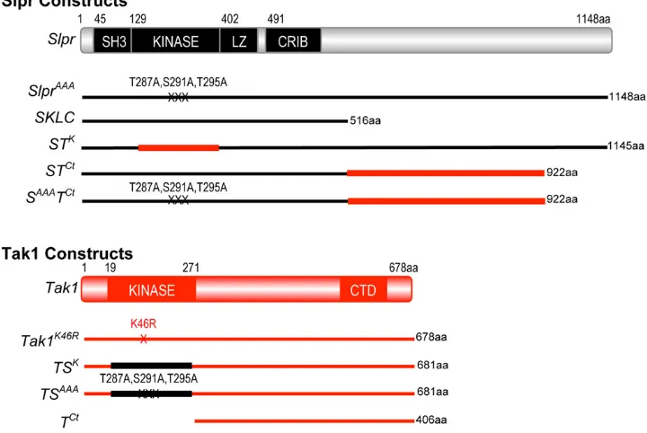
1989), the following chimeric constructs were created: the Slpr-Tak kinase swap, STK, consists of Slpr aa 1–128, Tak1 aa 19–271, and Slpr aa 383–1148, in that order. The Slpr-Tak C-terminus swaps, STCtand SAAATCt, consist of Slpr aa 1–516, with either a wild-type kinase domain or with activation loop alanine mutations, respectively (Garlena et al. 2010), fol-lowed directly by Tak1 aa 272–678. The alternate Tak-Slpr kinase swap chimeras, TSKand TSAAA, consist of Tak1 aa 1– 18, Slpr wild-type or triple alanine mutant kinase domain aa 128–385, followed directly by Tak1 aa 272–678. Finally, we also generated the Tak1 C terminus alone, TCt encoding aa 272–678, with the 59 UTR and starting methionine codon from the wild-type Tak1 transcript upstream. All constructs were verified by DNA sequencing.
Fly strains
Stocks were maintained at 22°on cornmeal–molasses–agar medium. Crosses were raised at 25°in 50 610% relative humidity unless noted otherwise.w1118was used as a
con-trol. For mutants and transgenics, Bloomington (BL) Stock Center numbers are given if appropriate: UAS-Slpr, UAS-SlprAAA, and UAS-SKLC (Garlena et al. 2010), slprBS06
(Polaski et al. 2006), Tak12 BL# 26272 (Vidal et al.
2001), UAS-Tak1 and UAS-Tak1K46R (Mihaly et al. 2001), egrGS9830 (UAS-eiger) (Igakiet al. 2002), pucE69 (puc-lacZ) (Ring and Martinez Arias 1993), and UAS-srcEGFP BL# 5432. For constructs under the control of UAS sequences, expression was regulated by the Gal4 transcription factor (Brand and Perrimon 1993). arm-Gal4 BL# 1560 (Sanson
et al. 1996) and da-Gal4 BL# 5460 (Wodarz et al. 1995) were used for ubiquitous expression; pnr-Gal4 BL# 3039 (Callejaet al. 1996) was used for expression in the dorsal ectoderm of the embryo, though it directs expression in other cells and tissues throughout development; Yp1-Gal4
(yolk-Gal4) (Vidal et al. 2001) was used for expression in the adult female fat body starting around day 2 after eclosion; r4-Gal4 BL# 33832 (Lee and Park 2004) drives expression in the larval and adult fat body of both sexes; and GMR-Gal4 BL# 1104 was used for expression in the developing eye tissue (Freeman 1996). The genetic rescue
experiment with thegtX11slpr921double mutant chromosome has been described previously (Stronach and Perrimon 2002).
Tissue immunofluorescence, X-gal staining, and immunoblot
Embryos were collected overnight on grapejuice plates, dechorionated, washed, and thenfixed at room temperature for 20 min with equal volumes of 4% formaldehyde in PEM buffer (100 mM Pipes, 2 mM EGTA, 1 mM MgSO4) and hep-tane. After devitellinization in methanol, subsequent washes and processing were done in PBS plus 0.1% Triton X-100. For immunofluorescent staining of fat body, larvae were coarsely dissected andfixed in PBS plus 4% formaldehyde overnight at 4°. Subsequent washes and incubations were in PBS plus 0.1% Tween-20. The following antibodies and dilutions were used: mousea-HA (16B12, Covance) at 1:500–1:1000, rabbit
a-b-galactosidase preadsorbed at 1:1000 (Cappel), mouse
a-fasciclin 3 (7G10, Developmental Studies Hybridoma Bank) at 1:50, and rat a-Tak1 peptide antibody at 1:250 (custom antibody services, GenScript). The immunogenic peptide sequence was 440-SSTNAKSDGRERLT-453. Second-ary antibodies were FITC- or TxRed-conjugates from Jackson ImmunoResearch Laboratories, used at 1:200 or were Alexa Fluor conjugates from Invitrogen/Molecular Probes used at 1:500–1:750. For detection of thepuc-lacZ reporter in adult fat body, 3- to 4-day-old mated females were collected and their abdomens were cut off in cold PBS with fine tissue scissors. Then while grasping the terminalia with a forceps, an incision was made through the cuticle at the dorsal mid-line with scissors. The tissue wasfixed and then stained with X-Gal reagent overnight at 25°according to a published pro-tocol (Romeo and Lemaitre 2008). The stained abdominal tissue was washed,filleted open, and mounted in 70% glyc-erol in PBS. Protein lysates for Western immunoblots were made by homogenizing, in 150ml RIPA buffer, four wander-ing third instar larvae, programmed to express transgenic proteins with the r4-Gal4 driver. An equal volume of lysate was separated by SDS–PAGE and blots were probed with mouse a-HA (16B12, Covance) diluted 1:1000 or mouse
a-GFP (GF28R, Pierce) at 1:1500. Expression was quantified by chemiluminescent imaging using the analysis tools pro-vided with the ProteinSimple FluorChem E system software.
Image capture and processing
Images of adult flies were obtained with NIS-Elements software using a Nikon DS-Fi1 digital camera mounted on a Nikon SMZ1500 stereomicroscope. Fluorescent images of stained embryos and larval tissues were obtained by laser-scanning confocal microscopy using an Olympus FV1000 Fluoview system on an IX81 compound inverted microscope and assembled in Adobe Photoshop. For quantification of puc-lacZ induction in the embryo as a measure of JNK signaling intensity,b-galactosidase-positive nuclei fromfive consecutive segments along the leading edge were marked using the
was obtained by selecting a 1003100 pixel region of interest along the central ventral section of the image in the red chan-nel only and measuring “integrated density”in Adobe Photo-shop. Values from 5–22 specimens were averaged. Graphing and statistical analysis was performed with GraphPad Prism.
Innate immune assays
Crosses betweenTak12;da-Gal4females andw1118/Y;
UAS-transgene males were reared at 22°. Newly eclosed adults were aged 2–4 days at 25°. For infection, adults were pricked once below the wing with a needle dipped in a loose pellet of overnight Escherichia coli DH5acell culture. Flies were then maintained at 29°and monitored daily for viabil-ity. Data from multiple trials with two independent insertion lines were combined, plotted as survival curves, and ana-lyzed using the log-rank test (Mantel–Cox) in GraphPad Prism. A control cross between da-Gal4and UAS-GFP
con-firmed that the Gal4 line directs expression ubiquitously throughout development and we note in particular that GFP is expressed highly in newly eclosed adults. Adults with the genotypes da-Gal4 . UAS-Tak1WT or da-Gal4.
UAS-SlprWTwere not recovered in sufficient quantity to test.
cDNA synthesis and quantitative real-time PCR
Crosses were raised at 25°and 2- to 4-day-old adult mated females (Yp1-Gal4.UAS-transgene) were collected, at which time, half of them were infected as described above. After 6 hr at 29°, 7–10flies were homogenized in 300ml of TRIzol (Invitrogen). RNA was extracted according to the manufac-turer’s recommendations and suspended in 20–25 ml of water. First strand cDNA was synthesized by transcribing 2mg of RNA template using the Maxima Reverse Transcriptase kit (Thermo Scientific) and random primers. In an Applied Bio-systems 7900HT thermal cycler, transcript amplification was monitored with Sybr green dye (Thermo Scientific) using
100 ng input cDNA. The following primer pairs were used: RpL32 (forward) 59-ACCGCAGTACCCACTCAATC-39and (reverse) 59-CAATCTCCTTGCGCTTCTTG-39, Diptericin (for-ward) 59-ACCGCAGTACCCACTCAATC-39 and (reverse) 59 -ACTTTCCAGCTCGGTTCTGA-39. Four biological replicates (consisting of two independent transgenic lines per construct) were collected for each genotype exceptTak1K46R, which had three replicates. Relative gene expression, compared to ano transgene control, was calculated by normalizing to RpL32 expression levels according to the comparative Ct method (Schmittgen and Livak 2008). Infive instances out of 86 data points total (11 genotypes, three or four trials, and two probes), a trial was excluded as an outlier if values exceeded the mean of the remaining values by a factor offive.
Results
Design and construction of MAP3K chimeras
If the primary functions of a kinase catalytic domain are to recognize, bind, and phosphorylate substrate, then two
kinase domains that recognize and phosphorylate the same substrate are predicted to be interchangeable. To test this assertion, we engineered Slpr and Tak1 proteins with kinase domain swaps. For example, we generated a full-length Slpr construct with the kinase domain from Tak1 swapped in to replace the endogenous Slpr kinase domain and vice versa, creatingSTKandTSK, respectively (Figure 1). Given that one of the assays used to monitor a requirement for Tak1 is based on dominant interference of endogenous activity, we also generated a kinase domain swap in Tak1,TSAAA, using a Slpr kinase domain mutated in the activation loop to pre-vent activating phosphorylation. Our previous work demon-strated that this combination of alanine mutations disrupts phosphorylation and renders Slpr nonfunctional due to its inability to activate downstream JNK signaling (Garlena
et al.2010).
The ability of Slpr to localize to the cell cortex in embryonic epithelium is attributed to the C-terminal half of the protein, and though this activity was nonessential in mutant rescue experiments, it contributed to maximal Slpr function (Garlenaet al.2010). The C terminus of the Tak1 protein harbors a putative regulatory domain identifiable by an island of sequence conservation among homologs (Takatsu et al.2000; Mihalyet al.2001). This region may contribute to Tak1 localization or protein interactions with signaling partners, as suggested by cell culture and biochem-ical assays (Takaesu et al. 2000; Zhou et al. 2005; Besse
et al. 2007; Guntermann and Foley 2011). Based on this evidence, we reasoned that sequences encompassing this domain might direct Tak1 to specific signaling complexes for which Slpr is excluded, as a specificity-determining mechanism. To test this idea, we replaced amino acids C terminal to the CRIB domain of Slpr with Tak sequences beginning immediately after the kinase domain (Figure 1), both in the context of a wild-type (STCt) and a nonphosphor-ylatable Slpr kinase domain (SAAATCt). This part of Tak1, lacking the kinase domain, was also expressed on its own (TCt). Using these transgenic reagents, we tested protein localization, function, and specificity in both Slpr-dependent and Tak1-dependent processes during Drosophila develop-ment, cell death, and immunity.
Differential localization of chimeric proteins in two tissue contexts is attributable to C-terminal sequences
All transgenic proteins generated in this study were detect-able by indirect immunofluorescence with antiserum di-rected against the C-terminal HA tag and were therefore expressed as full-length proteins. Wild-type Slpr, SlprAAA, and STK displayed strong enrichment at the cell cortex in embryonic epithelia (Figure 2, A and B and Garlena et al.
characterized construct, SKLC (Garlenaet al.2010), which is truncated directly after the CRIB domain of Slpr, suggesting that the Tak1 C-terminal replacement had a minimal effect on localization beyond the loss of the Slpr C terminus. Neverthe-less, to determine if the cytoplasmic localization of the chi-meras reflected that of the Tak1 C terminus, we assessed the distribution of this portion of Tak1 in isolation. Indeed, the TCtprotein had a similar distribution predominantly in the cy-toplasm, but in addition appeared to localize partially in the nucleus, though it was not enriched there (Figure 2G). To-gether, these results align with our previous studies demon-strating that the C-terminal half of the Slpr protein directs its enrichment at the plasma membrane (Garlenaet al.2010).
Since the C-terminal portion of Tak1 was detected in the cytoplasm and nucleus, we next determined whether this distribution reflected that of the full-length Tak1 protein and Tak/Slpr chimeras. To that end, immunofluorescence was performed using either the anti-HA antiserum to detect the chimeras or an anti-Tak1 antibody to detect the untagged Tak1K46R transgenic protein, a kinase-dead form of Tak1 (Mihaly et al. 2001). In the embryonic epidermis, overex-pressed Tak1K46Rlocalized in the cytoplasm, absent from nu-clei. In addition, we observed some association with the cell cortex, as evidenced by a prominent signal at cell boundaries upon completion of dorsal closure (Figure 2H). We did not attempt to localize overexpressed wild-type Tak1 due to its strong proapoptotic effects and disruption of epithelial integ-rity. Also, we note here that under conditions suitable for de-tection of the transgenic Tak1 protein, appreciable levels of endogenous Tak1 were not observed, though maternal, and later, ubiquitious expression is reported in FlyBase (Drysdale and FlyBase 2008; Graveleyet al.2011). Finally, the distribu-tions of the chimeric transgenes replacing the kinase domain of Tak1 with that of Slpr appeared identical to that of
Tak1K46R, with prominent cytoplasmic staining and occasional cortical localization (Figure 2, E and F). Taken together these localization data suggest that the determinants of subcellular location likely reside outside the kinase domains.
(Figure 3J). All the transgenic proteins were overexpressed relative to their endogenous counterparts based on both immunofluorescence and RT-PCR analysis of transcripts (Supporting Information,Figure S2). Altogether, from these localization studies, we conclude that the cellular distribu-tion of Slpr and Tak1 is distinct and primarily determined by the protein sequences, not the tissue contexts tested here.
Rescue of Slpr-dependent dorsal closure and mutant lethality demonstrates kinase specificity
Among all of theDrosophilaMAP3K proteins, the function of Slpr is selectively required in the activation of JNK signaling to orchestrate morphogenesis of epithelial tissues during embryonic development and adult metamorphosis. This is borne out by genetic analysis ofslprmutants. Zygotic lethal alleles of slprcause a failure of dorsal closure, leaving the embryonic epidermis unclosed, resulting in embryonic death (Stronach and Perrimon 2002; Polaskiet al.2006). Animals mutant for another allele, slprBS06, transition through
em-bryogenesis but emerge as adults with reduced Mendelian
frequency of 5–10% of normal (Polaskiet al.2006). The mutant adults that do eclose variably display defects in mor-phogenesis of the adult thorax, genitalia, and maxillary palps, as well as reduced longevity (Polaski et al. 2006; Gonda et al. 2012). Using slpralleles of different severity, it was possible to test for the ability of the ubiquitously expressed transgenes to rescue Slpr function acutely during embryonic dorsal closure or throughout development, re-storing survival to adulthood. For example, only three trans-genes improved survival over the course of development relative to no transgene expression (Figure 4A). These were SlprWT as expected, SKLC, as shown previously (Garlena
et al.2010), and STCt. Expression of all the other transgenes depressed the frequency of slprBS06 adult recovery to a
greater extent than without transgene expression, effec-tively acting as dominant negative proteins.
A requirement to rescueslprBS06mutants to adulthood is
a stringent criterion for function and only the wild-type Slpr transgene provided significant rescuing function. Thus, to measure functional properties of the expressed transgenes over a shorter developmental time period, we asked whether each protein was capable of rescuing the dorsal closure phenotype of the embryonic lethalslpr921allele
(Fig-ure 4B). Mirroring the previous rescue experiment, we found that SlprWT, SKLC, and STCtprovided substantial res-cuing function compared to no transgene expression, reduc-ing the percentage of embryos with a severe dorsal open (DO) phenotype (solid), while increasing the recovery of embryos with no dorsal closure defects or only head defects (open). Only one additional construct, STK, showed an im-provement in phenotype upon expression, though to a lesser extent than those mentioned. Thus, the N-terminal half of Slpr, namely the SKLC domains, provided nearly full func-tional rescue of embryogenesis and some rescue to adult-hood, implying that the C terminus is nonessential for function under conditions of high level expression. The prence of the Tak C terminus attached to Slpr SKLC was es-sentially neutral in both assays acting similarly to SKLC alone. Interestingly, while the Slpr/Tak kinase swap, STK, provided some function during embryogenesis compared to the control, it did not suffice to functionally compensate for all Slpr functions throughout development (compare A and B in Figure 4). Importantly, the ability to rescue devel-opmental defects in the short or long term was independent of transgene expression level.
Localized and specific kinase sequences are key to optimal JNK signaling during dorsal closure
To delve into the basis for the rescue data, we assessed the effect of transgene expression on the expression ofpuc-lacZ, a molecular reporter for JNK pathway activity used exten-sively inDrosophila.puc-lacZis an enhancer trap allele of the puckeredgene encoding JNK phosphatase, a negative feed-back regulator (Martin-Blanco et al.1998). As benchmarks for comparison,puc-lacZinduction was assessed in embryos expressing wild-type or dominant negativeslprconstructs in Figure 2 Differential localization of transgenic proteins in embryonic
dorsal epidermis maps to the C terminus. (A–G) Anti-HA and (H) anti-Tak1 immunostaining. The indicated constructs were expressed in the embryo with the pnr-Gal4 driver. Images are single confocal slices
the dorsal epidermis usingpnr-Gal4as the driver. As shown in Figure 5, B–Bii and quantified, SlprWTinduced a twofold increase in the number of cells expressing puc-lacZ away from the leading edge of the dorsal epidermis at mid and late stages of dorsal closure compared with control embryos that expresspuc-lacZin one row of dorsalmost cellsflanking the central amnioserosa tissue (Figure 5, A–Aii). In contrast, SlprAAA inhibited JNK-dependent puc-lacZ expression com-pletely (Figure 5, C–Cii). Deleting the C-terminal half of Slpr (SKLC construct) or replacing it with that of Tak1 (STCt construct) resulted in similar rescuing ability but a minimal effect on puc-lacZ expression (Figure 5, E–Eii and Garlena et al. 2010). Notably, if the kinase catalytic domain of Slpr was mutant, however, the presence of the Tak1 C terminus made the SAAATCt protein a less effective inhibitor of puc-lacZ induction than full-length SlprAAA (compare Fii and Cii in Figure 5), presumably due to mis-localization in the cytosol. Expression of Slpr with the Tak1 kinase domain (STK) induced mild ectopicpuc-lacZ expres-sion beyond the dorsalmost cells, demonstrating catalytic
competency, though not to the extent of SlprWT, consistent with the embryonic rescue data (Figure 5, D–Dii). Expres-sion of the Tak1 derivative constructs, including the C ter-minus alone (TCt), kinase dead (Tak1K46R), and the kinase swaps (TSK and TSAAA), were also nearly neutral in this assay, neither inducing nor inhibiting puc-lacZ relative to controls (Figure 5, G–Jii), though they were highly expressed. These data attest to the specificity of Slpr func-tion in the embryonic epidermis and suggest that the Tak1 kinase domain cannot compensate for that of Slpr, nor can the nonkinase domains of Tak1 engage the protein in pro-ductive signaling complexes in those cells under conditions where they are normally responsive to Slpr.
Eiger/tumor necrosis factor-induced cell death engages the Tak1 C terminus
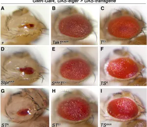
TNF (Igakiet al.2002; Geukinget al.2005). This results in cell death of the developing eye tissue, such that the adult eye is severely reduced in size (Figure 6A). Loss of Tak1 signaling by mutation, RNA interference, or expression of dominant negative constructs, suffices to block Eiger-induced cell death (Igakiet al. 2002; Morenoet al. 2002), restoring adult eye tissue (Figure 6B); and this effect is specific to Tak1 in comparison with Slpr (Polaski et al.
2006). Thus, we turned to this assay to define domains that
are essential for Eiger signaling in this context. Upon cross-ing the experimental transgenic lines to a GMR-Gal4, UAS-eigertester stock, in which high levels ofeigerexpression are induced in the developing larval eye imaginal discs (Igaki
et al. 2002), we observed a striking pattern of results. Ex-pression of the C-terminal region of Tak1 alone (Figure 6C) or in combination with any other sequences (Figure 6, E, F, H, and I) showed strong inhibition of cell death, whether the linked kinase domain was wild type or not. For example, even the Tak/Slpr kinase swap (TSK), wherein the Slpr ki-nase domain is wild type, blocked the cell death phenotype. In contrast, Slpr constructs characterized as dominant neg-ative or the Slpr/Tak kinase swap (STK) failed to interfere with Eiger signaling (Figure 6, D and G). Moreover, expres-sion of these constructs in the absence of Eiger did not phe-nocopy Eiger overexpression (not shown). In fact, none of the forms of Slpr we have expressed inflies are sufficient to dominantly suppress Eiger-induced cell death. Thus, we conclude that the region responsible for integration of Tak1 into the Eiger/TNF signaling network resides down-stream of the kinase domain, in the C-terminal region. Given that Tab2 binds to the C terminus of Tak1 and that Tab2 is required for Eiger-JNK signaling (Takaesuet al.2000; Geuking
et al. 2005; Zhuang et al. 2006), we speculate that excess transgenic protein may sequester Tab2 or other binding partners in unproductive complexes.
Probing Tak1-dependent innate immune response
Tak1 mutants are viable as adults but susceptible to Gram-negative bacterial infection (Vidal et al.2001). This obser-vation along with numerous other studies have defined the so-called immune deficiency (Imd) pathway (Lemaitreet al.
1995), in which Tak1 plays a central role in the induction of antimicrobial and stress defenses through the activation of Relish (Rel)/NFkB- and JNK-dependent transcriptional pro-grams (Georgel et al. 2001; Vidal et al. 2001; Silverman
et al. 2003; Aggarwal and Silverman 2008). To test the specificity of MAP3K signaling in this process, both infection susceptibility and target gene expression were monitored in adults expressing the various transgenic proteins. First, we generated a stock of theTak12allele, encoding an early stop
codon (Vidalet al.2001), in combination with a ubiquitous driver, da-Gal4. It was then possible to cross females from this stock to theUAStransgenic lines. From this cross, male progeny hemizygous mutant for Tak12 were assessed for
rescue of the immune deficiency upon challenge withE. coli. In parallel, female progeny heterozygous for Tak12 were
also challenged to test whether expression of any transgenic constructs dominantly enhanced the heterozygous loss of Tak1 signaling. Results of these experiments are given in Figure 7. In our hands, more than half of theTak1 mutant males died over the course of a week after challenge (Figure 7A). Though we were unable to complement the suscepti-bility by expressing wild-type Tak1 due to early embryonic lethality, none of the transgenic proteins were sufficient to rescue the mutant susceptibility, including TSK. Among the Figure 4 Rescue ofslprmutant viability or dorsal closure demonstrates
kinase specificity. (A) Floating bar plot showing the degree of rescue provided by expression of the indicated transgenes (x-axis), as a ratio of slprBS06mutant to siblingFM7cmaleflies (y-axis). Bars span minimum to
maximum values and horizontal lines indicate the mean ratio for three to six independent trials exceptSlprAAAandSAAATCt, which were each two
trials, testing a minimum of two different transgenic insertions per geno-type. In the absence of a UASconstruct (no Tg), the eclosion ratio is
0.05. The total number (N) of males counted is shown below each bar. Expression of HA-tagged SlprWT provides a significant degree of
Figure 5 Specificity of Slprvs.Tak1 signaling in activation of JNK target gene expression during dorsal closure. Early and late progression of dorsal closure (stage 13–14, left; stage 15, right) is shown in merged panels (A–J) and in individual channels, with immunostaining for either Fas3 (Ai–Ji) or b-gal to detectpuc-lacZenhancer trap expression (Aii–Jii). Transgenes indicated in the lower left of each panel (A–J) are expressed in the dorsal ectoderm and amnioserosa under the control ofpnr-Gal4. Embryos are shown dorsally with anterior to the left. Bar, 20mm. Quantification ofpuc-lacZin stage 15 embryos as a proxy for JNK pathway activity is given in the rightmost panels as the mean number ofb-gal positive nuclei perfive hemisegments
experiments with females (Figure 7B), the heterozygotes were normal, demonstrating that Tak1 is not haploinsuffi -cient, but the homozygous individuals were susceptible as expected. Intriguingly, expression of only two transgenic constructs showed any significant perturbation of the im-mune response in the heterozygous background. One was
Tak1K46R, a dominant negative form of Tak1. Although this result was anticipated (Vidalet al.2001), its expression did not fully recapitulate the homozygous mutant phenotype. The other transgene that depressed the immune response in females similar to the dominant negative construct was
SAAATCt. Given that the mutant kinase domain of Slpr in the context of the full-length Slpr protein (SlprAAA) did not show an effect, this result seems to point to the juxtaposition of the mutant kinase with the Tak1 C terminus, which
de-fined a different spatial context for the chimera according to the localization results (Figure 2 and Figure 3). However, TSAAAexpression also had no effect. The only sequence dif-ference between the constructs, SAAATCt and TSAAA, is the N-terminal nonkinase domains of Slpr, including the SH3, LZ, and CRIB domains, which in combination with an in-active kinase domain, might disrupt some important step in the activation of the pathway by the remaining endogenous Tak1 protein. We also note that expression of the Tak1 C terminus alone with da-Gal4 or a fat body-specific Gal4 driver, r4-Gal4, did not inhibit the immune response, con-trasting with the context of Eiger-dependent cell death.
A second approach to assess the effects of Slpr and Tak1 in the immune signaling pathways involved monitoring induction of Rel and JNK pathway target genes. It has been demonstrated that ectopic expression of Tak1 or an up-stream activator, imd, can dominantly induce antimicrobial
peptide (AMP) expression even in the absence of challenge (Georgelet al.2001; Vidal et al.2001), though expression levels are below that induced by bacterial infection. Based on this evidence, we assessed induction of a Rel target AMP encoded by Diptericin (Dpt), using quantitative real-time PCR upon expression of the wild-type or chimeric constructs in the adult fat body withYp1-Gal4as a driver (Figure 8 and
Figure S1). We observed significant induction of basal Dpt levels upon expression of wild-type Tak1, with an average eightfold increase compared to no transgene (Figure 8, A and B). In contrast, expression of the other transgenes failed to induce ectopic Dpt expression under basal conditions (Figure 8B). To determine instead whether the transgenic proteins specifically potentiated or interfered with Tak1-dependent signaling under induced conditions, the experiment was also performed after immune challenge with E. coli. Pairwise comparisons of the individual transgenic linesfirst revealed that onlyTak1WTand theno transgenecontrol sam-ples significantly activated Dpt expression upon challenge (Figure 8A). Among the challenged samples, kinase-dead Tak1 significantly inhibited Dpt upregulation as expected, along with the other Tak1 C-terminal domain-bearing trans-genics (STCt, SAAATCt, TSK, TSAAA, and TCt) (Figure 8A) similar to their effects on Eiger signaling. Although Dpt induction was also reduced by expression of SlprWT and
though induced Dpt expression was dampened in flies expressing many of these transgenes, there was not a strict correlation with overall susceptibility to immune challenge as shown in Figure 7 or with relative expression levels of the constructs (Figure 3 andFigure S2), thus the full response to expression of the chimeras undoubtedly involves regulation of additional genes or pathways.
With respect to the JNK signaling axis, rather than measuring small and transient changes in puckered tran-script expression at the population level with real-time PCR, we chose to monitor induction of thepuc-lacZreporter construct in individual females, again using Yp1-Gal4 as a tissue-specific driver (Figure S1). Unlike Dpt, however, pairwise comparisons of individual lines revealed no signif-icant stimulation of JNK activity after bacterial challenge, including those flies expressing no transgene (Figure 9, A and Ai). Regardless of infection, though, we observed that the wild-type forms of Tak1 and Slpr induced robust JNK reporter expression in the fat body (Figure 9, A and B), whereasTak1K46R-expressingflies resembled those with no transgene in having the lowest puc-lacZ expression. The other trasngenes spurred intermediate reporter expression. Notably, SlprWTwas the only transgene to activatepuc-lacZ
in the oenocytes, an early component of the Yp1-Gal4 ex-pression pattern, as well as fat body (Figure 9B andFigure S1). Also,flies with ectopic Tak1 expression were noticeably unhealthy and showed altered organization and loss of fat body tissue over the course of a few days (Figures 9Bi and
Figure S3) consistent with other observations on the detri-mental consequences of wild-type Tak1 overexpression. Thus, for this experiment, the chimeras with domain swaps were determined to be nonequivalent to the parental wild-type forms in their ability to ectopically activate JNK signal-ing, whereas dominant negative Tak1 was the most effective inhibitor ofpuc-lacZ expression.
Discussion
Biological responses to developmental, immune, and cell death signals, are mediated in part by the activation of JNK signaling through numerous upstream MAP3K and MAP2K transducers. Genetic analyses in model organisms and biochemical studies in cultured cells have revealed that different JNK-dependent responses require selective use of various MAP3K proteins (Chenet al.2002; Stronach 2005; Cuevas et al.2007; Craig et al.2008; Cronanet al.2012). Figure 7Tak1-dependent antibacterial defense in the ab-sence or preab-sence of ectopic chimera protein expression. (A) Survival curves ofTak12mutant males after infection
withE. coli, without or with expression of indicated trans-genes under the control of da-Gal4. Mutant males are susceptible to infection (red) and expression of the trans-genic proteins did not significantly rescue the susceptibil-ity. The total number (N) of adultflies tested is shown. (B) Survival curves of females homozygous forTak12or
het-erozygous mutant plus expression of chimeric proteins with the ubiquitous da-Gal4 driver and infected with E. coli. In the absence of transgene expression, homozy-gousTak12females are significantly more susceptible to
infection (red) than the heterozygous females (gray), which are not. Expression of dominant-negativeTak1K46R
(light blue) orSAAATCt(purple) transgenes renders the
het-erozygousTak12females modestly, but significantly, more
Understanding the factors that determine selective or com-binatorial action of upstream transducers is important for the prospect of therapeutic intervention in diseases of un-regulated JNK signaling (Manning and Davis 2003). Sequences that contribute to selective functionsin vivowere investigated here using molecular chimeras of theDrosophila
MAP3K family members, Slpr, a MLK homolog, and Tak1. Three different contexts were examined including embryonic dorsal closure morphogenesis, Eiger/TNF-dependent cell death during eye development, and systemic innate immu-nity in adults, asking what protein domains are required by Slpr and Tak1 to inhibit endogenous JNK signaling or to induce ectopic signaling.
Kinase domain specificity
It has been established that Tak1 and Slpr/MLK both transduce signals directly to Hep/MKK7 protein kinase as an intermediate to JNK activation (Sathyanarayana et al.
2003; Geukinget al.2009), but that Tak1 can phosphorylate other substrates as well to activate the Rel/NF-kB pathway (Silvermanet al.2003). Given the different contexts where both MAP3Ks are expressed, we investigated what controls the use of one transducer over the other and whether the kinase activity of one MAP3K would suffice for the other. Our findings indicate that the kinase domains of Slpr and
Tak1 do not functionally compensate for one another, even when introduced into the alternate signaling context by way of additional nonkinase domains. STKwas feeble in rescuing the embryonic function ofslprmutants and detrimental over the course of development (Figure 4). Yet, the localization of the transgenic protein was indistinguishable from wild-type Slpr in two tissue contexts (Figure 2 and Figure 3) and overexpression resulted in ectopic induction of puc-lacZ in the embryo, an indication that catalytic activity was intact, though perhaps not maximal (Figure 5). Similarly, TSKdid not support Tak1-mediated immune or cell death responses (Figure 6 and Figure 7), nor did it induce robust Tak1-dependent transcriptional targets (Figure 8 and Figure 9). The catalytic activity of TSK is unknown; however, the pro-tein was expressed highly and localized comparably with Tak1K46R protein in the cytosol (Figure 1, Figure 2, and Figure 3). These data suggest that precise exchange of the kinase domains between Tak1 and Slpr does not reconsti-tute functional signal transducers contrasting with studies of protein kinase C catalytic domain swaps, which reconsti-tuted functional enzymes with altered specificity (Walker
et al. 1995). In that case, the degree of conservation was much higher, whereas the kinase domains of MLK and Tak1 are only 32% identical. We suggest that the mechanics of catalytic activation may have been uncoupled from the Figure 8 The C-terminal region of Tak1 is sufficient to inhibit induction of Rel target gene,Diptericin, in adult females challenged withE. coli. (A) Quantitative real-time PCR results of relative Dipter-icin(Dpt) antimicrobial gene expression in females expressing the indicated transgenes relative to the Yp1-Gal4 driver-alone control (no Tg) in the ab-sence and preab-sence of bacterial chal-lenge. Values were normalized against RpL32 expression to control for varia-tion in input cDNA and shown as the means 6SEM for three to four inde-pendent biological replicates. Statistical comparisons were first performed on each pair (control vs. +Ec) using one-way ANOVA with Bonferroni’s multiple comparisons test. Asterisks indicate sig-nificant differences (****P,0.001) in Dptinduction upon challenge. One-way ANOVA with Bonferroni’s post-test was also used to compare only the values of E. colichallenged groupsvs.the control (no Tg +Ec) indicating significant de-pression ofDpt induction (##P,0.01, #P , 0.05). (B) Bar graph displaying
mean Dpt expression 6 SEM values taken from graph in A to compare rela-tiveDptexpression levels in the indicated groups under basal (unchallenged) con-ditions only. ANOVA analysis comparing all groups to theno Tgcontrol highlights significant induction by Tak1WT only
kinase domains in our swaps. To elaborate, ubiquitylation is required at multiple steps during Tak1-dependent innate immune signaling to regulate protein activation and degra-dation (Park et al. 2004; Tsuda et al. 2005; Zhou et al.
2005). It has also been shown that Tak1 catalysis can be
reconstituted in vitro by unanchored K63-polyubiquitin chains bound to Tab2/3 (Kanayama et al. 2004; Xiaet al.
2009). Though the precise details of this mechanism are still unclear, the Tab2–ubiquitin complexes may be ineffective toward the activation of the Slpr kinase domain even in the context of the remaining Tak1 sequences. The kinase domains are also sites of interaction with unique protein partners likely to contribute to specific responses. For in-stance, mammalian Tak1 signaling is regulated by Tab1, a pseudophosphatase, via interaction with the kinase do-main (Shibuya et al. 1996; Sakurai et al. 2000; Conner
et al. 2006). MLKs on the other hand, have the potential to bind numerous regulators at the kinase domain including Rho GTPase (Neisch et al. 2010), a RhoGEF (Swenson-Fields et al. 2008), Pak kinase (Poitras et al. 2003), and an Hsp90/p50 co-complex (Zhang et al.2004). Thus, the differential kinase functions observed in our studies could be attributable to nonoverlapping cohorts of binding partners, modifications, activation mechanisms, and possibly spatial context within the cell.
Contributions of nonkinase domains
In regard to subcellular spatial localization as a possible contributor to signaling specificity, the C-terminal half of the Slpr protein facilitates cortical subcellular localization in both epithelia and fat body tissue (Figure 2 and Figure 3). Com-paring SlprWTto SKLC or STCtunder conditions of overexpres-sion, the C-terminal region was not absolutely essential for viability, but clearly bolstered Slpr function, including activa-tion ofpuc-lacZin the embryo and the adult (Figure 4, Figure 5, and Figure 9). Swapping the Slpr C terminus for that of Tak1 did not alter Slpr specificity in dorsal closure or immu-nity. Instead, STCtsupported a moderate degree of signaling, as evidenced by the slpr rescue experiments, and SAAATCt showed limited interference with endogenous JNK signaling during dorsal closure (Figure 4 and Figure 5), indicating re-sidual functional interactions with the SH3, kinase, LZ, and CRIB domains of Slpr. In the context of innate immune sig-naling, addition of the Tak1 C terminus to Slpr SKLC to make STCtalso failed to impart the ability to respond systemically or transcriptionally (Figure 7 and Figure 8). Altogether, with re-spect to Slpr-dependent JNK activation, we argue that locali-zation at the cortex of the cell, mediated by sequences in the C-terminal half of the Slpr protein, coupled with the presence of the SH3, LZ, and CRIB domains, which allow interactions with upstream activators (Garlenaet al. 2010), are required for optimal signaling and target gene expression during dorsal closure. Since Tak1 lacks these interaction domains and local-ization at the membrane, endogenous Tak1 and the Tak1-based chimeric transgenes are unproductive in engaging JNK signaling during dorsal closure. This is not likely to reflect the absence of appropriate signaling partners, however. Given that overexpression of wild-type Tak1 robustly induces JNK-dependent cell death in the epidermis simi-lar to its effect in simi-larval imaginal discs (Takatsu et al.
2000; Mihaly et al. 2001), the machinery for productive Figure 9 JNK-dependentpuc-lacZinduction by Slpr and Tak1 in adult
female fat body. (A) X-gal staining of adult female abdominal fillets showing induction of puc-lacZas indicated by the blue product upon expression of various transgenes compared to a Gal4-only control (no Tg) in the absence (left column) or presence (right column) ofE. coli infection. Cells of the dorsal vessel have endogenous galactosidase activ-ity. (Ai) Quantification ofb-gal staining intensity in arbitrary units is shown as afloating bar graph representing minimum to maximum values for 5– 22 individuals with a vertical line at the mean. Data from two indepen-dent transgenes were combined. Transgene iindepen-dentities are aligned with the corresponding stained images from A. All pairwise comparisons of puc-lacZinduction, with and without E.colichallenge, are not signifi -cantly different; however, all the individual means compared to the con-trol (without infection) are significantly different exceptTak1K46R. Analysis
Tak1-dependent JNK signaling is presumably present, but latent.
Just as the C terminus of Slpr is important for maximal Slpr function, the Tak1 C-terminal region was key to participation in Eiger-dependent cell death. The small eye phenotype resulting from ectopic Eiger expression was strongly sup-pressed by coexpression with any construct that contained the C-terminal portion of Tak1, suggesting that interactions within this region are rate limiting for Eiger signaling. One explana-tion for these results is sequestraexplana-tion of Tab2, whose levels are critical for appropriate signal transduction from Eiger (Geuking et al.2005). In line with these results, cytokine-stimulated Tak1 signaling in cultured human and mouse cells is also dependent on functional interactions with Tab2/3, which map to residues in the C terminus of Tak1 (Besseet al.2007). Our additionalfindings that no individ-ual Slpr mutant or deletion constructs were sufficient to dominantly block Eiger signaling (Figure 6 and Polaski
et al. 2006) are also consistent; these constructs lacked docking sites for Tak1 C-terminal binding partners, trump-ing residual interactions with the substrate Hep kinase. Another factor possibly contributing to the unsuccessful phe-notypic suppression of Eiger by transgenic Slpr proteins is the MAP2K, Mkk4, which is required in a nonredundant manner with Hep/Mkk7 downstream of Tak1 (Geuking
et al.2009).Mkk4mutants are viable, however, suggesting a lack of functional requirements in Slpr-dependent develop-mental signaling contexts. Thus, the genetic requirements and binding interactions of Mkk4 and Tab2 with Tak1 in JNK acti-vation would provide a feasible explanation for the context-dependent selective signaling of Tak1, rather than Slpr, downstream of Eiger/TNF. Lastly, recent studies implicate Eiger-dependent JNK signaling associated with endocytic compart-ments (Igaki et al.2009), which may also facilitate specificity through spatial separation of transducers. Taken together, these data indicate that the C-terminal regions of Slpr and Tak1 con-tribute to localization and selective integration into the appropri-ate signaling pathways in a context-dependent manner.
Intriguingly, in the context of the innate immune re-sponse, which requires Tak1-dependent activation of JNK and Rel signaling in combination with Tab2 (Kleino et al.
2005; Zhuanget al.2006), expression of the Tak1 C-terminal region on its own did not impair an effective immune re-sponse againstE. coliinfection, even in a heterozygousTak1 mutant background (Figure 7). Yet, phenotypic susceptibil-ity was observed with expression of Tak1K46Rand SAAATCt. To get a handle on the extent to which the phenotypes reflected effects on AMP expression, we evaluated basal and inducedDiptericinlevels inflies expressing the various transgenes. Basal immune signaling is actively repressed, but overexpression of Tak1 is sufficient for Rel-dependent AMP inductionin vivoin the absence of bacterial challenge (Vidal et al. 2001; Leulier et al. 2002). Our findings also demonstrate that Tak1 can induce constitutiveDpt expres-sion above basal levels as expected, but the other chimeras and SlprWThad no effect (Figure 8). The latter observation
is consistent with the absence of immunity phenotypes of slprmutants (not shown), the resistance of adults express-ing dominant negative SlprAAAtoE.coliinfection (Figure 7), and previous reports that expression of activated Hep failed to induce ectopicdptexpression without bacterial challenge (Delaneyet al.2006). Thus, in the context of the Rel signal-ing branch, Tak1 is highly specific vs.Slpr. Upon infection, Dptexpression levels increased a 100-fold or more in several hours. Under these conditions,SlprWTandSTKhad a minor insignificant effect, but SlprAAA blocked full induction. Tak1Ct-bearing proteins inhibited induction ofDptat least as well as Tak1K46R, whose expression was actually far greater based on RT-PCR amplification with Tak1 gene-specific primers (Figure 8 andFigure S2). Thus, there was a partial disconnect between Dpt regulation and infection susceptibility vis-à-vis expression of the TCtand SlprAAA con-structs, the latter of which might be due to its influence on JNK signaling, resulting in submaximal AMP induction upon infection as noted by others (Kallioet al.2005; Delaneyet al.
2006). Given that innate immune signaling is highly com-plex and regulated at many levels to prevent unnecessary activation or prolonged response (Schneider 2007), it is perhaps not surprising that the effects onDptinduction did not fully account for the overall systemic response.
With respect to the JNK signaling arm,pucis known to be upregulated transiently and at relatively low levels in the event of infection (Boutros et al. 2002; Park et al. 2004; Guntermann and Foley 2011). Here, both Tak1 and Slpr in-ducedpuc-lacZlevels significantly in the fat body regardless of infection (Figure 9), indicating that these cells have the capa-bility to activate JNK signaling in response to more than one MAP3K. However, the effects of Tak1 were much more severe, presumably attributable to activation of other factors like Rel. No other construct induced a response similar to their parental constructs consistent with results on basalDptinduction.
In summary, Tak1 is dispensable in the Slpr-dependent process of dorsal closure; it does not induce or inhibit morphogenetic JNK signaling. Similarly, Slpr is dispensable for Eiger/TNF-induced cell death and innate immune re-sponse mediated by Tak1. In exploring the protein contri-butions to this context-dependent specificity, our findings substantiate the following conclusions. First, the kinase catalytic domains are distinct in the chimeras, inferring that they contribute to inherent specificity of the proteins and pathways in which they function. Second, the C-terminal regions direct integration of the proteins into proper signaling contexts spatially and through interactions with relevant activators. Third, the properties afforded by certain domains,e.g., the C-terminal region of Tak1, are also subject to context-specific influences such that interactions that are rate limiting in one signaling context may not be in another.
Acknowledgments
contri-butions andfly stock maintenance during the course of this work. We also appreciate the generosity of thefly commu-nity including L. Kockel, M. Miura, N. Silverman, E. Spana, and the Bloomington Stock Center for stocks used in this study. Fas3 antibody was acquired from the Developmental Studies Hybridoma Bank, developed under the auspices of the National Institute of Child Health and Human Develop-ment and maintained by the University of Iowa, DepartDevelop-ment of Biology. This work was funded by the National Institutes of Health (HD045836).
Literature Cited
Aggarwal, K., and N. Silverman, 2008 Positive and negative regu-lation of the Drosophila immune response. BMB Rep 41: 267–277. Alexander, J., D. Lim, B. A. Joughin, B. Hegemann, J. R. Hutchins
et al., 2011 Spatial exclusivity combined with positive and negative selection of phosphorylation motifs is the basis for con-text-dependent mitotic signaling. Sci. Signal. 4: ra42.
Anisimov, A., V. M. Leppanen, D. Tvorogov, G. Zarkada, M. Jeltsch
et al., 2013 The basis for the distinct biological activities of vascular endothelial growth factor receptor-1 ligands. Sci. Sig-nal. 6: ra52.
Besse, A., B. Lamothe, A. D. Campos, W. K. Webster, U. Maddineni
et al., 2007 TAK1-dependent signaling requires functional in-teraction with TAB2/TAB3. J. Biol. Chem. 282: 3918–3928. Bisson, N., M. Tremblay, F. Robinson, D. R. Kaplan, S. P. Truskoet al.,
2008 Mice lacking both mixed-lineage kinase genes Mlk1 and Mlk2 retain a wild type phenotype. Cell Cycle 7: 909–916. Bock, B. C., P. O. Vacratsis, E. Qamirani, and K. A. Gallo,
2000 Cdc42-induced activation of the mixed-lineage kinase SPRK in vivo. Requirement of the Cdc42/Rac interactive binding motif and changes in phosphorylation. J. Biol. Chem. 275: 14231–14241. Boutros, M., H. Agaisse, and N. Perrimon, 2002 Sequential acti-vation of signaling pathways during innate immune responses in Drosophila. Dev. Cell 3: 711–722.
Brancho, D., J. J. Ventura, A. Jaeschke, B. Doran, R. A. Flavellet al., 2005 Role of MLK3 in the regulation of mitogen-activated pro-tein kinase signaling cascades. Mol. Cell. Biol. 25: 3670–3681. Brand, A. H., and N. Perrimon, 1993 Targeted gene expression as
a means of altering cell fates and generating dominant pheno-types. Development 118: 401–415.
Calleja, M., E. Moreno, S. Pelaz, and G. Morata, 1996 Visualization of gene expression in living adult Drosophila. Science 274: 252–255.
Chang, L., and M. Karin, 2001 Mammalian MAP kinase signalling cascades. Nature 410: 37–40.
Chen, J., E. M. Miller, and K. A. Gallo, 2010 MLK3 is critical for breast cancer cell migration and promotes a malignant pheno-type in mammary epithelial cells. Oncogene 29: 4399–4411. Chen, W., M. A. White, and M. H. Cobb, 2002 Stimulus-specific
requirements for MAP3 kinases in activating the JNK pathway. J. Biol. Chem. 277: 49105–49110.
Collins, C. A., Y. P. Wairkar, S. L. Johnson, and A. Diantonio, 2006 Highwire restrains synaptic growth by attenuating a MAP kinase signal. Neuron 51: 57–69.
Conner, S. H., G. Kular, M. Peggie, S. Shepherd, A. W. Schuttelkopf
et al., 2006 TAK1-binding protein 1 is a pseudophosphatase. Biochem. J. 399: 427–434.
Craig, E. A., M. V. Stevens, R. R. Vaillancourt, and T. D. Camenisch, 2008 MAP3Ks as central regulators of cell fate during devel-opment. Dev. Dyn. 237: 3102–3114.
Cronan, M. R., K. Nakamura, N. L. Johnson, D. A. Granger, B. D. Cuevaset al., 2012 Defining MAP3 kinases required for
MDA-MB-231 cell tumor growth and metastasis. Oncogene 31: 3889– 3900.
Cuevas, B. D., A. N. Abell, and G. L. Johnson, 2007 Role of mito-gen-activated protein kinase kinase kinases in signal integration. Oncogene 26: 3159–3171.
Delaney, J. R., S. Stoven, H. Uvell, K. V. Anderson, Y. Engstrom
et al., 2006 Cooperative control of Drosophila immune re-sponses by the JNK and NF-kappaB signaling pathways. EMBO J. 25: 3068–3077.
Drysdale, R., and C. Flybase, 2008 FlyBase: a database for the Drosophila research community. Methods Mol. Biol. 420: 45–59. Du, Y., B. C. Bock, K. A. Schachter, M. Chao, and K. A. Gallo, 2005 Cdc42 induces activation loop phosphorylation and membrane targeting of mixed lineage kinase 3. J. Biol. Chem. 280: 42984–42993.
Freeman, M., 1996 Reiterative use of the EGF receptor triggers differentiation of all cell types in the Drosophila eye. Cell 87: 651–660.
Gallo, K. A., and G. L. Johnson, 2002 Mixed-lineage kinase control of JNK and p38 MAPK pathways. Nat. Rev. Mol. Cell Biol. 3: 663–672. Garlena, R. A., R. L. Gonda, A. B. Green, R. M. Pileggi, and B. Stronach, 2010 Regulation of mixed-lineage kinase activation in JNK-dependent morphogenesis. J. Cell Sci. 123: 3177–3188. Georgel, P., S. Naitza, C. Kappler, D. Ferrandon, D. Zacharyet al., 2001 Drosophila immune deficiency (IMD) is a death domain protein that activates antibacterial defense and can promote apoptosis. Dev. Cell 1: 503–514.
Geuking, P., R. Narasimamurthy, and K. Basler, 2005 A genetic screen targeting the tumor necrosis factor/Eiger signaling path-way: identification of Drosophila TAB2 as a functionally con-served component. Genetics 171: 1683–1694.
Geuking, P., R. Narasimamurthy, B. Lemaitre, K. Basler, and F. Leulier, 2009 A non-redundant role for Drosophila Mkk4 and hemipterous/Mkk7 in TAK1-mediated activation of JNK. PLoS ONE 4: e7709.
Gonda, R. L., R. A. Garlena, and B. Stronach, 2012 Drosophila heat shock response requires the JNK pathway and phosphory-lation of mixed lineage kinase at a conserved serine-proline motif. PLoS ONE 7: e42369.
Graveley, B. R., A. N. Brooks, J. W. Carlson, M. O. Duff, J. M. Landolin et al., 2011 The developmental transcriptome of Drosophila melanogaster. Nature 471: 473–479.
Guntermann, S., and E. Foley, 2011 The protein Dredd is an es-sential component of the c-Jun N-terminal kinase pathway in the Drosophila immune response. J. Biol. Chem. 286: 30284– 30294.
Ho, S. N., H. D. Hunt, R. M. Horton, J. K. Pullen, and L. R. Pease, 1989 Site-directed mutagenesis by overlap extension using the polymerase chain reaction. Gene 77: 51–59.
Holland, P. M., M. Suzanne, J. S. Campbell, S. Noselli, and J. A. Cooper, 1997 MKK7 is a stress-activated mitogen-activated protein kinase kinase functionally related to hemipterous. J. Biol. Chem. 272: 24994–24998.
Hultmark, D., 1993 Immune reactions in Drosophila and other insects: a model for innate immunity. Trends Genet. 9: 178–183. Igaki, T., H. Kanda, Y. Yamamoto-Goto, H. Kanuka, E. Kuranaga
et al., 2002 Eiger, a TNF superfamily ligand that triggers the Drosophila JNK pathway. EMBO J. 21: 3009–3018.
Igaki, T., J. C. Pastor-Pareja, H. Aonuma, M. Miura, and T. Xu, 2009 Intrinsic tumor suppression and epithelial maintenance by endocytic activation of Eiger/TNF signaling in Drosophila. Dev. Cell 16: 458–465.
Jadrich, J. L., M. B. O’Connor, and E. Coucouvanis, 2006 The TGF beta activated kinase TAK1 regulates vascular development in vivo. Development 133: 1529–1541.
Johnson, G. L., and K. Nakamura, 2007 The c-jun kinase/stress-activated pathway: regulation, function and role in human dis-ease. Biochim. Biophys. Acta 1773: 1341–1348.
Kallio, J., A. Leinonen, J. Ulvila, S. Valanne, R. A. Ezekowitzet al., 2005 Functional analysis of immune response genes in Dro-sophila identifies JNK pathway as a regulator of antimicrobial peptide gene expression in S2 cells. Microbes Infect. 7: 811– 819.
Kanayama, A., R. B. Seth, L. Sun, C. K. Ea, M. Hong et al., 2004 TAB2 and TAB3 activate the NF-kappaB pathway through binding to polyubiquitin chains. Mol. Cell 15: 535–548. Kang, M. J., J. Chung, and H. D. Ryoo, 2012 CDK5 and MEKK1 mediate pro-apoptotic signalling following endoplasmic reticu-lum stress in an autosomal dominant retinitis pigmentosa model. Nat. Cell Biol. 14: 409–415.
Kant, S., W. Swat, S. Zhang, Z. Y. Zhang, B. G. Neel et al., 2011 TNF-stimulated MAP kinase activation mediated by a Rho family GTPase signaling pathway. Genes Dev. 25: 2069– 2078.
Keshet, Y., and R. Seger, 2010 The MAP kinase signaling cas-cades: a system of hundreds of components regulates a diverse array of physiological functions. Methods Mol. Biol. 661: 3–38. Kleino, A., S. Valanne, J. Ulvila, J. Kallio, H. Myllymaki et al., 2005 Inhibitor of apoptosis 2 and TAK1-binding protein are components of the Drosophila Imd pathway. EMBO J. 24: 3423–3434.
Korchnak, A. C., Y. Zhan, M. T. Aguilar, and D. N. Chadee, 2009 Cytokine-induced activation of Mixed Lineage Kinase 3 requires TRAF2 and TRAF6. Cell Signal.21:1620–1625. Kuranaga, E., H. Kanuka, T. Igaki, K. Sawamoto, H. Ichijo et al.,
2002 Reaper-mediated inhibition of DIAP1-induced DTRAF1 degradation results in activation of JNK in Drosophila. Nat. Cell Biol. 4: 705–710.
Lee, G., and J. H. Park, 2004 Hemolymph sugar homeostasis and starvation-induced hyperactivity affected by genetic manipula-tions of the adipokinetic hormone-encoding gene in Drosophila melanogaster. Genetics 167: 311–323.
Lemaitre, B., E. Kromer-Metzger, L. Michaut, E. Nicolas, M. Meister
et al., 1995 A recessive mutation, immune deficiency (imd), defines two distinct control pathways in the Drosophila host defense. Proc. Natl. Acad. Sci. USA 92: 9465–9469.
Leulier, F., S. Vidal, K. Saigo, R. Ueda, and B. Lemaitre, 2002 Inducible expression of double-stranded RNA reveals a role for dFADD in the regulation of the antibacterial response in Drosophila adults. Curr. Biol. 12: 996–1000.
Manning, A. M., and R. J. Davis, 2003 Targeting JNK for thera-peutic benefit: from junk to gold? Nat. Rev. Drug Discov. 2: 554– 565.
Manning, G., D. B. Whyte, R. Martinez, T. Hunter, and S. Sudarsanam, 2002 The protein kinase complement of the human genome. Science 298: 1912–1934.
Martin-Blanco, E., A. Gampel, J. Ring, K. Virdee, N. Kirovet al., 1998 puckered encodes a phosphatase that mediates a feed-back loop regulating JNK activity during dorsal closure in Drosophila. Genes Dev. 12: 557–570.
Mihaly, J., L. Kockel, K. Gaengel, U. Weber, D. Bohmann et al., 2001 The role of the Drosophila TAK homologue dTAK during development. Mech. Dev. 102: 67–79.
Moreno, E., M. Yan, and K. Basler, 2002 Evolution of TNF signal-ing mechanisms: JNK-dependent apoptosis triggered by Eiger, the Drosophila homolog of the TNF superfamily. Curr. Biol. 12: 1263–1268.
Neisch, A. L., O. Speck, B. Stronach, and R. G. Fehon, 2010 Rho1 regulates apoptosis via activation of the JNK signaling pathway at the plasma membrane. J. Cell Biol. 189: 311–323.
Omori, E., K. Matsumoto, H. Sanjo, S. Sato, S. Akira et al., 2006 TAK1 is a master regulator of epidermal homeostasis
involving skin inflammation and apoptosis. J. Biol. Chem. 281: 19610–19617.
Park, J. M., H. Brady, M. G. Ruocco, H. Sun, D. Williams et al., 2004 Targeting of TAK1 by the NF-kappa B protein Relish regulates the JNK-mediated immune response in Drosophila. Genes Dev. 18: 584–594.
Poitras, L., S. Jean, N. Islam, and T. Moss, 2003 PAK interacts with NCK and MLK2 to regulate the activation of jun N-terminal kinase. FEBS Lett. 543: 129–135.
Polaski, S., L. Whitney, B. W. Barker, and B. Stronach, 2006 Genetic analysis of slipper/mixed lineage kinase reveals requirements in multiple Jun-N-terminal kinase-dependent mor-phogenetic events during Drosophila development. Genetics 174: 719–733.
Ring, J. M., and A. Martinez Arias, 1993 puckered, a gene in-volved in position-specific cell differentiation in the dorsal epi-dermis of the Drosophila larva. Dev. Suppl. 251–259.
Romeo, Y., and B. Lemaitre, 2008 Drosophila immunity: methods for monitoring the activity of Toll and Imd signaling pathways. Methods Mol. Biol. 415: 379–394.
Rorth, P., 1998 Gal4 in the Drosophila female germline. Mech. Dev. 78: 113–118.
Ryabinina, O. P., E. Subbian, and M. S. Iordanov, 2006 D-MEKK1, the Drosophila orthologue of mammalian MEKK4/MTK1, and Hemipterous/D-MKK7 mediate the activation of D-JNK by cad-mium and arsenite in Schneider cells. BMC Cell Biol. 7: 7. Sabapathy, K., 2012 Role of the JNK pathway in human diseases.
Prog. Mol. Biol. Transl. Sci. 106: 145–169.
Sakurai, H., 2012 Targeting of TAK1 in inflammatory disorders and cancer. Trends Pharmacol. Sci. 33: 522–530.
Sakurai, H., H. Miyoshi, J. Mizukami, and T. Sugita, 2000 Phosphorylation-dependent activation of TAK1 mito-gen-activated protein kinase kinase kinase by TAB1. FEBS Lett. 474: 141–145.
Sanchez-Hernandez, D., J. Sierra, J. R. Ortigao-Farias, and I. Guerrero, 2012 The WIF domain of the human and Drosophila Wif-1 secreted factors confers specificity for Wnt or Hedgehog. De-velopment 139: 3849–3858.
Sanson, B., P. White, and J. P. Vincent, 1996 Uncoupling cad-herin-based adhesion from wingless signalling in Drosophila. Nature 383: 627–630.
Sathyanarayana, P., M. K. Barthwal, C. N. Kundu, M. E. Lane, A. Bergmann et al., 2002 Activation of the Drosophila MLK by ceramide reveals TNF-alpha and ceramide as agonists of mam-malian MLK3. Mol. Cell 10: 1527–1533.
Sathyanarayana, P., M. K. Barthwal, M. E. Lane, S. F. Acevedo, E. M. Skoulakis et al., 2003 Drosophila mixed lineage ki-nase/slipper, a missing biochemical link in Drosophila JNK sig-naling. Biochim. Biophys. Acta 1640: 77–84.
Schmittgen, T. D., and K. J. Livak, 2008 Analyzing real-time PCR data by the comparative C(T) method. Nat. Protoc. 3: 1101– 1108.
Schneider, D. S., 2007 How and why does afly turn its immune system off? PLoS Biol. 5: e247.
Shibuya, H., K. Yamaguchi, K. Shirakabe, A. Tonegawa, Y. Gotoh
et al., 1996 TAB1: an activator of the TAK1 MAPKKK in TGF-beta signal transduction. Science 272: 1179–1182.
Shim, J. H., C. Xiao, A. E. Paschal, S. T. Bailey, P. Rao et al., 2005 TAK1, but not TAB1 or TAB2, plays an essential role in multiple signaling pathways in vivo. Genes Dev. 19: 2668–2681. Silva, R. M., C. Y. Kuan, P. Rakic, and R. E. Burke, 2005 Mixed lineage kinase-c-jun N-terminal kinase signaling pathway: a new therapeutic target in Parkinson’s disease. Mov. Disord. 20: 653– 664.