ISSN(Online): 2320-9801 ISSN (Print): 2320-9798
I
nternational
J
ournal of
I
nnovative
R
esearch in
C
omputer
and
C
ommunication
E
ngineering
(An ISO 3297: 2007 Certified Organization)
Vol. 3, Issue 4, April 2015
Generation of Vessel Tree Structure from
Retinal Fundus Image
Madhumantee Naskar
M.Tech Student, Dept. of Computer Science & Engineering, IEM, Kolkata, India
ABSTRACT: This paper presents the mathematical morphology method to detect and the blood vessels and Minutiae Points.And based on this Minutiae Points generate vessel tree. Over the last few decades, analysis of retinal vascular images has gained its popularity among the cyber scientists. Retina is unique in nature because of its most important feature, bifurcation point, crossover points, terminal points serving reliable authentication basis. Using these points many problem of authentication could be handled easily. Literature has shown that in a retinal vascular structure, bifurcation & ridge ending points need to be separated for the purpose of authentication. With this motivation in mind, we want to propose a novel method to create a hierarchical structure using the detected vascular bifurcation points and ridge ending points in any retinal image.
KEYWORDS: Retinal fundus image analysis, retinal blood vessels Structure, Minutiae Points detection, Generate Vessel Tree .
I. INTRODUCTION
In today’s advanced digital technology world, there is an increased requirement of security measures leading to the development of many biometrics based personal authentication systems. With the immense growth of digital automation, the demand for the automated computer-aided analysis of medical images has been emerging as a potential research domain. The multifarious growth of Internet technology in modern cyber age has been producing alarming threat to the traditional security system. To ensure better security, cyber scientists are searching for biometric features as the basis of authentication. The motive behind using the biological features is its uniqueness in nature. Biometrics is the science of uniquely recognizing humans based upon one or more intrinsic physical (e.g., fingerprint, iris, face, retina etc.) or behavioral (e.g., gait, signature etc.) traits. Retina is one of the most secured, reliable, trustworthy sources of biometric authentication. Retina is unique because of its anatomical structure of retinal nerves.
Fig. 1 Minutiae Points. (a) ridge ending (b) bifurcation
ISSN(Online): 2320-9801 ISSN (Print): 2320-9798
I
nternational
J
ournal of
I
nnovative
R
esearch in
C
omputer
and
C
ommunication
E
ngineering
(An ISO 3297: 2007 Certified Organization)
Vol. 3, Issue 4, April 2015
• Those that work on binarized fingerprint images
• Those that work directly on gray-scale fingerprint images
Figure3.Image from DRIVE database Figure4. Vessel segmented image
II. RELATED WORK
A class of popular approaches for vessel segmentation is based filtering methods [2], which work by maximizing response as ship-structures. Mathematical morphology [3] is another approach by applying morphological operators. Trace-based methods [1] to map out the global network of blood vessels after edge detection by tracing the centerlines of vessels. Such methods are highly dependent on the result of edge detection. Machine learning based methods [1, 4-5] have also been proposed and can be divided into two groups: supervised methods [1, 5] and unsupervised methods [4]. Supervised methods exploit some labeling information before deciding whether a pixel belongs to a ship or not, while unsupervised methods do vessel segmentation without any prior knowledge of labeling. In previous transform based edge detection and Segmentation like simple and global thresholding algorithm was used and there were some drawbacks in the existing methods like
1. In this method blood vessel detection process in the retinal images are difficult. 2. In Curved edges, the accuracy of edge localization in wavelet transform is small.
3. Poor Edge detection and Enhancement. In wavelet transform it cannot enhance the curved structure of the retinal images.
4. Not possible to cluster a fundus Exudates.
In an improvement was done to the tracking based method reported in by proposing a four step algorithm namely: 1.matched filtering,
2. local entropy
3.thresholding, length filtering
4. vascular intersection detection for detection and extraction of blood vessels in retina images.
The blood vessels were first enhanced by the use of matched filtering, based on the assumption that blood vessels usually have lower reflectance compared with the background. Entropy based threshold was then used to distinguished between background and vessels in the generated matched filter response (MFR) image of step one. Length filtering was later employed to eliminate misclassified pixels before the application of a 3x3 windows to probe for branching points and intersection or crossovers. The algorithm very well with the only problem of sever performance degradation in the presence of lesions.
ISSN(Online): 2320-9801 ISSN (Print): 2320-9798
I
nternational
J
ournal of
I
nnovative
R
esearch in
C
omputer
and
C
ommunication
E
ngineering
(An ISO 3297: 2007 Certified Organization)
Vol. 3, Issue 4, April 2015
Correlation Based Technique [84, 85, 86] : Let I(Δx, Δy, θ) represent a rotation of the input image I by an angle θ around the origin (usually the image center) and shifted by Δx and Δy pixels in directions x and y, respectively. Then
the similarity between the two fingerprint images T and I can be measured as :
(1) where CC(T, I) = TTI is the cross-correlation between T and I. The cross-correlation is a well known measure of image similarity. It allows us to find the optimal registration. The direct application of eq. (1) rarely leads to acceptable results, mainly due to the following problems: a) Non-linear distortion makes impressions of the same finger significantly different in terms of global structure; the use of local or block-wise correlation techniques can help to deal with this problem
III.PROPOSED ALGORITHM
One of the pixels from ridges until rides are one pixel wide. Ridge ending point are those pixels in an image which have only one neighbour in a 3*3 neighbourhood .Obtain the point by using HIT or MISS transform. The results is generated by Matlab software. Unique features of retina is the Bifurcation point and ridge ending point. We use images of retina collected from DRIVE database. The scheme takes fundus image then extract vessel segmented binary image.(Fig3).after that some noise removal process are running if required and convert it to Skeletonised binary image, also called Thin image. The thinning process an value’0’ for the skeleton image. The total work flow shown below.
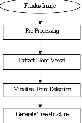
Figure2. General flow chart for generate Vessel Tree
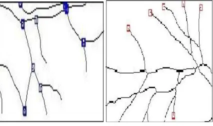
Figure 5. Skeletonized binary image Figure 6. Ridge Ending Point
Fundus Image
Pre-Processing
Extract Blood Vessel
Minutiae Point Detection
ISSN(Online): 2320-9801 ISSN (Print): 2320-9798
I
nternational
J
ournal of
I
nnovative
R
esearch in
C
omputer
and
C
ommunication
E
ngineering
(An ISO 3297: 2007 Certified Organization)
Vol. 3, Issue 4, April 2015
IV.PSEUDO CODE
Step1. Take a rgb retinal image as an input image (Fig2).
Step2. Convert into gray scale and extract blood vessel using some morphological operation. Step3. Then convert the binary to Skeletonized binary image, called thin image(Fig5).
Step4. Identify the ridge ending points , bifurcation points of each retinal blood vessels from thin image and marked on the image(Fig6).
Step5.Consider the bifurcation points as a parent node and ridge ending point as a child node and given different name to identify each node.
Step6.Based on this two major points create a hierarchical tree structure of each vessels and represent whole the binary image as a geometrical shape
Step7:End
V. SIMULATION RESULTS
The simulation studies involve the deterministic small network topology with 5 nodes as shown in Fig.1. The proposed energy efficient algorithm is implemented with MATLAB. We transmitted same size of data packets through source node 1 to destination node 5. Proposed algorithm is compared between two metrics Total Transmission Energy and Maximum Number of Hops on the basis of total number of packets transmitted, network lifetime and energy consumed by each node. We considered the simulation time as a network lifetime and network lifetime is a time when no route is available to transmit the packet. Simulation time is calculated through the CPUTIME function of MATLAB. Our results shows that the metric total transmission energy performs better than the maximum number of hops in terms of network lifetime, energy consumption and total number of packets transmitted through the network.
The network showed in Fig. 1 is able to transmit 22 packets if total transmission energy metric is used and 17 packets if used maximum number of hops metric. And the network lifetime is also more for total transmission energy. It clearly shows in figure that the metric total transmission energy consumes less energy than maximum number of hops. As the network is MANET means nodes are mobile and they change their locations. After nodes have changed their location the new topology is shown in Fig .6 and energy consumption of each node is shown in Fig.7. Our results shows that the metric total transmission energy performs better than the maximum number of hops in terms of network lifetime, energy consumption and total number of packets transmitted through the network.
ISSN(Online): 2320-9801 ISSN (Print): 2320-9798
I
nternational
J
ournal of
I
nnovative
R
esearch in
C
omputer
and
C
ommunication
E
ngineering
(An ISO 3297: 2007 Certified Organization)
Vol. 3, Issue 4, April 2015
VI.CONCLUSION AND FUTURE WORK
In this paper, we propose a novel method to create a vascular tree based on bifurcation points and ending point from the retinal images by analyzing neighborhood connectivity of non-vascular regions around a junction point on retinal blood-vessels. In future we can implement many types of tree based on this point as per needed. It is evident from the result that our scheme is effective in detecting Prominent bifurcation points with reasonable accuracy and time. By using the concept, in near future, we can go for the authentication base on the local feature around the retinal
The simulation results showed that the proposed algorithm performs better with the total transmission energy metric than the maximum number of hops metric. The proposed algorithm provides energy efficient path for data transmission and maximizes the lifetime of entire network. As the performance of the proposed algorithm is analyzed between two metrics in future with some modifications in design considerations the performance of the proposed algorithm can be compared with other energy efficient algorithm. We have used very small network of 5 nodes, as number of nodes increases the complexity will increase. We can increase the number of nodes and analyze the performance.
REFERENCES
1. M.Kavitha and S.Palani,”Retinal blood vessel segmentation algorithm for diabetic retinopathy and abnormality classification by
supervised machine learning”, International Journal of Neural Networks and Applications.vol.5,no.1,Jan-June 2012.ISSN 0974-6048.
2. Sujitkumar S B, Vipula Singh,”Automatic detection of diabetic retinopathy in Non-dilated RGB Retinal fundus images” ,International
journal of computer applications(0975-888),vol.47,no.19,june 2012.
3. Thomas Walter,Jean-Claude Klein,Pascale Massin,and Ali Erginay,”A Contribution of image processing to the diagnosis of diabetic
retinopathy detection of exudates in colour fundus images of the human retina”,IEEE Transaction on medical imaging,vol.21,no.10,October2002.
4. The DRIVE database, .Image sciences institute, university medical center utrecht,. The Netherlands. http://www.isi.uu.nl /Research
/Databases/DRIVE/, last accessed on 7th July, 2007
5. R.Radha1 and Bijee Lakshman “RETINAL IMAGE ANALYSIS USING MORPHOLOGICAL PROCESS AND CLUSTERING
TECHNIQUE”, SIPIJ ,Vol4.
6. V. Bevilacqua, S. Cambo, L. Cariello, and G. Mastronardi, ìa combined method to detect retinal fundusfeatures,î Proceedings of European
Conference on Emergent Aspects in Clinical Data Analysis, pp. 1ñ6, 2005.
7. Sourav Saha, Nilanjana Dutta Roy “AUTOMATIC DETECTION OF BIFURCATION POINTS IN RETINAL FUNDUS-IMAGES”
International Journal of Latest Research in Science and Technology ,Volume 2,Issue 2 :Page No.105-108 , March - April (2013).
8. Neera Singh, Ramesh Chandra Tripath,” Automated Early Detection of Diabetic Retinopathy Using Image Analysis Techniques”,
International Journal of Computer Applications (0975 – 8887) Volume 8– No.2, October 2010.
9. Kande, G.B.,,(2008), Segmentation of Exudates and Optic Disc in Retinal Images, IEEE Transactions in Computer Vision, Graphics &
Image Processing, pgs:535 – 542, Conference Publications.
10. Kavitha, D. , (2005), Automatic detection of optic disc and exudates in retinal images, IEEE , Intelligent Sensing and Information
Processing, Proceedings of International Conference , Page(s): 501 – 506, Conference Publications
11. Sunita Lokhande, et. al. (2013), Wavelet Packet Based Iris Texture Analysis For Person Authentication, Signal & Image Processing: An
International Journal (SIPIJ), Vol.4, No.2, April 2013.
12. Eunhwa Jung and Kyungho Hong, Automatic retinal vasculature structure tracing and vascular landmark extraction from human eye
image, Proceedings of IEEE International Conference on Hybrid Information Technology, vol. 12, pp. 161167, 2006.
BIOGRAPHY