| INVESTIGATION
Maintenance of Heterochromatin by the Large Subunit
of the CAF-1 Replication-Coupled Histone Chaperone
Requires Its Interaction with HP1a Through a
Conserved Motif
Baptiste Roelens,1Marie Clémot,2Mathieu Leroux-Coyau,2Benjamin Klapholz,3and Nathalie Dostatni4 Institut Curie, Paris Sciences et Lettres Research University, Centre national de la recherche scientifique, Unité mixte de recherche 3664, 75248, Paris, France and Sorbonne Universités, Université Pierre-et-Marie-Curie, 75005 Paris, France ORCID ID: 0000-0003-0118-0229 (B.R.)
ABSTRACTIn eukaryotic cells, the organization of genomic DNA into chromatin regulates many biological processes, from the control of gene expression to the regulation of chromosome segregation. The proper maintenance of this structure upon cell division is therefore of prime importance during development for the maintenance of cell identity and genome stability. The chromatin assembly factor 1 (CAF-1) is involved in the assembly of H3-H4 histone dimers on newly synthesized DNA and in the maintenance of a higher order structure, the heterochromatin, through an interaction of its large subunit with the heterochromatin protein HP1a. We identify here a conserved domain in the large subunit of the CAF-1 complex required for its interaction with HP1a in theDrosophilafruitfly. Functional analysis reveals that this domain is dispensable for viability but participates in two processes involving heterochromatin: position-effect variegation and long range chromosomal interactions during meiotic prophase. Importantly, the identification in the large subunit of CAF-1 of a domain required for its interaction with HP1 allows the separation of its functions in heterochromatin-related processes from its function in the assembly of H3-H4 dimers onto newly synthesized DNA.
KEYWORDSheterochromatin; variegation; HP1; CAF-1;Drosophila
I
N eukaryotic cells, the chromatin is partitioned into two cytologically and functionally distinct structures: hetero-chromatin and euhetero-chromatin. Heterohetero-chromatin was initially defined as the part of the genome that remains condensed during the whole cell cycle and stains intensively with DNA dyes. Heterochromatin is generally gene poor; rich in repeated sequences and transposable elements (Hoskinset al.2007). Initially considered to correspond to “junk DNA,” hetero-chromatin contains essential protein-coding genes whoseexpression depends on the neighboring heterochromatin structure (Schulzeet al.2005). It encodes essential chromo-somal structures such as centromeres (Sun et al.1997) or telomeres (Mason et al.2008) and is required for essential chromosomal functions such as homolog pairing during mei-osis (Dernburget al.1996; Karpenet al.1996) . While essen-tial for the biology of the genome, many of these structures are not directly encoded in the sequence of these regions and epigenetic mechanisms are likely required for their mainte-nance through generations.
The chromatin assembly factor-1 (CAF-1) is a heterotrimeric complexfirst isolated as a histone chaperone able to deposit H3-H4 dimers onto newly synthesized DNA during replication or repair (Smith and Stillman 1989; Gaillardet al.1996). Its large subunit interacts directly with PCNA (Shibahara and Stillman 1999; Moggs et al.2000) and the CAF-1 complex is foundin vivoat the replication foci (Krude 1995; Taddei
et al.1999). The large subunit of CAF-1 has also been asso-ciated to the maintenance of heterochromatin: it was shown to be essential for the stable inheritance of gene silencing in
Copyright © 2017 by the Genetics Society of America doi: 10.1534/genetics.116.190785
Manuscript received April 22, 2016; accepted for publication October 30, 2016; published Early Online November 11, 2016.
Supplemental material is available online atwww.genetics.org/lookup/suppl/doi:10. 1534/genetics.116.190785/-/DC1.
1Present address: Stanford University School of Medicine, Stanford, CA 94305. 2These authors contributed equally to this work.
3Present address: The Gurdon Institute and Department of Physiology, Development and Neuroscience, University of Cambridge, Cambridge CB2 1QN, United Kingdom.
subtelomeric regions in Saccharomyces cerevisiae (Monson
et al.1997); its absence infission yeast led to defective main-tenance of silencing at both the centromeres and mating type loci, accompanied with a decrease of HP1 ortholog Swi6p binding in these regions (Dohkeet al.2008); and in mice, it is required for the duplication and maintenance of pericentric heterochromatin (Quivyet al.2004, 2008). This function of mouse P150 is independent of the known function of CAF-1 in histone deposition and has been linked to its ability to interact with HP1 proteins (Quivyet al.2008). The large subunit of CAF-1 is therefore an important and conserved factor required for maintenance of multiple levels of chromatin organization. It however remains to be determined whether the two appar-ently separate biochemical activities of CAF-1 ensure common or independent functions during development.
InDrosophila, the large subunit of CAF-1, P180 (Tyleret al.
2001; Songet al. 2007; Klapholzet al.2009), is essential for larval development (Songet al.2007; Klapholzet al.2009) and is required for the following: (i) proliferation of mitotic and endocycling cells (Tyleret al.2001; Songet al.2007; Klapholz
et al.2009), (ii) assembly of nucleosomes on newly synthesized DNA (Klapholzet al.2009; Tyleret al.2001), and (iii) replica-tion of euchromatic regions in larval endocycling cells (Klapholz
et al.2009). These properties together with genetic interactions between mutant alleles of Caf1-180 (hereafter referred to as
p180), which encodes P180, andasf1(Klapholzet al.2009), encoding the histone chaperone ASF1, suggest that the function of P180 essential for viability inDrosophilais related to CAF-1-dependent histone deposition. Whether P180 is also required for proper maintenance of heterochromatic regions was initially less clear: although two mutant alleles ofp180were shown to act as dominant suppressors of position-effect variegation (PEV) (Songet al.2007), quantitative analysis of the efficiency of the replication of heterochromatic regions did not show, in contrast to euchromatic regions, major defects upon the loss of P180 activity in larval endocycling cells (Klapholzet al.2009). More recently, an RNA interference approach showed that P180 reg-ulates, in a dose-dependent manner, the structure of pericentric heterochromatin by affecting H3K9Me and H4K20Me levels to-gether with the recruitment of HP1a on polytene chromosomes; thereby conclusively showing the conservation of this function in
flies (Huanget al.2010). Moreover, as the artificial targeting of HP1a to chromosomes induces the accumulation of P180 at these ectopic positions (Huanget al.2010), it was proposed that the role of CAF-1 in heterochromatin maintenance inflies was also likely mediated by an interaction between P180 and HP1a. In this study, we identify a conserved domain in the Dro-sophilaCAF-1 large subunit required for itsin vitrointeraction with HP1a. We show that this domain is not essential for viability but is required for proper heterochromatin mainte-nance in germ cells and participates in two processes that require proper heterochromatin structure in flies: PEV and persistence of pairing between heterochromatic chromosomal regions in developing oocytes. Our results therefore demon-strate that the interaction between the large subunit of CAF-1 and HP1a is not essential forDrosophilalarval development
and viability, but participates in the maintenance of hetero-chromatin organization.
Materials and Methods
Fly stocks
Fly stocks include thep1803[caf1-180(3)] (Klapholzet al.2009),
Su(var)2055[Su(var)205(5)] (Eissenberget al.1992), and
Mei-W681[Mei-W68(1)] (McKimet al.1998) alleles; theDp(1;Y)
B[S]-markedYchromosome; theP{mw,HSBGHb3Bcd3-lacZ} in-sertion on theXchromosome (Simpson-Broseet al.1994); the FM7i,P{ActGFP}JMR3,TM3,Sb,TM3,Sb SerandSM6bbalancer chromosomes; theT(2;3)SbV(T(2;3)Sb[V]) translocation; and
theC(2)ENandC(4)RM,ci[1],ey[R] compound chromosomes. Ubiquitous expression was obtained using the Gal4/UAS system (Brand and Perrimon 1993) withP{Act5C-Gal4}25FO1insertion as a Gal4 driver. Upstream activation sequence (UAS) transgenes include UAS-p180 (Klapholz et al. 2009) and UAS-p180DHIM,
which were obtained by germline transformation of thepUASp
vector (Rorth 1998) containing thep180orp180DHIM
comple-mentary DNA (cDNA) (see theVectors and cloningsection).
Genetics
PEV was assessed by analyzing the progeny of females that were or were not carrying mutant alleles ofp180andSu(var)
205crossed withSbV/TM3,Sb,Ser(Figure 4C) or SbV/TM3,
Ser(Supplemental Material,Figure S3) males. The 12 macro-chaetae in notum and scutellum ofSbVoffspring were scored
to quantify the Sb phenotype.
Meiotic segregation defects ofXchromosomes were mon-itored using dominant markers located on the paternalX(mw from P{mw,HSBGHb3Bcd3-LacZ}) andY(BarfromDp(1;Y)B[S]) chromosomes as described in McKim et al. (2009). Briefly, females that were or were not carrying mutant alleles of
p180were also heterozygous for theymarker to allow the discrimination of meiosis I from meiosis II segregation de-fects. Wheny+,p180+or2/FM7,y,B1females were crossed to y,{mw}/BSY males, regular progeny include females with
normal or mildly Bar (B1heterozygotes) eyes and males with
severe Bar (BS) eyes.Xchromosome segregation defects during
female meiosis generate oocytes with two Xchromosomes or none: the corresponding exceptional progeny are females with severe Bar eyes (XXY,BSfemale) or males with normal or mildly
Bar eyes (XO male). Segregation defects during meiosis I lead to nonyellow-bodiedXXYfemales that are heterozygous for the
ymarker, whereas segregation defects in meiosis II lead to an equal number of yellow-bodied and nonyellow-bodiedXXY fe-males. Because of increased meiotic segregation defects inXXY
The frequency of segregation defects was normalized to ac-count for unviable XXX and OY genotypes, and calculated as 23(exceptional progeny)/[23(exceptional progeny) + (regular progeny)].
Statistical tests
The low level of exceptional progeny observed did not allow us to use the classicalx2test. Rates of segregation defects were therefore compared using Fisher’s exact test which is more suitable to low effectives. For all tests, a two-tailedP-value of lower than 5% was considered statistically significant. A recent study provided tools to compare rates of segregation defects of theXchromosome using a multinomial Poisson hierarchy model (Zeng et al.2010). Treatment of our data with the Fisher’s
exact test or the Poisson hierarchy model reached similar conclusions.
Sequence alignment and conservation
Sequences were aligned using the MUSCLE algorithm (Edgar 2004a,b). Conservation at any given position was computed as the average of a numerical index reflecting the conserva-tion across the selected species of physico-chemical proper-ties (Livingstone and Barton 1993) on 11 residues centered on the position: for example the value at position 15 reflects the conservation of residues 10–21. When,11 values were available for a given position, conservation was computed on the available values within the +5/25 distance.
Vectors and cloning
A cDNA library ofDrosophilaembryos (Clontech) was used to generate the threeDrosophilaHP1a GST constructs using three pairs of primers: ssHP1-a (TTAAGAATTCATGGGCAAGAAAATC GACAACCC) and ssHP1-b (TTAAGAATTCGCTCGCCTCGTACT GCTGG) to amplify the sequence encoding thefirst 72 residues of HP1a; ssHP1-a and ssHP1-c (TTAAGAATTCGCCGCGATCGAATC CGGTAGATCC) to amplify the sequence encoding the first 146 residues of HP1a; ssHP1-a and ssHP1-d (TTAAGAATT CATCTTCAATATCAGAGTACCAGG) to amplify the full-length HP1a coding sequence. These PCR products were then cloned into theEcoRI restriction site of thepGEX-4T-1plasmid (GE Healthcare). The wild-type and mutant P180-His proteins were produced in bacteria using thepET30A+plasmid (Novagen), in which the corresponding cDNA was cloned in frame with the sequence encoding the histidine tag. The various cDNAs encoding the truncated P180 proteins used to identify the HP1-interacting motif (HIM) were obtained using standard cloning methods as follows: for p180DNt, ligation of LG1 (TATGCACGCTGGCG
TAGTTG) and LG2 (GATCCAACTACGCCAGCGTGCA) oligo-nucleotides in the p180 cDNA digested with NdeI and
BamHI; forp180DNtD(883–1101), ligation of PSKP1 (GCAGGAG TTCGCTGATGCG) and PSKP2 (GTACCGCATCAGCGAACTCC TGCTGCA) oligonucleotides in thep180DNtcDNA digested with
PstI and KpnI; for p180DNtD(1110–1183), ligation of KPNO1 (GTACCAGCCGCCTCGCCCGC) and KPNO2 (GTACCGCA TCAGCGAACTCCTGCTGCA) oligonucleotides in thep180DNt
cDNA digested withNotI andKpnI; forp180DNtD(883–959),
self-ligation of the p180DNt cDNA digested with PstI; for
p180DNtD(883–986), ligation in thep180DNtcDNA digested with
PstI andKpnI of the product obtained by PCR amplification of the p180cDNA with the PSKP DELTA2959 (CCGCTGCAGGAG TACCTGAAAACCCAAGCGA) and p180-39KpnI (GCGGCTGGTA CCGCATCCGCTG) primers; forp180DNtD(883–1010), ligation in the
p180DNtcDNA digested withPstI andKpnI of the product obtained
by PCR amplification of thep180cDNA with the PSKP DELTA3030 (CCGCTGCAGAAATTCGACGAGCTTGCCAG) and p180-39KpnI (GCGGCTGGTACCGCATCCGCTG) primers; forp180DNtD(88321040), ligation in thep180DNtcDNA digested withPstI andKpnI of
the product obtained by PCR amplification of thep180cDNA with the PSKP DELTA3130 (CCGCTGCAGAAACCGAAGAA GCGCCTCTG) and p180-39KpnI (GCGGCTGGTACCGCATCC GCTG) primers; forp180DNtDHIM, ligation in thePstI digested
p180DNtD(883–986)cDNA of the fragment obtained by PstI di-gestion of thep180cDNA; forp180D(959–986), ligation in the
p180 cDNA digested with PstI and KpnI of the product obtained by PCR amplification of thep180cDNA with the PSKP DELTA2959 (CCGCTGCAGGAGTACCTGAAAACCCA AGCGA) and p180-39KpnI (GCGGCTGGTACCGCATCCG CTG) primers; and for p180 DHIM, ligation of the
frag-ment obtained by PstI digestion of the p180 cDNA in the
p180D(959–986)cDNA digested withPstI. The P180-His was pro-duced in bacteria using the pET30A+ plasmid (Novagen), in which the p180 cDNA was cloned in frame with the se-quence encoding the histidine tag.
Co-immunoprecipitation on Schneider S2 cells
Schneider S2 cells were incubated for 15 min on ice in a lysis buffer containing 10 mM HEPES, 10 mM KCl, 0.1 mM EDTA, 0.1 mM EGTA, 1 mM DTT, 0.5 mM PMSF, 5mg/ml leupeptin, 2 mg/ml pepstatin, and 15 mg/ml aprotinin. NP-40 was added to a final concentration of 0.5% and the cells were strongly agitated. Nuclei were centrifuged for 5 min at 100 3 g. The nuclear pellet was resuspended in nuclear extraction buffer (NEB) 0.42 (20 mM HEPES, 25% glycerol, 1 mM EDTA, 1 mM EGTA, 1 mM DTT, 0.5 mM PMSF, 5mg/ml leupeptin, 2 mg/ml pepstatin, and 15 mg/ml aprotinin) with 0.42 M NaCl and incubated for 20 min at 4°on a wheel before a centrifugation of 30 min at 20,0003g. Immuno-precipitation (IP) was performed on the supernatant with a 1:100 dilution of a polyclonal rabbit anti-P180, kindly pro-vided by Tyleret al.(2001), and Sepharose-Protein A beads (Upstate) in NEB 0.2 (same as NEB 0.42 but containing 0.2 M NaCl). The detection of co-immunoprecipitated pro-teins was performed by western blots using a 1:8000 di-lution of the same anti-P180, a 1:1000 didi-lution of polyclonal rabbit anti-P105 kindly provided by Tyleret al.
Recombinant proteins and GST pull downs
GST-fusion and His-tagged proteins were produced in
Escherichia coliBL21-DE3 upon induction with 0.4 mM isopropyl 1-thio-b-D-galactopyranoside. Pellets were suspended in buffer
Y(13PBS, 1 mM EDTA, 1% Triton X-100, 1 mM PMSF, 5mg/ml leupeptin, 2mg/ml pepstatin, and 10mg/ml aprotinin) and son-icated. Lysates were centrifuged at 20,000 3gfor 30 min to remove aggregates. GST fusions were purified using Glutathione Sepharose 4B beads (GE Healthcare). After washes in buffer
Y, bound proteins were suspended in buffer (PBS/EDTA, 0.5 M, pH 8.0). The relative concentration of proteins was es-timated by Coomassie staining of SDS-PAGE for GST fusion and by western blotting for His-tagged proteins. For GST pull-down assays, beads of Glutathione Sepharose bound to GST fusions were suspended in immuno-precipitation (IP) buffer (50 mM Tris-HCl, pH 7.0, 0.25 mM EDTA, 0.03% NP-40, 5mg/ml leu-peptine, 2mg/ml pepstatine, and 10mg/ml aprotinine) and in-cubated with lysates containing the His-tagged proteins at 4°for 3 hr under gentle agitation. The Sepharose beads were collected by centrifugation at 8003gand washed four times with IPTAG buffer containing 150 mM NaCl. Pulled-down proteins were de-tected by western blotting with a 1:500 dilution of a His-probe mouse monoclonal antibody (H-3, #sc-8036, Santa Cruz Biotech-nology), a 1:10,000 dilution of a secondary goat peroxidase-conjugated anti-mouse antibody (#115-036-062, Jackson ImmunoResearch), and SuperSignal West Pico Chemilumi-nescent Substrate (Thermo Scientific).
Detection of P180 in larval extracts
Larval protein extracts were prepared as described in Klapholz
et al. (2009). P180 proteins were detected using an antibody raised in rabbit against a peptide corresponding to the residues 198–421 fused to GST. As this peptide is located in the amino-terminal region of the protein, the deletion of the HIM is unlikely to affect recognition of this epitope in denaturing conditions.
Immunodetection on salivary gland polytene chromosomes
Staining was performed as described in Paro (2000). P180 was detected using a FLAG-tag fused to the P180 protein in the UAS-p180transgene using either a monoclonal mouse (M2, Sigma Aldrich, Figure 2B) or a polyclonal rabbit antibody (F7425, Sigma-Aldrich, Figure 2, A and C). Using the M2 antibody, a nonspecific staining was recurrently observed at the chromo-center, even inflies not expressing the P180-Flag protein. The other antibodies used were: mouse monoclonal PC10 (Dako) specific for PCNA, rabbit polyclonal R20/12 specific for acety-lated H4K12 (Turneret al.1992), and mouse monoclonal C1A9 (Developmental Studies Hybridoma Bank) detecting HP1a.
Immunodetection on ovaries
Ovaries of 4- to 5-day-old females were dissected in PBS and
fixed in 4% paraformaldehyde for 15 min. Samples were then permeabilized in PBS with 0.2% Triton X-100 (PBT) for at least 30 min, incubated at 4° overnight with primary antibodies diluted in PBT (rabbit anti-P180, 1:1000, described in the
section Detection of P180 in larval extracts; and mouse anti-HP1 C1A9, Developmental Studies Hybridoma Bank, 1:100), washed three times for 15 min in PBT, incubated for 2 hr with secondary antibodies diluted in PBT (Alexa-568 conjugated anti-rabbit and Alexa-488 conjugated anti-mouse, Life Tech-nologies, 1:1000), and washed three times for 15 min in PBT. For DNA staining, samples were subsequently stained with DAPI (0.8mg/ml in PBT) for 10 min. Samples were mounted in Vectashield (Vector Laboratories, Burlingame, CA).
Imaging of germaria and egg chambers was performed with a Carl Zeiss (Thornwood, NY) LSM780 confocal micro-scope using identical acquisition settings for all analyzed genotypes. Image projections were performed with six to eight images containing the upper half of the germarium or
five to six images containing the oocytes for the egg chambers. Images were separated by 0.8–1mm along thez-axis.
Quantification of HP1a signal in oocytes
The total HP1a signal in oocytes was evaluated using Fiji, by (i) identifying the stacks containing the oocyte nucleus based on the DAPI staining, (ii) performing a sum-intensity projection of the typicallyfive to six stacks containing the oocyte’s nucleus, and (iii) summing the value of every pixel contained within a region of interest designed to encompass the oocyte nucleus.
FISH staining and imaging
The 359-bp repeat probe was obtained by PCR amplification of genomic DNA using primers (359 bp-1, CGGTCATCAAATAAT CATTTATTTTGC; and 359 bp-2, CGAAATTTGGAAAAACA GACTCTGC) designed from the published sequence (Hsieh and Brutlag 1979). Probes were digested with AluI and then tailed with digoxigenin using the DIG-oligonucleotide tailing kit (Roche). FISH was performed as described (Dernburget al.
1996). After hybridization, probes were detected using a sheep anti-digoxigenin primary antibody (1:1500, Roche) and the Alexa Fluor 568 donkey anti-sheep (1:1000, Molecular Probes, Eugene, OR), DNA was stained with DAPI, and the samples were mounted in Vectashield. Image acquisitions and processing were performed using a Delta Vision microscope (GE Healthcare Life Sciences) with 403and 1003objectives and the SoftWorx software. The settings for acquisition and deconvolution were con-stant for each staining. Each quantification was performed on at least two independent experiments. Image projections were per-formed with 4–10 images separated by 0.2mm along thez-axis.
Data availability
Strains are available upon request.Table S1contains accession numbers for P180 orthologs used for conservation analysis.
Results
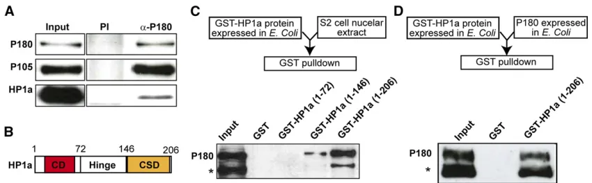
The Drosophila large subunit of CAF-1 and HP1a interact directly
P180 and HP1a have been shown to genetically interact in
of this interaction in the maintenance of heterochromatic structure, we aimed to characterize its molecular details. We
first recapitulated this interaction by performing an IP of P180 from a nuclear extract ofDrosophilaSchneider S2 cells. P180 coprecipitated with P105, the medium subunit of CAF-1, and, in agreement with a study by Jiao and colleagues (Huanget al.
2010), with HP1a (Figure 1A). Additional GST pull-down as-says on S2 nuclear extracts with truncated forms of HP1a (Figure 1B) fused at their N-terminal end to the GST indicated that (a) P180 does not interact with the chromodomain of HP1a, (b) the hinge of HP1a is able to interact weakly with P180, and (c) the full interaction between P180 and HP1a requires both the hinge and the chromo shadow domain of HP1a (Figure 1C). Finally, GST pull-down assays using recombinant P180 and GST-HP1a proteins showed that HP1a was able to pull-down P180 on its own (Figure 1D) and that the interaction between the two pro-teins was therefore direct.
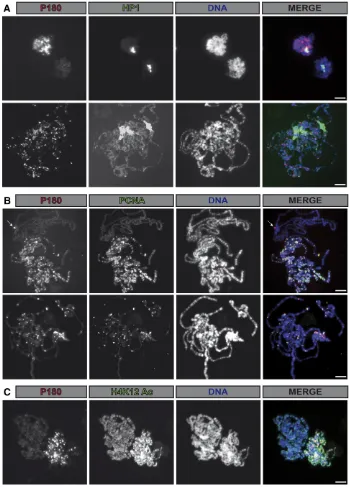
Colocalization between P180 and HP1a is only observed on replicating polytene chromosomes
In the experiment described Figure 1A, we noticed that only a minor fraction of HP1a was coprecipitated with P180. This could be due to the fact that HP1 proteins interact with other partners that could potentially compete with P180 for bind-ing to HP1 in nuclear extracts (Kellum 2003). In addition, as CAF-1 is mostly required duringSphase of the cell cycle, this interaction might also be restricted during this phase of the cell cyclein vivo. We tested this hypothesis by analyzing the localization of both P180 and HP1a on salivary gland poly-tene chromosomes. As described previously (James and Elgin 1986; Jameset al.1989), HP1a was detected at the chromo-center, a DAPI-dense region containing the pericentric and centric regions (Figure 2A). Codetection of P180 using a FLAG-tagged transgene indicated that localization of P180 only poorly correlated with the binding of HP1a but was de-tected on polytene chromosomes also stained by PCNA
(Figure 2B) or showing high levels of newly incorporated histones (H4K12Ac, Figure 2C); two markers of chromatin undergoing replication. Accordingly, polytene chromosomes not undergoing replication and therefore not stained by PCNA, or with lower signals of H4K12Ac, were poorly bound by P180. This suggests that binding of P180 at the HP1a bound loci might occur in a replication-dependent manner and raises the possibility that the interaction between these two factors might be cell-cycle regulated.
A 27-residue domain is required for the direct interaction between recombinant P180 and HP1a proteins
We next searched for a peptide motif in the P180 protein sequence which could mediate its interaction with the chromo shadow domain of HP1a. Surprisingly, P180 lacks the canonical consensus PXVXL motif found in many HP1-interacting proteins, including vertebrate CAF-1 large sub-units (Murzinaet al.1999; Smothers and Henikoff 2000). P180 also does not contain the HP1-interacting peptide motif of the heterochromatin protein HP2 (Stephens
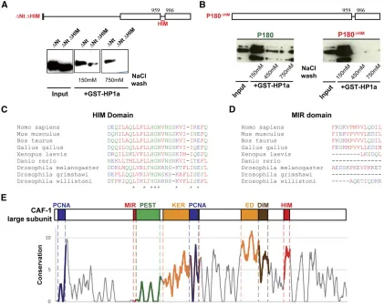
et al.2005), indicating that the direct interaction between P180 and HP1a inDrosophilawas mediated through a do-main not previously described. As we were able to detect a direct interaction between recombinant P180 and HP1a, we performed additional GST pull-down experiments on truncated versions of P180 expressed in bacteria and iden-tified, in the C-terminal, a 27-amino acid motif likely in-volved in this interaction (Figure S1 and Figure 3A). Further experiments using a full-length P180 protein lack-ing only the identified 27 residues confirmed that this do-main, the HIM dodo-main, was required for P180 to interact with HP1a in vitro under stringent conditions (450 mM NaCl) (Figure 3, A and B). Of note, these stringent condi-tions are commonly used to detect specific interactions with HP1 proteins (Maisonet al.2002). Furthermore, many
cells and tissues have been reported to have a high nuclear so-dium and potassium concentrations, up to 250 mM NaCl and 280 mM KCl in frog oocyte nuclei (Naoraet al.1962; Hooper and Dick 1976; Moore and Morrill 1976), and such stringent salt conditions can therefore be achieved in the nucleus. Altogether, these observations indicate that the robust interaction we ob-servedin vitrobetween the CAF-1Drosophilalarge subunit and HP1a involves both the hinge and chromo shadow domain of HP1a and requires the HIM domain, a short domain of 27 resi-dues localized in the C-terminal part of the CAF-1 large subunit.
The HIM is conserved in vertebrates and insects
We next assessed the HIM conservation by aligning available sequences of CAF-1 large subunits in various species (Figure
S2and Table S1). We noticed that the HIM was well con-served in insects and vertebrates (Figure 3C). The MIR do-main, which contains the PXVXL motif and was identified as the domain mediating the interaction between the murine large subunit of CAF-1 and HP1b protein (Murzina et al.
1999), is conserved in mammals and the chicken (Takami
et al. 2007), but is lacking inXenopusor zebrafish (Figure 3D). In Drosophilaspecies, the MIR domain is either com-pletely lacking (Drosophila grimshawi) or highly divergent (D. willistoniandD. melanogaster). Interestingly, when conser-vation was assessed along the whole length of the protein using a method computing the conservation of physico-chemical properties in the alignment (Livingstone and Barton 1993), the HIM domain clearly corresponded to a local maximum of
Figure 2 Binding of P180 on polytene chro-mosomes occurs in a replication-dependent manner and only poorly correlates with HP1a localization. (A–C) Immunofluorescent staining allows detection of (A–C) P180-FLAG fusion protein, (A) HP1a, (B) PCNA, or (C) H4 acetylated on lysine 12 on salivary gland polytene chromosome in p1803 /p1803;Act-Gal4,UAS-p180-FLAG/CyO.
conservation with a score comparable to other domains pre-viously identified as important for the function of the large subunit of CAF-1 in replication-coupled nucleosome assembly, such as the acidic KER and ED domains, which are thought to interact with the histones (Kaufmanet al.1995); the dimeriza-tion domain (Quivyet al.2001); or the PCNA interacting motifs (Shibahara and Stillman 1999; Moggset al.2000) (Figure 3E). In contrast, in this same analysis, the MIR domain was not highlighted, confirming its poor conservation.
The HIM domain of P180 is not essential for viability
We then intended to test the functional significance of the HIM
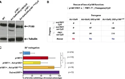
in vivoand constructed, using the Gal4/UAS system (Brand and Perrimon 1993), a strain expressing a mutant version of P180, P180DHIM, deleted for the 27 residues identified
pre-viously (Figure 4A). Interestingly, ubiquitous expression of P180DHIM using an Act5C-Gal4 driver rescued the viability ofp1803mutant males at least as efficiently as the wild-type
P180 (Figure 4B). We had previously reported that mater-nally provided P180 is undetectable by the end of embryonic development (Klapholzet al.2009) and therefore the trans-gene is the sole source of P180 in the rescued p1803
hemi-zygous males at least from larval development onwards. Importantly, the rescued individuals showed no obvious de-velopmental defects and in particular none that could resem-ble the defects previously described upon downregulation of any of the CAF-1 subunits (Huanget al.2010; Yuet al.2013). These observations indicate that the HIM domain of P180 is largely dispensable for Drosophilalarval development and supports a model in which the ability to interact with HP1a
is not an essential feature of P180, at least after embryogen-esis. The P180DHIMconstruct can therefore be used to directly testin vivothe role of the interaction between the large sub-unit of CAF-1 and HP1a during these stages, without affect-ing CAF-1 essential functions.
The HIM domain of P180 is required for P180-induced modification of PEV
In a previous study, mutant alleles of p180were shown to act as dominant suppressors of PEV, thereby highlighting the role of P180 in the maintenance of heterochromatin (Huang
et al.2010). We therefore wondered whether transgenic ex-pression of P180 or its mutant version, P180DHIM, was able to counteract this effect. As a reporter of PEV, we used the T(2;3)SbV translocation (hereafter referred to asSbV) which
juxtaposes the dominantSb1mutation and a portion of the
centric heterochromatin of the second chromosome. This in-duces mosaicflies with short (stubble) or long (normal) bris-tles which can be rigorously quantified. Changing the genetic context from wild type to p1803/+ in SbV flies induced a
significant increase in the frequency of short bristles (Figure 4C, compare the blue and the red histograms), indicating that this allele also behaves as a dominant suppressor of PEV. Interestingly, this suppression of PEV was rescued, albeit par-tially, by Gal4-induced expression of wild-type P180 (Figure
4C, compare red and yellow histograms), while Gal4-induced expression of the P180DHIM mutant did not rescue and indeed further suppressed the variegation in ap1803/+
back-ground (compare the red and orange histograms). In contrast, expression of either the wild-type or the HIM-depleted versions of P180 inflies with two wild-type copies ofp180induced only a mild suppression of SbV variegation of similar magnitude
(Figure S3). Altogether, these observations support a role for the HIM domain of P180 in p1803-induced suppression of
variegation.
The HIM domain of P180 contributes to heterochromatin-mediated pairing in oocytes
In Drosophila oocytes, homologous chromosomes remain associated along their pericentromeric regions throughout meiotic prophase and, when homologs have failed to form crossovers during meiotic recombination, this persisting as-sociation promotes their correct segregation according to the homologous achiasmate segregation system (Dernburget al.
1996; Karpenet al.1996). This pairing process relies on the integrity of pericentric regions that are heterochromatic and, accordingly, mutations in components of heterochromatin have altered levels of achiasmate segregation (Verni et al. 2000; Peng and Karpen 2009; Subramanian and Bickel 2009). More specifically, a recent study was able to demonstrate that partial
Figure 4 The HIM domain is not essential for viability but contributes top180-induced PEV. (A) Expression of P180 in larvae of the indicated genotype assessed by western blotting on larval extracts using an anti-tubulin antibody and ana-P180 antibody raised against a peptide covering residues 198–
421 of P180. WT, wild type. (B) Rescue efficiency of the various transgenes was quantified using a test cross betweenp1803/FM7ifemales andCyO
males carrying the indicated combination of transgenes. Rescue was evaluated in the progeny of the test cross by the presence of rescued males (non-FM7, non-CyO) only expressing the transgenic P180. (C) Rescue ofSbVvariegation induced byp1803heterozygosity by transgenic expression of P180.
Flies heterozygous for theSbVchromosomal aberration and expressing either the wild-type or the HIM-deleted P180 were scored for the number of
macrochaetae showing aSbphenotype inp1803/+background.nspecifies the number offlies analyzed for each genotype and*indicates a significant
depletion of HP1a, or its paralog Rhino in germ cells, induced defects in maintenance of pericentric associations of a pair of achiasmateXchromosomes, which resulted in elevated segre-gation defects (Giauque and Bickel 2016). We therefore aimed to test the functional impact of the loss of the HIM domain of P180 in this process and analyzed the pairing status of hetero-chromatic repeats of an achiasmate pair composed of a wild-typeXand aFM7balancer chromosome in females expressing various doses of wild-type or HIM-deleted versions of P180.
Wefirst compared pairing efficiencies between wild-type andp1803heterozygous females. FISH using a probe specific
for the pericentric heterochromatin sequences of the wild-typeXchromosome (359-bp repeats) indicated that oocytes produced by females carrying one wild-type and onep1803
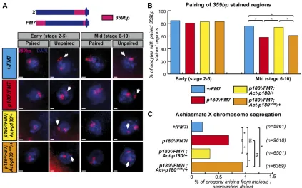
allele were significantly impaired in maintaining the pairing ofXchromosome-heterochromatic 359-bp sequences during oogenesis when compared to their wild-type counterparts (Figure 5, A and B, compare red and blue histograms in midstage egg chambers). In theFM7chromosome, the heterochro-matic 359-bp repeats are split in a large subtelomeric region and a smaller pericentromeric region (Figure 5A). The defects in pairing could thus involve only one of those sequences or
both. By assessing the size of the unpaired sequence, which allows distinguishing defects in pairing involving either the subtelomeric (large FISH signal) or the pericentric sequences (small FISH signal), we observed that the defects in pairing observed upon reduction in the dose of P180 affect both sequences in proportions correlated to their respective sizes (seeFigure S4for quantification).
As some of these pairing events have been proposed to contribute to the correct segregation of homologous achiasmate chromosomes, we analyzed this process inp1803heterozygous
females. In agreement with their pairing defects, these females produced oocytes with a modest but significant increase in the segregation defects of this pair of nonrecombining chromo-somes (Figure 5C, compare red and blue histograms). These segregation defects are specific to nonrecombining somes: similar defects were observed with achiasmate chromo-somes II and IV but were not observed with chiasmate chromosomes II (Figure S5).
Importantly, ubiquitous expression of wild-type P180 but not P180DHIM, using the Gal4/UAS system, was able to res-cue both the pairing maintenance defects and segregation defects of oocytes produced byp1803heterozygous females
Figure 5 The HIM domain participates in heterochromatin-mediated pairing in germ cells. (A) FISH staining on oocytes allows the detection ofX pericentric regions (359 bp) and total DNA (DAPI) in females of the indicated genotype. Based on previously described detection of synaptonemal complex components (Resnicket al.2009), two different regions of the ovariole were analyzed: the early region from vitelline stage 2–5 in which the synaptonemal complex, maintaining aligned homologs, is fully assembled; and the middle region containing egg chambers from stage 6–10, in which the synaptonemal complex is undergoing disassembly and therefore likely requires additional mechanisms to maintain pericentric interactions. Scale bar represents 1mm. (B) Pairing of 359-bp regions of homologousXchromosomes was quantified in females of the indicated genotype. Numbers of oocytes scored for stages 2–5 (early):+/FM7i, 64;p1803/FM7i, 68;p1803/FM7;Act-p180/+, 80; andp1803/FM7i;Actp180DHIM/+, 126. Number of oocytes scored
for stages 6–10 (late):+/FM7i, 75;p1803/FM7i, 63;p1803/FM7;Act-p180/+, 57; andp1803/FM7i;Actp180DHIM/+, 99. (C) Rate of exceptional oocytes arising
(Figure 5, A–C, compare yellow and orange histograms). This argues that the HIM domain, which is central to the direct interaction between HP1a and the large subunit of CAF-1, is also important for the maintenance of pericentric pairing during meiotic prophase; a process relying on proper HP1a loading and function (Giauque and Bickel 2016).
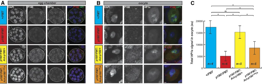
The HIM domain of P180 contributes to the loading of HP1 at the oocyte chromocenter
One possible way P180 could contribute to heterochromatin maintenance would be by regulating the loading of HP1a to chromosomes. As we identified the HIM as an important domain for the interaction between P180 and HP1a, we tested this possibility by analyzing thein vivodistribution of P180 and HP1a in egg chambers isolated from females expressing a wild-type dose ofp180. We observed a robust P180 signal in nurse cell nuclei (Figure 6A, second column, blue genotype) but not in oocytes (Figure 6B, second column, blue genotype). As nurse cells are actively replicating and oocytes are postrepli-cative and undergoing the meiotic prophase program, this ob-servation is consistent with staining on salivary gland polytene chromosomes indicating that P180 localizes to the chromatin duringSphase. In contrast, P180 expression was detected at similar levels in oocytes from females expressing the wild-type or the HIM-depleted transgene (Figure 6B, second column, yellow and orange genotypes); indicating that at least some degree of P180 accumulation duringSphase is due to tran-scriptional regulation, which is not recapitulated by the ubiq-uitous Act5C-Gal4 driver. As the samples were prepared, stained, and imaged under identical conditions, we could also compare the overall levels of HP1a accumulated in the oocyte nucleus in the different genotypes. We could robustly observe that levels of HP1a were reduced in oocytes ofp1803
hetero-zygous females when compared to females with two wild-type alleles ofp180(Figure 6, B and C, third column, compare blue
and red genotypes). This decrease was rescued by trans-genic expression of the wild-type P180 (Figure 6, B and C, third column, yellow genotype) whereas expression of the HIM-depleted P180 protein resulted in only partial rescue (Figure 6, B and C, third column, orange genotype). Similar trends were observed on the relative level of HP1a in nurse cells, depending on the genotype (Figure S6). Thus the HIM-depleted P180 appears defective in loading HP1 in the DAPI-dense regions of the oocyte nucleus. Altogether, these experiments indicate that P180 is required for the loading or maintenance of HP1a in germ cells and that the HIM domain contributes to this effect.
Discussion
The CAF-1 complex was originally isolated and characterized as a histone chaperone that assembles histone H3-H4 dimers onto newly synthesized DNA (Smith and Stillman 1989). Further studies showed that the interaction between the large subunit of CAF-1 and the HP1 proteins was required for the proper duplication and maintenance of heterochro-matin regions in mitotically dividing cells (Dohkeet al.2008; Quivyet al.2008). These two functions of CAF-1’s large
sub-unit, which involve different levels of chromatin organiza-tion, are also conserved in Drosophila. First, the large subunit of CAF-1 is involved in H3-H4 dimer deposition dur-ing DNA replication (Tyler et al. 2001): this function was shown to be essential for larval development and viability (Song et al. 2007; Klapholz et al. 2009). Second, CAF-1’s
large subunit interacts with HP1a (Huanget al.2010). We have shown that this interaction is direct and have identified the HIM domain in CAF-1’s large subunit required for this interaction. Interestingly,flies expressing only a mutant form of CAF-1’s large subunit deleted for the HIM are viable. This indicates that the ability of CAF-1’s large subunit to interact
Figure 6 Altered loading of HP1a in germ-cells of females expressing either low dose of P180 or P180DHIM. (A and B) Immunodetection of P180, HP1a,
with HP1a is not strictly required for viability inflies and that additional mechanisms, such as the one involving the inter-action between HP1a and the H3.3 chaperone XNP (Bassett
et al.2008), might contribute to proper heterochromatin for-mation or maintenance and ensure successful development. As this function is essential in mammalian cells (Houlard
et al.2006; Quivyet al.2008),D. melanogasterrepresents a unique model to understand its role during development.
Interestingly, although we could obtain individuals, males and females, expressing only the HIM-deleted forms of CAF-1 large subunit, we realized that the HIM deletion resulted in female sterility (Marie Clémot, unpublished data). This could indicate either that the HIM domain is strictly required for some aspect offly reproduction, either via its role in het-erochromatin maintenance or via some other functions of CAF-1’s large subunit, or that the HIM is required during embryogenesis for the initial formation of heterochromatin structure, a possibility we could not directly test because of the important maternal contribution and the inability of
p1803 germline clones to develop into mature oocytes
(Klapholzet al.2009).
Our work also provides new insights into the mechanisms of heterochromatin maintenance: previous studies in
S. pombeand mammalian cells had led to propose a model in which CAF-1 is recruited to the replication forks through its interaction with PCNA and transfers HP1 to the repli-cated chromatin at these sites to locally maintain the chro-matin structure (Quivyet al.2004; Dohkeet al.2008; Quivy
et al.2008). Consistently, we only detected colocalization of CAF-1’s large subunit with HP1a on salivary gland poly-tene chromosomes upon replication of the HP1a-enriched regions. However, although it is now clearly established that CAF-1 participates in the maintenance of heterochro-matin in eukaryotes, very little is known on the contribu-tion of the different domains to this funccontribu-tion. In mammals, the MIR domain, which contains a PXVXL motif, is essential for heterochromatin maintenance (Quivy et al. 2004; Dohkeet al.2008; Quivyet al.2008). The MIR domain is not conserved in the yeastsS. cerevisiaeand S. pombe, al-though a role in heterochromatin maintenance has been reported for CAF-1’s large subunits (Monson et al.1997; Dohkeet al.2008). Similarly, in chicken cultured cells, the MIR domain does not seem to be involved in heterochro-matin maintenance since a point mutant in the PXVXL motif could support normal cell proliferation with no detectable defects in heterochromatin structure (Takamiet al.2007). In this study, we coupled biochemistry, genetics, and cytol-ogy to identify thefirst domain ofDrosophilaCAF-1’s large
subunit involved in heterochromatin maintenance. Al-though our data suggest a similar role for the MIR and the HIM domains in transferring the HP1 protein upon rep-lication of heterochromatin regions, further studies are re-quired to elucidate the precise molecular mechanisms by which the HIM domain contributes to the maintenance of heterochromatin and to determine whether it is function-ally conserved in other species.
Acknowledgments
We thank Geneviève Almouzni, Valérie Borde, Jean-Pierre Quivy, Edith Heard, Angela Taddei, Aude Porcher, and all members of the Unité Mixte de Recherche 3664 for discus-sions. We are grateful to the Bloomington Stock Center, De-velopmental Studies Hybridoma Bank, Stéphane Ronsseray, and Jessica Tyler for providing reagents. We also gratefully acknowledge Fabiana Heredia for the polytene chromosome staining and Patricia Le Baccon and the Plateforme d’Imagerie Cellulaire et Tissulaire - Infrastructures en Biologie Santé et Agronomie PICT-IBiSA of the Institut Curie. This work was supported by funds from Association pour la Recherche Contre le Cancer (ARC) (3735); Electricité de France (EDF,32030) Ligue Nationale Contre le Cancer (LNCC, 24059); the “Inves-tissements d’Avenir”launched by the French Government and implemented by ANR with the references ANR-11-LABX-0044 Development, Epigenesis, Epigenetics and life-time Poten-tial Laboratoire d’Excellence and Paris Sciences et Lettres ANR-10-IDEX-0001-02. B.R. was supported by Fondation pour la Recherche Médicale (FRM); B.K. was supported by ARC and FRM.
Literature Cited
Bassett, A. R., S. E. Cooper, A. Ragab, and A. A. Travers, 2008 The chromatin remodelling factor dATRX is involved in heterochro-matin formation. PLoS One 3: e2099.
Brand, A. H., and N. Perrimon, 1993 Targeted gene expression as a means of altering cell fates and generating dominant pheno-types. Development 118: 401–415.
Dernburg, A. F., J. W. Sedat, and R. S. Hawley, 1996 Direct evi-dence of a role for heterochromatin in meiotic chromosome segregation. Cell 86: 135–146.
Dohke, K., S. Miyazaki, K. Tanaka, T. Urano, S. I. Grewal et al., 2008 Fission yeast chromatin assembly factor 1 assists in the replication-coupled maintenance of heterochromatin. Genes Cells 13: 1027–1043.
Edgar, R. C., 2004a MUSCLE: a multiple sequence alignment method with reduced time and space complexity. BMC Bioinfor-matics 5: 113.
Edgar, R. C., 2004b MUSCLE: multiple sequence alignment with high accuracy and high throughput. Nucleic Acids Res. 32: 1792–1797. Eissenberg, J. C., G. D. Morris, G. Reuter, and T. Hartnett, 1992 The heterochromatin-associated protein HP-1 is an es-sential protein in Drosophila with dosage-dependent effects on position-effect variegation. Genetics 131: 345–352.
Gaillard, P. H., E. M. Martini, P. D. Kaufman, B. Stillman, E. Moustacchi et al., 1996 Chromatin assembly coupled to DNA repair: a new role for chromatin assembly factor I. Cell 86: 887–896.
Giauque, C. C., and S. E. Bickel, 2016 Heterochromatin-associated proteins HP1a and Piwi collaborate to maintain the association of achiasmate homologs inDrosophilaoocytes. Genetics 203: 173–189. Hooper, G., and D. A. Dick, 1976 Nonuniform distribution of
so-dium in the rat hepatocyte. J. Gen. Physiol. 67: 469–474. Hoskins, R. A., J. W. Carlson, C. Kennedy, D. Acevedo, M.
Evans-Holmet al., 2007 Sequencefinishing and mapping of Drosoph-ila melanogasterheterochromatin. Science 316: 1625–1628. Houlard, M., S. Berlivet, A. V. Probst, J. P. Quivy, P. Hery et al.,
Hsieh, T., and D. Brutlag, 1979 Sequence and sequence variation within the 1.688 g/cm3 satellite DNA of Drosophila mela-nogaster. J. Mol. Biol. 135: 465–481.
Huang, H., Z. Yu, S. Zhang, X. Liang, J. Chen et al., 2010 DrosophilaCAF-1 regulates HP1-mediated epigenetic si-lencing and pericentric heterochromatin stability. J Cell Sci. 123: 2853–2861.
James, T. C., and S. C. Elgin, 1986 Identification of a nonhistone chromosomal protein associated with heterochromatin in Dro-sophila melanogaster and its gene. Mol. Cell. Biol. 6: 3862– 3872.
James, T. C., J. C. Eissenberg, C. Craig, V. Dietrich, A. Hobsonet al., 1989 Distribution patterns of HP1, a heterochromatin-associated nonhistone chromosomal protein of Drosophila. Eur. J. Cell Biol. 50: 170–180.
Karpen, G. H., M. H. Le, and H. Le, 1996 Centric heterochromatin and the efficiency of achiasmate disjunction inDrosophila fe-male meiosis. Science 273: 118–122.
Kaufman, P. D., R. Kobayashi, N. Kessler, and B. Stillman, 1995 The P150 and P60 subunits of chromatin assembly factor I: a molecular link between newly synthesized histones and DNA-replication. Cell 81: 1105–1114.
Kellum, R., 2003 HP1 complexes and heterochromatin assembly. Curr. Top. Microbiol. Immunol. 274: 53–77.
Klapholz, B., B. H. Dietrich, C. Schaffner, F. Heredia, J. P. Quivy et al., 2009 CAF-1 is required for efficient replication of eu-chromatic DNA inDrosophilalarval endocycling cells. Chromo-soma 118: 235–248.
Krude, T., 1995 Chromatin assembly factor 1 (CAF-1) colocalizes with replication foci in HeLa cell nuclei. Exp. Cell Res. 220: 304– 311.
Livingstone, C. D., and G. J. Barton, 1993 Protein sequence align-ments: a strategy for the hierarchical analysis of residue conser-vation. Comput. Appl. Biosci. 9: 745–756.
Maison, C., D. Bailly, A. H. Peters, J. P. Quivy, D. Roche et al., 2002 Higher-order structure in pericentric heterochromatin involves a distinct pattern of histone modification and an RNA component. Nat. Genet. 30: 329–334.
Mason, J. M., R. C. Frydrychova, and H. Biessmann, 2008 Drosophila telomeres: an exception providing new insights. Bioessays 30: 25– 37.
McKim, K. S., B. L. Green-Marroquin, J. J. Sekelsky, G. Chin, C. Steinberg et al., 1998 Meiotic synapsis in the absence of re-combination. Science 279: 876–878.
McKim, K. S., E. F. Joyce, and J. K. Jang, 2009 Cytological anal-ysis of meiosis infixedDrosophilaovaries. Methods Mol. Biol. 558: 197–216.
Moggs, J. G., P. Grandi, J. P. Quivy, Z. O. Jonsson, U. Hubscher et al., 2000 A CAF-1-PCNA-mediated chromatin assembly pathway triggered by sensing DNA damage. Mol. Cell. Biol. 20: 1206–1218.
Monson, E. K., D. de Bruin, and V. A. Zakian, 1997 The yeast Cac1 protein is required for the stable inheritance of transcriptionally repressed chromatin at telomeres. Proc. Natl. Acad. Sci. USA 94: 13081–13086.
Moore, R. D., and G. A. Morrill, 1976 A possible mechanism for concentrating sodium and potassium in the cell nucleus. Bio-phys. J. 16: 527–533.
Murzina, N., A. Verreault, E. Laue, and B. Stillman, 1999 Heterochromatin dynamics in mouse cells: interaction between chromatin assembly factor 1 and HP1 proteins. Mol. Cell 4: 529–540.
Naora, H., H. Naora, M. Izawa, V. G. Allfrey, and A. E. Mirsky, 1962 Some observations on differences in composition be-tween the nucleus and cytoplasm of the frog oocyte. Proc. Natl. Acad. Sci. USA 48: 853–859.
Paro, R., 2000 Mapping protein distributions on polytene chromosomes by immunostaining, pp. 131–139 in Drosoph-ila Protocols, edited by W. Sullivan, M. Ashburner, and R. Hawley. Cold Spring Harbor Laboratory Press, Cold Spring Harbor, NY.
Peng, J. C., and G. H. Karpen, 2009 Heterochromatic genome stability requires regulators of histone H3 K9 methylation. PLoS Genet. 5: e1000435.
Quivy, J. P., P. Grandi, and G. Almouzni, 2001 Dimerization of the largest subunit of chromatin assembly factor 1: importance in vitro and during Xenopusearly development. EMBO J. 20: 2015–2027.
Quivy, J. P., D. Roche, D. Kirschner, H. Tagami, Y. Nakataniet al., 2004 A CAF-1 dependent pool of HP1 during heterochromatin duplication. EMBO J. 23: 3516–3526.
Quivy, J. P., A. Gerard, A. J. Cook, D. Roche, and G. Almouzni, 2008 The HP1-p150/CAF-1 interaction is required for pericen-tric heterochromatin replication and S-phase progression in mouse cells. Nat. Struct. Mol. Biol. 15: 972–979.
Resnick, T. D., K. J. Dej, Y. Xiang, R. S. Hawley, C. Ahn et al., 2009 Mutations in the chromosomal passenger complex and the condensin complex differentially affect synaptonemal com-plex disassembly and metaphase I configuration in Drosophila female meiosis. Genetics 181: 875–887.
Rolef Ben-Shahar, T., A. G. Castillo, M. J. Osborne, K. L. Borden, J. Kornblattet al., 2009 Two fundamentally distinct PCNA inter-action peptides contribute to chromatin assembly factor 1 func-tion. Mol. Cell. Biol. 29: 6353–6365.
Rorth, P., 1998 Gal4 in the Drosophila female germline. Mech. Dev. 78: 113–118.
Schulze, S. R., D. A. Sinclair, K. A. Fitzpatrick, and B. M. Honda, 2005 A genetic and molecular characterization of two proxi-mal heterochromatic genes on chromosome 3 ofDrosophila mel-anogaster. Genetics 169: 2165–2177.
Shibahara, K., and B. Stillman, 1999 Replication-dependent marking of DNA by PCNA facilitates CAF-1-coupled inheritance of chromatin. Cell 96: 575–585.
Simpson-Brose, M., J. Treisman, and C. Desplan, 1994 Synergy between the hunchback and bicoid morphogens is required for anterior patterning in Drosophila. Cell 78: 855–865.
Smith, S., and B. Stillman, 1989 Purification and characterization of CAF-I, a human cell factor required for chromatin assembly during DNA replication in vitro. Cell 58: 15–25.
Smothers, J. F., and S. Henikoff, 2000 The HP1 chromo shadow domain binds a consensus peptide pentamer. Curr. Biol. 10: 27– 30.
Song, Y., F. He, G. Xie, X. Guo, Y. Xuet al., 2007 CAF-1 is essential forDrosophiladevelopment and involved in the maintenance of epigenetic memory. Dev. Biol. 311: 213–222.
Stephens, G. E., E. E. Slawson, C. A. Craig, and S. C. Elgin, 2005 Interaction of heterochromatin protein 2 with HP1
de-fines a novel HP1-binding domain. Biochemistry 44: 13394– 13403.
Subramanian, V. V., and S. E. Bickel, 2009 Heterochromatin-mediated association of achiasmate homologs declines with age when cohesion is compromised. Genetics 181: 1207– 1218.
Sun, X., J. Wahlstrom, and G. Karpen, 1997 Molecular structure of a functionalDrosophilacentromere. Cell 91: 1007–1019. Taddei, A., D. Roche, J. B. Sibarita, B. M. Turner, and G. Almouzni,
1999 Duplication and maintenance of heterochromatin do-mains. J. Cell Biol. 147: 1153–1166.
Turner, B. M., A. J. Birley, and J. Lavender, 1992 Histone H4 iso-forms acetylated at specific lysine residues define individual chromosomes and chromatin domains in Drosophila polytene nuclei. Cell 69: 375–384.
Tyler, J. K., K. A. Collins, J. Prasad-Sinha, E. Amiott, M. Bulger et al., 2001 Interaction between the Drosophila CAF-1 and ASF1 chromatin assembly factors. Mol. Cell. Biol. 21: 6574– 6584.
Verni, F., R. Gandhi, M. L. Goldberg, and M. Gatti, 2000 Genetic and molecular analysis of wings apart-like (wapl), a gene con-trolling heterochromatin organization in Drosophila mela-nogaster. Genetics 154: 1693–1710.
Xiang, Y., and R. S. Hawley, 2006 The mechanism of secondary nondisjunction in Drosophila melanogaster females. Genetics 174: 67–78.
Yu, Z., H. Wu, H. Chen, R. Wang, X. Liang et al., 2013 CAF-1 promotes Notch signaling through epigenetic control of target gene expression duringDrosophiladevelopment. Development 140: 3635–3644.
Zeng, Y., H. Li, N. M. Schweppe, R. S. Hawley, and W. D. Gilliland, 2010 Statistical analysis of nondisjunction assays in Drosoph-ila. Genetics 186: 505–513.
A
1
P180
1-7 623
P18Q~Nt
P18Q~Nt
~
(883-1101)
~
P18Q~Nt
~
(1101-1183)
~
P18Q~Nt
~
(883-959)
~
P18Q~Nt
~
(883-986)
~
P18Q~Nt ~(883-1010)1183
1183
883 1101
I
D
1101
~
883 959
H
883 986
H
883 1010
B
GST-HP1 a proteinex ressed in E. Coli
Drosophila melanogaster
Drosophila sechellia
Drosophila yakuba
Drosophila erecta
Drosophila ananassae
Drosophila pseudoobscura
Drosophila willistoni
Drosophila mojavensis
Drosophila virilis
Drosophila grimshawi
Aedes aegypti
Anopheles gambiae
Tribolium castaneum
Dania rerio
Xenopus laevis
Gallus gallus
Mus musculus
Bos taurus
Homo sapiens
Caenorhabditis elegans
Caenorhabditis briggsae
Caenorhabditis remanei
Arabidopsis thaliana
Saccharomyces cerevisiae
DD
R
LM
LV
R
L
THGN
R
NS
K
T
FLI
N
E
Y
L
DD
R
LM
LV
R
L
THGN
R
NS
K
MFLI
N
E
Y
L
DD
R
H
M
LV
R
L
THGN
R
NS
K
MFLI
N
E
Y
L
DD
R
LM
LV
R
L
THGN
R
NS
K
MFLV
N
E
Y
L
DE
R
LL
LV
R
L
THGN
R
NS
K
VFLI
N
E
Y
L
DE
R
LL
LV
R
L
THGN
R
N
A
K
AFLI
S
E
Y
L
D
T
P
R
I
LI
K
LI
HGN
R
NS
K
VFLI
S
E
FL
DE
Q
LL
LV
R
LI
HGNSN
A
K
MFLIA
E
Y
L
DE
Q
LL
LV
R
LI
HGNSN
A
K
AFLI
S
E
Y
L
DE
PLL
LV
R
LV
HGNS
K
S
K
AFLI
S
E
Y
L
V
DE
G
V
K
E
LI
T
LI
HGS
AL
N
RK
FLI
K
E
FL
T
DE
AV
C
D
LA
R
LV
HGN
V
NN
RK
FLV
R
E
F
H
R
ED
LIPAFL
K
LI
QGN
V
N
KRK
MIV
DE
FI
N
E
K
LL
T
MLLPLL
HGN
V
NSN
K
VII
T
E
FL
D
R
Q
IL
S
K
LVPLL
HGN
V
NGS
K
IMI
Q
E
F
Q
D
IL
GQ
LLPLL
HGN
V
NGS
K
VII
Q
E
F
Q
D
QH
ILA
Q
LLPLL
HGN
V
NGS
K
VII
H
E
F
Q
D
ILA
Q
LLPLL
HGN
V
NGS
K
VII
R
E
F
Q
DE
Q
ILA
Q
LLPLL
HGN
V
NGS
K
VII
R
E
F
Q
P
SNS
K
A
K
IIP
---
D
S
D
LL
T
VV
ST
I
Sbv variegation
}
}
+/FM7
p18()3/FM7
p18()3/FM7; Act-p180/+
p18()3/FM7; Act-p1BfY'H
1Mf+
A
c
Chiasmate Chromosome II
+/+
p18Q3/+
p18Q3/+; Act-p180!+
MeiW68'
Number of viable descendant per female crossed
(n=278)
(n=179)
(n=121)
(n=111)
Achiasmate Chromosome IV
(n=2734)
*
8
Achiasmate Chromosome IINumber of viable descendant per female crossed
(n=100)
(n=135)
(n=119)
_ bLowP180 _ j
-
~
HighP180
c::::::::J
- •oooo
'~
'
~
=
b b
-
~
-
b
•oooo
-'~
'~
~
=6
- DAPI
Table S1:
Species
GenBank Accession number
Aedes aegypti
XP_001662838
Anopheles gambiae
EAA01417
Arabidopsis thaliana
BAA77811
Bos taurus
AAI47897
Caenorhabditis briggsae
CAP31419
Caenorhabditis elegans
CAB63306
Caenorhabditis remanei
XP_003112214
Culex quinquefasciatus
EDS42701
Danio rerio
AAI25917
Drosophila ananassae
EDV35238
Drosophila erecta
EDV46344
Drosophila grimshawi
EDV92135
Drosophila melanogaster
AAF46399
Drosophila mojavensis
EDW06763
Drosophila sechellia
EDW51345
Drosophila virilis
EDW65561
Drosophila willistoni
EDW86482
Drosophila yakuba
EDX02228
Drosophila_pseudoobscura
EAL32030
Gallus gallus
CAG31858
Homo sapiens
AAH67093
Mus musculus
AAH53740
Saccharomyces cerevisiae
DAA11444
Schizosaccharomyces pombe
CAB44771
Tribolium Castaneum
EFA13257
Xenopus laevis
AAK31811
Supporting Information:
Figure S1:
Identification of the HIM domain
.
A:
Structure of P180 deleted constructs used to
identify the HIM.
B:
The GST pull‐down strategy described in Figure 1D was used to identify
the residues critical for this interaction. Truncation of the residues 959 to 986 abolished the
interaction between the carboxy‐terminal part of P180 and GST‐HP1a after washing with
450mM NaCl. These blots were performed similarly as in Figure 2A.
Figure S2:
Conservation of the HIM domain.
Available
sequences of CAF‐1 large subunits
orthologs were collected (see table S1 for accession numbers) and alignment was calculated
on whole protein sequences using the MUSCLE algorithm (E
DGAR2004b; E
DGAR2004a). Only
the sequences aligned with
D. melanogaster
HIM domain are shown here. The sequence of
this domain seems well conserved across eukaryotes and especially across metazoans with
some exceptions: the nematodes
C. elegans, C. briggsae and C. remanei
and the fly
D.
simulans
. In these four species, the predicted protein sequence seems to have lost all of the
carboxy‐terminus when compared to orthologous sequences. As the carboxy‐terminal region
of CAF‐1 large subunit has been shown in human to be responsible for the interaction with
the medium subunit of the complex
(K
AUFMANet al.
1995), loss of these sequences would likely
strongly impair CAF‐1 function in replication‐coupled histone deposition. As a consequence,
we think that the absence of the C‐terminal sequences in these species is either due to a
defective genome sequence or genome annotation or to evolution of a different interaction
platform between the medium and the large subunits of CAF‐1. However, as these C‐terminal
related to
D. melanogaster
, we rather support the hypothesis of defective genome annotation
for
D. simulans
.
Figure S3:
Both the wild‐type and the HIM‐depleted P180 are mild suppressors of PEV.
Flies
heterozygous for the
Sb
Vchromosomal aberration and expressing either the wild‐type or the
HIM‐deleted P180 were scored for the number of macrochaetes showing a
Sb
phenotype in a
p180
+/+background. Because of the sensitivity of the assay,
Sb
Vvariegation was recorded both
in flies expressing the transgenes and, as a reference, in flies expressing one or two doses of
p180
. Number of flies analyzed (n),
+/+
, 117;
p180
3/+
, 133;
Act‐Gal4,UAS‐p180/+
, 118 and
Act‐
Gal4,UAS‐p180
HIM, 72. Differences are indicated to be statistically significant (*, p < 0.05; **,
p<0.0001) or not (ns, p > 0.05) using the Mann‐Whitney‐Wilcoxon test.
Figure S4: Pairing defects observed between the 359bp sequences of a wild‐type X and a FM7
balancer involve both the subtelomeric or the pericentric 359bp repeats of the FM7 chromosome. In
the highly rearranged FM7 chromosome, the pericentric 359bp repeats are split in two blocks: a small
one remains in a pericentric positon and a larger one is located in a subtelomeric position. As a
consequence, defective association of all 359bp sequences as observed by FISH in Figure 6A does not
necessarily reflect defective maintenance of pericentric association. This can however be assessed by
analyzing the distribution of the FISH signal as described in (SUBRAMANIAN AND BICKEL 2009): the
presence of two FISH signals of the same size likely reflect association between the X and FM7
pericentric regions and exclusion of the sub‐telomeric 359bp block (orange) whereas the presence of
two FISH signals of very different size and intensity likely reflect association between the X
chromosome pericentric region with the subtelomeric 359bp repeat block of the FM7 with exclusion
of the FM7 pericentric region (red). Only the latter configuration with defective maintenance of pairing
achiasmate segregation system (DERNBURG et al. 1996; KARPEN et al. 1996). The dataset used for this
quantification is the same as in Figure 5B.
Figure S5: Dose‐dependent requirement of P180 for the proper segregation of achiasmate
chromosomes. A and B: Segregation efficiency of 2nd chromosomes was estimated as described in
(MCKIM et al. 2009), using a cross between males carrying the compound C(2)EN chromosomes and
females of the indicated genotype. In such cross, the viable descendants can only arise from maternal
2nd chromosome segregation defect. The total number of viable descendants was normalized by the
number of females crossed. In panel B, most 2nd chromosomes are achiasmate due to heterozygosity
for the SM6
balancer chromosome. C: Rate of exceptional progeny arising from 4th chromosomesegregation defect in females of the indicated genotype also homozygous for the svspa‐pol 4th
chromosome recessive marker. Meiotic segregation defects were assayed on the 4th chromosome
using a classical test cross between females, carrying or not mutant alleles of p180 and homozygous
for the svspa‐pol recessive mutation, and males, carrying the compound 4th chromosome C(4)RM, which
consists of two covalently linked 4th chromosomes carrying the eyR and ci1 recessive mutations. Regular
progeny svspa‐pol/C(4)RM with three 4th chromosomes have normal eyes and wing venation whereas
exceptional progeny with two 4th chromosomes, svspa‐pol/svspa‐pol have rough eyes and normal wing
venation and C(4)RM exhibit wing venation defects. Individuals with no 4th chromosome are not viable
and flies with four 4th chromosomes are both rare and indistinguishable from regular progeny. Flies
with one 4th chromosome have a Minute phenotype and were not included in our calculations. The
frequency of segregation defects was therefore calculated as (exceptional progeny)/[(normal progeny
+ exceptional progeny)]. In every crosses, due to lethality of p1803 hemizygous males, normal progeny
number was adjusted by estimating that the number of p1803/Y males was equal to their +/Y siblings.
In panels A and B, n indicates the number of females crossed for the indicated genotype and in panel
C, n indicates the number of progeny adjusted for unviable genotypes as described in Materials and
Figure S6: Altered level of HP1a in nurse cells of females expressing either low dose of P180 or
P180ΔHIM. Distribution of fluorescent DAPI, HP1a and P180 signals were analyzed in nurse cells from
similarly staged egg chambers, by averaging for each position on the horizontal axis of the depicted
boxes the values of all pixels of the region of interest located at the vertical of this position. Analyzed
images are maximum intensity projections of an identical number of slices. As at this stage, nurse cell
nuclei have different P180 levels, likely dependent on their cell‐cycle state, analysis was performed on
two nuclei of the same egg‐chamber with either relatively low (yellow box) or relatively high (red box)
levels of P180.