Original Research Article
A study of cardiovascular reflexes in diabetic autonomic neuropathy
patients at a tertiary care hospital
P. Dasharatham
1, E. Ashok Kumar
2*, Arun Kumar Ponna
3INTRODUCTION
Diabetes is a unique disease with its own risk factors on one side and diabetes itself acting as a risk factor for various other conditions on the other hand. The risk factors of diabetes have been classified as modifiable and non-modifiable. The non-modifiable risk factors are age, sex, family history etc. The modifiable risk factors are many and are listed as obesity, weight gain, excessive food intake, sedentary life style, alcohol consumption etc. Diabetes is a silent disease. It occurs in an individual
without any signs and symptoms. Thus, it is called as an iceberg disease in which the individual is having the disease but shows no signs and symptoms. Hence, he will never seek the medical help as there are no signs and symptoms. By the time the patient develops signs and symptoms, it would have already passed a time period of 5-10 years depending upon the individual threshold limit of blood sugar levels. This asymptomatic and undetected period is very harmful and damages many organs and systems. Hence there is an importance of early detection of diabetes to prevent complications. The various well
ABSTRACT
Background: Cardiovascular autonomic neuropathy (CAN) is a serious complication of diabetes mellitus which is generally neglected by the treating doctor as well as patient. The objective of the study was to study cardiovascular reflexes in diabetic autonomic neuropathy at a tertiary care hospital.
Methods: Present hospital based cross sectional study was conducted at Osmania Medical College and General Hospital which is a tertiary care hospital for a period of two years among 80 known cases of diabetes. Institutional Ethics Committee permission was obtained, and informed consent was taken.
Results: In the present study, maximum i.e. 60% were males. Postural hypotension was noted in the long-standing diabetics. The average duration of diabetes showing postural hypotension was 13.5 years. The highest recorded postural hypotension was 50 mmHg. 30% showed the autonomic damage. 45% showed normal variation. 25% showed border line variation. 30% had abnormal response (A Valsalva ratio of 1.10 and less is abnormal). 35% showed abnormal response (If the ratio is 1.00 or less, vagal damage is probably present).
Conclusions: The average duration of diabetes showing postural hypotension was 13.5 years. The highest recorded postural hypotension was 50 mmHg. 25% of the patients had blood pressure fall of 30 mmHg and more. In 55% of the cases, the fall was between 10-20 mmHg. 30% showed the autonomic damage. 30% had abnormal Valsalva ratio.
Keywords: Autonomic damage, Postural hypotension, Vagal damage
1Department of General Medicine, MediCiti Institute of Medical Sciences, Ghanpur, Medchal, Telangana, India 2Department of General Medicine, Malla Reddy Institute of Medical Sciences, Suraram, Hyderabad, Telangana, India 3Department of Nephrology, Osmania Medical College, Koti, Hyderabad, Telangana, India
Received: 22 September 2017 Accepted: 31 October 2017
*Correspondence: Dr. E. Ashok Kumar,
E-mail: ponnadasharatham@gmail.com
Copyright: © the author(s), publisher and licensee Medip Academy. This is an open-access article distributed under the terms of the Creative Commons Attribution Non-Commercial License, which permits unrestricted non-commercial use, distribution, and reproduction in any medium, provided the original work is properly cited.
know complications of diabetes mellitus are diabetic neuropathy, diabetic nephropathy, diabetic retinopathy, diabetic foot, diabetic cardiopathy, hypoglycemia due to treatment for diabetes and infections. But the complications like cardiovascular autonomic neuropathy (CAN) are not well known which is more dangerous.1
If the diabetes mellitus is of long duration, then it is associated with many more complications. Diabetic autonomic neuropathy is one such complication. Diabetic autonomic neuropathy can produce problems in various systems functioning notably in genitourinary system, gastrointestinal system, or even cardiovascular system. Diabetic autonomic neuropathy is one of the most commonly neglected complications. This is due to the fact that the symptoms of Diabetic autonomic neuropathy are mild, and they do not bother the patient much.1
Cardiovascular autonomic neuropathy (CAN) is a serious complication which is generally neglected by the treating doctor as well as patient. The natural history of cardiovascular autonomic neuropathy (CAN) and the overall epidemiology is still to be studied in detail and this fact leads to overall negligence of this complication of diabetes mellitus.2
Various studies have focused that a number of various factors may be found to be associated with cardiovascular autonomic neuropathy (CAN). It has also been found that the prevalence of cardiovascular autonomic neuropathy (CAN) increases with age. Cardiovascular autonomic neuropathy (CAN) is also associated with duration of diabetes. Cardiovascular autonomic neuropathy (CAN) is also found to be associated with improper control of blood sugar.3
Cardiovascular autonomic neuropathy (CAN) is also found to be present with conditions like microangiopathy of diabetes and distal symmetric polyneuropathy. The risk factors of reduced heart rate variability among patients with type 2 diabetes mellitus are increasing age, prolonged duration with the disease, history of smoking and being overweight or obese.4
For patients with type 1 diabetes mellitus, the risk factors for cardiovascular autonomic neuropathy (CAN) are being hypertensive, retinopathy, high blood sugar levels, abnormal HbA1c levels, and distal symmetrical polyneuropathy.5
Hence, present study was carried out to study cardiovascular reflexes in diabetic autonomic neuropathy at a tertiary care hospital.
METHODS
Present hospital based cross sectional study was conducted at Osmania Medical College and General Hospital which is a tertiary care hospital for a period of two years among 80 known cases of diabetes.
Institutional Ethics Committee permission was obtained for the present study with following the standard guidelines and protocol of the Institutional Ethics Committee. Eligible subjects were asked for their willingness to participate in the present study to avoid ethical bias.
On admission, the detailed history and thorough clinical examination was carried out with special emphasis on symptoms and signs suggestive of autonomic neuropathy and recorded in the pre-designed, pre tested, and semi structured questionnaire for the present study.
Inclusion criteria
• Known cases of diabetes mellitus,
• Patients willing to give informed consent to participate in the present study.
Exclusion criteria
• Patients with chronic obstructive lung disease, • Hypertensive on anti-hypertensive medication, • Known cases of myocardial infarction.
All the subjects underwent tests like postural hypotension, beat to beat (R-R interval) variation of heart rate, heart rate changes with Valsalva maneuver and immediate heart rate response to standing.
The first test was carried out by measuring blood pressure using sphygmomanometer in resting and standing position. The other tests were carried out through continuous ECG recording.
Postural hypotension
The patient lay in bed quietly for three minutes. The pulse rate and blood pressure were recorded in resting state. The patient was asked to stand up suddenly and in that position immediately the pulse and blood pressure was measured. A difference in standing and resting systolic blood pressure was calculated and correlated with symptoms of postural hypotension. Care was taken to avoid blood pressure recording immediately after insulin injection, as insulin is known to produce vasodilatation thus lowering blood pressure.
Beat to beat (R-R interval) variation of heart rate
period of deep breath and record the difference in heart rate. The second method was used in this study.
Heart rate changes with Valsalva maneuver
The patient lay in bed and was asked to take deep breath and hold it till asked to release. At the same time pressure was exerted over the abdomen with two hands. The patient was instructed to try to expire with nose and mouth closed and not release breath till the pressure over the abdomen has been released. The whole procedure was done within 15 seconds. A mark was made of the time the pressure was exerted and also when it was released. A continuous ECG recording followed this procedure. The Valsalva ratio was calculated from longest R-R interval after maneuver (reflecting overshoot bradycardia) to the shortest R-R interval during the maneuver (reflecting the bradycardia during strain). This test has advantage of being simple, easy to perform, non-invasive and reproducible. It is effort dependent and cheating is possible, but provided these minor limitations are appreciated it is useful test.
Immediate heart rate response to standing
The subject laid for three minutes then stood up (within 5 seconds) and remains motionless for two minutes. A continuous ECG record was made. A minimum of 50 beats are recorded. The R-R interval was calculated at 30 and 15 beats which usually have long and short intervals in normal persons. Measurement of 30/15 ratio gives a simple numerical value that reflects the presence or absence of bradycardia. In normal persons the ratio is 1.03 or above, if the ratio is 1.00 or less vagal damage is probably present.
Statistical analysis
The data was entered in Microsoft Excel Worksheet and analyzed using proportions.
RESULTS
Present hospital based cross sectional study was conducted at Osmania Medical College and General Hospital which is a tertiary care hospital for a period of two years among 80 known cases of diabetes.
Table 1: Age and sex wise distribution of study subjects.
Age (years)
Male Female Total
No. % No. % No. % 15-24 08 16.7 04 12.5 12 25-34 08 16.7 04 12.5 12 35-44 04 08.2 04 12.5 08 45-54 08 16.7 16 50 24 > 55 20 41.7 04 12.5 24 Total 48 60 32 40 80 100
In the present study, maximum i.e. 60% were males. Among males, maximum belonged to age group of above 55 years while among female’s maximum belonged to 45-54 years and above 55 years of age.
Table 2: Blood pressure recordings during lying down and standing positions.
Systolic blood pressure from lying
down to standing position (mmHg) No. %
Fall of > 30 mmHg 20 25 Fall of 10-20 mmHg 44 55 Rise of 10 mmHg 16 20
Total 80 100
Postural hypotension was noted in the long-standing diabetics. The average duration of diabetes showing postural hypotension was 13.5 years. The highest recorded postural hypotension was 50 mmHg. 25% of the patient’s blood pressure fall of 30 mmHg and more. In 55% of the cases, the fall was between 10-20 mmHg. 20% of the patients show a rise of blood pressure on standing not exceeding 10 mmHg.
Table 3: Beat to beat variations among the study subjects.
Beat to beat variation No. %
< 10 beats variation (autonomic damage) 24 30 Normal variation 36 45 Border line variation 20 25
Total 80 100
In normal persons, the beat to beat variation with respiration are usually 15 and above per minute. If the difference is less than 10 per minute, autonomic damage is probably present. In the present study, 30% showed the autonomic damage. 45% showed normal variation. 25% showed border line variation.
Table 4: Heart rate changes during Valsalva and Valsalva ratio.
Valsalva ratio No. %
> 1.21 (normal response) 44 55 1.20 to 1.11 (border line response) 12 15 < 1.10 (abnormal response/autonomic
damage)
24 30
Total 80 100
A Valsalva ratio of 1.21 and above is taken as normal response. The ratio 1.20 to 1.11 is taken as border line response. A ratio of 1.10 and less is abnormal and indicates autonomic damage. In our series, 30% had abnormal response. 15% had border line response and majority i.e. 55% had normal response.
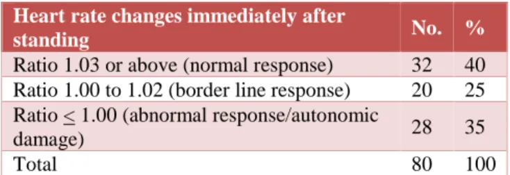
present study, 40% showed normal response, while 35% showed abnormal response. 25% showed border line response.
Table 5: Heart rate changes immediately after standing among study subjects.
Heart rate changes immediately after
standing No. %
Ratio 1.03 or above (normal response) 32 40 Ratio 1.00 to 1.02 (border line response) 20 25 Ratio < 1.00 (abnormal response/autonomic
damage) 28 35
Total 80 100
DISCUSSION
Present hospital based cross sectional study was conducted at Osmania Medical College and General Hospital which is a tertiary care hospital for a period of two years among 80 known cases of diabetes. In the present study, maximum i.e. 60% were males. Among males, maximum belonged to age group of above 55 years while among female maximum belonged to 45-54 years and above 55 years of age. Postural hypotension was noted in the long-standing diabetics. The average duration of diabetes showing postural hypotension was 13.5 years. The highest recorded postural hypotension was 50 mmHg. 25% of the patients had blood pressure fall of 30 mmHg and more. In 55% of the cases, the fall was between 10-20 mmHg. 20% of the patients show a rise of blood pressure on standing not exceeding 10 mmHg. In normal persons, the beat to beat variation with respiration are usually 15 and above per minute. If the difference is less than 10 per minute, autonomic damage is probably present. In the present study, 30% showed the autonomic damage. 45% showed normal variation. 25% showed border line variation. A Valsalva ratio of 1.21 and above is taken as normal response. The ratio 1.20 to 1.11 is taken as border line response. A ratio of 1.10 and less is abnormal and indicates autonomic damage. In our series, 30% had abnormal response. 15% had border line response and majority i.e. 55% had normal response. In normal person, the ratio is 1.03 or above. If the ratio is 1.00 or less, vagal damage is probably present. In the present study, 40% showed normal response, while 35% showed abnormal response. 25% showed border line response.
Birajdar SV et al found that abnormal E:I ratio was present in around 24 patients.6 34 patients had abnormal
Valsalva ratio, while 38 patients showed abnormal 30:15 ratio. Only 8% of the patients showed postural hypotension. 10 patients showed abnormal sustained hand grip test. 58% had cardiac autonomic neuropathy. There was statistically significant association between cardiac autonomic neuropathy and autonomic neuropathy. But there was no association between nephropathy, retinopathy and autonomic neuropathy. The author concluded that 58% had cardiac autonomic
neuropathy and the parasympathetic nervous system dominated over sympathetic nervous system.
Arif ZA et al in their study found that 36.7% of the patients had poor control over their diabetic status of type 2 and they were noted to be suffering from cardiovascular autonomic neuropathy (CAN).2 Thus, the authors
concluded that cardiovascular autonomic neuropathy (CAN) was common among those who had poor glycemic control.
Khoharo HK et al in their study had 13 years as mean duration of diabetes of the patients they studied.7 All
patients were found to have QTc prolonged. Cardiovascular autonomic neuropathy (CAN) was common with patients having duration of diabetes of more than five years as compared to patients with duration of diabetes of less than five years. And this was found to be statistically significant. The postural hypotension was significantly associated with prolonged duration of diabetes. The duration of diabetes was also found to be a significant risk factor for variation of heart rate. There was good correlation between QTc and factors like prolonged duration of diabetes, variation of heart rate and postural hypotension.
Pappachan JM et al found a prevalence of 60% of CAN.8
They reported that older people had 15.75 times more risk of developing CAN compared to younger ones. They observed that those with prolonged QTc were 5.55 times more at risk of having CAN than those with normal QTc interval. They found that those having diabetes for more than 10 years had twice the risk of developing CAN compared to those with lesser duration of diabetes. They also noted that those with peripheral neuropathy had 5.55 times more risk of having CAN than those who had no neuropathy.
Khoharo HK et al had more males in their study similar to the present study.9 Their study subjects had more mean
duration of diabetes and elevated levels of HbA1c. The authors noted an incidence 24% of CAN in their study subjects.
Kumhar MR et al found in their study that QTc dispersion is a sensitive, non-invasive, simple and cost-effective predictor of cardiac dysautonomia.10
Khoharo HK et al in their study had equal number of males and females in contrast to the present study where the number of males is more than females.11 The
incidence of CAN was 40% as reported by authors in their study. Heart rate variability was found to be significantly associated with increased duration of diabetes.
Khandoker AH et al found that in CAN group; the values of Poincare plot patterns were lesser.12 They concluded
CONCLUSION
Maximum i.e. 60% were males. The average duration of diabetes showing postural hypotension was 13.5 years. The highest recorded postural hypotension was 50 mmHg. 25% of the patients had blood pressure fall of 30 mmHg and more. In 55% of the cases, the fall was between 10-20 mmHg. 30% showed the autonomic damage. 30% had abnormal Valsalva ratio.
Funding: No funding sources Conflict of interest: None declared
Ethical approval: The study was approved by the institutional ethics committee
REFERENCES
1. Khandelwal E, Jaryal AK, Deepak KK. Pattern and prevalence of cardiovascular autonomic neuropathy in diabetics visiting a tertiary care referral center in India. Indian J Physiol Pharmacol. 2011;55(2):119-27.
2. Arif ZA, Shaikh IA, Masood N. Cardiovascular autonomic neuropathy (CAN) in patients of type 2 diabetes mellitus: A tertiary care hospital based study. Indian Heart J. 2014;66(6):751-4.
3. Vinik AI, Ziegler D. Diabetic cardiovascular autonomic neuropathy. Contemporary reviews in cardiovascular medicine. Circulation. 2007;115:387-97.
4. Maser RE, Mitchell BD, Vinik AI, Freeman R. The association between cardiovascular autonomic neuropathy and mortality in individuals with diabetes: a meta-analysis. Diabetes Care. 2003;6:1895-1901.
5. Maser R, Lenhard M, DeCherney G. Cardiovascular autonomic neuropathy: the clinical significance of its determination. Endocrinologist. 2000;10:27-33.
6. Birajdar SV, Chavan SS, Munde SA, Bende YP. A Study of autonomic nervous system dysfunction among patient with diabetes mellitus: a cross sectional study. Int J Adv Med. 2017;4:406-11. 7. Khoharo HK, Halepoto AW. QTc-interval, heart
rate variability and postural hypotension as an indicator of cardiac autonomic neuropathy in type 2 diabetic patients. J Pak Med Assoc. 2012;62(4):328-31.
8. Papachan JM, Sebastian J, Bino BC, Jayaprakash K, Vijaykumar K, Sujathan P, et al. Cardiac autonomic neuropathy in diabetes mellitus: prevalence, risk factors and utility of corrected QT interval in the ECG for its diagnosis. Postgrad Med J. 2008;84(990):205-10.
9. Khoharo HK, Ansari S, Ali Shaikh I, Qureshi F. Cardiac autonomic neuropathy (CAN) in type-1 diabetes mellitus patients and its association with the duration of disease and glycemic control. J Coll Physicians Surg Pak. 2009;19(4):232-5.
10. Kumhar MR, Agarwal TD, Singh VB, Kochar DK, Chadda VS. Cardiac autonomic neuropathy and its correlation with QTc dispersion in type 2 diabetes. Indian Heart J. 2000;52(4):421-6.
11. Khoharo HK, Qureshi F. Frequency of cardiac autonomic neuropathy in patients with type 2 diabetes mellitus reporting at a teaching hospital of Sindh. J Coll Physicians Surg Pak. 2008;18(12):751-4.
12. Khandoker AH, Jelinek HF, Palaniswami M. Identifying diabetic patients with cardiac autonomic neuropathy by heart rate complexity analysis. Biomed Eng Online. 2009;8:3.