Flushing the Liver With Urokinase
Before Transplantation Does Not
Prevent Nonanastomotic Biliary
Strictures
Lars C. Pietersen,1,2 A. Claire den Dulk,3 Andries E. Braat,1 Hein Putter,4
Kerem Sebib Korkmaz,3 Andre G. Baranski,1 Alexander F. M. Schaapherder,1 Jeroen Dubbeld,1 Bart van Hoek,3* and Jan Ringers1*
1Department of Transplant Surgery, Leiden University Medical Center, Leiden, the Netherlands 2Eurotransplant International Foundation, Leiden, the Netherlands, Departments of 3Gastroenterology and Hepatology, 4Medical Statistics, Leiden University Medical Center, Leiden, the Netherlands
The aim of the present study was to assess whether flushing the donor liver with urokinase immediately before implanta-tion reduces the incidence of nonanastomotic biliary strictures (NASs) after liver transplantaimplanta-tion, without causing increased blood loss, analyzed as a historical cohort study. Between January 2005 and October 2012, all liver (re-)transplantations were included. Of the 185 liver transplant recipients included, 63 donor livers between January 2010 and October 2012 received urokinase (study group), whereas the donor liver of 122 consecutive recipients, who served as a historical control group, between January 2005 and January 2010 did not receive urokinase. Basic donor (Eurotransplant donor risk index) and recipient (age, body mass index, laboratory Model for End-Stage Liver Disease score) characteristics did not signifi-cantly differ in both groups. Thirty-three recipients developed NASs: 22 in the control group (18%) and 11 (17.5%) in the study group (P50.68). Analyzed separately for donation after circulatory death (P50.42) or donation after brain death (P50.89), there was no difference between the groups in incidence of NAS. Of all the recipients developing NAS, 7 (21%) needed retransplantation and all others were treated conservatively. Autologous blood transfusion requirements did not differ significantly between both groups (P50.91), whereas interestingly, more heterologous blood transfusions were needed in the control group (P<0.001). This study has its limitations by its retrospective character. A multi-institutional prospective study could clarify this issue. In conclusion, arterial flushing of the liver with urokinase immediately before implantation did not lead to a lower incidence of NAS in this study, nor did it lead to increased blood loss.
Liver Transplantation 22 420-426 2016 AASLD
Received June 30, 2015; accepted October 30, 2015.
Biliary complications are a well-known, major cause of morbidity and graft failure in recipients after liver transplantation.(1,2) The most troublesome are the
so-called nonanastomotic biliary strictures (NASs), with an incidence of 5%-15% reported in most current studies,(3,4) and in up to 30% of patients receiving a
Abbreviations: aPTT, activated partial thromboplastin time; BMI, body mass index; CIT, cold ischemia time; DBD, donation after brain death; DCD, donation after circulatory death; ET-DRI, Eurotransplant donor risk index; FWIT, first warm ischemia time; HAT, hepatic artery thrombosis; HTK, histidine tryptophan ketoglutarate; labMELD, laboratory Model for End-Stage Liver Disease; NAS, nonanastomotic biliary stricture; PTT, partial thromboplastin time; RBC, red blood cell; SD, standard deviation; tPA, tissue plasminogen activator; WIT, warm ischemia time.
Address reprint requests to Andries E. Braat, M.D., Ph.D., Department of Transplant Surgery, Leiden University Medical Center, Albinusdreef 2, 2333 ZA Leiden, the Netherlands. Telephone:131 71 526 6188; E-mail: a.e.braat@lumc.nl
CopyrightVC2015 by the American Association for the Study of Liver Diseases
View this article online at wileyonlinelibrary.com. DOI 10.1002/lt.24370
liver from donation after circulatory death (DCD).(5) With direct treatment of strictures, by using endo-scopic or percutaneous cholangiographic dilatations and stenting, more than 50% of patients with NASs can be treated successfully.(6-11)
The pathophysiology of NAS development still remains unknown. Over the years, several risk factors have been indicated, suggesting that its origin may be multifactorial. In addition to immunological injury and bile salt–induced injury, it is suggested that ischemia injury to the peribiliary vascular plexus plays a critical role.(12) During the donor procedure, the peribiliary arterial plexus may not be completely flushed out. Because the blood supply to the biliary tract is solely dependent on arterial inflow, these microcirculatory disturbances in the peribiliary plexus may lead to obstruction and may subsequently result in insufficient bile duct preservation.(13,14)
Three previous studies with historical controls sug-gest that adding a thrombolytic agent, such as uroki-nase or tissue plasminogen activator (tPA) to the preservation fluid (after trimming of the donor liver, on the back table, or before completion of the portal vein anastomosis), seems to reduce the incidence of NAS. The hypothesis was that this might be the result from dissolving microthrombi in the microvascular sys-tem of the biliary tree.(15-17)
The aim of the present study was to assess whether flushing the donor liver with urokinase directly before transplantation reduces the incidence of NAS without causing increased blood loss.
Patients and Methods
Between January 2005 and October 2012, all ortho-topic liver transplantations at the Leiden University Medical Center (Leiden, the Netherlands) were included in this study.
Exclusion criteria were domino, split, or auxiliary liver transplantations. Clinical information was obtained from a prospectively collected database. Covariates included donor demographics, recipient demographics, pretransplant information, intraoperative data, and post-operative outcomes. Calculated laboratory Model for End-Stage Liver Disease (labMELD) scores were included in the recipient analysis.
The labMELD score was calculated using laboratory data (creatinine, bilirubin, international normalized ratio) and did not include exception points that were given for liver malignancies or other medical conditions.
On the basis of existing literature, a protocol change was made as of January 2010 to flush the donor liver with urokinase, directly before transplantation. This protocol change was approved by the institutional ethics committee.
DEFINITION OF NAS
NAS was defined as described by Ten Hove et al.(18) NAS was any stricture or irregularity of the intrahepatic or extrahepatic bile ducts of the liver graft that was at least 1 cm above the anastomosis, with or without dilation and with or without biliary sludge formation, and treated endoscopically with endoscopic retrograde cholangiopan-creatography and dilation and/or stenting, percutaneously with percutaneous transhepatic cholangiography and bili-ary drainage or by surgical intervention. Therefore, all these NASs were clinically significant. Hepatic artery thrombosis (HAT) by either Doppler ultrasound, or con-ventional angiography, as well as isolated strictures/steno-sis at the bile duct anastomostrictures/steno-sis and related dilations were, by definition, excluded from analysis.
OPERATIVE TECHNIQUES
was done according to protocol. Before January 1, 2010, the same protocol was carried out, only without administration of urokinase. After consultation of the medical ethics committee, recipients did not have to give informed consent because the administration of urokinase was implemented as a new center protocol.
STATISTICAL ANALYSIS
Continuous variables were presented as median (range) and standard deviation, whereas categorical variables were presented as number and percentage. Patient and graft sur-vival curves and the cumulative incidence of NAS were cal-culated using the Kaplan-Meier method and compared using the log-rank test. Categorical variables were com-pared with the Pearson’s chi-square test or Fisher’s exact test, where appropriate. Characteristics of the donor, trans-plantation, and recipient were analyzed using the 2-tailed Studentttest. Blood loss was analyzed using the Mann-Whitney U test. The level of significance was set at 0.05. Statistical analyses were performed using SPSS software version 22.0 for Windows (SPSS Inc., Chicago, IL).
POWER ANALYSIS
With an anticipated reduction of NAS from 45% to 10% on the basis of previous studies on DCD liver
transplantation,(15-17)the power of this study would be 83.2% when comparing 28 DCD livers in the study group to 17 DCD livers in the control group.
With an anticipated reduction of NAS from 20% to 5% on the basis of previous studies concerning DBD liver transplantation, the power of this study would be 80.1% when comparing 94 DBD livers in the study group to 46 DBD livers in the control group.
Results
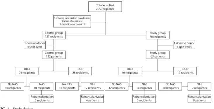
Of the 205 patients who received a liver transplanta-tion between January 2005 and October 2012, 5 recipients were excluded based on missing informa-tion on receiving urokinase, 3 recipients were excluded based on protocol deviation (Fig. 1). Of the 197 liver recipients remaining for the study, 127 donor livers did not receive urokinase (historic control group), and 70 donor livers received urokinase (study group).
In the historic control group, 5 recipients were excluded (4 split-liver transplantations, 1 domino donor), leaving 122 recipients in this group. In the study group, 7 recipients were excluded (6 split-liver transplantations, 1 domino donor), leaving 63 recipients.
FIG. 1. Study design.
DONOR AND RECIPIENT
CHARACTERISTICS
Table 1 shows the basic donor and recipient character-istics of both groups. The mean Eurotransplant donor risk index (ET-DRI)(19) in the control group was 1.860.3 (range, 1-3.1), in the study group 1.860.4 (range, 1-2.6; P50.56). Of 3 donors, the ET-DRI could not be calculated. Of the donors in the control group, 51% were female versus 48% in the study group (P50.76). Donor body mass index was lower in the control group than in the study group. The mean cold ischemia time (CIT) of the transplanted livers in the control group was 5726142 minutes (224-1090 minutes), in the study group 5356129 minutes (range, 230-850 minutes; P50.09). The mean first warm ischemia time in the control group was 16.765
minutes (range, 11-31 minutes), in the study group 17.666 minutes (range, 9-31 minutes; P50.60). The mean labMELD score in the control group was 16.668.7 (range, 6-40), in the study group 16.668.9 (range, 6-40;P50.99).
BILIARY COMPLICATIONS
In total, 33 (17.8%) recipients developed NASs, of which 22 (18%) recipients were in the control group, and 11 (17.5%) recipients were in the study group. None of the recipients had evidence of HAT or stenosis. The mean follow-up in the control group was 154361049 days (range, 1-3278 days) versus 6756495 days (range, 1-1434 days) in the study group. The median number of days of follow-up was 173161049 days (range, 312-2356 days) in the study group versus 7316495 days (range, 119-1109 days) in the control group (Table 1).
In the control group, the mean number of days until NAS was diagnosed was 2956363 days (range, 22-1454 days). In the study group, the mean number of days was 1896202 days (range, 30-723 days;P50.38). Graft survival, censored for death, shows equal results for both groups (P50.68; Fig. 2). In the control group, the median number of days until NAS was diagnosed was 1726363 days (range, 71-346) compared to 1196202 days (range, 48-216) in the study group.
Comparison of liver transplantations from DCD donors only also showed equal graft survival; 7 (41%) recipients in the study group developed NAS versus 12 (43%) recipients in the control group (P50.42).
In the control group, 10 (11%) recipients who received a liver allograft from DBD donors developed NAS versus 4 (9%) recipients in the study group. This was not different (P50.89). Of all cases, 7 (21%) recipients needed retransplantation for NAS.
TABLE 1. Donor, Transplant, and Recipient Characteristics
Urokinase Group (n563) Controls (n5122) P Value
ET-DRI 1.860.3 1.860.3 0.56
Donor age, years 49.4615.0 46.9614.2 0.27
Donor BMI, kg/m2 25.263.3 24.063.3 0.03
FWIT, minutes 17.666.0 16.764.5 0.6
CIT, minutes 535.06129.0 572.06142.2 0.09
Recipient WIT, minutes 35.968.5 33.868.4 0.12
Recipient age, years 51.9611.6 52.3610.8 0.80
Recipient BMI, kg/m2 25.864.6 26.264.8 0.53
LabMELD 16.668.9 16.668.7 0.99
Time to NAS diagnosis, days 1196202 1726363
Time of follow-up, days 7316495 173161049
NOTE: Data are presented as mean6SD. Time to NAS diagnosis and time of follow-up are presented as median6SD.
FIG. 2. Graft survival, censored for death.
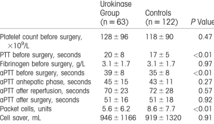
POSTREPERFUSION BLOOD LOSS
Table 2 shows the hematological and coagulation parameters of both groups preoperative; the activated partial thromboplastin time (aPTT) values during anhepatic phase, after reperfusion, and after surgery; the number of packed red blood cells (RBCs) trans-fused during the first 24 hours from incision; and vol-ume of autologous blood transfused.
Most remarkably, the mean preoperative aPTT in the control group was 34.567.5 versus 38.567.5 seconds in the study group (P0.01), whereas aPTT did not differ between the study group and control group in the anhepatic phase after reperfusion and after surgery.
The mean packed RBCs transfused in the control group was 8.667.7 versus 5.666.2 units in the study group (P<0.01). The mean volume of autologous blood transfused in the control group was 91961320 versus 94661166 mL in the study group (P50.91).
Discussion
The present retrospective cohort study demonstrates that flushing the donor liver with urokinase immedi-ately before liver transplantation is safe but does not prevent the development of NAS. It also did not lead to an increase of transfusion requirements or disturbed clotting.
In contrast to previous studies(15-17) describing a decrease in the rate of NAS after thrombolysis before liver transplantation, this study could not find a decrease in NAS rate. The first study by Hashimoto et al.(15)was a retrospective study, with the injection of tPA in the donor hepatic artery on the back table in
DCD liver transplantations, resulting in significantly less NAS development without increased blood loss. Lang et al.(16)described a prospective study with dou-ble perfusion of the donor liver with urokinase in DCD liver transplantation, resulting in significantly less NAS after 1 year of follow-up. Seal et al.(17) described a retrospective analysis of DCD liver trans-plantations with an intraoperative tPA injection, which minimized the incidence of NAS without increasing the need of intraoperative blood transfusion. When looking closely at the dosage, type, and timing of the thrombolytic agent, our study has some minor differ-ences compared to the studies previously published. In the study by Hashimoto et al.,(15)heparin was given to the donor before withdrawal of life support, and the liver allograft was perfused on the back table with 0.5 mg/100 g graft tPA in the donor hepatic artery. Lang et al.(16)perfused the arterial system of the donor liver twice. First, they used a dosage of 2000 mL HTK solution that contained 2 MU urokinase for perfusion through the arterial system. After trimming of the donor liver, the arterial system was perfused again with 1 MU urokinase. In the study by Seal et al.,(17)heparin was given to the donor before withdrawal of life sup-port and, based on donor’s weight and before comple-tion of the portal vein anastomosis, 100 mg/kg tPA was perfused in the donor liver to account for varia-tions in graft size.(15-17) In our study, the donor liver was perfused through the hepatic artery after inspec-tion on the back table, with a high fixed dose of 250,000 IU of urokinase, manually pressurized, in order to dissolve possible microthrombi.
To our knowledge, no previous studies have been published that show superiority for tPA, compared to urokinase, in low temperature circumstances.
Furthermore, the CIT in the study by Hashimoto et al.(15) and Seal et al.(17) was much shorter. They described a CIT of 422696 minutes and 5.161.2 hours, whereas the CIT in this study was 5726142 minutes in the control group and 5356129 minutes in the study group. Lang et al.(16) described a CIT between 2 and 13.5 hours but did not mention a mean value.
The effect of thrombolytic agents has been reported in the experimental as well as clinical transplantation of various organs from DCD donors,(20-23)suggesting that pretreatment with thrombolytic agents could be helpful in human liver transplantation. A possible explanation for not seeing a decrease in the rate of NAS in the cur-rent study could be the timing of the intervention. The therapeutic principle of administrating urokinase is to TABLE 2. Hematological, Coagulation, and Transfusion
Parameters Before, During, and After Surgery
Urokinase Group (n563)
Controls
(n5122) P Value
Platelet count before surgery, 3109/L
128696 118690 0.47
PTT before surgery, seconds 2068 1765 <0.01
Fibrinogen before surgery, g/L 3.161.7 3.161.7 0.97
aPTT before surgery, seconds 3968 3568 <0.01
aPTT anhepatic phase, seconds 45615 43611 0.27
aPTT after reperfusion, seconds 70623 72628 0.57
aPTT after surgery, seconds 51616 51618 0.92
Packet cells, units 5.666.2 8.667.7 <0.01
Cell saver, mL 94661166 91961320 0.91
dissolve possible microthrombi from the peribiliary microcirculation. It may be that administrating uroki-nase immediately after organ procurement may be bene-ficial, whereas late administration of urokinase may not be able to prevent or dissolve microthrombosis. How-ever, this hypothesis is not supported by Seal et al.,(17) who administered tPA before completion of the portal vein anastomosis in order to limit the effects of hypo-thermia and dilution. The administration of urokinase in the donor liver is not part of the liver transplantation protocol in many other centers in the Eurotransplant region. For that reason, we were not able to administer urokinase during the donation procedure.
The absence of microthrombi in the microvascular system of the biliary tree also has to be considered as a possible explanation why urokinase in our hands did not prevent NAS. In a study by op den Dries et al.,(24) biopsies were taken from the donor bile duct in 128 liver transplant procedures. In the peribiliary plexus, thrombi were found in only 2.7% of these bile ducts from the livers that developed NAS, suggesting that thrombosis may not play a critical role in the develop-ment of NAS.
In a study by Hansen et al.,(25) histological evalua-tions of 93 donor common bile ducts, received after recirculation of the hepatic artery and before biliary end-to-end anastomosis in LT, were performed. With regard to NAS, they found that necrosis of the bile duct wall, arteriolonecrosis, vascular lesions, and intra-mural bleeding were statistically relevant associated factors. Thrombosis was not of statistical influence on occurrence of NAS. This also supports the theory that thrombosis does not play a critical role in the develop-ment of NAS.
Furthermore, Burlage et al.(26) believed that other factors, such as increased experience, explain the observed differences found in the study by Seal et al.(17)
The use of allogeneic and autologous blood as a measurement of blood loss has been described before by Hendriks et al.(27) Surprisingly, in this study, there was significantly more need for packed RBC transfu-sion in the control group, even though this group had significantly better coagulation parameters preopera-tively. A possible explanation for this may be a positive effect of increasing experience within the transplanta-tion team, as has been described previously.(27,28) During the study period, the transfusion protocol has not changed.
The extent and severity of NAS after liver trans-plantation determines its prognosis and management. Diffuse strictures have worse prognosis than local
strictures because of a lack of therapeutic options. Even though we did not see a significant difference in the rate of NAS, the number of retransplantations due to NAS in the first year after transplantation could potentially show a difference because of different sever-ity of NAS. Of all cases of NAS in the control group, 6 (27%) recipients required retransplantation in the first year after liver transplantation. Five (23%) recipi-ents received a retransplantation in the first year after liver transplantation and 1 (5%) recipient was placed on the waiting list for retransplantation. In the study group, none of the recipients received a retransplanta-tion in the first year after liver transplantaretransplanta-tion, and 1 (9%) recipient was placed on the waiting list for retransplantation. This difference was not significant (P50.32). All other cases of NAS were treated con-servatively by using balloon dilatation of the bile duct combined with the placement of an intraductal stent. The median number of interventions for NAS was 5.062.4 (range, 2.0-7.0) in the control group versus 4.564.2 (range, 2.0-8.0) in the study group.
Even though the median number of days of follow-up is shorter in the study grofollow-up, compared to the control group, we do not believe this difference had an influence on the outcome because the median number of days until NAS diagnosis in both groups (1726363 days [range, 71-346 days] in the control group versus 1196202 days [range, 48-216 days] in the study group) is much shorter compared to the median number of days of follow-up in both groups (173161049 days in the study group versus 7316495 days in the control group). Because NAS is more common after DCD liver transplantation, compared to DBD liver transplantation, both groups were analyzed separately. When analyzed separately, no difference was found in the incidence of NAS after DCD liver transplantation (P50.42). Also, no difference was found in the incidence of NAS after DBD liver transplantation (P50.89).
This study has its limitations by its retrospective character. A multi-institutional prospective study could clarify this issue.
To conclude, arterial flushing of the donor liver with urokinase immediately before implantation did not lead to a lower incidence of NAS in this study, nor did it lead to increased blood loss.
REFERENCES
conduit between the donor and recipient common bile ducts. Ann Surg 1976;184:605-609.
2) Starzl TE, Marchioro TL, Vonkaulla KN, Hermann G, Brittain RS, Waddell WR. Homotransplantation of the liver in humans. Surg Gynecol Obstet 1963;117:659-676.
3) Sawyer RG, Punch JD. Incidence and management of biliary complications after 291 liver transplants following the intro-duction of transcystic stenting. Transplantation 1998;66: 1201-1207.
4) Pascher A, Neuhaus P. Biliary complications after deceased-donor orthotopic liver transplantation. J Hepatobiliary Pancreat Surg 2006;13:487-496.
5) Dubbeld J, Hoekstra H, Farid W, Ringers J, Porte RJ, Metselaar HJ, et al. Similar liver transplantation survival with selected car-diac death donors and brain death donors. Br J Surg 2010;97: 744-753.
6) Gopal DV, Pfau PR, Lucey MR. Endoscopic management of biliary complications after orthotopic liver transplantation. Curr Treat Options Gastroenterol 2003;6:509-515.
7) Guichelaar MM, Benson JT, Malinchoc M, Krom RA, Wiesner RH, Charlton MR. Risk factors for and clinical course of non-anastomotic biliary strictures after liver transplantation. Am J Transplant 2003;3:885-890.
8) Li S, Stratta RJ, Langnas AN, Wood RP, Marujo W, Shaw BW Jr. Diffuse biliary tract injury after orthotopic liver trans-plantation. Am J Surg 1992;164:536-540.
9) Rerknimitr R, Sherman S, Fogel EL, Kalayci C, Lumeng L, Chalasani N, et al. Biliary tract complications after orthotopic liver transplantation with choledochocholedochostomy anastomo-sis: endoscopic findings and results of therapy. Gastrointest Endosc 2002;55:224-231.
10) Sanchez-Urdazpal L, Gores GJ, Ward EM, Maus TP, Buckel EG, Steers JL, et al. Diagnostic features and clinical outcome of ischemic-type biliary complications after liver transplantation. Hepatology 1993;17:605-609.
11) Ward EM, Kiely MJ, Maus TP, Wiesner RH, Krom RA. Hilar biliary strictures after liver transplantation: cholangiography and percutaneous treatment. Radiology 1990;177:259-263.
12) Buis CI, Verdonk RC, Van der Jagt EJ, van der Hilst CS, Slooff MJ, Haagsma EB, Porte RJ. Nonanastomotic biliary strictures after liver transplantation, part 1: Radiological features and risk factors for early vs. late presentation. Liver Transpl 2007;13:708-718. 13) Pirenne J, Van Gelder F, Coosemans W, Aerts R, Gunson B,
Koshiba T, et al. Type of donor aortic preservation solution and not cold ischemia time is a major determinant of biliary strictures after liver transplantation. Liver Transpl 2001;7:540-545. 14) Canelo R, Hakim NS, Ringe B. Experience with hystidine
tryp-tophan ketoglutarate versus University Wisconsin preservation solutions in transplantation. Int Surg 2003;88:145-151. 15) Hashimoto K, Eghtesad B, Gunasekaran G, Fujiki M, Uso TD,
Quintini C, et al. Use of tissue plasminogen activator in liver transplantation from donation after cardiac death donors. Am J Transplant 2010;10:2665-2672.
16) Lang R, He Q, Jin ZK, Han DD, Chen DZ. Urokinase perfu-sion prevents intrahepatic ischemic-type biliary leperfu-sion in donor livers. World J Gastroenterol 2009;15:3538-3541.
17) Seal JB, Bohorquez H, Reichman T, Kressel A, Ghanekar A, Cohen A, et al. Thrombolytic protocol minimizes ischemic-type biliary complications in liver transplantation from donation after circulatory death donors. Liver Transpl 2015;21:321-328. 18) Ten Hove WR, Korkmaz KS, op den Dries S, de Rooij BJ, van
Hoek B, Porte RJ, et al. Matrix metalloproteinase 2 genotype is associated with nonanastomotic biliary strictures after orthotopic liver transplantation. Liver Int 2011;31:1110-1117.
19) Braat AE, Blok JJ, Putter H, Adam R, Burroughs AK, Rahmel AO, et al.; for European Liver and Intestine Transplant Associa-tion (ELITA) and Eurotransplant Liver Intestine Advisory Com-mittee (ELIAC). The Eurotransplant donor risk index in liver transplantation: ET-DRI. Am J Transplant 2012;12:2789-2796. 20) Yamauchi JI, Richter S, Vollmar B, Menger MD, Minor T.
Warm preflush with streptokinase improves microvascular pro-curement and tissue integrity in liver graft retrieval from non-heart-beating donors. Transplantation 2000;69:1780-1784. 21) Minor T, Hachenberg A, Tolba R, Pauleit D, Akbar S.
Fibrino-lytic preflush upon liver retrieval from non-heart beating donors to enhance postpreservation viability and energetic recovery upon reperfusion. Transplantation 2001;71:1792-1796.
22) Sugimoto R, Date H, Sugimoto S, Okazaki M, Aokage K, Inokawa H, et al. Post-mortem administration of urokinase in canine lung transplantation from non-heart-beating donors. J Heart Lung Transplant 2006;25:1148-1153.
23) Gok MA, Shenton BK, Buckley PE, Peaston R, Cornell C, Soomro N, et al. How to improve the quality of kidneys from non-heart-beating donors: a randomised controlled trial of thrombolysis in non-heart-beating donors. Transplantation 2003;76:1714-1719. 24) op den Dries S, Westerkamp AC, Karimian N, Gouw AS, Bruinsma
BG, Markmann JF, et al. Injury to peribiliary glands and vascular plexus before liver transplantation predicts formation of non-anastomotic biliary strictures. J Hepatol 2014;60:1172-1179. 25) Hansen T, Hollemann D, Pitton MB, Heise M,
Hoppe-Lotichius M, Schuchmann M, et al. Histological examination and evaluation of donor bile ducts received during orthotopic liver transplantation--a morphological clue to ischemic-type bili-ary lesion? Virchows Arch 2012;461:41-48.
26) Burlage LC, Karangwa SA, Lisman T, Martins PN, Porte RJ. Thrombolytic protocol minimizes ischemic-type biliary complica-tions in liver transplantation from donation aftercirculatory death donors. Liver Transpl 2015;21:1231-1232.
27) Hendriks HG, van der Meer J, Klompmaker IJ, Choudhury N, Hagenaars JA, Porte RJ, et al. Blood loss in orthotopic liver trans-plantation: a retrospective analysis of transfusion requirements and the effects of autotransfusion of cell saver blood in 164 consecutive patients. Blood Coagul Fibrinolysis 2000;11(suppl 1):S87-S93. 28) Deakin M, Gunson BK, Dunn JA, McMaster P, Tisone G,