SHORT COMMUNICATION
Potent neutralization of SARS-CoV-2 by human antibody heavy-chain variable
domains isolated from a large library with a new stable scaffold
Zehua Suna*, Chuan Chena*, Wei Lia*, David R. Martinezb*, Aleksandra Drelichc*, Du-San Baeka, Xianglei Liua,
John W. Mellorsa,d, Chien-Te Tsengc, Ralph S. Baricb, and Dimiter S. Dimitrova,d
aCenter for Antibody Therapeutics, Division of Infectious Diseases, Department of Medicine, University of Pittsburgh Medical School, Pittsburgh, PA, USA; bDepartment of Epidemiology, University of North Carolina at Chapel Hill, Chapel Hill, NC, USA; cDepartment of Microbiology & Immunology, Centers for Biodefense and Emerging Diseases, Galveston National Laboratory, Galveston, TX, USA; dAbound Bio, Pittsburgh, PA, USA
ABSTRACT
Effective therapies are urgently needed for COVID-19. Here we describe the identification of a new stable human immunoglobulin G1 heavy-chain variable (VH) domain scaffold that was used for the construction of a large library, lCAT6, of engineered human VHs. This library was panned against the receptor-binding domain (RBD) of the SARS-CoV-2 spike (S) glycoprotein. Two VH domains (VH ab6 and VH m397) were selected and fused to Fc for increased half-life in circulation. The VH-Fc ab6 and m397 specifically neutralized SARS-CoV-2 with high potencies (50% neutralization at 0.35 µg/ml and 1.5 µg/ml, respectively) as measured by two independent replication-competent virus neutralization assays. Ab6 and m397 competed with ACE2 for binding to RBD, suggesting a competitive mechanism of virus neutralization. These VH domains may have potential applications for prophylaxis and therapy of COVID-19 alone or in combination, as well as for diagnosis and as tools for research.
ARTICLE HISTORY
Received 18 May 2020 Revised Vxx xxx xxxx Accepted 20 May 2020
KEYWORDS
Therapeutic antibodies; coronaviruses; SARS-CoV-2
Introduction
As of early 2020, the severe acute respiratory syndrome cor-onavirus 2 (SARS-CoV-2)1 has spread worldwide and became a global pandemic.2,3 Recent statistics showed that more than 3 million people were confirmed positive of virus infection globally, and the number is growing. Safe and effective preven-tions and therapies are therefore urgently needed. Before an effective vaccine is available, passive antibody therapy will play an important role in medical care. Human monoclonal anti-bodies (mAbs), which are typically target-specific and nontoxic to humans, are promising therapeutics4,5 as COVID-19 inter-ventions based on the experience from human mAbs against other emerging viruses, including SARS-CoV, MERS-CoV, and henipaviruses.6-8 More than 80 antibody therapeutics have been granted marketing approval and more than 570 antibody-derived candidates are being investigated in clinical trials.9,10 Here, we describe the potent neutralization of SARS- CoV-2 by human VH domains isolated from a large phage- displayed library with a new stable scaffold.
Results
Phage display is well established among in vitro mAb selection technologies,11-13 and more than 12 antibodies isolated by phage display of different format libraries have been approved for therapy.14 Human VH domains are relatively small and
could interact with amino acid residues in the target which are not accessible by larger antibody fragments. Importantly, their small size would allow better penetration in normal and dis-eased tissue. Previously, we demonstrated the construction of a human antibody variable heavy-chain (VH) domain library based on a VH scaffold from IGHV3-23, and isolated neutra-lizers against HIV-1 from this library.15 In recent studies, more VH scaffolds, including IGHV3-7, IGHV4-34, IGVH1-69, IGVH3-15, and IGVH5-51, were chosen for library construc-tion based on their frequency in the memory compartment of the immune repertoires.16,17
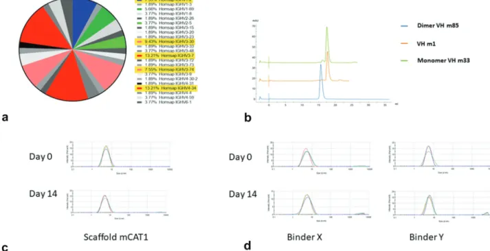
A major concern for generation of VH-based therapeutics is their stability. To identify a relative stable scaffold for VH libraries but without introducing mutations leading to poten-tial immunogenicity we screened natively occurring stable VH scaffolds using a heat denaturation and refolding strategy.18 We constructed a small VH phage-displayed library (size ~1 × 10⁷) from 3 healthy human donors’ peripheral blood mononuclear cells (PBMCs) and heated it at 80°C for 20 min-utes. Chilled phages were spun down and supernatant was collected for bacterial TG1 infection. After three rounds of heating, refolding, and amplification, we evaluated a panel of stable VH scaffolds (Figure 1(a)). One VH scaffold, mCAT1, which is of IGHV3-30, was selected. This scaffold was mono-meric as determined by size exclusion chromatography (SEC) (Figure 1(b)) and dynamic light scattering (DLS) (Figure 1(c)). It was stable at 37°C and showed less than 1% aggregation after
CONTACT Dimiter S. Dimitrov [email protected] ScD Center for Antibody Therapeutics, Division of Infectious Diseases, Department of Medicine, University of Pittsburgh Medical School, Pittsburgh, PA 15261, USA; Zehua Sun [email protected] Center for Antibody Therapeutics, Division of Infectious Diseases, Department of Medicine, University of Pittsburgh Medical School, S848 Scaife Hall, 3550 Terrace Street, Pittsburgh, PA 15261, USA
*These authors contributed equally to this work.
Supplemental data for this article can be accessed on the publisher’s website. 2020, VOL. 12, NO. 1, 1–6
https://doi.org/10.1080/19420862.2020.1778435
© 2020 University of Pittsburgh. Published with license by Taylor & Francis Group, LLC.
incubation for 14 days (Figure 1(c)). Other highly enriched scaffolds, IGHV3-7, IGHV4-34, are shown in Fig. S1.
A VH domain library, lCAT6, was constructed by grafting naturally occurring CDR2 s and CDR3 s from human PBMCs to mCAT1 as well as to IGHV3-7-, IGHV4-34-, and IGVH3- 23-based scaffolds following the strategy described previously.15 The IGHV3-7-, IGHV4-34-, and IGVH3-23- based scaffolds were added to increase diversity although the mCAT1 scaffold was still dominant representing 40% of the clones. The library construction is shown in Fig.S2. No repeat-ing sequences were found in 100 clones indicatrepeat-ing good diver-sity. In addition, only 21% of the clones were nonproductive (10% of clones had stop codons, and 11% of clones were out of frame). The size of the library was estimated to be 7.9 × 10¹⁰ after subtracting the nonproductive clones based on the titra-tion of transformants. The library functitra-tionality was tested by panning it against a human protein antigen that is unrelated to the RBD of the SARS-CoV-2 spike protein; two high-affinity binders, VH X and VH Y, were selected which did not aggre-gate after incubation at 37°C for 14 days as measured by DLS (Figure 1(d)).
We used ICAT6 for the selection of high-affinity VH domains by panning it against recombinant SARS-CoV-2 RBD developed in our laboratory. We identified a panel of binders, two of which, ab6 and m397, were further character-ized for affinity, competition with the SARS-CoV-2 receptor ACE2, and neutralization.
Both VH ab6 and m397 bound cell surface-associated native S glycoprotein, confirming that their epitopes are accessible and not changed on the native S protein (Figure 2 (a)). The equilibrium dissociation constant, Kd, of these VH
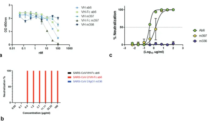
binders (ab6 and m397) to recombinant RBD as measured by BLItz was 4.6 nM and 29.2 nM, respectively (Figure 2(b)). Both ab6 and m397 competed with ACE2 for binding to the RBD (Figure 3(a)), indicating that the virus could be neutra-lized by preventing its binding to the receptor. To extend their
in vivo half-lives and potentially their neutralizing activity, we converted ab6 and m397 to the Fc fusion proteins VH-Fc ab6 and VH-Fc m397, respectively. Both VH-Fc ab6 and VH-Fc m397 bound cell-surface-associated native S glycoprotein (Figure 2(c)). The Kd of the VH-Fc ab6 and VH-Fc m397 to recombinant RBD as measured by BLItz was 11 nM and 9.6 nM, respectively (Figure 2(d)). Interestingly and rather unusual, the VH-Fc ab6 exhibited higher Kd (11 nM) to RBD compared to VH ab6 (4.6 nM) mostly due to its significantly decreased association rate constant ka – about 10-fold – smaller than that for m397 (about fourfold decrease). Part of this decrease is due to the larger size of the VH-Fc compared to VH resulting from the smaller ratio of the paratope to the total area of the molecule and decreasing the number of effective encounters by Brownian motion as is also for m397. However, significant portion could be due to other factors which remain to be elucidated. In addition, the gain in the dissociation rate constant (about four times, due to avidity because of increased local concentration) for ab6 is smaller than that for m397 which is about 10 times. The unusually large decrease in the association rate constant com-bined with the relatively small decrease in the dissociation rate constant resulted in the relatively rare situation when avidity of binding is weaker than affinity, one should also take into account that there are two VHs in Fc fusion protein and that avidity depends on the surface concentration of the
Figure 2. Binding characterization of VH ab6 and m397 with or without human IgG1 Fc. (a) Flow cytometry analysis for binding of VH ab6 and m397 to S-glycoprotein expressed on the surface of HEK293 T cell (293 T-S). (b) Representative sensorgrams for VH ab6 and m397. Equilibrium dissociation constants (Kd) = 4.6 nM (ka = 4.4E4 1/
Ms, kd = 2.0E-4 1/s) and 29.2 nM (ka = 1.3E5 1/Ms, kd = 3.9E-3 1/s), respectively. (c) Flow cytometry analysis for binding of VH-Fc ab6 and m397 to S-glycoprotein
expressed on the surface of HEK293 T cell. (d) Representative sensorgrams for VH-Fc ab6 and m397. Equilibrium dissociation constants (Kd) = 11 nM (ka = 4.6E3 1/Ms, kd
= 5.3E-5 1/s), and 9.6 nM (ka = 3.4E4 1/Ms, kd = 3.3E-4 1/s), respectively.
target molecules. We plan to study the mechanism of this unusual finding in detail including dose-dependent binding to native S protein expressed in virions and on the cell surface. Consistent with weaker binding compared to VH ab6, VH-Fc ab6 showed decreased competition with ACE2 for binding to RBD compared to VH ab6, while VH-Fc m397 exhibited stronger competition to ACE2 compared to VH m397 due to its enhanced binding avidity to RBD (Figure 3(a)).
VH-Fc ab6 was further characterized by both a standard microneutralization-based assay and a luciferase reporter gene assay to determine its neutralizing activity against live SARS- CoV-2 virus, whereas VH-Fcm397 was evaluated only by the luciferase reporter gene assay. VH-Fc ab6 and VH-Fc m397 exhibited potent activity, neutralizing 50% of the virus at con-centrations of 0.35 µg/ml (≈4.6 nM) and 1.5 µg/ml (≈20 nM), respectively, as measured by the luciferase reporter gene assay; VH-Fc ab6 neutralized 100% live virus at 0.4 µg/ml and 0% at 0.1 µg/ml as measured by the microneutralization-based assay (Figure 3(b,c)) which is consistent with the result from the reporter gene assay. In agreement with the specificity of bind-ing to the SARS-CoV-2 RBD and not to the SARS-CoV RBD, neither VH-Fc ab6 nor VH-Fc m397 neutralized live SARS- CoV. One control, our IgG1 m336,19 which is a potent neu-tralizer of MERS-CoV (neutralized 50% live MERS-CoV at 0.07 μg/ml), did not exhibit any neutralizing activity against SARS-CoV-2. Interestingly, VH-Fc ab6, which showed weaker competition to ACE2 than VH-Fc m397, more potently neu-tralized live SARS-CoV-2 than VH-Fc m397, indicating an additional ACE competition independent neutralization mechanism for VH-Fc ab6 which could be through affecting the S protein conformation. This interesting neutralization phenomenon also suggests that ab6 targets a unique epitope on RBD perhaps which could be very different from that of m397. Future experiments are being planned to further char-acterize the neutralizing activity of these mAbs in vitro and
in vivo alone or in combination with inhibitors as well as to extensively characterize their biophysical properties including their structures in complex with the S protein.
Discussion
Human antibody VH domains are promising therapeutic can-didates. In this study, we constructed a large nonimmune human VH domain antibody phage-displayed library, ICAT6, based on relatively stable VH scaffolds, from which two potent SARS-CoV-2-binding antibodies ab6 and m397 were selected and characterized. Recently, two articles described the isolation of antibody domains against the SARS-CoV-2.20,21 Those domains were of relatively modest activity and tested only by using pseudovirus assays. The VH-Fc ab6 and m397 exhibited very high inhibitory activity against live virus. VH-Fc ab6 was tested by using two different assays in two different laboratories with essentially the same results. The high affinity/avidity of these mAbs, and their high efficacy in neutralizing live virus, suggest their potential application for prophylaxis and therapy of SARS-CoV-2 infections alone or in combination, as well as for research studies to explore mechanisms of antibody–anti-gen interactions and for diagnosis.
Materials and methods
Library construction and panning
A large phage-displayed human VH library (≈8.9 × 10¹⁰ clones) was constructed by grafting of naturally occurring heavy-chain CDR2 s and CDR3 s from cDNA of a 10 donor-PBMC pool to VH framework scaffolds (Figure 1) as previously described.15 After library construction, we sequenced 100 clones and did not observe any repetitive sequences, indicating a good diversity of the library. The antibody phage-displayed libraries were preab-sorbed on streptavidin-M280-Dynabeads in phosphate-buffered saline (PBS) for 1 h at room temperature (RT) and incubated with 50 nM biotinylated SARS-CoV-2 RBD (residues 330–532) for 2 h at RT with gentle agitation. Phage particles binding to biotinylated antigen were separated from the phage library using streptavidin-M280-Dynabeads and a magnetic separator (Dynal). After washing 20 times with 1 ml of PBS containing 0.1% Tween-20 and another 20 times with 1 ml of PBS, bound phage particles were eluted from the beads using 100 mM triethanolamine followed by neutralization with 1 M, pH7.5 Tris-HCl. For the second round of panning, 10 nM (2 nM for the third round) of biotinylated antigen was used as antigen. After the third round of panning against 2 nM biotinylated antigen, 96 individual clones were screened for binding to RBD- Fc fusion protein by phage ELISA. Panels of VHs were selected and sequenced, and 16 unique sequences were identified.
Expression, purification of SARS-CoV-2 RBD-his, SARS-CoV-2 RBD-Fc, ACE2-FC, and VH binders, and conversion of the VH binders to Fc fusion proteins
ELISA
Antigen protein was coated on a 96-well plate (Costar) at 50 ng/well in PBS overnight at 4°C. For the soluble VH binding assay, horseradish peroxidase (HRP)-conjugated mouse anti- FLAG tag antibody (A8592, Sigma–Aldrich) was used to detect VH binding. For the competition ELISA, 2 nM of biotinylated human ACE2-Fc was incubated with serially diluted VH or Fc- fused VH, and the mixtures were added to antigen (RBD)- coated wells. After washing, bound biotinylated human ACE2- Fc was detected by HRP-conjugated streptavidin (P9170, Sigma–Aldrich).
BLItz
Both affinities (ab6 and m397) and avidities (VH-Fc ab6 and VH-Fc m397) of anti-RBD VHs were analyzed by biolayer interferometry BLItz (ForteBio, Menlo Park, CA). To measure affinity, protein A biosensors (ForteBio:18–5010) were coated with RBD-Fc for 2 min and incubated in Dulbecco’s PBS (DPBS) (pH = 7.4) to establish baselines. Different doses of VHs were used for association. To measure avidity, RBD-Fc was biotinylated with EZ-link sulfo-NHS-LC-biotin (Thermo Fisher Scientific, Waltham, MA) (RBD-Fc-Bio), and streptavi-din biosensors (ForteBio:18–5019) were coated with RBD-Fc- Bio for 2 min and incubated in DPBS (pH = 7.4) to establish baselines. Biosensor only with antibodies and antigen-coated biosensor with PBS were served as reference controls. Different doses of VHs-Fc were recruited for association. The association was monitored for 2 min and dissociation was monitored in DPBS for 4 min.
Flow cytometry
Full-length S protein of SARS-CoV-2 with replacement of native signal peptide by the CD5 signal peptide was codon- optimized and synthesized (IDT). S gene-containing plasmid was used for transient transfection of 293 T cells cultured in Dulbecco’s Modified Eagle’s Medium with 10% fetal bovine serum. The transient expression level was tested by fluores-cence-activated cell sorting (FACS) using the recombinant hACE2-Fc and IgG1 CR3022. And 1 μM antibodies were added to 1 × 10⁶ cells for staining. Phycoerythrin (PE)- conjugated anti-human Fc antibody (P9170, Sigma–Aldrich) and viability dye were used to detect antibody-bound cells. For detecting VH antibodies, DYKDDDDK Antibody (130–101- 576, Miltenyi Biotec) and viability dye were used. FACS ana-lysis was performed on a BD LSR II (San Jose, CA). Single cells were gated first, followed by gating of live cells. PE-A+ popula-tion was gated based on live cells. FACS binding analysis was performed using FlowJo_V10_CL.
Micro-neutralization assay
The standard live virus-based microneutralization assay was used for measurement.22 Serially diluted test antibodies were prepared in 96-well microtiter plates with a final volume of 60 μl per well before adding 120 infectious units of SARS-CoV or SARS-CoV-2 in 60 μl to individual wells. After 2 h culturing
at room temperature, 100 μl of the antibody-virus mixtures were transferred into designated wells of confluent Vero E6 cells grown in 96-well microtiter plates. Vero E6 cells with or without virus infection served as positive and negative con-trols, and Vero E6 cells treated with the MERS-CoV RBD- specific neutralizing m336 mAb were included as an additional control. Virus-induced formation of cytopathic effect was detected by microscopy after four days of incubation at 37°C. The work was performed in the Department of Microbiology & Immunology, Centers for Biodefense and Emerging Diseases, Galveston National Laboratory, 301 University Blvd, Galveston, Texas 77550, USA.
Reporter gene neutralization assay
Full-length viruses expressing luciferase were designed and recovered.23 Viruses were titered in Vero E6 USAMRID cells before experiments. Vero E6 USAMRID cells were plated at 20,000 cells per well in clear-bottom black-walled 96-well plates (Corning 3904) one day before experiments. Antibody samples were fourfold serially diluted from 100 μg/ml. SARS-CoV- UrbaninLuc and SARS-CoV-2-SeattlenLuc viruses were mixed with serially diluted antibodies according to the University of North Carolina safety rules and were incubated at 37°C with 5% CO2 for one hour. After incubation, growth media was removed from cells and virus-antibody dilution complexes were added to the cells. Virus-only controls and cell-only controls were included in each assay. After infection, cells were incubated at 37°C with 5% CO2 for 48 hours and then were lysed and luciferase activity was measured via Nano-Glo Luciferase Assay System (Promega). Experiments were performed in duplicate and IC50 was analyzed by Graphpad Prism 7 under “sigmoidal dose-response (variable slope)” equation.
Acknowledgments
We would like to thank the members of the Center for Antibody Therapeutics Doncho Zhelev, Cynthia Adams and Xioajie Chu for their help with some of the experiments and helpful discussions. We also thank Rui Gong from the Institute of Virology in Wuhan and Rachel Fong from Integral Molecular for helpful suggestions. This work was supported by the University of Pittsburgh Medical Center. David R. Martinez is funded by an NIH NIAID T32 AI007151 and a Burroughs Welcome Fund Postdoctoral Enrichment Program Award. RSB is supported by grants from the NIH AI132178 and AI108197. Some monoclonal antibodies were generated by the University of North Carolina Protein Expression and Purification core facility, which is funded by NIH grant P30CA016086.
Author contributions
DSD, RSB, CTT, JWM and ZS conceived and designed the research; ZS and WL identified and characterized antibodies; CC made recombinant antigens and characterized them; DB and XL made and characterized reagents and performed some of the experiments; AD and DM performed the neutralization assays; DSD and ZS wrote the first draft of the article, and all authors discussed the results and contributed to the manuscript.
Disclosure of Potential Conflicts of Interest
Funding
This work was supported by the University of Pittsburgh Medical Center. [University of Pittsburgh Medical Center.]; NIH [AI132178 and AI108197].
References
1. Zhou P, Yang X-L, Wang X-G, Hu B, Zhang L, Zhang W, Si H-R, Zhu Y, Li B, Huang C-L, et al. A pneumonia outbreak associated with a new coronavirus of probable bat origin. Nature.
2020;579:270–73. doi:10.1038/s41586-020-2012-7.
2. Schuklenk U. COVID19: why justice and transparency in hospital triage policies are paramount. Bioethics. 2020;34:325–27. doi:10.1111/bioe.12744.
3. Thao TTN, Labroussaa F, Ebert N, V’kovski P, Stalder H, Portmann J, Kelly J, Steiner S, Holwerda M, Kratzel A, et al. Rapid reconstruction of SARS-CoV-2 using a synthetic genomics platform. Nature. 2020. doi:10.1038/s41586-020-2294-9.
4. Chen L, Xiong J, Bao L, Shi Y. Convalescent plasma as a potential therapy for COVID-19. Lancet Infect Dis. 2020;20:398–400. doi:10.1016/S1473-3099(20)30141-9.
5. Hoffmann M, Kleine-Weber H, Schroeder S, Krüger N, Herrler T, Erichsen S, Schiergens TS, Herrler G, Wu N-H, Nitsche A, et al. SARS-CoV-2 cell entry depends on ACE2 and TMPRSS2 and is blocked by a clinically proven protease inhibitor. Cell.
2020;181:271–280.e8. doi:10.1016/j.cell.2020.02.052.
6. Zhu Z, Dimitrov AS, Bossart KN, Crameri G, Bishop KA, Choudhry V, Mungall BA, Feng Y-R, Choudhary A, Zhang M-Y, et al. Potent neutralization of Hendra and Nipah viruses by human monoclonal antibodies. J Virol. 2006;80:891–99. doi:10.1128/ JVI.80.2.891-899.2006.
7. Zhu Z, Bossart KN, Bishop KA, Crameri G, Dimitrov AS, McEachern JA, Feng Y, Middleton D, Wang L-F, Broder CC, et al. Exceptionally potent cross-reactive neutralization of Nipah and Hendra viruses by a human monoclonal antibody. J Infect Dis.
2008;197:846–53. doi:10.1086/528801.
8. Prabakaran P, Gan J, Feng Y, Zhu Z, Choudhry V, Xiao X, Ji X, Dimitrov DS. Structure of severe acute respiratory syndrome cor-onavirus receptor-binding domain complexed with neutralizing antibody. J Biol Chem. 2006;281:15829–36. doi:10.1074/jbc. M600697200.
9. Iyer S, Zaheer S, Vandre D, Long C, Montgomery R. Characterization of human interferon species using gel extraction and monoclonal antibodies: implications on clinical use of inter-feron preparations. J Biol Response Mod. 1986;5:548–61. 10. Kung PC, Berger CL, Estabrook A, Edelson RL. Monoclonal
anti-bodies for clinical investigation of human T lymphocytes. Int J Dermatol. 1983;22:67–74. doi:10.1111/j.1365-4362.1983. tb03318.x.
11. Kumar R, Parray HA, Shrivastava T, Sinha S, Luthra K. Phage display antibody libraries: a robust approach for generation of recombinant human monoclonal antibodies. Int J Biol Macromol. 2019;135:907–18. doi:10.1016/j.ijbiomac.2019.06.006.
12. Ubah O, Palliyil S. Monoclonal antibodies and antibody like frag-ments derived from immunised phage display libraries. Adv Exp Med Biol. 2017;1053:99–117. doi:10.1007/978-3-319-72077-7_6. 13. Bhardwaj D, Singh SS, Abrol S, Chaudhary VK. Monoclonal
anti-bodies against a minor and the major coat proteins of filamentous phage M13: their application in phage display. J Immunol Methods. 1995;179:165–75. doi:10.1016/0022-1759(94)00280-a. 14. Lu RM, Hwang Y-C, Liu I-J, Lee -C-C, Tsai H-Z, Li H-J, Wu H-C.
Development of therapeutic antibodies for the treatment of diseases. J Biomed Sci. 2020;27:1. doi:10.1186/s12929-019-0592-z. 15. Chen W, Zhu Z, Feng Y, Dimitrov DS. Human domain antibodies
to conserved sterically restricted regions on gp120 as exceptionally potent cross-reactive HIV-1 neutralizers. Proc Natl Acad Sci USA.
2008;105:17121–26. doi:10.1073/pnas.0805297105.
16. Van Blarcom T, Lindquist K, Melton Z, Cheung WL, Wagstrom C, McDonough D, Valle Oseguera C, Ding S, Rossi A, Potluri S, et al. Productive common light chain libraries yield diverse panels of high affinity bispecific antibodies. mAbs. 2018;10:256–68. doi:10.1080/19420862.2017.1406570.
17. Tiller T, Schuster I, Deppe D, Siegers K, Strohner R, Herrmann T, Berenguer M, Poujol D, Stehle J, Stark Y, et al. A fully synthetic human Fab antibody library based on fixed VH/VL framework pairings with favorable biophysical properties. mAbs.
2013;5:445–70. doi:10.4161/mabs.24218.
18. Dudgeon K, Rouet R, Kokmeijer I, Schofield P, Stolp J, Langley D, Stock D, Christ D. General strategy for the generation of human antibody variable domains with increased aggregation resistance. Proc Natl Acad Sci USA. 2012;109:10879–84. doi:10.1073/ pnas.1202866109.
19. Ying T, Du L, Ju TW, Prabakaran P, Lau CCY, Lu L, Liu Q, Wang L, Feng Y, Wang Y, et al. Exceptionally potent neutralization of Middle East respiratory syndrome coronavirus by human monoclonal antibodies. J Virol. 2014;88:7796–805. doi:10.1128/ JVI.00912-14.
20. Wu Y, Li C, Xia S, Tian X, Kong Y, Wang Z, Gu C, Zhang R, Tu C, Xie Y, et al. Identification of human single-domain antibodies against SARS-CoV-2. Cell Host Microbe. 2020. doi:10.1016/j. chom.2020.04.023.
21. Wrapp D, De Vlieger D, Corbett KS, Torres GM, Wang N, Van Breedam W, Roose K, van Schie L, Hoffmann M, Pöhlmann S, et al. Structural basis for potent neutralization of betacoronaviruses by single-domain camelid antibodies. Cell. 2020;181:1004–1015.e15. doi:10.1016/j.cell.2020.04.031.
22. Agrawal AS, Ying T, Tao X, Garron T, Algaissi A, Wang Y, Wang L, Peng B-H, Jiang S, Dimitrov DS, et al. Passive transfer of A germline-like neutralizing human monoclonal antibody pro-tects transgenic mice against lethal Middle East respiratory syn-drome coronavirus infection. Sci Rep. 2016;6:31629. doi:10.1038/ srep31629.
23. Scobey T, Yount BL, Sims AC, Donaldson EF, Agnihothram SS, Menachery VD, Graham RL, Swanstrom J, Bove PF, Kim JD, et al. Reverse genetics with a full-length infectious cDNA of the Middle East respiratory syndrome coronavirus. Proc Natl Acad Sci U S A.