Address for correspondence
Omar Hamadah
E-mail: omar.hamadah@gmail.com
Funding sources
None declared
Conflict of interest
None declared
Acknowledgements
The authors would like to kindly thank Dr. Michaela Goodson for her linguistic assistance.
Received on April 29, 2018 Reviewed on June 14, 2018 Accepted on June 27, 2018
Abstract
The objective of this study was to investigate the in vivo effectiveness of laser in the prevention of enamel
demineralization during orthodontic treatment.
A search of electronic databases (PubMed, ScienceDirect, Google Scholar, Scopus, the Cochrane Central
Register of Controlled Trials – CENTRAL, OpenGrey, and ProQuest Dissertations and Theses – PQDT Open
from ProQuest) was carried out. In vivo studies, randomized and/or controlled clinical trials regarding the
use of laser treatment to prevent enamel demineralization during orthodontic treatment were included.
The risk of bias of the studies included was assessed independently by 2 authors according to Cochrane
guidelines.
Eight articles were identified, comprising a total of 183 patients. Significant differences were observed in
enamel demineralization between laser-irradiated and control groups for all laser types: argon laser, CO
2laser, neodymium-doped yttrium aluminum garnet (Nd:YAG) laser, and Optodan
®laser, except for argon
laser application for curing bracket adhesives, where no statistically significant differences were noted.
Laser irradiation may be effective in inhibiting demineralization during orthodontic treatment, but there is
a need for further randomized, controlled clinical trials, utilizing different laser systems to determine real
clinical efficacy of the technique.
Key words:
prevention, laser, orthodontics, white spot lesions, demineralization
Słowa kluczowe:
zapobieganie, laser, ortodoncja, białe plamy próchnicowe, demineralizacja
DOI
10.17219/dmp/92636
Copyright
© 2018 by Wroclaw Medical University and Polish Dental Society
This is an article distributed under the terms of the Creative Commons Attribution Non-Commercial License (http://creativecommons.org/licenses/by-nc-nd/4.0/)
Effectiveness of laser irradiation in preventing enamel demineralization
during orthodontic treatment: A systematic review
Skuteczność promieniowania laserowego w zapobieganiu
demineralizacji szkliwa podczas leczenia ortodontycznego
– systematyczny przegląd piśmiennictwa
Tuqa Rashad Raghis
1,A–D,F, Ghiath Mahmoud
1,B–F, Omar Hamadah
1,2,C,E,F1 Department of Orthodontics, Faculty of Dental Medicine, University of Damascus, Syria 2 Higher Institute for Laser Research and Applications, University of Damascus, Syria
A – research concept and design; B – collection and/or assembly of data; C – data analysis and interpretation; D – writing the article; E – critical revision of the article; F – final approval of the article
T. Raghis, G. Mahmoud, O. Hamadah. Laser for enamel demineralization prevention 322
Introduction
Enamel demineralization or white spot lesion (WSL)
development during orthodontic treatment with fixed
ap-pliances is a common clinical problem in modern
orth-odontic practice.
1Fixed attachments may encourage
pro-longed plaque accumulation, particularly in patients with
poor oral hygiene, compliancy or disability.
2In addition,
a prolonged period of fixed orthodontic treatment
in-creases the risk of WSL formation.
3The prevalence of WSLs in patients undergoing
orth-odontic treatment is about 68.4%, so professional preventive
procedures are recommended for fixed orthodontic
treat-ment patients.
3The responsibility of the orthodontist is to
minimize decalcification through education and motivation
of the patient to maintain good oral hygiene.
4Topical
fluo-ride (high-fluofluo-ride toothpaste, fluofluo-ride mouthwashes, gels
and varnishes) is effective in caries prevention and
manage-ment of WSLs during and after orthodontic treatmanage-ment.
5There is evidence in the literature that laser irradiation
modifies the enamel structure, making it more resistant
to acid dissolution,
6so laser application may serve as
a preventive measure for WSL formation for orthodontic
patients without relying on patient compliancy.
Several types of laser beams have been used to increase
enamel resistance to decalcification during orthodontic
treatment. These include CO
2, neodymium-doped yttrium
aluminum garnet (Nd:YAG), erbium-doped yttrium
alumi-num garnet (Er:YAG), erbium, chromium:
yattrium-scandi-um-gallium-garnet (Er, Cr:YSGG), diode, and argon lasers.
7,8The effectiveness of different lasers in decreasing the
susceptibility of the enamel surface to caries have been
investigated mostly by in vitro studies and a handful of in
vivo studies,
8,9but the clinical evidence about laser
effec-tiveness is still unclear. There is only 1 published
system-atic review investigating the effect of lasers in preventing
demineralization during orthodontic treatment; however,
this study did not involve all types of laser beams that
could be applied for this purpose.
10Equally, there are no
reports about the effective and safe laser parameters for
clinical use in managing WSL formation.
The aim of this systematic review is to investigate the in
vivo effectiveness of different types of laser in preventing
enamel demineralization during orthodontic treatment.
A secondary aim is to evaluate, using published reports,
the effective and safe laser settings that can be used to
manage demineralization during orthodontic treatment.
Material and methods
Protocol and registration
This systematic review was conducted following the
Preferred Reporting Items for Systematic Reviews and
Meta-Analyses (PRISMA) guidelines.
11Review questions:
1. Does laser irradiation significantly increase enamel
resis-tance to demineralization during orthodontic treatment?
2. What are the most effective and safest lasers for the
preven-tion of demineralizapreven-tion related to orthodontic treatment?
Table 1. Review questions – PICO study design
Review questions – PICO study design
Population
Eligibility criteria:
– healthy patients with permanent teeth, receiving orthodontic treatment with fixed orthodontic appliances (no predetermined restrictions on initial malocclusion or indications for treatment);
– patients of any age; – patients of both genders; – patients of any ethnic group. Exclusion criteria:
– syndromic patients;
– patients with any systemic disease;
– patients with teeth with enamel imperfections or restorations
Intervention Application of different laser beams on enamel during orthodontic treatment.
Comparison Formation of WSLs or enamel demineralization – comparison between laser-irradiated enamel and non-manipulated enamel, or with other preventive procedures applied.
Outcome
Primary outcome:
– formation or no formation of WSLs, assessed by clinical diagnosis or on digital images; – degree of decalcification;
– changes in the enamel structure after laser application.
Study design
Eligibility criteria:
– in vivo studies (human studies); – RCTs;
– CCTs;
– no predetermined restrictions on language, year of publication or publication status. Exclusion criteria:
– case reports or case series;
– editorials, personal opinions, reviews, and technique description articles, without a reported sample; – in vitro studies and animal studies.
These review questions were developed according to
the population, intervention, comparison, and outcome
(PICO) study design (Table 1).
Types of publications
This review included all publications, regardless
of language, about the clinical application of
differ-ent laser types to prevdiffer-ent WSLs or enamel
deminer-alization during fixed orthodontic treatment. Personal
opinions, editorials, literature reviews, and abstracts
were excluded.
Eligibility criteria of the population were:
– healthy patients with permanent teeth, receiving
orthodontic treatment with fixed orthodontic
appli-ances;
– patients of any age;
– patients of both genders;
– patients of any ethnic groups.
Exclusion criteria of the population were:
– syndromic patients;
– patients with any systemic disease;
– patients with teeth with enamel imperfections or
resto-rations.
Information sources
The search strategy incorporated searching
elec-tronic databases, supplemented by hand searching.
The electronic search was performed in PubMed
(Na-tional Library of Medicine – NLM, Na(Na-tional Center
for Biotechnology Information – NCBI),
ScienceDi-rect, Google Scholar, Scopus, the Cochrane Central
Register of Controlled Trials (CENTRAL), OpenGrey
(to identify the grey literature), and ProQuest
Dis-sertations and Theses (PQDT Open) from ProQuest
(to identify dissertations and theses). The references
of each relevant study were screened to discover
ad-ditional relevant publications and to improve the
sensitivity of the search. ClinicalTrials.gov and the
World Health Organization International Clinical
Tri-als Registry Platform Search Portal (ICTRP) were Tri-also
screened to evaluate any unpublished studies or
cur-rent accomplished research work.
Hand searching was carried out in the following
jour-nals: “American Journal of Orthodontics and
Dentofa-cial Orthopedics”; “Australasian Orthodontic Journal”;
“Caries Research”; “European Journal of
Orthodon-tics”; “Journal of Biomedical OpOrthodon-tics”; “Lasers in
Medi-cal Science”; “Lasers in Surgery and Medicine”; “Laser
Therapy”; “Orthodontics and Craniofacial Research”;
“Photomedicine and Laser Surgery”; “Seminars in
Or-thodontics”; “Angle Orthodontist”; “Journal
of Ortho-dontics”; and “Korean Journal of Orthodontics”.
Search
PubMed, Scopus, ScienceDirect, Google Scholar, and
Cochrane databases were explored through advanced
searches. The search was conducted in June 2017, using the
following keywords: (laser therapy) OR (laser irradiation)
OR (laser application) AND (enamel caries prevention)
OR (enamel resistance) OR (enamel decalcification) OR
(enamel demineralization) OR (white spot lesions WSLs)
OR (enamel dissolution) OR (enamel microhardness) AND
(orthodontics) OR (orthodontic treatment) OR
(orthodon-tic brackets) OR (fixed appliances). The full electronic
search strategy is presented in Supplementary Material 1.
Study selection
The obtained articles were independently subjected to
clear inclusion and exclusion criteria by 2 authors (TRR
and GM).
Inclusion criteria for the studies were:
– in vivo studies (human studies);
– randomized controlled trials (RCTs);
– controlled clinical trials (CCTs).
Exclusion criteria for the studies were:
– case reports or case series;
– editorials, personal opinions, reviews, and technique
description articles, without a reported sample;
– in vitro studies and animal studies.
Sequential search strategy
Firstly, all article titles were screened and the irrelevant
articles, reviews, case reports, and in vitro studies were
excluded. Then, abstracts of the remaining articles were
screened to eliminate studies based on data obtained
from abstracts. Finally, the full text of the remaining
ticles was screened to confirm the acceptability of the
ar-ticles depending on the inclusion and exclusion criteria.
The authors compared their decisions and resolved
dif-ferences through discussion, consulting a third author
(OH) when consensus could not be reached. The 3
rdau-thor was an experienced senior reviewer.
Data extraction
The data was extracted from the studies according to
the aims of the systematic review by the same 2 authors
(TRR and GM) independently and were arranged in the
following fields: general information (name of author and
year of publication); study characteristics (study design
and treatment comparison); sample description (size,
age and sex); laser application (type of laser beam, laser
parameters and details of irradiation protocol); and
out-comes (primary outout-comes, methods of primary outcome
measurement, and statistical significance of the reported
differences between treated and control groups).
T. Raghis, G. Mahmoud, O. Hamadah. Laser for enamel demineralization prevention 324
Supplementary Material 1. Literature databases searched with a search strategy (last search on June 30, 2017)
Database Site Search strategy Search
builder Limits Items
Items involved
after excluding irrelevant articles
PubMed http://www.ncbi.nlm.nih. gov/pubmed/
(laser therapy) OR (laser irradiation) OR (laser application) AND (enamel caries prevention) OR (enamel resistance) OR (enamel decalcification) OR (enamel demineralization) OR (white spot lesions WSLs) OR (enamel dissolution) OR (enamel microhardness) AND (orthodontics) OR (orthodontic treatment) OR (orthodontic brackets) OR (fixed appliances)
all fields – 42,045 106
CENTRAL (Cochrane Library)
http:// www.cochranelibrary. com/
(laser therapy) OR (laser irradiation) OR (laser application) AND (enamel caries prevention) OR (enamel resistance) OR (enamel decalcification) OR (enamel demineralization) OR (white spot lesions WSLs) OR (enamel dissolution) OR (enamel microhardness) AND (orthodontics) OR (orthodontic treatment) OR (orthodontic brackets) OR (fixed appliances)
title, abstract, keywords
trials 8,650 27
ScienceDirect https://www.sciencedirect. com/
(laser therapy) OR (laser irradiation) OR (laser application) AND (enamel caries prevention) OR (enamel resistance) OR (enamel decalcification) OR (enamel demineralization) OR (white spot lesions WSLs) OR (enamel dissolution) OR (enamel microhardness) AND (orthodontics) OR (orthodontic treatment) OR (orthodontic brackets) OR (fixed appliances)
title, abstract, keywords
– 7 6
Scholar https://scholar.google.com/
(laser therapy) OR (laser irradiation) OR (laser application) AND (enamel caries prevention) OR (enamel resistance) OR (enamel decalcification) OR (enamel demineralization) OR (white spot lesions WSLs) OR (enamel dissolution) OR (enamel microhardness) AND (orthodontics) OR (orthodontic treatment) OR (orthodontic brackets) OR (fixed appliances)
– – 1,520 53
Scopus http://www.scopus.com/
(laser therapy) OR (laser irradiation) OR (laser application) AND (enamel caries prevention) OR (enamel resistance) OR (enamel decalcification) OR (enamel demineralization) OR (white spot lesions WSLs) OR (enamel dissolution) OR (enamel microhardness) AND (orthodontics) OR (orthodontic treatment) OR (orthodontic brackets) OR (fixed appliances)
title, abstract, keywords
– 118 22
OpenGrey http://www.opengrey.eu/
(laser therapy) OR (laser irradiation) OR (laser application) AND (enamel caries prevention) OR (enamel resistance) OR (enamel decalcification) OR (enamel demineralization) OR (white spot lesions WSLs) OR (enamel dissolution) OR (enamel microhardness) AND (orthodontics) OR (orthodontic treatment) OR (orthodontic brackets) OR (fixed appliances)
– orthodontics 416 5
PQDT Open (from ProQuest)
http://pqdtopen.proquest. com/
(laser therapy) OR (laser irradiation) OR (laser application) AND (enamel caries prevention) OR (enamel resistance) OR (enamel decalcification) OR (enamel demineralization) OR (white spot lesions WSLs) OR (enamel dissolution) OR (enamel microhardness) AND (orthodontics) OR (orthodontic treatment) OR (orthodontic brackets) OR (fixed appliances)
– – 0 0
ClinicalTrials.
gov https://clinicaltrials.gov/
Condition or disease: (enamel caries prevention) OR (enamel resistance) OR (enamel decalcification) OR (enamel demineralization) OR (white spot lesions WSLs) OR (enamel dissolution) OR (enamel microhardness)
Other terms: (laser therapy) OR (laser irradiation) OR (laser application) AND (orthodontics) OR (orthodontic treatment) OR (orthodontic brackets) OR (fixed appliances)
all – 8 1
WHO ICTRP http://apps.who.int/ trialsearch/
(laser therapy) OR (laser irradiation) OR (laser application) AND (enamel caries prevention) OR (enamel resistance) OR (enamel decalcification) OR (enamel demineralization) OR (white spot lesions WSLs) OR (enamel dissolution) OR (enamel microhardness) AND (orthodontics) OR (orthodontic treatment) OR (orthodontic brackets) OR (fixed appliances)
all – 776 2
Overall 53,540 222*
* There were 53,540 items identified from electronic databases. 473 items were added through hand searching and references screening. After excluding irrelevant articles, there were 222 items involved from electronic search and 150 items from hand searching and references screening. Then, after filtering for duplication, there were 304 items left (155 items from electronic databases and 149 items from hand searching and references screening).
Assessment of methodological quality
The risk of bias of the included trials was also
as-sessed independently by the same 2 authors (TRR and
GM), using the recommended approach for assessing
the risk of bias in studies included in Cochrane
re-views.
12The studies were evaluated in the following
fields as low, high or unclear risk of bias: sequence
generation (selection bias); allocation concealment
(selection bias); blinding of participants and
person-nel (performance bias); blinding of outcome assessors
(detection bias); incomplete outcome data addressed
(attrition bias); selective outcome reporting
(report-ing bias); and other bias types. The overall risk of bias
of the included trials was assessed according to the
following criteria: low risk of bias – if all fields were
assessed as low risk of bias; unclear risk of bias – if at
least 1 field was assessed as unclear risk of bias; and
high risk of bias – if at least 1 field was assessed as high
risk of bias.
Synthesis of results and statistical analysis
Relevant data related to the previously stated variables
was collected and organized into tables. No
meta-analy-ses could be performed due to the heterogeneity of study
designs, treatment protocols and outcomes.
Results
Study selection
Article review and data extraction was performed
according to the PRISMA flow diagram. The initial
search identified a total of 54,013 references. Following
the screening of the article titles, 304 potentially relevant
articles were identified. Independent screening of the
abstracts resulted in the selection of 23 publications and
1 protocol (for ongoing study) for possible inclusion.
The inclusion and exclusion criteria were applied to the
23 full-text articles. Finally, 8 articles that met the
pre-defined criteria were included in the current systematic
review. The PRISMA flow chart (Fig. 1) illustrates the
search methodology and results.
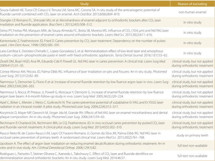
Exclusion of studies
The reasons for excluding studies after full-text
assess-ment were as follows: use of non-human enamel (n = 1),
in vitro studies (n = 4), the clinical aspect not applied
through orthodontic treatment (n = 7), studies on
pri-mary teeth (n = 1), full text non-available (n = 2). The
ex-cluded studies, together with the reasons of excluding, are
outlined in Supplementary Material 2.
Quality assessment
The quality assessment of the included studies revealed
unclear risk of bias (for 1 or more key domains) in the
8 studies included. Blinding of participants and blinding
during outcome assessment were the most problematic
fields (unclear risk of bias in 87.5% and 75% of studies,
re-spectively). The overall risk of bias for the included
stud-ies is summarized in Fig. 2 and 3.
Study characteristics
The studies were compared regarding the sample size,
study design, type and parameters of the laser applied,
and the main outcomes. The 8 articles were published
between 2000 and 2015. They involved 183 patients, and
the main inclusion criterion was healthy patients in need
of orthodontic treatment without caries,
demineraliza-tion or restorademineraliza-tions on the facial surfaces of teeth, except
for the trial by Harazaki et al., which included orthodontic
patients with early demineralization.
13Intervention in all
trials was the application of different laser types; 4
stud-ies applied an argon laser, 2 studstud-ies applied a CO
2laser,
1 study applied an Nd:YAG laser, and 1 study applied an
Optodan
®laser. The characteristics of the 8 studies are
summarized in Table 2.
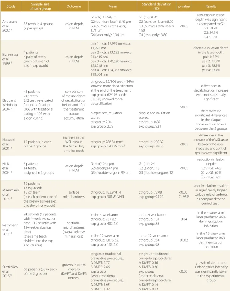
Results of individual studies
Four clinical studies applied an argon laser
14–17and
3 of them reported a significant reduction in the lesion
depth, measured on microphotographs of the polarized
light microscopy, for the argon laser-irradiated groups
of teeth compared to the control groups (p ≤ 0.05).
14–16One study did not find a significant effect of argon laser
curing on enamel WSL formation, evaluated on the basis
of photographs, in the laser group compared to the
con-trol group (p ≥ 0.05).
17Two studies applied a CO
2laser to enamel around
orth-odontic brackets and reported that CO
2laser irradiation
produced marked demineralization inhibition in short
and medium follow-up terms, as it led to significantly
higher enamel microhardness compared to the control
non-irradiated enamel (p ≤ 0.04).
18,19One clinical study applied an Nd:YAG laser to enamel
with WSLs and showed that it was effective in inhibiting
the development of dental caries, as the increase of the
WSL area was significantly lower compared to the control
group (p ≤ 0.05).
13One clinical trial used an Optodan low-intensity
la-ser around orthodontic brackets and reported that the
growth index in dental and surface caries intensity was
significantly lower in the laser therapy group than in the
control group (p ≤ 0.001).
20The results of the studies
in-cluded are summarized in Table 3.
T. Raghis, G. Mahmoud, O. Hamadah. Laser for enamel demineralization prevention 326
Fig. 1. PRISMA fl ow diagram illustrating the literature search protocol PRISMA – Preferred Reporting Items for Systematic Reviews and Meta-Analyses.
Supplementary Material 2. Studies excluded after full text reading with the reasons of excluding
Study Reason of excluding
Souza-Gabriel AE, Turssi CP, Colucci V, Tenuta LM, Serra MC, Corona SA. In situ study of the anticariogenic potential of fluoride varnish combined with CO2 laser on enamel. Arch Oral Biol. 2015;60(6):804–810.
non-human enamel
Stangler LP, Romano FL, Shirozaki MU, et al. Microhardness of enamel adjacent to orthodontic brackets after CO2 laser
irradiation and fluoride application. Braz Dent J. 2013;24(5):508–512. in vitro study Seino PY, Freitas PM, Marques MM, de Souza Almeida FC, Botta SB, Moreira MS. Influence of CO2 (10.6 μm) and Nd:YAG laser
irradiation on the prevention of enamel caries around orthodontic brackets. Lasers Med Sci. 2015;30(2):611–616. in vitro study Kantorowitz Z, Featherstone JD, Fried D. Caries prevention by CO2 laser treatment: Dependency on the number of pulses
used. J Am Dent Assoc. 1998;129(5):585–591. in vitro study
Lara-Carrilloa E, Doroteo-Chimalb C, Lopez-Gonzaleza S, et al. Remineralization effect of low-level laser and amorphous
sodium–calcium–phosphosilicate paste in teeth with fixed orthodontic appliances. Tanta Dental Journal. 2016;13(1):55–62. in vitro study Zezell DM, Boari HGD, Ana PA, Eduardo Cde P, Powell GL. Nd:YAG laser in caries prevention: A clinical trial. Lasers Surg Med.
2009;41(1):31–35.
clinical study, but not applied during orthodontic treatment Correa-Afonso AM, Pécora JD, Palma-Dibb RG. Influence of laser irradiation on pits and fissures: An in situ study. Photomed
Laser Surg. 2013;31(2):82–89.
clinical study, but not applied during orthodontic treatment Nammour S, Demortier G, Florio P, et al. Increase of enamel fluoride retention by low fluence argon laser in vivo. Lasers Surg
Med. 2003;33(4):260–263.
clinical study, but not applied during orthodontic treatment Nammour S, Rocca JP, Pireaux JJ, Powell G, Morciaux Y, Demortir G. Increase of enamel fluoride retention by low fluence
argon laser beam: A 6-month follow-up study in vivo. Lasers Surg Med. 2005;36(3):220–224.
clinical study, but not applied during orthodontic treatment Apel C, Birker L, Meister J, Weiss C, Gutknecht N. The caries-preventive potential of subablative Er:YAG and Er:YSGG laser
radiation in an intraoral model: A pilot study. Photomed Laser Surg. 2004;22(4):312–317.
clinical study, but not applied during orthodontic treatment Korytnicki D, Mayer MP, Daronch M, Singer Jda M, Grande RH. Effects of Nd:YAG laser on enamel microhardness and dental
plaque composition: An in situ study. Photomed Laser Surg. 2006;24(1):59–63.
clinical study, but not applied during orthodontic treatment Rechmann P, Charland DA, Rechmann BM, Le CQ, Featherstone JD. In vivo occlusal caries prevention by pulsed CO2 laser
and fluoride varnish treatment: A clinical pilot study. Lasers Surg Med. 2013;45(5):302–310.
clinical study, but not applied during orthodontic treatment Raucci-Neto W, de Castro-Raucci LM, Lepri CP, Faraoni-Romano JJ, Gomes da Silva JM, Palma-Dibb RG. Nd:YAG laser in
occlusal caries prevention of primary teeth: A randomized clinical trial. Lasers Med Sci. 2015;30(2):761–768. study on primary teeth Jacobson A. The effect of argon laser irradiation on reducing enamel decalcification during orthodontic treatment: An in
vitro and in vivo study. Am J Orthod Dentofacial Orthop. 2006;129(1):82. full text non-available Rodrigues L, Parisotto T, Steiner-Oliveira C, Azevedo L, Tabchoury C. Effect of CO2 laser and fluoride dentifrice on
demineralization around orthodontic brackets: An in situ study. Lasers Surg Med. 2014;46:57. full text non-available
T. Raghis, G. Mahmoud, O. Hamadah. Laser for enamel demineralization prevention Dent Med Probl. 2018;55(3):321–332
328 329
Table 2. Characteristics of the trials included
Study
Methods
Participants
Intervention
Follow-up time Primary outcomes Methods of measurement of primary outcomes study
design treatment comparison
type of laser
beam
laser parameters details of irradiation protocol
Anderson et al. 200214
RCT
amount of decalcification in the control group and the argon laser-irradiated groups of teeth
(non-pumiced-non-etched group, pumiced group and
etched group)
9 patients scheduled for orthodontic treatment with the extraction of 4 premolars; 36 premolars allocated in 4 groups
(inclusion criteria: teeth without enamel defects or decalcification)
argon laser
– beam: 325 mW; – time: 60 s;
– fluence (energy density): 100 J/cm2;
– beam diameter: 5 mm; irradiated through a wand handled at a distance of about 3 mm from the facial surface of the tooth
in 3 lased groups, the laser was applied alone or after pumicing or after pumicing
and etching; then modified orthodontic bands with pockets to plaque accumulation
were fitted on the premolars
5 weeks; then the teeth were extracted
lesion depth measurement [μm]; lesion
area measurement [μm2]
polarized light microscopy – digital microscope images were examined and measured
Blankenau et al. 199915
clinical pilot study
demineralization of enamel in the laser-irradiated and control
teeth
4 patients needing orthodontic treatment with bilateral premolar extraction; 4 pairs of premolars from each participant (1 experimental and 3 control)
argon laser
– beam: 250 mW; – time:10 s; – fluence: 12 J/cm2;
– beam diameter: 5 mm
after experimental teeth irradiation, modified orthodontic bands were fitted on
the lased and control teeth
5 weeks; then the teeth
were extracted lesion depth polarized light microscopy
Elaut and Wehrbein 200417
RCT (SP)
bracket bonding failure and enamel decalcification in argon
laser-cured and conventional light-cured bracket adhesives through orthodontic treatment
45 patients (28 girls and 17 boys), average age: 12 years 11 months, 742 teeth; in each patient, teeth with odd numbers received argon laser curing and teeth with
even numbers had conventional light curing of bracket adhesives; the maxillary anterior teeth (212) were evaluated for decalcification
(inclusion criteria: patients with fully erupted and restoration-free contralateral pairs of teeth)
argon laser
– beam: 250 mM (continuous mode) – time: 5 s from the incisal side and
5 s from the gingival side; – fluence: 12 J/cm2;
– beam diameter: 5 mm
thermoformed plastic/aluminium foil was used to cover the control teeth during
argon laser curing
14 months for enamel decalcification (after removing brackets from
the maxillary anterior teeth), 12 months for plaque accumulation; the bonding failure rate
was evaluated for 4–5 weeks after bonding
absence or degree of WSLs on enamel on the facial surfaces of maxillary anterior teeth; plaque
accumulation on the maxillary anterior teeth; the bonding failure rate during the study period
enamel decalcification and plaque accumulation were evaluated through comparing
digital images before and after the study period by a team of 7 examiners
Harazaki et al. 200113
clinical trial
increase in WSLs in the laser-irradiated and the control patients; enamel changes in the irradiated and non-manipulated parts of enamel
of the premolar
in vivo part of the study: 10 patients undergoing orthodontic treatment, with enamel WSLs on their teeth, the other 10 patients were a control group;
the focus was on maxillary incisors
in vitro part of the study: laser irradiation was applied to 1 extracted premolar
Nd:YAG laser
– pulse width: 0.3 ms; – pulse energy: 0.75 J; – power: 2 × 10 W; – repeated 20 pps; – time: 5 s; – fluence: 40 J/cm2
in vivo study: the experimental group of 10 patients was administered a black liquid agent, then it was irradiated with laser, and
finally the APF gel was applied in vitro study: one part of the extracted premolar was painted with a black liquid, and
then irradiated with laser, while the other part of the premolar was used as a control
1 year
increase in the area of WSLs after 1 year in the experimental and control groups; enamel surface changes in the irradiated
and control parts of the extracted premolar
WSLs were traced by taking photographs and with tracing
paper, and the total WSL area was calculated before laser irradiation and after 1 year; enamel surface changes were
observed by SEM
Hicks et al. 200416
clinical pilot study
lesion depth in argon laser irradiated-teeth in the argon
and fluoride group and the control (non-treatment) group
5 patients (3 females, 2 males), age: 19–28 years, requiring orthodontic treatment with tooth extraction (14 teeth); the teeth were caries-free on the buccal surfaces
argon laser
– beam: 250 mW; – time: 10 s; – fluence: 12 J/cm2
the teeth were assigned in 3 groups: argon laser, topical fluoride followed by argon laser
irradiation, and no treatment (control), then modified orthodontic bands with plaque
retentive slots were placed on the teeth
5 weeks; then the teeth were extracted
lesion depth in the 3
groups polarised light microscopy
Miresmaeili et al. 201419
RCT (SP)
enamel surface microhardness in the treated and control
premolars
16 patients (11 females, 5 males) scheduled for the extraction of at least 2 premolars. (1 first or second premolar treated with laser – 16 teeth, the premolar from the other
side in the same patient served as a control – 16 teeth) (inclusion criteria: age <25 years, complete eruption of teeth, no lesions on the enamel surfaces, moderate to good oral hygiene; exclusion criteria: patients with
enamel lesions or cracks on the buccal surfaces)
CO2 laser
– wave length: 10.6 μm; – pulse duration: 3 s; – pulse repetition rate: 5 Hz; – beam diameter: 0.2 mm; – power: 0.7 W
the experimental teeth were irradiated with laser and the control premolars were exposed to non-theraputic light; then orthodontic brackets were attached
to both premolars and the T-loop was engaged to the brackets to increase plaque
accumulation
at least 2 months after laser irradiation,
then the teeth were extracted (1 tooth from each group was extracted after 1 week
of laser irradiation for the SEM evaluation)
enamel surface microhardness around
orthodontic brackets; enamel surface changes
after laser therapy
Vickers diamond microhardness testing machine was used to evaluate enamel surface microhardness; SEM was used
to observe enamel surface changes Rechmann et al. 201118 RCT (PG) enamel demineralization around orthdontic brackets in
the laser-irradiated area and other area in the same tooth as
a control
24 patients
(13 females, 11 males), average age:14.9 ±2.2 years, randomly assigned to 4-week (average age: 14.6 ±2.3 years) and 12-week (average age: 15.2 ±2.1 years) study arms
(inclusion criteria: healthy patients, aged 12–18 years, scheduled to premolar extraction
for orthodontic reasons, teeth without caries or restorations on the facial surface; exclusion criteria: systematic diseases, medication affecting oral flora or salivary flow,
fluoride treatment in the last 3 months)
CO2 laser
– wave length: 9.6 μm; – pulse duration: 20 μs; – pulse repetition rate: 20 Hz; – beam diameter: 1,100 μm; – fluence per pulse: 3.3–4.4 J/cm2;
irradiated through a straight laser handpiece
the laser beam was applied on enamel, cervical to the bracket of the premolar on one side of an imaginary line, perpendicular
to the bracket slot, while the other side of the line in the same tooth served as a control; each spot of the testing area was irradiated with 20 pulses and laser
irradiation was applied only once
4 weeks for one group (12 patients) and 12 weeks for the other
group (12 patients); then the teeth were
extracted
overall relative mineral loss in the 4-week and 12-week arms in the irradiated and control
enamel cross-sectional microhardness testing Suetenkov et al. 201520 RCT
dental caries and oral hygiene in the group with traditional preventive measures and the group with laser therapy and traditional preventive measures
60 patients (20 girls, 40 boys), age: 12–13 years, divided into 2 groups (30 patients in each group)
Optodan (low-intensity)
laser
– wave length: 0.98–0.85 nm; – power: 0.5–1.0 W;
– pulse repetition rate: 2000 Hz; – time: 2 min for each segment
the irradiated area included 2 segments (upper and lower teeth aligments); irradiation was applied after professional
oral hygiene measures were taken, and there were 4 courses per year, every
3 months
1 year (after orthodontic treatment
completion and removing orthodontic
brackets)
oral hygiene and caries intensity before and after orthodontic treatment in
both groups
visual examination to determine OHI-S, DMFT index and DMFS index before and after the treatment
for both groups
APF – acidulated phosphate fluoride; DMFT – count of Decayed, Missing and Filled Teeth; DMFS – count of Decayed, Missing and Filled tooth Surfaces; Nd: YAG – neodymium-doped yttrium aluminum garnet; OHI-S – simplified oral hygiene index; PG – parallel groups; pps – pulse per second; RCT – randomized controlled trial; SEM – scanning electron microscopy; SP – split-mouth design; WSL – white spot lesion.
T. Raghis, G. Mahmoud, O. Hamadah. Laser for enamel demineralization prevention 330
Table 3. Summary of the results of the studies included
Study Sample size
of each group Outcome Mean
Standard deviation
(SD) p-value Results
Anderson et al. 200214
36 teeth in 4 groups (9 per group)
lesion depth in PLM
G1 (ctr): 15.69 μm G2 (pumice+laser): 6.45 μm G3 (pumice+etch+laser): 1.71 μm
G4 (laser only): 1.34 μm
G1 (ctr): 9.30
G2 (pumice+laser): 8.70 G3 (pumice+etch+laser): 4.80
G4 (laser only): 3.80
<0.05
reduction in lesion depth was significant
as compared to G1: G2: 58.9% G3: 89.1% G4: 91.6% Blankenau et al. 199915 4 patients 4 pairs of teeth (each patient 1 ctr and 1 exp tooth)
lesion depth in PLM
pair 1 – ctr: 17,959 nm/exp: 11,976 nm
pair 2 – ctr: 313,622 nm/exp: 213,445 nm
pair 3 – ctr: 178,528 nm/exp: 128,218 nm
pair 4 – ctr: 154,163 nm/exp: 118,004 nm
decrease in lesion depth in the lased tooth:
pair 1: 33% pair 2: 31.9% pair 3: 28.1% pair 4: 23.4%
Elaut and Wehrbein 200417
45 patients 742 teeth
212 teeth evaluated for decalcification (106 with traditional curing + 106 with argon curing)
comparison of the incidence of decalcification
before and after the treatment
plaque accumulation
ctr group: 85/106 teeth (54%) showed more decalcification at the end of the treatment exp group: 62/106 teeth (58.5%) showed more decalcification
>0.05
differences in decalcification increase
were not statistically significant
plaque accumulation scores:
ctr group: 2.34 exp group: 2.39
plaque accumulation scores:
ctr group: 0.86 exp group: 9.81
there were no significant differences
in the plaque accumulation scores between the 2 groups
Harazaki et al. 200113
10 patients in each of the 2 groups
increase in the WSL area in the 6 maxillary
anterior teeth
ctr group: 286.84 mm2
exp group: 140.76 mm2
ctr group: 209.37
exp group: 38.05 <0.05
differences in the increase of the WSL areas
between the laser-irradiated and control groups were significant
Hicks et al. 200416
5 patients 14 teeth,
assigned in 3 groups
lesion depth in PLM
G1 (ctr): 261 μm G2 (argon):147 μm G3 (fluoride+argon): 99 μm
G1 (ctr): 24 G2 (argon): 18 G3 (fluoride+argon): 12
<0.05
reduction in lesion depth: G2 vs G1: 44% G3 vs G1: 62% G3 vs G2: 32%
Miresmaeili et al. 201419
16 patients 16 exp teeth 16 ctr teeth
(in each patient, one of the premolars was exp and the other was ctr)
surface microhardness
ctr group: 183.9 VHN exp group: 301.81 VHN
ctr group: 72.08 exp group: 94.29
<0.001 CI: 95%
laser irradiation resulted in significantly higher surface microhardness as compared to the
control teeth
Rechmann et al. 201118
24 patients (12 patients with 4-week evaluation time + 12 patients with 12-week evaluation time)
(the same teeth divided into the exp and ctr area)
sectional microhardness (overall relative mineral loss)
in the 4-week arm: ctr group: 737 ΔZ exp group: 402 ΔZ
in the 4-week arm: ctr group: 131 exp group: 85
0.04
in the 4-week arm: laser produced 46% demineralization
inhibition
in the 12-week arm: ctr group: 1,076 ΔZ exp group: 135 ΔZ
in the 12-week arm: ctr group: 254 exp group: 98
0.002
in the 12-week arm: laser produced 86% demineralization
inhibition
Suetenkov et al. 201520
60 patients (30 in each of the 2 groups)
growth in caries intensity (DMFT and DMFS
indices)
ctr group (traditional preventive procedure): Δ DMFT: 2.77 Δ DMFS: 2.66 exp group (laser+traditional preventive procedure): Δ DMFT: 1.05 Δ DMFS: 1.37
ctr group (traditional preventive procedure): Δ DMFT: 0.56 Δ DMFS: 0.30 exp group (laser+traditional preventive procedure): Δ DMFT: 0.14 Δ DMFS: 0.13
<0.001
growth of dental and surface caries intensity was significantly lower in the experimental
group
CI – confidence interval; ctr – control; DMFT: count of Decayed, Missing and Filled Teeth; DMFS: count of Decayed, Missing and Filled tooth Surfaces; Δ DMFT and Δ DMFS – growth in caries intensity indices; exp – experimental; G – group; PLM – polarized light microscopy; VHN – Vickers hardness number (measurement unit for surface microhardness); WSL – white spot lesion; ΔZ – overall relative mineral loss (measured by plotting normalized volume percent mineral against distance from the enamel surface; vol% × μm).
The most eff ective and safest laser types
and parameters for the prevention
of enamel demineralization
Studies that compared the effectiveness of 2 or more
laser beams in demineralization inhibition during
orth-odontic treatment were all in vitro.
21,22Studies that
com-pared the effect of different parameters of the same laser
type were also in vitro and did not concern orthodontic
treatment. There were no studies undertaken during
orthodontic treatment comparing the improvement in
demineralization resistance among different laser types
or different laser settings.
Discussion
The prevention of demineralization or WSL formation
during orthodontic treatment is one of the most difficult
challenges orthodontists have to face. Many preventive
procedures have been used in the literature for this
pur-pose. Laser irradiation has been widely studied in vitro
and showed its effectiveness in increasing enamel
resis-tance to decalcification, suggesting that it could be useful
during orthodontic treatment. As presented in the
litera-ture, many laser types have been used to prevent enamel
demineralization around orthodontic appliances,
includ-ing Er:YAG,
23–25Nd:YAG,
13,21,22CO
2
,
18–22,26–28diode,
29,30and argon laser.
14–17,31,32Although the clinical application
of lasers during orthodontic treatment for a preventive
purpose is still limited, the present review showed
clini-cal effectiveness of laser irradiation in inhibiting enamel
demineralization.
There were no clinical trials that applied Er:YAG or
di-ode lasers during orthodontic treatment to prevent WSL
formation.
In 3 studies, the application of argon laser irradiation on
the enamel surface showed significant reduction in lesion
depth in comparison with non-irradiated teeth, and its
ef-fect was significantly higher when it was combined with
fluoride application,
14–16but the sample sizes in these
studies were small, with short follow-up periods. The
ef-fect of irradiation with an argon laser on WSL formation
while curing the adhesives of orthodontic brackets was
evaluated in 1 RCT lasting 1 year, but no significant effect
on enamel demineralization was observed.
17Irradiation with a CO
2laser had a significant effect on
enamel microhardness around orthodontic brackets and
it decreased mineral loss in comparison with
non-irradi-ated enamel in 2 RCTs.
18,19The wave lengths applied
clini-cally were 9.6 μm and 10.6 μm, respectively. However, the
effect of CO
2lasers during orthodontic treatment was not
evaluated for a long follow-up period.
The effect of Nd:YAG laser irradiation on existing
WSLs was studied in only 1 clinical trial, with a 1-year
follow-up.
13The increases in the WSL area were
signifi-cantly lower in the laser-irradiated group of patients in
comparison with the control group. This type of laser had
not been previously applied clinically on sound enamel
during orthodontic treatment to prevent decalcification.
The effect of an Optodan laser on enamel
demineraliza-tion was studied in a RCT by comparing the development
of tooth caries intensity (growth of the Decayed, Missing
and Filled Teeth index – ΔDMFT, and growth of the
De-cayed, Missing and Filled tooth Surfaces index – ΔDMFS)
between the laser group and the control group for a 1-year
follow-up period, and it showed significantly lower caries
intensity in the lased group as compared to the control
non-irradiated group.
20Changes in the enamel structure after laser irradiation
were evaluated in 2 of the included studies by scanning
electron microscopy. Miresmaeili et al. evaluated enamel
surface changes by extracting 2 premolars (irradiated and
control) of 1 patient after 1 week of CO
2laser irradiation;
the lased tooth showed melting of the enamel surface.
19As studied in the literature, the prevention of caries by
CO
2laser irradiation could stem from reduced enamel
permeability and solubility as a result of melting.
33,34Ha-razaki et al. studied enamel changes after Nd:YAG
irra-diation in vitro.
13The irradiated portion of the tooth had
a smooth surface with a small number of cracks.
The limitations of this review are related primarily to the
lack of high-level evidence from RCTs and the
heteroge-neity among studies in irradiation protocols, outcomes,
follow-up periods, and methods of outcome measurement.
Conclusions
This review showed that laser irradiation may be
effec-tive in preventing demineralization during orthodontic
treatment, but further studies are needed, including RCTs
using different lasers, to evaluate which is the most
ef-fective laser and what settings should be used. There is
also a need for longer follow-up periods to evaluate the
longevity of treatment.
References
1. Bergstrand F, Twetman S. A review on prevention and treatment of post-orthodontic white spot lesions – evidence-based methods and emerging technologies. Open Dent J. 2011;5:158–162.
2. Sudjalim TR, Woods MG, Manton DJ. Prevention of white spot lesions in orthodontic practice: A contemporary review. Aust Dent J. 2006;51(4):284–289,quiz 347.
3. Sundararaj D, Venkatachalapathy S, Tandon A, Pereira A. Critical evaluation of incidence and prevalence of white spot lesions dur-ing fixed orthodontic appliance treatment: A meta-analysis. J Int Soc Prev Community Dent. 2015;5(6):433–439.
4. Zabokova-Bilbilova E, Popovska L, Kapusevska B, Stefanovska E. White spot lesions: Prevention and management during the orth-odontic treatment. Pril (Makedon Akad Nauk Umet Odd Med Nauki). 2014;35:161–168.
5. Lapenaite E, Lopatiene K, Ragauskaite A. Prevention and treatment of white spot lesions during and after fixed orthodontic treatment: A systematic literature review. Stomatologija. 2016;18(1):3–8.
T. Raghis, G. Mahmoud, O. Hamadah. Laser for enamel demineralization prevention 332
6. Featherstone JD. Lasers in dentistry 3. The use of lasers for the prevention of dental caries [in Dutch]. Ned Tijdschr Tandheelkd. 2002;109(5):162–167.
7. Karandish M. The efficiency of laser application on the enamel sur-face: A systematic review. J Lasers Med Sci. 2014;5(3):108–114. 8. Rodrigues LKA, de Freitas PM, Nobre-dos-Santos M. Lasers in caries
prevention. In: Freitas PM, Simões A, eds. Lasers in Dentistry: Guide for Clinical Practice. Hoboken, NJ: Wiley-Blackwell; 2015:126–130. 9. Rezaei Y, Bagheri H, Esmaeilzadeh M. Effects of laser irradiation on
caries prevention. J Lasers Med Sci. 2011;2(4):159–164.
10. Sadr Haghighi H, Skandarinejad M, Abdollahi AA. Laser applica-tion in prevenapplica-tion of demineralizaapplica-tion in orthodontic treatment.
J Lasers Med Sci. 2013;4(3):107–110.
11. Moher D, Liberati A, Tetzlaff J, Altman DG; PRISMA Group. Preferred Reporting Items for Systematic Reviews and Meta-Analyses: The PRISMA statement. Int J Surg. 2010;8(5):336–341.
12. Higgins JP, Altman DG, Gøtzsche PC, et al.; Cochrane Bias Methods Group, Cochrane Statistical Methods Group. The Cochrane Collab-oration’s tool for assessing risk of bias in randomised trials. BMJ. 2011;343:d5928.
13. Harazaki M, Hayakawa K, Fukui T, Isshiki Y, Powell LG. The Nd-YAG laser is useful in prevention of dental caries during orthodontic treatment. Bull Tokyo Dent Coll. 2001;42(2):79–86.
14. Anderson AM, Kao E, Gladwin M, Benli O, Ngan P. The effects of argon laser irradiation on enamel decalcification: An in vivo study. Am J Orthod Dentofacial Orthop. 2002;122(3):251–259. 15. Blankenau RJ, Powell G, Ellis RW, Westerman GH. In vivo caries-like
lesion prevention with argon laser: Pilot study. J Clin Laser Med Surg.
1999;17(6):241–243.
16. Hicks J, Winn D 2nd, Flaitz C, Powell L. In vivo caries formation in
enamel following argon laser irradiation and combined fluoride and argon laser treatment: A clinical pilot study. Quintessence Int. 2004;35(1):15–20.
17. Elaut J, Wehrbein H. The effects of argon laser curing of a resin adhesive on bracket retention and enamel decalcification: A pro-spective clinical trial. Eur J Orthod. 2004;26(5):553–560.
18. Rechmann P, Fried D, Le CQ, et al. Caries inhibition in vital teeth using 9.6-μm CO2-laser irradiation. J Biomed Opt. 2011;16(7):071405.
19. Miresmaeili A, Farhadian N, Rezaei-soufi L, Saharkhizan M, Veisi M. Effect of carbon dioxide laser irradiation on enamel surface micro-hardness around orthodontic brackets. Am J Orthod Dentofacial Orthop. 2014;146(2):161–165.
20. Suetenkov DY, Petrova AP, Kharitonova TL. Photo activated disin-fection efficiency of low-intensity laser and comprehensive pre-vention of caries and gingivitis in adolescents using bracket sys-tem. J Innovat Opt Health Sci. 2015;8(3):1541002.
21. Seino PY, Freitas PM, Marques MM, de Souza Almeida FC, Botta SB, Moreira MS. Influence of CO2 (10.6 μm) and Nd:YAG laser irradiation
on the prevention of enamel caries around orthodontic brackets.
J Lasers Med Sci. 2015;30(2):611–616.
22. Paulos RS, Seino PY, Fukushima KA, et al. Effect of Nd:YAG and CO2
laser irradiation on prevention of enamel demineralization in ortho-dontics: In vitro study. Photomed Laser Surg. 2017;35(5):282–286. 23. Ulkur F, Sungurtekin Ekçi E, Nalbantgil D, Sandalli N. In vitro effects
of two topical varnish materials and Er:YAG laser irradiation on enamel demineralization around orthodontic brackets. Sci World J. 2014;2014:490503.
24. Fornaini C, Brulat N, Milia G, Rockl A, Rocca JP. The use of sub-abla-tive Er:YAG laser irradiation in prevention of dental caries during orthodontic treatment. Laser Ther. 2014;23(3):173–181.
25. Garma NM, Jasim ES. The effect of Er:YAG laser on enamel resis-tance to caries during orthodontic treatment: An in vitro study.
J Bagh Coll Dentistry. 2015;27(1):182–188.
26. de Souza-e-Silva CM, Parisotto TM, Steiner-Oliveira C, Kamiya RU, Rodrigues LK, Nobre-dos-Santos M. Carbon dioxide laser and bonding materials reduce enamel demineralization around orth-odontic brackets. JLasers Med Sci. 2013;28(1):111–118.
27. Mirhashemi AH, Hakimi S, Ahmad Akhoundi MS, Chiniforush N. Pre-vention of enamel adjacent to bracket demineralization following carbon dioxide laser radiation and titanium tetra fluoride solution treatment: An in vitro study. J Lasers Med Sci. 2016; 7(3):192–196.
28. Stangler LP, Romano FL, Shirozaki MU, et al. Microhardness of enamel adjacent to orthodontic brackets after CO2 laser
irradia-tion and fluoride applicairradia-tion. Braz Dent J. 2013;24(5):508–512. 29. Lacerda ÂS, Hanashiro FS, de Sant’Anna G, Steagall W Júnior,
Barbosa P, de Souza-Zaroni WC. Effects of near infrared laser radi-ation associated with photoabsorbing cream in preventing white spot lesions around orthodontic brackets: An in vitro study. Pho-tomed Laser Surg. 2014;32:686–693.
30. Lara-Carrilloa E, Doroteo-Chimalb C, Lopez-Gonzaleza S, et al. Remineralization effect of low-level laser and amorphous sodi-um–calcium–phosphosilicate paste in teeth with fixed orthodon-tic appliances. Tanta Dent J. 2016;13(1):55–62.
31. Miresmaeili A, Etrati Khosroshahi M, Motahary P, et al. Effect of argon laser on enamel demineralization around orthodontic brackets: An in vitro study. J Dent (Tehran). 2014;11(4):411–417. 32. Noel L, Rebellato J, Sheats RD. The effect of argon laser irradiation on
demineralization resistance of human enamel adjacent to orthodon-tic brackets: An in vitro study. Angle Orthod. 2003;73(3):249–258. 33. Stern RH, Vahl J, Sognnaes RF. Lased enamel: Ultrastructural
observations of pulsed carbon dioxide laser effects. J Dent Res. 1972;51(2):455–460.
34. Borggreven JM, van Dijk JW, Driessens FC. Effect of laser irradia-tion on the permeability of bovine dental enamel. Arch Oral Biol. 1980;25:831–832.