Address for correspondence
Dr. Maryam Khalili, Assistant Professor, Department of Dermatology,
Afzalipour Hospital,
Kerman University of Medical Sciences, Iran Post code: 7616913911
Email: [email protected]
Original Article
The efficacy of combined topical niosomal dapsone
gel and intralesional injection of meglumine
antimoniate in comparison with intralesional
meglumine antimoniate and cryotherapy in the
treatment of cutaneous leishmaniasis
Introduction
Leishmaniasis is a protozoan infectious disease
caused by several species of the
Leishmania
and
transmitted by infected sandfly bite.
1The three
mainstay clinical forms of the disease including
cutaneous leishmaniasis (CL), mucocutaneous
leishmaniasis and visceral leishmaniasis have
Mahin Aflatoonian*, Alireza Fekri**, Zahra Rahnam*, Maryam Khalili*, Abbas Pardakhti¶, Payam Khazaeli¶, Kambiz Bahadini¶¶* Department of Dermatology, School of Medicine, Kerman University of Medical Sciences, Iran ** Department of Dermatology, Faculty of Medicine, University of Medical Sciences, Kerman, Iran
* Department of Dermatology, School of Medicine, Kerman University of Medical Sciences, Iran ¶ Department of Pharmaceutics, School of Pharmacy, University of Medical Sciences, Kerman, Iran
¶¶Medical Informatics Research Center, Institute of future studies in health, Kerman University of Medical Sciences, Kerman, Iran
Abstract
Objective To evaluate the efficacy of niosomal dapsone gel and intralesional meglumine antimoniate with cryotherapy and intralesional meglumine antimoniate in cutaneous leishmaniasis. Methods This was a randomized clinical trial with 73 participants that were divided into two groups including, case group (weekly intralesional meglumine antimoniate and twice a day niosomal dapsone gel) and control group (weekly intralesional meglumine antimoniate with biweekly cryotherapy). The treatment course continued until 16 weeks or complete cure, whatever occurred earlier and participants were followed up in 4th, 8th, 12th and 16th weeks of the treatment.Results Overall, 68 patients (33 males and 35 female) completed the study. Age, sex, size and duration of the lesions were not statistically different between two groups. At the end of the study, 82.9% of patients in case group showed complete response.
Conclusion Niosomal dapsone gel has promising results with fewer adverse effects, so, it can be used as an alternative treatment modality, especially in children and patients with contraindication of systemic drugs.
Key words
been recognized.
2Cutaneous
Old
World
leishmaniasis
is
categorized into two groups, anthroponotic
cutaneous leishmaniasis (ACL) and zoonotic
cutaneous
leishmaniasis
(ZCL)
which,
classically are caused by
L. tropica
and
L.
major
, respectively.
2,3,4Annual
incidence
of
the
disease
is
approximately 1.5 to 2 million.
5,6It affects more
than half of Iran’s provinces and there is an
increase in geographical spreading of the disease
in Kerman, southeast of Iran.
3,7,8,9Although CL is not a fatal disease, the treatment
is necessary in order to prevent disease
transmission and scar formation especially in
exposed sites.
10,11Currently, based on recommendation of WHO,
gold standard of the disease treatment is
intramuscular
pentavalent
antimonial
with
cryotherapy, but, there is limitation to the
administration of standard treatment due to poor
availability, high cost, serious side effects and
painful injection of the drug.
2,4,8,11,12In recent years, several treatment modalities
such as systemic antifungal drugs, amphotericin,
topical paromomycin and physical therapies
were evaluated with different results.
10,13-18One
of the systemic drugs which can be used as an
alternative treatment for leishmaniasis is
dapsone.
This
drug
affects
alternative
complement pathway and can enhance cellular
immunity through cytokines.
19,20Absorption of dapsone after oral intake is slow
and after absorption, it is found in internal
organs such as liver, muscle, kidney and skin.
Side effects of this drug are dose-dependent such
as hemolysis, methemoglobinemia, peripheral
neuropathy,
allergic
dermatitis,
headache,
anemia, hepatitis and agranulocytosis.
19,20Investigations show that oral intake of dapsone
in the treatment of different infections leads to
low efficacy and therapeutic index of the drug,
due to lower concentration at the site of
infection.
19,20Based upon recently gathered data, some of the
topical treatment regimens had superior effects
compared to systemic ones or at least had equal
efficacy. Topical treatment options such as
niosomal drug delivery systems for the
leishmaniasis have increased in recent years.
21-24Considering the good efficacy and safety of 5%
dapsone gel in the treatment of acne,
20,25,26we
decided for the first time, to assess the efficacy
of niosomal form of dapsone in the treatment of
leishmaniasis.
Methods
This open randomized clinical trial was
performed from December 2011 to October
2013 in dermatology clinic of Afzalipour
hospital – Kerman (southeast of Iran). A total of
73 patients were enrolled in the study after their
written consent. Patients aged ≥ 7 years and
diagnosed with positive smear or demonstration
of Leishman-Donovan (LD) bodies in skin
biopsy were included in the study.
sporotrichoid
or
lupoid
forms
of
the
leishmaniasis.
Demographic information including age, sex,
size and site of the lesions were completed and
induration of the lesions were assessed with
ruler through Sokal method. Patients were
randomly assigned to group A (intralesional
meglumine
antimoniate
and
biweekly
cryotherapy) and B (niosomal dapsone gel twice
a day and weekly intralesional meglumine
antimoniate).
Cryotherapy
was
conducted
by
dipstick
technique with liquid nitrogen. Cotton-tipped
applicator was applied on the lesion until 1 mm
white halo formation around the lesion.
Meglumine antimoniate (Glucantime™,
Rhone-Poulenc, France) was injected until blanching of
the lesion.
Considering the fact that dapsone is soluble in
lipid, we chose gel as drug vehicle, in order to
better drug delivery into skin. In order to prepare
niosomal dapsone by conventional hydrated film
method, cholesterol and nonionic surfactant and
dapsone were dissolved in chloroform and
acetone respectively, then, the solvent was
evaporated by rotary evaporator, next, the
resultant lipid film was hydrated with 5ml
deionized water at 70. At last, niosome was
mixed with 1% carbomer934 with equal rates.
Particle’s size analysis, physical stability,
encapsulation efficacy, percentage of drug
delivery and penetration into rat’s abdominal
skin have been done in pharmaceutical research
center.
Participants were treated for 16 weeks or until
complete healing, whatever happened earlier.
They were evaluated in the 4th, 8th, 12th, 16th
weeks of the treatment and response rate was
recorded according to ESL (evaluation of lesions
scheme) as follows:
Cured (complete re-epithelialization without
induration); partially cured (more than 50%
re-epithelialization
and
decrease
induration);
improved (less than 50% re-epithelialization and
decrease induration in the lesion); no change
(without any change in the size of the lesion) or
worsening (increase in the size of the lesions).
In this study we, categorized participants into
three groups as: complete response (100%
improvement), partial response (50%-99%
improvement) and no response (improvement
less than 50%). None of the patients in this study
had increase in the size of the lesion.
This proposal was approved by the ethics
committee of research center of medical science
university of Kerman with approval code
K/90/503.
Statistical analysis
Data analysis was performed by SPSS 17.0
software (SPSSINC, Chicago, USA) and
Random effects mixed model, Mann-Whitney
test, t test and chi-square were applied for
analysis.
Results
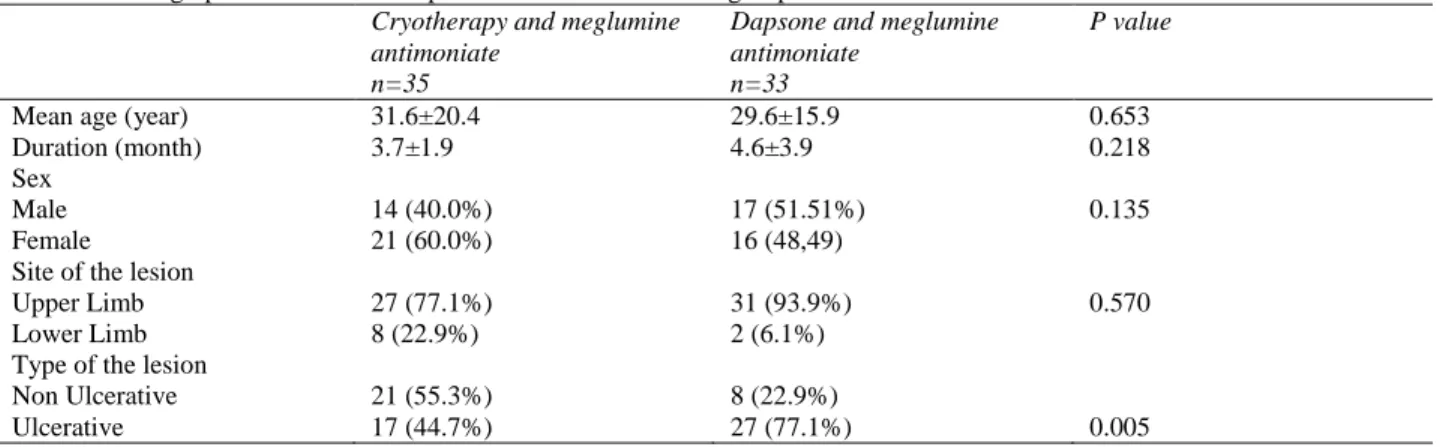
Table 1 Demographic features and response rate in two treatment groups Cryotherapy and meglumine
antimoniate n=35
Dapsone and meglumine antimoniate
n=33
P value
Mean age (year) 31.6±20.4 29.6±15.9 0.653
Duration (month) 3.7±1.9 4.6±3.9 0.218
Sex
Male 14 (40.0%) 17 (51.51%) 0.135
Female 21 (60.0%) 16 (48,49)
Site of the lesion
Upper Limb 27 (77.1%) 31 (93.9%) 0.570
Lower Limb 8 (22.9%) 2 (6.1%)
Type of the lesion
Non Ulcerative 21 (55.3%) 8 (22.9%)
Ulcerative 17 (44.7%) 27 (77.1%) 0.005
Table 2 Treatment response according to percent improvement during the treatment in 2 groups, cryotherapy and meglumine antimoniate (group A) and niosomal dapsone gel and meglumine antimoniate (group B).
Treatment Response
4 week n (%)
8 week n (%)
12 week n (%)
16 week n (%)
A B A B A B A B
Complete Response
- - 11
(31.4%) 7 (18.4%)
23 (65.7%)
23 (60.5%)
29 (82.9%)
33 (86.8%) Partial
Response
26 (74.3%) 26 (68.4%)
23 (65.7%)
30 (78.9) 11 (31.4) 14 (36.8%)
5 (14.3%)
4 (10.5%) Without
Response
9 (25.7) 12 (31.6%)
1 (2.9%)
1 (2.6%) 1 (2.9%) 1 (2.6%) 1 (2.9%) 1 (2.6%)
Table 3 Treatment response comparison between two groups. Response rate OR (95% CI) P value
Total 1.03 (0.64-1.65) 0.885
Sex 1.04 (0.64-1.69) 0.866
Age 1.01 (0.62-1.64) 0.952
Type of the lesion 1.32 (0.8-2.18) 0.271 Site of the lesion 1.15 (0.68-1.93) 0.595
Size 1.14 (0.73-1.79) 0.545
duration of the disease and size of the lesions.
Table 2
shows the treatment response in two
groups at week 4, 8, 12 and 16 of follow-up.
29 (82.9%)patients in group A (meglumine
antimoniate and cryotherapy) showed complete
response as compared to 33 (86.8%) in group B
(meglumine antimoniate and niosomal dapsone
gel)
.
During the twelve months post-treatment
follow-up, no recurrence was reported in the
case group, but in the control group one patient
developed lupoid leishmaniasis and was treated
with systemic meglumine antimoniate.
No side effect with application of niosomal
dapsone gel during the treatment and follow up
was observed.
Discussion
Intralesional infiltration of the pentavalent
antimony compounds is the treatment of choice
for leishmaniasis which had variable efficacy in
different studies from 81% to 97%, depending
on duration of the treatment,
Leishmania
species
and definition treatment response.
1,27,28Asilian
et al.
evaluated the efficacy of
cryotherapy
and
intralesional
meglumine
antimoniate on
L. major
. Ninety percent of the
patients showed complete response. This result
was compatible with our results (86.8% in
control group), however recent conducted
studies demonstrated that
L. tropica
is more
refractory to treatment than
L. major
.
27,29Dapsone is an antibacterial drug that inhibits
synthesis
of
dihydrofolic
acid
through
competition with para-aminobenzoate. This drug
also
has
anti-inflammatory
and
immunomodulatory effects by myeloperoxidase
blocking.
38In one study by Dogra
et al.
efficacy of oral
dapsone in leishmaniasis was evaluated and oral
dapsone had 82% response rate in comparison
with placebo.
13. In another study by Dogra
et al.
oral dapsone was used in dose of 2 mg/kg/day
for 21 days and 80% of patients responded to
treatment and there was no recurrence in the
next 6 months.
Due to adverse effects of oral dapsone, 5%
topical dapsone gel was used in several studies.
Advantage of this form of dapsone is low
systemic absorption with plasma peak which is
100 times lower than oral types. Topical
dapsone gel has been evaluated in the treatment
of acne with good efficacy and favorable
results.
20,25,26One of the newest structural drug delivery
systems is niosomal form. This form consists of
hydrated cholesterol and nonionic surfactants
such as alkyl ether, alkyl ester and alkyl acid
that induce more stability, higher absorption of
drug in the skin and slow release of effective
substance.
41,42Niosomes have structure similar to lipids layers
of biologic membrane. This structure leads to
increase of drug penetration into skin through
melting of the niosome in stratum corneum and
delivery of drug in deeper layer of epidermis. On
the other hand, this structure enhances
penetration of different substances in cells such
as Langerhans cells, thus, it has been used and
assessed for intracellular infection such as
leishmaniasis. Other advantages of this form are
lower drug toxicity and cost, as well as, easy
manufacturing of the drug.
41,42Our study is the first study to evaluate the
efficacy of niosomal dapsone gel in the
treatment of CL. Our results showed that
efficacy of niosomal dapsone gel combined with
intralesional
meglumine
antimoniate
was
comparable
with
the
standard
treatment
including
cryotherapy
and
intralesional
meglumine
antimoniate.
There
was
no
significant difference in partial and complete
response rate between the two groups; also, both
treatments had similar increase in efficacy
during follow-up. During the twelve months of
observation, no recurrence was reported in the
case group whereas in the control group one
patient developed lupoid leishmaniasis.
In this study we were not able to demonstrate
Leishmania
species for all of the patients, but, in
the previous study in Kerman, most of the
Leishmania
species were reported as
L.
tropica.
43Strengths of our study are randomization,
adequate sample size and similarity between
demographic features in the two groups, which
increase reliability and stability of our results.
We were not able to conduct a double-blind
clinical trial, due to different treatment
modalities.
regimens.
Conclusion
Niosomal dapsone gel can be used as an
alternative and adjunctive drug with lower cost
and approximately without any adverse effect in
the treatment of CL, especially in children, due
to painful injection of meglumine antimoniate. It
can be applied as an alternative treatment,
instead of cryotherapy, in sites such as ear, nose
and fingers because of possible risk of scar
formation secondary to vascular compromise
and in individual with darker skin type due to
complications such as hypo/ hyperpigmentation.
References
1. Ramtin Hadighi R, Mehdi Mohebali M, Patrick Boucher P, Homa Hajjaran H, Ali Khamesipour A, Marc Ouellette M. Unresponsiveness to glucantime treatment in Iranian cutaneous leishmaniasis due to drug-resistant Leishmania tropica parasites. PLoS Med. 2006;3:e162.
2. Desjeux P. The increase in risk factors for leishmaniasis worldwide. Trans R Soc Trop Med Hyg. 2001;95:239-43.
3. William H, Markle MD, Khaldoun M. Cutaneous leishmaniasis: recognition and treatment. Am Fam Physician. 2004;69:1455-60.
4. Al-Jaser M, el-Yazigi A, Kojan M, croft SL. Distribution and elimination of antimony following administration of sodium stibogluconate to patients with cutaneous leishmaniasis. Antimicrob Agents Chemother. 1995;39:516-9.
5. Jeronimo SB, Sousa A, Pearson RD. Leishmaniasis species. In: Mandell GL, Bennett JE, Dolin R, eds. Principles and Practice of Infectious Diseases. 6th ed. Philadelphia: Churchill-Livingstone 2005; 45-65.
6. Dowlati Y. Cutaneous leishmaniasis: clinical aspect. Clin Dermatol. 1996;14:425-31.
7. Mohajery M, Bolursaz M, Shamsian S. A survey on the prevalence of cutaneous leishmaniasis in the students of Mashhad
district. J Mashhad Faculty Med. 2001;44(72):54-60.
8. Azizi F, Hatam H, Janghorbani M. Epidemiology and control of common disorders in Iran. Tehran: Khosravi Publications 2003. P. 524-32.
9. Athari A, Jalalloo N. Epidemiology of five year of cutaneous leishmaniasis in Iran (2001-2005). Isfahan Faculty Med J. 2006;24:8-13.
10. Vardy D, Barenholz Y, Cohen R, Zvulunov A, Biton A, Klaus S. Topical amphotericin B for cutaneous leishmaniasis. Arch Dermatol. 1999;135:856-7.
11. Al-Jaser MH. Treatment trends of cutaneous leishmaniasis in Saudi Arabia. Saudi Med J. 2005;26:1220-4.
12. Akilov OE, Kosaka B. Parasiticidal effect of delta-aminolevulinic acid-based photodynamic therapy for cutaneous leishmaniasis in direct and mediated through the killing of host cell. Exp Dermatol. 2007;16:651-60.
13. Dogra J. A double-blind study on the efficacy of oral dapsone in cutaneous leishmaniasis. Trans R Soc Trop Med Hyg. 1991;85:212-3.
14. Esfandiarpour I, Alavi AF. Evaluating the efficacy of allopurinol and meglumine antimoniate (Glucantime) in the treatment of cutaneous leishmaniasis: a pilot study. Int J Dermatol. 1997;36:59-60.
15. Shamsi Meimandi S, Zandi S, Dabiri SH. Efficacy of topical 5% imiquimod with cryotherapy versus intralesional meglumine antimoniate in treatment of anthroponotic cutaneous leishmaniasis. Iran J Dermatol. 2001;14:42-47.
16. Magill AJ. Cutaneous leishmaniasis in the returning traveler. Infect Dis Clin North Am. 2005;19:241-66.
17. Dogra J, Lal BB, Misra SN. Dapsone in the treatment of cutaneous leishmaniasis. Int J Dermatol. 1986;25:398-400.
18. Al-Mutairi N, Alshiltawy M, El Khalawany
M, Joshi A, Eassa BI, Manchanda Y et al.
Tropical medicine rounds: Treatment of old world cutaneous leishmaniasis with dapsone, itraconazole, cryotherapy and imiquimod alone and in combination. Int J Dermatol. 2009;48:862-9.
20. Raimer S, Maloney JM, Bourcier M, Wilson D. Efficacy and safety of dapsone gel 5% for the treatment of acne vulgaris in adolescents. Cutis. 2008;81:171-8.
21. Croft SL, Sundar S, Fairlamb AH. Drug resistance in leishmaniasis. Clin Microbiol Rev. 2006;19:111-26.
22. Faghihi G, Tavakoli‐kia R. Treatment of cutaneous leishmaniasis with either topical paromomycin or intralesional meglumine antimoniate. Clin Exp Dermatol. 2003;28:13-6.
23. Shazad B, Abbaszadeh B, Khamesipour A. Comparison of topical paromomycin sulfate (twice/day) with intralesional meglumine antimoniate for the treatment of cutaneous leishmaniasis caused by L. major. Eur J Dermatol. 2005;15:85-7.
24. Alavi-Naini R, Fazaeli A, O’Dempsey T. Topical treatment modalities for old world cutaneous leishmaniasis: a review. Prague Med Report. 2012;113:105-18.
25. Draelos ZD, Carter E, Maloney M. Two randomized studies demonstrate the efficacy and safety of dapsone gel 5%, for the treatment of acne vulgaris. J Am Acad Dermatol. 2007;56:439.e1-10.
26. Lucky AW, Roberts J, Maloney M, Ling M. Dapsone gel 5% for the treatment of acne vulgaris: safety and efficacy of long-term (1 year) treatment. J Drugs Dermatol. 2007;6:981-7.
27. Khatami A, Firooz A, Gorouhi F, Dowlati Y. Treatment of acute Old World cutaneous leishmaniasis: a systematic review of the randomized controlled trials. J Am Acad Dermatol. 2007;57:335. e1-29. E.
28. Munir A, Janjua SA, Hussain I. Clinical efficacy of intramuscular meglumine antimoniate alone and in combination with intralesional meglumine antimoniate in the treatment of old world cutaneous leishmaniasis. Acta Dermatovenerologica Croatica. 2008;16(2):60-4.
29. Asilian A, Sadeghinia A, Faghihi G, Momeni A. Comparative study of the efficacy of combined cryotherapy and intralesional meglumine antimoniate (Glucantime) vs. cryotherapy and intralesional meglumine antimoniate (Glucantime) alone for the treatment of cutaneous leishmaniasis. Int J Dermatol. 2004;43:281-3.
30. Nilforooshzadeh MA, Jaffari F, Malekafzali B. Efficacy of combined triple therapy (paromomycin ointment, cryotherapy and
intralesional Glucantime) in comparison with intralesional Glucantime for the treatment of acute cutaneous leishmaniasis. Iran J Dermatol. 2004;7:136-9.
31. Asilian A, Sadeghinia A, Faghihi G, Momeni A, Amini Harandi A. The efficacy of treatment with intralesional meglumine antimoniate alone, compared with that of cryotherapy combined with the meglumine antimoniate or intralesional sodium stibogluconate, in the treatment of cutaneous leishmaniasis. Ann Trop Med Parasitol. 2003;97:493-8.
32. Layegh P, Rajabi O, Jafari MR, Emamgholi Tabar Malekshah P, Moghiman T, Ashraf H et al. Efficacy of topical liposomal amphotericin B versus intralesional meglumine antimoniate (Glucantime) in the treatment of cutaneous leishmaniasis. J Parasitol Res. 2011;2011:523-656.
33. Larbi EB, al-Khawajah A, al-Gindan Y, Jain S, Abahusain A, al-Zayer A. A randomized, double-blind, clinical trial of topical clotrimazole versus miconazole for treatment of cutaneous leishmaniasis in the eastern province of Saudi Arabia. Am J Trop Med Hyg. 1995;52:166-8.
34. El-On J, Halevy S, Grunwald MH, Weinrauch L. Topical treatment of Old World cutaneous leishmaniasis caused by Leishmania major: A double-blind control study. J Am Acad Dermatol. 1992;27:227-31.
35. Asilian A, Davami M. Comparison between the efficacy of photodynamic therapy and topical paromomycin in the treatment of Old World cutaneous leishmaniasis: a placebo-controlled, randomized clinical trial. Clin Exp Dermatol. 2006;31:634-7.
36. Carneiro G, Santos DC, Oliveira MC, Fernandes AP, Ferreira LS, Ramaldes GA et al. Topical delivery and in vivo antileishmanial activity of paromomycin-loaded liposomes for treatment of cutaneous leishmaniasis. J Liposome Res. 2010;20:16-23.
37. Asadi AP, Moshafi M, I Sharifi M. Preparation and in vivo administration of paromomycin niosomes in balb/c mice. Res Pharm Sci. 2012;7:S373.
39. Paniker U, Levine N. Dapsone and sulfapyridine. Dermatol Clin. 2001;19:79-86.
40. Ji B, Perani EG, Petinom C, N'Deli L,
Grosset JH. Clinical trial of ofloxacin alone
and in combination with dapsone plus clofazimine for treatment of lepromatous leprosy. Antimicrob Agents Chemother. 1994;38:662-7.
41. Raeiszadeh M. Preparation and characterization of dapsone-loaded niosomal gel for local treatment of cutaneous leishmaniasis. Kerman, Iran: School of
Pharmacy, Kerman University of Medical Sciences; 2012.
42. Manconi M, Sinico C, Valenti D, Lai F,
Fadda AM. Niosomes as carriers for
tretinoin: III. A study into the in vitro cutaneous delivery of vesicle-incorporated tretinoin. Int J Pharm. 2006;311:11-9. 43. Fazaeli A, Fouladi B, Sharifi I. Emergence