Am. J. Respir. Cell Mol. Biol. Vol. 20, pp. 595–604, 1999 Internet address: www.atsjournals.org
Mucin Gene Expression during Differentiation of Human Airway
Epithelia
In Vitro
MUC4 and MUC5B Are Strongly Induced
Susan H. Bernacki, Andrew L. Nelson, Lubna Abdullah, John K. Sheehan, Ann Harris,
C. William Davis, and Scott H. Randell
School of Medicine, Cystic Fibrosis/Pulmonary Research and Treatment Center, University of North Carolina at Chapel Hill, Chapel Hill, North Carolina; Wellcome Trust Centre for Cell-Matrix Research, School of Biological Sciences, University of Manchester, Manchester; and Paediatric Molecular Genetics, Institute of Molecular Medicine, John Radcliffe Hospital, Oxford, United Kingdom
Mucus hypersecretion is characteristic of chronic airway diseases. However, regulatory mechanisms are poorly understood. Human airway epithelial cells grown on permeable supports at the air–liquid interface (ALI) develop a mucociliary morphology resembling that found in vivo. Such cultures provide a model for studying secretory cell lineage, differentiation, and function, and may provide insight regarding events leading to mucus hypersecretion. The mucin gene expression profile of well-differentiated human airway epithelial cells in culture has not yet been established. We compared expression of all the currently de-scribed mucin genes in poorly differentiated (conventional cultures on plastic) and well-differentiated (ALI) human nasal and bronchial epithelial cells. Differentiation-dependent upregulation of MUC3, MUC5AC, MUC5B, and MUC6 messenger RNA (mRNA) was demonstrated using reverse transcriptase–polymerase chain reaction (RT–PCR). Northern blot analysis showed a similar increase for MUC4 and demonstrated that induction of MUC4 and MUC5B expression depended on retinoic acid. MUC1, MUC2, MUC7, and MUC8 mRNAs were also detected by RT–PCR, but these genes did not appear to be strongly regulated as a function of differentiation. Mucin gene expression was similar in bronchial and nasal cells. Thus, muco-ciliary differentiation of human airway epithelia in vitro entails upregulation of several mucin genes. Bernacki, S. H., A. L. Nelson, L. Abdullah, J. K. Sheehan, A. Harris, C. W. Davis, and S. H. Ran-dell. 1999. Mucin gene expression during differentiation of human airway epithelia in vitro: MUC4 and MUC5B are strongly induced. Am. J. Respir. Cell Mol. Biol. 20:595–604.
An overlying mucous layer protects the airway epithelium and traps particulates for mucociliary clearance. This layer is composed primarily of large, gel-forming, oligomeric mucin glycoproteins that are produced by both airway sur-face goblet cells and submucosal gland mucous cells. In chronic airway diseases such as asthma, chronic bronchitis, and cystic fibrosis, submucosal glands become enlarged (hypertrophy), the number of goblet cells in the airway surface epithelium increases (hyperplasia), and goblet cells appear in distal airways where they are not normally present (metaplasia). As part of the overall disease pro-cess, these cellular changes often result in mucus
hyperse-cretion and/or the production of mucus with altered physi-cal properties, which in turn may increase resistance to air flow and impair mucociliary clearance (see 1, 2, and refer-ences therein). In certain cases, the resulting physiologic airway obstruction may lead to chronic infection. A more thorough understanding of mucin gene expression may permit the development of novel therapeutic approaches for treating chronic airway diseases.
To date, nine mucin genes have been identified in hu-mans. Four of these, MUC2, MUC5AC, MUC5B, and MUC6, are clustered at chromosomal location 11p15.5 and are structurally related (3). The complete complemen-tary DNA (cDNA) sequence and at least partial genomic structure have been determined for the membrane-bound mucin MUC1 (4, 5), the intestinal mucin MUC2 (6–8), and the small salivary mucin MUC7 (9, 10).
MUC4 (11), MUC5AC (12, 13), MUC5B (14), and MUC8 (15) were originally cloned from airway epithelial cDNA libraries. MUC1, MUC2, MUC3, MUC6, and MUC7 are also expressed in airway epithelia (16–20), al-though MUC3 and MUC6, strongly expressed in intestine (Received in original form June 1, 1998 and in revised form August 26, 1998)
Address correspondence to: Dr. Scott H. Randell, UNC CF Center, CB #7248, 4006 Thurston-Bowles Bldg., Chapel Hill, NC 27599-7248. E-mail: randell@med.unc.edu
596 AMERICAN JOURNAL OF RESPIRATORY CELL AND MOLECULAR BIOLOGY VOL. 20 1999
and stomach, respectively, are probably present at very low levels.
The predominant mucins in adult respiratory secretions appear to be MUC5AC and MUC5B (21–24). MUC2 is expressed in airway epithelial cells and appears to be regu-lated by various factors in culture (25–27) and in certain disease states (7, 28); however, it may be a minor compo-nent of airway epithelial secretions in most cases (22). MUC4 is also expressed in airway and may contribute to respiratory secretions. The tissue distribution of the MUC4 messenger RNA (mRNA) has been well characterized (18, 29, 30), but the MUC4 gene is known only by a 48-base-pair (bp) repeat sequence (11), and the biochemical nature of the gene product is poorly understood.
Recent technical advances now permit the routine pro-duction of human airway epithelial cell cultures with a
mu-cociliary morphology resembling the in vivo epithelium.
These culture techniques, together with improved trans-fection protocols, will facilitate the study of mucosecretory cell differentiation and the control of mucin gene expres-sion. The tissue dissociation process yields primary cells derived principally from the airway surface as opposed to the submucosal glands, and an important consideration is how the cultures compare with the known expression in native adult epithelia, both at the level of gene expression and in the composition of the secreted proteins. A funda-mental step in this direction is to define patterns of expres-sion for the mucin genes in the in vitro system.
In the current study, we evaluated gene expression for MUC1–MUC8. We compared mucus-secreting cultures grown on permeable supports at the air–liquid interface (ALI) with cultures grown submerged on tissue-culture plastic and determined the expression patterns of the known mucin genes. The submerged cultures exhibit a rel-atively uniform morphology when compared with the mu-cociliary phenotype seen both in native tissues and in cul-tures grown at the ALI, and are used as examples of un- or predifferentiated airway epithelial cells. We also com-pared nasal and tracheobronchial cell cultures. We report for the first time the differentiation- and retinoic acid (RA)– dependent expression of MUC4, normally expressed in both bronchi and bronchioles, and MUC5B, a submucosal gland mucin, in cultures of normal human airway cells.
Materials and Methods
Cell Culture
Well-differentiated cultures from passage 1 (p1) or pas-sage 2 (p2) airway epithelial cells were grown using proce-dures modified as follows from those described by Gray and colleagues (31). Normal lung and nasal tissues were obtained from patients undergoing thoracic surgery or elective nasal surgical procedures as per Institutional Re-view Board–approved protocols. Excised airways, from which excess connective tissue had been removed, were rinsed in cold Joklik’s minimum essential medium plus an-tibiotics and then incubated in 0.1% protease (Sigma Type XIV) for 16 to 48 h at 48C (32–34). Ten percent serum was added to neutralize the protease, and cells were freed by gentle scraping and agitation. The cells were washed, re-suspended, counted, and then plated at a density of 1 to
2 3 106 cells/100-mm-diameter collagen-coated tissue-cul-ture dish in modified LHC9 medium (35), termed bron-chial epithelial growth medium (BEGM). The modifica-tions included increasing the epidermal growth factor (EGF) concentration to 25 ng/ml, adjusting the RA con-centration to 5 3 1028 M and the gentamicin
concentra-tion to 40 mg/ml, and adding 0.5 mg/ml bovine serum
al-bumin, 0.8% bovine pituitary extract (see Reference 36), 50 U/ml penicillin, 50 mg/ml streptomycin, and 0.125 mg/ml amphotericin. Collagen-coated dishes were prepared by incubating dishes for 2 h at 378C with 40 mg/ml Vitrogen 100 (Collagen Biomaterials, Palo Alto, CA) in distilled water, in such a volume as to give 1 mg Vitrogen/cm2 sur-face area. The Vitrogen solution was then aspirated, and the dishes were dried and sterilized for 10 min under ultra-violet (UV) light. At approximately 75% confluence, the cells were harvested by trypsinization and subpassaged at a density of 0.1 to 0.25 3 106 cells/cm2 on 24-mm Transwell-COL inserts (T-Transwell-COL; Costar, Cambridge, MA) in ALI me-dium. ALI is similar to BEGM except that a 50:50 mixture of LHC Basal and Dulbecco’s modified Eagle’s medium-H is used as the base, amphotericin and gentamicin are omitted, and the EGF concentration is reduced to 0.5 ng/ ml. Upon reaching confluence (3 to 7 d), the apical surface was rinsed with phosphate-buffered saline, and medium was replaced only in the bottom compartment of the cul-ture. When standard tissue-culture wells (2 ml/well) were used, medium was changed daily; in some cases Deep Well Plates (12 ml/well; Becton Dickinson, Franklin Lakes, NJ) were used and medium was changed twice weekly. For studies of mucin gene expression, RNA was isolated from both near confluent primary cells on plastic and from well-differentiated p1 or p2 cells grown on T-COL membranes at an ALI for at least 14 d.
Histology
For histologic evaluation, specimens were prepared using
one of two methods: (1) fixation with 2% formaldehyde–
2% glutaraldehyde and embedding in glycol methacrylate for 1- to 2-mm-thick plastic sections; or (2) fixation with
1% solution of OsO4 dissolved in perfluorocarbon (37)
followed by direct immersion in 100% ethanol, conven-tional embedding in epon/araldite resin, and preparation of 0.5- to 1.0-mm-thick sections. Richardson’s stain was used as per standard protocol.
RNA Isolation
Total RNA was isolated from cultured cells using TRI Re-agent (Molecular Research Center, Inc., Cincinnati, OH) according to the manufacturer’s instructions. Because of the high mucin content of some samples, we included the optional steps suggested for samples with high carbohy-drate levels, and this was found to increase RNA yield and purity. RNA was quantitated spectrophotometrically.
Polymerase Chain Reaction Template Preparation
RNA samples were digested with RQ1 ribonuclease (RNase)-free deoxyribonuclease (DNase) in the presence of RNasin RNase inhibitor (both from Promega, Madison,
WI) in the recommended buffer for 30 min at 378C. The
Bernacki, Nelson, Abdullah, et al.: Mucin Gene Expression in Human Airway Epithelia 597
kit (Qiagen, Chatsworth, CA) using the protocol titled “RNA Cleanup” in the product manual. Integrity of the
DNase-treated RNA was verified by running 2 mg of each
preparation on an ethidium bromide (EBr)–containing agarose gel and visualizing the ribosomal RNA bands with a UV transilluminator.
RNA prepared as described previously was reverse transcribed using Random Primer oligonucleotides and
SuperScript II RNase H2 Reverse Transcriptase (both
from GIBCO BRL, Grand Island, NY) as per the manu-facturer’s instructions. To reduce variability between poly-merase chain reaction (PCR) reactions, enough first-strand cDNA was made at one time to provide template for all PCR amplifications. PCR conditions were optimized em-pirically using AmpliTaq Gold DNA polymerase (Perkin– Elmer, Branchburg, NJ) and a PTC-100 thermal cycler (MJ Research, Inc., Watertown, MA). PCR analysis of the templates with an intron-spanning primer pair for g-actin
indicated no detectable DNA contamination (see Figure
1). For each primer pair, the identity of the PCR product was confirmed by digesting with a specific restriction en-donuclease to give products of predicted sizes.
For each tissue type and culture condition (bronchial plastic, bronchial T-COL, nasal plastic, nasal T-COL), RNA was pooled from different individuals (4, 12, 4, and 6 cultures, respectively; equal quantities of RNA from each culture), for a total of four pools. (RNA from these same pools was used for Northern analysis; seebelow). To mini-mize experimental error, all pools were handled identi-cally and processed at the same time. For a given mucin primer pair, all four template pools were amplified simul-taneously. Negative controls, consisting of reaction mix-tures containing all components except template, were in-cluded with each PCR run.
PCR Primers
The primers used are listed in Figure 1. Primers for MUC1, MUC2 and MUC5AC, and MUC7 were from ref-erences (38), (39), and (9), respectively. All other primers were designed using GCG Wisconsin Package Software from Genetics Computer Group, Inc. (Madison, WI).
PCR Data Analysis
The linear range of the amplification was determined by sampling the PCR reactions every three cycles, starting at cycle 15 to 21 and continuing through cycle 39. PCR reac-tions were run on NuSeive agarose gels (FMC BioProd-ucts, Rockland, ME), and the gels were stained with EBr. Digitized images of the gels were obtained using an Image-Store 7500 Gel Documentation System (UVP, Upland, CA) and were analyzed using the public-domain program NIH Image. To compensate for possible irregularity in UV illumination and charged coupled device (CCD) camera sensitivity, gels were positioned consistently on the UV light box before imaging. Once the linear range was deter-mined for a given primer pair, the PCR reactions were run twice more for the appropriate number of cycles. Band in-tensity varied considerably between the different primer pairs and slightly from run to run for the same primer pair (probably due to variations in EBr staining). Band inten-sity was normalized for each of the three runs by setting the average intensity of the bands from the four different template pools to “1” and adjusting the values for the bands accordingly. Normalized values from the three runs were averaged, and the standard deviation was calculated as a measure of experimental error. The average values were then standardized with respect to the cyclophilin mRNA content for each of the four pools (standardized relative intensity; see Figure 2).
Northern Analysis
Northern analysis was performed according to standard procedures as outlined (40). For the airway epithelial cell lanes, RNA from multiple cultures was pooled (the same RNA preparations used for PCR analysis). Dermal fibro-blast RNA was prepared from cultures of human foreskin fibroblasts. RNA from trachea, intestine, and salivary gland was obtained from Clontech (Palo Alto, CA). After denaturing agarose gel electrophoresis on a 1.2% gel,
RNA was transferred to Hybond-N1 by capillary transfer.
Blots were hybridized in Rapid-hyb solution with 32
P-labeled random primed probes for MUC5AC and MUC5B
(redi-prime; all from Amersham, Arlington Heights, IL)
598 AMERICAN JOURNAL OF RESPIRATORY CELL AND MOLECULAR BIOLOGY VOL. 20 1999
and riboprobe for MUC4 (Riboprobe Gemini System;
Promega). MUC5AC was detected using an EcoRI/BamHI
547-bp fragment of NP3a (12) (kindly provided by Dr. Mary Rose, Children’s Hospital Medical Center, Washing-ton, DC). For detection of MUC5B mRNA, a PCR prod-uct from the carboxy terminus of MUC5B (Accession no. S80993, bases 74-586) was TA cloned into the pCRII-TOPO vector (Invitrogen, Carlsbad, CA). Identity and integrity of the cloned fragment were confirmed by DNA sequenc-ing. The MUC4 probe consisted of 75 bases from the tandem repeat region of the MUC4 gene (Accession no. M64594, bases 111–184) cloned into pBluescript as previously de-scribed (18). After hybridization, blots were exposed to BioMax MS film (Eastman Kodak, Rochester, NY).
Results
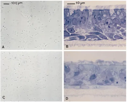
Recent technical improvements permit the reproducible production of well-differentiated human airway epithelial cell cultures. Dissociated cells are seeded on T-COL mem-branes and grow to confluence within 3 to 7 d. After con-fluence, no bulk leakage of medium from the basolateral to the apical surface of the epithelium occurs, and the cells grow at an ALI. By 14 d after confluence, extensive ciliary beating is apparent and collections of mucoid material are often visible on the apical surface. In late stage cultures (. 21 d), cells typical of native epithelium, including cili-ated, basal, and secretory cells, are apparent (Figure 3). Well-differentiated cultures often persist for more than 2 mo, but pores eventually form in the epithelia and dis-rupt barrier function. Primary airway epithelial cells cul-tured on plastic assume a mostly uniform morphology without recognizable ciliated or secretory cells.
PCR was used to screen nasal and bronchial cell cul-tures for expression of all described mucins except for the predominantly airway epithelial mucin MUC4 (41), which was screened by Northern blot. For MUC4, the only pub-lished nucleotide sequence is from the repetitive region of the gene, which is unsuitable for the design of PCR prim-ers (11).
For comparative PCR, in which expression levels of a single gene are compared between samples, PCR time courses were generated for all primer pairs to determine the linear range of the PCR reactions. The time courses for the 11p15 cluster mucins, MUC2, MUC5AC, MUC5B,
and MUC6, and the standard, cyclophilin, are shown in Figure 2. These time courses illustrate the expected in-crease in PCR product quantity.
Cyclophilin was chosen as an internal standard to cor-rect for variations in the starting concentrations of tem-plate cDNA. Cyclophilin is a cytosolic protein that binds cyclosporin A, and it also may be involved in protein fold-ing. It is abundant in eukaryotic cells, and the level ap-pears stable in most tissues and cell lines (42). The levels of cyclophilin mRNA in airway epithelial cells appear to remain constant under conditions that affect the level of MUC2 expression (7).
Bar graphs comparing expression levels for mucin genes in nasal and bronchial cultures grown on tissue-cul-ture plastic and on T-COL membranes are shown in Fig-ure 4. Of the primary secreted respiratory mucins, MUC5AC and MUC5B show differentiation-dependent changes in expression, with the level increasing in the T-COL cul-tures. MUC3 and MUC6 also show this pattern. MUC5B and MUC3 show the most dramatic increases, with ap-proximately 4-fold more signal in T-COL cultures as com-pared with plastic. MUC1 and MUC7 may have slight increases in expression in T-COL cultures. The level of MUC2 remains fairly constant. For all mucin genes, simi-lar patterns of expression were found in bronchial and na-sal epithelial cell cultures.
Northern blot analysis was used to determine the ex-pression pattern for MUC4, and to confirm exex-pression of the important airway mucins MUC5AC and MUC5B. All three of these mucins were detected in nasal and bronchial cells grown on T-COL membranes, but not in those grown on plastic (Figure 5). The high molecular-weight smear seen for these mucins is typical for RNA prepared by con-ventional methods. Debailleul and associates (43) have demonstrated that this polydispersity is most likely due to mechanical shearing of the very large mRNA transcripts (up to 24 kb), rather than to physiologic mechanisms or nuclease degradation. This would explain the difference in molecular-weight profile seen between the MUC4 and MUC5B signals in Figures 5 and 6, because the RNA preps used in Figure 6 were handled considerably more than those in Figure 5. The commercially prepared tra-cheal RNA also shows more smearing than the RNA pre-pared from cell cultures in Figure 5, and this is probably due to differences in preparative technique. As expected Figure 2.
Differentiation-depen-dent expression of 11p15.5 clus-ter mucins for bronchial and na-sal cells grown on plastic (Pl) and T-COL membranes (T), as determined by reverse transcrip-tase (RT)–PCR. Order of panels depicts the gene order on the chromosome. PCR product band intensity in EBr-stained agarose gels shows the linear range of
the reactions for MUC6, MUC2, MUC5AC, MUC5B, and the internal standard, cyclophilin. The cycle numbers (indicated near top of
figure) used for triplicate determinations are circled. RT–PCR assays for MUC1, MUC3, MUC7, and MUC8 were analyzed the same
Bernacki, Nelson, Abdullah, et al.: Mucin Gene Expression in Human Airway Epithelia 599
from the established tissue distribution, MUC4, MUC5AC, and MUC5B were strongly expressed in human trachea, and were not detected in dermal fibroblasts or intestine.
The time course of MUC4 and MUC5B expression and the RA dependence are shown in Figure 6. Six days after plating on T-COL membranes, no MUC5B was detected. By 13 d, MUC5B was expressed in the presence of RA. For MUC4, high molecular-weight transcripts also appear by 13 d. The significance of the low molecular-weight smear in the 6-d sample is not clear. Examination of the ri-bosomal RNA bands indicate that the 6-d RNA sample is not more degraded than samples at the later time points. The band across all lanes at approximately 4.4 kb corre-sponds in position and intensity to the large ribosomal subunit, and probably represents nonspecific binding of the probe. In the absence of RA, both MUC4 and MUC5B expression were completely suppressed. Figure 7 shows the morphology of the cultures from 6 to 27 d in the pres-ence and abspres-ence of RA. Omission of RA results in loss of ciliated and goblet cells and in the development of squa-mous metaplasia.
Discussion
In the normal human airway, mucus glycoproteins are pro-duced by goblet cells in the surface epithelium and by mu-cous cells in the submucosal glands. This study is an initial characterization of mucin gene expression patterns in cul-tures of human airway epithelial cells that in many ways resemble native epithelia. Such primary cell cultures will facilitate the study of mucin gene expression because they more closely mimic the in vivo situation than do most
es-tablished cell lines. Expression of the MUC2 gene, for ex-ample, appears to be at least an order of magnitude lower in some cell lines than in native tissue (8). Our ultimate goal is to use these cultures for transfections and other ma-nipulations to study airway epithelial cell differentiation and the regulation of mucin gene expression, protein syn-thesis and glycosylation, and secretion.
Transcripts from all mucin genes tested—MUC1, MUC2, MUC3, MUC5AC, MUC5B, MUC6, MUC7, and MUC8— were detected by PCR in airway cultures, and MUC4 was detected by Northern blot. The respiratory mucins MUC4, MUC5AC, and MUC5B were upregulated in differenti-ated cultures, whereas MUC2 was not. MUC3 and MUC6, although not considered important respiratory mucins, were also upregulated upon differentiation. Expression patterns for the mucin genes were similar in bronchial and nasal cultures. The upregulation of MUC4 and MUC5B mRNA expression was shown by Northern blot to depend on RA.
All of these mucins have been previously described in the airway (15, 16, 18, 44, 45, 47). MUC3, a predominantly intestinal mucin, was not detected by Northern blot in cul-tures similar to these (46), or by in situ hybridization in normal airway (18); however, it is detectable by antibodies in lung tissue (47) and has been shown to be upregulated in lung adenocarcinoma (19). MUC6 is primarily ex-pressed in the stomach, gallbladder, and intestine, and al-though it is not detectable by Northern blot in airway, faint staining of the tracheal surface epithelium (but not submucosal glands) has been seen with MUC6 anti-bodies (20, 48).
Comparing actual abundance of the different mucin Figure 3. Human airway
epi-thelial cells in vitro. On tissue-culture plastic, passage 1 bron-chial (A) and nasal (C) cells (5 d after seeding) assume a poorly differentiated morphology. At an ALI on T-COL membranes, bronchial (B) and nasal (D) cells (. 21 d after seeding) exhibit the well-organized, ciliated, pseu-dostratified phenotype charac-teristic of native airway epithelia containing ciliated, secretory, and basal cells. The T-COL membranes are visible below the cells. Cells grown on plastic were viewed unfixed by phase micros-copy. T-COL cultures were fixed with OsO4-PFC and processed
as described in MATERIALSAND
600 AMERICAN JOURNAL OF RESPIRATORY CELL AND MOLECULAR BIOLOGY VOL. 20 1999
mRNAs is problematic with either PCR or Northern blot techniques because of inherent differences in binding of primers and probes and to the highly complex structures of the mucin genes. Our PCR method was not designed to compare the levels of gene expression of different genes, but rather to compare the expression of a particular gene in dif-ferent types of cultures. MUC7 and MUC8 are probably expressed at very low levels, as PCR gave faint bands at 39 cycles for these genes. The MUC7 and MUC8 primers gave strong signals with RNA from salivary gland and the lung carcinoma cell line NCI-H292, respectively (not shown), suggesting that the primers are reasonably efficient.
These types of assays measure only steady-state levels
of mRNA, and therefore may not accurately represent de
novo RNA synthesis. The half-lives of MUC2-6 mRNAs
have been shown to be well in excess of 12 h in some cell lines (8, 43), and it is possible that mucin mRNAs synthe-sized prior to reestablishment of the mucociliary pheno-type persist in some cultures. Endotoxin contamination of the medium is also a potential problem. Endotoxin upreg-ulates the MUC2 gene (7), and any dependence of expres-sion on differentiation could be masked if MUC2 has al-ready been induced.
Experimental variability is a common concern when PCR is used to obtain quantitative results. Factors such as reagent variability, inconsistent block temperature, and pi-petting errors can contribute to experimental error. In this study, the results of three independent runs were averaged for each primer pair. The standard deviations for these av-erages were small, indicating that experimental error was not a major problem in these reactions. The higher vari-ability in the MUC7 and MUC8 samples was due to the low intensity of the PCR product bands.
MUC5AC and MUC5B appear to be the primary com-ponents of adult airway secretions (21–24). Airway mucus is secreted by both the goblet cells of the surface epithe-lium and the mucous cells of the submucosal glands. Gob-let cells and mucous cells are antigenically distinct (13), and some of the differences may be due to differential pression of mucin genes. MUC5AC is predominantly ex-pressed in surface goblet cells and in some gland neck cells, and MUC5B is expressed by the mucous cells of the glands and in bronchioles (16, 49). Although MUC4 is ex-pressed in airway epithelia, its contribution to airway secretions has not been determined. MUC4 is circumstan-tially linked to secretions of the transformed human tra-cheal gland cell line MM-39, which expresses the MUC4 gene and secretes mucin-like material but does not express MUC2, MUC3, MUC5AC, or MUC5B. However, these cells are cultured on tissue-culture plastic and do not ex-hibit a well-differentiated morphology (50).
The method we used to isolate primary cells probably yields predominantly surface rather than gland epithelial cells, so the high level of MUC5B expression suggests changes in gene expression as the epithelium becomes re-established in culture. Airway epithelial cells are known to have great phenotypic plasticity. For example, serous cells may differentiate into mucosecretory cells after injury, which the dissociation process may mimic (51). During de-velopment, MUC5B mRNA is expressed in both surface tracheal epithelium and developing submucosal glands at 13 wk of gestation, but by 23 wk it is more prominent in the glands (18). In the adult, MUC5B is expressed in the mucous tubules of the submucosal glands (16) and MUC5AC remains confined to the surface goblet cells
(18). During gland morphogenesis in vivo, specialized
Figure 4. Effect of culture
Bernacki, Nelson, Abdullah, et al.: Mucin Gene Expression in Human Airway Epithelia 601
basal cells act as gland progenitors and equivalent cells may be present in the dissociated cell preparation used to initiate the cultures. The signals controlling the normal, in
vivo patterns of gene expression may not be present, or
may be spatially and/or temporally disrupted in the T-COL culture system. Airway epithelial cell cultures may re-semble an embryonic stage in which both MUC5B and MUC5AC are co-expressed, may mimic the healing pro-cess, or may have a pattern of gene expression not repre-sented in vivo. An intermediate phenotype for airway cell cultures has been described by Sommerhoff and
Fink-beiner (52), who report that proteins characteristic of ei-ther serous or mucous cells are co-expressed in individual cells in cultures from tracheal glands. Further studies are needed to determine whether MUC5AC and MUC5B are in the same or different cells in the T-COL cultures.
RA is required for mucociliary differentiation and for
the maintenance of the mucociliary phenotype. In vivo,
retinoid deficiency leads to loss of the normal phenotype and development of squamous metaplasia (reviewed in 53). Previous studies in cultured human bronchial epi-thelial cells have demonstrated that RA is necessary for mucociliary differentiation and for the expression of MUC5AC and MUC2 (25, 31, 39). The results obtained in the current study for MUC4 and MUC5B are in agree-ment with the established role of RA in these cells.
Although MUC3 and MUC6 are unlikely to be major secreted mucins in airway epithelial cell cultures, their dif-ferentiation-dependent expression is nevertheless intrigu-Figure 6. Time course and RA dependence of MUC4 and
MUC5B expression in human bronchial cells. Total RNA was isolated from cells grown on T-COL membranes or plastic in the presence (1RA) or absence (2RA) of 5 3 1028 M RA, and
ana-lyzed by Northern blot. Approximately 5 mg RNA was loaded per 3-mm lane. Lower panel shows ribosomal RNA bands in gel, visualized with EBr.
Figure 5. Differentiation dependence of MUC4, MUC5AC, and
602 AMERICAN JOURNAL OF RESPIRATORY CELL AND MOLECULAR BIOLOGY VOL. 20 1999
ing. MUC6 is clustered with MUC2, MUC5AC, and MUC5B (in that order) at chromosomal location 11p15.5 (3). All of these mucin genes except MUC2 show differen-tiation-dependent regulation in airway epithelial cell cul-tures. Regulation of this gene cluster is clearly complex, as all four of the mucin genes show different expression
pat-terns in vivo. However, the expression pattern in airway
cultures suggests the possibility of some co-regulatory fea-tures for the 11p15.5 mucin gene cluster. This type of regu-lation is also seen for MUC3 (7q22) (54, 55) and MUC4 (3q29) (11, 56) but is not pronounced for MUC1, MUC7, or MUC8, which map to 1q21, 4q13-q21, and 12q24.3, re-spectively (9, 15, 44). MUC3 and MUC4 may also share
some regulatory elements, even though their tissue distri-butions are different.
This study presents an expression profile for mucin genes in well-differentiated cultures of airway epithelial cells. Companion studies to determine the molecular com-position of the secreted mucous layer in these cells are cur-rently in progress. The model provided by these cultures will complement other experimental systems, for example, tracheal xenografts and in vitro developmental models, to further the scope of our knowledge of cell lineage and gene regulation in the airway epithelium. A greater under-standing of transcriptional regulation of mucin genes will provide insights into the pathologic processes characteris-Figure 7. Effect of RA on morphology of bronchial epithelial cells on T-COL membranes. Cells were grown in the presence (A–C) or
Bernacki, Nelson, Abdullah, et al.: Mucin Gene Expression in Human Airway Epithelia 603
tic of chronic airway diseases, and may suggest avenues for therapeutic interventions.
Acknowledgments: The authors acknowledge Dr. James R. Yankaskas, Diana Walstad, and Ron Kim of the UNC Cystic Fibrosis Center Tissue Culture Core for invaluable assistance in tissue procurement and cell isolation; Kim Burns and Tracy Bartolotta of the UNC Cystic Fibrosis Center Histology Core for their excellent work; and Colm Reid for useful contributions. This work was supported by grant Randel97Z0 from the Cystic Fibrosis Foundation (S.H.R.); the Cystic Fibrosis Trust (A.H.); and NIH grants HL58345 (S.H.R.) and DK-46589 (A.H.).
References
1. Phillips, G. J., S. L. James, and M. I. Lethem. 1997. Rheological properties and hydration of airway mucus. In Airway Mucus: Basic Mechanisms and Clinical Perspectives. D. F. Rogers and M. I. Lethem, editors. Birkhauser Verlag, Basel, Switzerland. 117–147.
2. Wells, U. M., and P. S. Richardson. 1997. Mucus hypersecretion and its role in the airway obstruction of asthma and chronic obstructive pulmonary disease. In Airway Mucus: Basic Mechanisms and Clinical Perspectives. D. F. Rogers and M. I. Lethem, editors. Birkhauser Verlag, Basel, Switzer-land. 275–300.
3. Pigny, P., V. Guyonnet-Duperat, A. S. Hill, W. S. Pratt, S. Galiegue-Zoui-tina, M. C. d’Hooge, A. Laine, I. Van-Seuningen, P. Degand, J. R. Gum, Y. S. Kim, D. M. Swallow, J. P. Aubert, and N. Porchet. 1996. Human mu-cin genes assigned to 11p15.5: identification and organization of a cluster of genes. Genomics 38:340–352.
4. Gendler, S. J., C. A. Lancaster, J. Taylor-Papadimitriou, T. Duhig, T. Peat, and J. Burchell. 1990. Molecular cloning and expression of a human tu-mor-associated polymorphic epithelial mucin. J. Biol. Chem. 265:15286– 15293.
5. Lan, M. S., S. K. Batra, W.-N. Qi, R. S. Metzgar, and M. A. Hollingsworth. 1990. Cloning and sequencing of a human pancreatic tumor mucin cDNA. J. Biol. Chem. 265:15294–15299.
6. Gum, J. R., Jr., J. W. Hicks, N. W. Toribara, B. Siddiki, and Y. S. Kim. 1994. Molecular cloning of human intestinal mucin (MUC2) cDNA. Identifica-tion of the amino terminus and overall sequence similarity to prepro-von Willebrand factor. J. Biol. Chem. 269:2440–2446.
7. Li, J. D., A. F. Dohrman, M. Gallup, S. Miyata, J. R. Gum, Y. S. Kim, J. A. Nadel, A. Prince, and C. B. Basbaum. 1997. Transcriptional activation of mucin by Pseudomonas aeruginosa lipopolysaccharide in the pathogenesis of cystic fibrosis lung disease. Proc. Natl. Acad. Sci. USA 94:967–972. 8. Gum, J. R., J. W. Hicks, and Y. S. Kim. 1997. Identification and
character-ization of the MUC2 (human intestinal mucin) gene 59-flanking region: promoter activity in cultured cells. Biochem. J. 325:259–267.
9. Bobek, L. A., J. Liu, S. N. Sait, T. B. Shows, Y. A. Bobek, and M. J. Levine. 1996. Structure and chromosomal localization of the human salivary mucin gene, MUC7. Genomics 31:277–282.
10. Bobek, L. A., H. Tsai, A. R. Biesbrock, and M. J. Levine. 1993. Molecular cloning, sequence, and specificity of expression of the gene encoding the low molecular weight human salivary mucin (MUC7). J. Biol. Chem. 268: 20563–20569.
11. Porchet, N., V. C. Nguyen, J. Dufosse, J. P. Audie, V. Guyonnet-Duperat, M. S. Gross, C. Denis, P. Degand, A. Bernheim, and J. P. Aubert. 1991. Molecular cloning and chromosomal localization of a novel human tra-cheo-bronchial mucin cDNA containing tandemly repeated sequences of 48 base pairs. Biochem. Biophys. Res. Commun. 175:414–422.
12. Meerzaman, D., P. Charles, E. Daskal, M. H. Polymeropoulos, B. M. Mar-tin, and M. C. Rose. 1994. Cloning and analysis of cDNA encoding a major airway glycoprotein, human tracheobronchial mucin (MUC5). J. Biol. Chem. 269:12932–12939.
13. Aubert, J.-P., N. Porchet, M. Crepin, M. Duterque-Coquillaud, G. Vergnes, M. Mazzuca, B. Debuire, D. Petitprez, and P. Degand. 1991. Evidence for different human tracheobronchial mucin peptides deduced from nucle-otide cDNA sequences. Am. J. Respir. Cell Mol. Biol. 5:178–185. 14. Dufosse, J., N. Porchet, J.-P. Audie, V. Guyonnet-Duperat, A. Laine, I.
Van-Seuningen, S. Marrakchi, P. Degand, and J.-P. Aubert. 1993. Degen-erate 87-base-pair tandem repeats create hydrophilic/hydrophobic alter-nating domains in human mucin peptides mapped to 11p15. Biochem. J. 293:329–337.
15. Shankar, V., P. Pichan, R. L. Eddy, Jr., V. Tonk, N. Nowak, S. N. Sait, T. B. Shows, R. E. Schultz, G. Gotway, R. C. Elkins, M. S. Gilmore, and G. P. Sachdev. 1997. Chromosomal localization of a human mucin gene (MUC8) and cloning of the cDNA corresponding to the carboxy terminus. Am. J. Respir. Cell Mol. Biol. 16:232–241.
16. Sharma, P., L. Dudus, P. A. Nielsen, H. Clausen, J. R. Yankaskas, M. A. Hollingsworth, and J. F. Englehardt. 1998. MUC5B and MUC7 are differ-entially expressed in mucous and serous cells of submucosal glands in hu-man bronchial airways. Am. J. Respir. Cell Mol. Biol. 19:30–37.
17. Chambers, J. A., M. A. Hollingsworth, A. E. Trezise, and A. Harris. 1994.
Developmental expression of mucin genes MUC1 and MUC2. J. Cell Sci. 107:413–424.
18. Reid, C. J., S. Gould, and A. Harris. 1997. Developmental expression of mu-cin genes in the human respiratory tract. Am. J. Respir. Cell Mol. Biol. 17:592–598.
19. Nguyen, P. L., G. A. Niehans, D. L. Cherwitz, Y. S. Kim, and S. B. Ho. 1996. Membrane-bound (MUC1) and secretory (MUC2, MUC3, and MUC4) mucin gene expression in human lung cancer. Tumour Biol. 17:176–192. 20. De Bolos, C., M. Garrido, and F. X. Real. 1995. MUC6 apomucin shows a
distinct normal tissue distribution that correlates with Lewis antigen ex-pression in the human stomach [comment]. Gastroenterology 109:723–734. 21. Hovenberg, H. W., J. R. Davies, and I. Carlstedt. 1995. Human tracheal mu-cins—is MUC5 more prominent in the epithelial surface than in the sub-mucosa? Biochem. Soc. Trans. 23:534S. (Abstr.)
22. Hovenberg, H. W., J. R. Davies, A. Herrmann, C. J. Linden, and I. Carl-stedt. 1996. MUC5AC, but not MUC2, is a prominent mucin in respiratory secretions. Glycoconj. J. 13:839–847.
23. Thornton, D. J., I. Carlstedt, M. Howard, P. L. Devine, M. R. Price, and J. K. Sheehan. 1996. Respiratory mucins: identification of core proteins and glycoforms. Biochem. J. 316:967–975.
24. Thornton, D. J., M. Howard, N. Khan, and J. K. Sheehan. 1997. Identifica-tion of two glycoforms of the MUC5B mucin in human respiratory mucus. Evidence for a cysteine-rich sequence repeated within the molecule. J. Biol. Chem. 272:9561–9566.
25. Yoon, J. H., T. Gray, K. Guzman, J. S. Koo, and P. Nettesheim. 1997. Regu-lation of the secretory phenotype of human airway epithelium by retinoic acid, triiodothyronine, and extracellular matrix. Am. J. Respir. Cell Mol. Biol. 16:724–731.
26. Finkbeiner, W. E., S. D. Carrier, and C. E. Teresi. 1993. Reverse transcrip-tion-polymerase chain reaction (RT-PCR) phenotypic analysis of cell cul-tures of human tracheal epithelium, tracheobronchial glands, and lung car-cinomas. Am. J. Respir. Cell Mol. Biol. 9:547–556.
27. Kai, H., K. Yoshitake, A. Hisatsune, T. Kido, Y. Isohama, K. Takahama, and T. Miyata. 1996. Dexamethasone suppresses mucus production and MUC-2 and MUC-5AC gene expression by NCI-H292 cells. Am. J. Phys-iol. 271:L484–L488.
28. Li, D., D. Wang, S. Majumdar, B. Jany, S. R. Durham, J. Cottrell, N. Ca-plen, D. M. Geddes, E. W. Alton, and P. K. Jeffery. 1997. Localization and up-regulation of mucin (MUC2) gene expression in human nasal biopsies of patients with cystic fibrosis. J. Pathol. 181:305–310.
29. Seregni, E., C. Botti, C. Lombardo, A. Cantoni, A. Bogni, I. Cataldo, and E. Bombardieri. 1996. Pattern of mucin gene expression in normal and neo-plastic lung tissues. Anticancer Res. 16:2209–2213.
30. Inatomi, T., S. Spurr-Michaud, A. S. Tisdale, Q. Zhan, S. T. Feldman, and I. K. Gipson. 1996. Expression of secretory mucin genes by human con-junctival epithelia. Invest. Ophthalmol. Vis. Sci. 37:1684–1692.
31. Gray, T. E., K. Guzman, C. W. Davis, L. H. Abdullah, and P. Nettesheim. 1996. Mucociliary differentiation of serially passaged normal human tra-cheobronchial epithelial cells. Am. J. Respir. Cell Mol. Biol. 14:104–112. 32. Wu, R., J. Yankaskas, E. Cheng, M. R. Knowles, and R. Boucher. 1985.
Growth and differentiation of human nasal epithelial cells in culture: se-rum-free, hormone-supplemented medium and proteoglycan synthesis. Am. Rev. Respir. Dis. 132:311–320.
33. Yankaskas, J. R., C. U. Cotton, M. R. Knowles, J. T. Gatzy, and R. C. Boucher. 1985. Culture of human nasal epithelial cells on collagen matrix supports: a comparison of bioelectric properties of normal and cystic fibro-sis epithelia. Am. Rev. Respir. Dis. 132:1281–1287.
34. Willumsen, N. J., C. W. Davis, and R. C. Boucher. 1989. Intracellular Cl- ac-tivity and cellular Cl- pathways in cultured human airway epithelium. Am. J. Physiol. 256:C1033–C1044.
35. Lechner, J. F., and M. A. LaVeck. 1985. A serum-free method for culturing normal human bronchial epithelial cells at clonal density. J. Tissue Cult. Meth. 9:43–48.
36. Kaartinen, L., P. Nettesheim, K. B. Adler, and S. H. Randell. 1993. Rat tra-cheal epithelial cell differentiation in vitro. In Vitro Cell. Dev. Biol. Anim. 29A:481–492.
37. Sims, D. E., and M. M. Horne. 1997. Heterogeneity of the composition and thickness of tracheal mucus in rats. Am. J. Physiol. 273:L1036–L1041. 38. Voynow, J. A., and M. C. Rose. 1994. Quantitation of mucin mRNA in
respiratory and intestinal epithelial cells. Am. J. Respir. Cell Mol. Biol. 11: 742–750.
39. Guzman, K., T. E. Gray, J. H. Yoon, and P. Nettesheim. 1996. Quantitation of mucin RNA by PCR reveals induction of both MUC2 and MUC5AC mRNA levels by retinoids. Am. J. Physiol. 271:L1023–L1028.
40. Ausubel, F. M., R. Brent, R. E. Kingston, D. D. Moore, J. G. Seidman, J. A. Smith, and K. Struhl. 1997. Current protocols in molecular biology. In Cur-rent Protocols. V. Chanda, editor. John Wiley & Sons, Inc., New York. 41. Porchet, N., P. Pigny, M. P. Buisine, V. Debailleul, P. Degand, A. Laine,
and J. P. Aubert. 1995. Human mucin genes: genomic organization and ex-pression of MUC4, MUC5AC and MUC5B. Biochem. Soc. Trans. 23:800– 805.
604 AMERICAN JOURNAL OF RESPIRATORY CELL AND MOLECULAR BIOLOGY VOL. 20 1999
43. Debailleul, V., A. Laine, G. Huet, P. Mathon, M. C. d’Hooghe, J. P. Aubert, and N. Porchet. 1998. Human mucin genes MUC2, MUC3, MUC4, MUC5AC, MUC5B, and MUC6 express stable and extremely large mRNAs and exhibit a variable length polymorphism: an improved method to analyze large mRNAs. J. Biol. Chem. 273:881–890.
44. Gendler, S. J., and A. P. Spicer. 1995. Epithelial mucin genes. Annu. Rev. Physiol. 57:607–634.
45. Shankar, V., M. S. Gilmore, R. C. Elkins, and G. P. Sachdev. 1994. A novel human airway mucin cDNA encodes a protein with unique tandem-repeat organization. Biochem. J. 300:295–298.
46. Van Klinken, B. J., T. C. Van Dijken, E. Oussoren, H. A. Buller, J. Dekker, and A. W. Einerhand. 1997. Molecular cloning of human MUC3 cDNA re-veals a novel 59 amino acid tandem repeat region. Biochem. Biophys. Res. Commun. 238:143–148.
47. Apostolopoulos, V., P. X. Xing, and I. F. McKenzie. 1995. Anti-peptide monoclonal antibodies to intestinal mucin 3. J. Gastroenterol. Hepatol. 10:555–561.
48. Toribara, N. W., A. M. Roberton, S. B. Ho, W. L. Kuo, E. Gum, J. W. Hicks, J. R. Gum, Jr., J. C. Byrd, B. Siddiki, and Y. S. Kim. 1993. Human gastric mucin: identification of a unique species by expression cloning. J. Biol. Chem. 268:5879–5885.
49. Audie, J. P., A. Janin, N. Porchet, M. C. Copin, B. Gosselin, and J. P. Au-bert. 1993. Expression of human mucin genes in respiratory, digestive, and
reproductive tracts ascertained by in situ hybridization. J. Histochem. Cy-tochem. 41:1479–1485.
50. Lo-Guidice, J. M., M. D. Merten, G. Lamblin, N. Porchet, M. C. Houve-naghel, C. Figarella, P. Roussel, and J. M. Perini. 1997. Mucins secreted by a transformed cell line derived from human tracheal gland cells. Biochem. J. 326:431–437.
51. Basbaum, C., and B. Jany. 1990. Plasticity in the airway epithelium. Am. J. Physiol. 259:L38–L46.
52. Sommerhoff, C. P., and W. E. Finkbeiner. 1990. Human tracheobronchial submucosal gland cells in culture. Am. J. Respir. Cell Mol. Biol. 2:41–50. 53. Jetten, A. M. 1991. Growth and differentiation factors in tracheobronchial
epithelium. Am. J. Physiol. 260:L361–L373.
54. Fox, M. F., F. Lahbib, W. Pratt, J. Attwood, J. Gum, Y. Kim, and D. M. Swallow. 1992. Regional localization of the intestinal mucin gene MUC3 to chromosome 7q22. Ann. Hum. Genet. 56:281–287.
55. Gum, J. R., J. W. Hicks, D. M. Swallow, R. L. Lagace, J. C. Byrd, D. T. Lamport, B. Siddiki, and Y. S. Kim. 1990. Molecular cloning of cDNAs de-rived from a novel human intestinal mucin gene. Biochem. Biophys. Res. Commun. 171:407–415.