Original Research Article
Incidence of neurosensory deficits following surgical removal of
mandibular third molars: a prospective clinical study
Thaufiq Ahamed M. I.
1*, Naveen Jayakumar
2, Neelakandan R. S.
3INTRODUCTION
It is well established that the oral and perioral regions have an outstanding tactile spatial acuity as determined by psychophysical methods (Weinstein, 1968).1 The oral
and maxillofacial regions are the areas with increased
concentration of peripheral receptors because of their remarkable importance in daily life. It is difficult to tolerate neurological disturbances in oral and maxillofacial areas compared to disturbances in other parts of the body.2
1Department ofDental and Maxillofacial surgery, Karpagam Faculty of Medical Sciences and Research, Coimbatore,
Tamil Nadu, India
2Department of Oral and Maxillofacial Surgery, Sri Ramachandra Dental College and Hospital, Porur, Chennai, Tamil
Nadu, India
3Department of Oral and Maxillofacial Surgery, Meenakshi Ammal Dental College Maduravoyal, Chennai, Tamil
Nadu, India
Received: 05 December 2018
Revised: 05 April 2019
Accepted: 11 April 2019
*Correspondence:
Dr. Thaufiq Ahamed M. I., E-mail: drthau@gmail.com
Copyright: © the author(s), publisher and licensee Medip Academy. This is an open-access article distributed under the terms of the Creative Commons Attribution Non-Commercial License, which permits unrestricted non-commercial use, distribution, and reproduction in any medium, provided the original work is properly cited.
ABSTRACT
Background: The aim of this prospective study was to determine the incidences of inferior alveolar nerve and lingual nerve deficit following surgical removal of impacted mandibular third molars and to evaluate the risk factors responsible for these postoperative neurosensory deficits.
Methods: A total of 80 patients who reported to department of oral and maxillofacial surgery, Meenakshi Ammal Dental College, Chennai, Tamil Nadu, India requiring surgical removal of impacted mandibular third molar were included in this cross-sectional study. Standard surgical procedure was performed. All patients were reassessed one week post-surgery. Subjectively reported altered sensations were recorded and objective assessments were performed with light touch test, two-point discrimination threshold and pin-pick pain threshold. The collected data was analyzed using the chi square test to find out any clinical relevance.
Results: There was no inferior alveolar nerve related neurosensory deficits and 6 (7.5%) resulted in lingual nerve related neurosensory deficits. The incidence of LN deficit for mesioangular, horizontal, distoangular was 1.3%, 3.8% and 2.5% respectively. Type of impaction assumed a mild statistical significance (p = 0.050).
Conclusions: This study highlights the importance of careful preoperative clinical and radiographic assessment of patients where third molar surgery is planned. The surgical technique of third molar removal is also likely to have great impact on the outcome.
Keywords: Inferior alveolar nerve, Lingual nerve, Nerve injury, Paraesthesia, Third Molar
Infraorbital, inferior alveolar, lingual and mental nerves centrally transmit sensations namely pain, temperature, touch and proprioception from the perioral regions. Each of these sensations is carried out by different types of sensory receptors and nerve fibers, each showing different susceptibility to injury and recovery.3
Mandibular third molars are the most frequently impacted teeth and its surgical removal remains the most common oral and maxillofacial procedure to be performed. 91.9% of the extractions are carried out without any serious complications. Injury to the lingual, inferior alveolar and sensory branch of the mylohyoid nerves caused by the surgical removal of mandibular third molars is an infrequent but unpleasant complication. The most common incidence of nerve injury is transient sensory disturbance. In some cases, hypoaesthesia, paraesthesia, or dysaesthesia can occur. These sensory impairments can cause problems with speech, mastication and may affect the patient’s quality of life.
Table 1: Seddon and Sunderland nerve injury
classification and incidence during 3rd molar removal.
Classification Type of
injury
Incidence during 3rd
molar removal
First degree /neuropraxia
Minor compression injuries
Elevation of a third molar with roots in close proximity to mandibular canal
Second degree/ axonotemesis
Crush injuries
More severe compression of lingual/inferior alveolar nerve by elevator
Third/fourth degree/ axonotemesis
Rupture of endoneurium /perineurium
While raising a lingual
mucoperiosteal flap
Fifth degree/ neurotmesis
Complete section of the nerve
Penetration of inferior alveolar nerve through the apices of third molar and is served during surgical removal. Rotating bur dividing lingual/IAN
Inferior alveolar nerve (IAN) injury has a reported incidence of 0.26-8.4% which usually presents with paresthesia of affected side lower lip, chin and buccal mucoperiosteum. Similarly, the lingual nerve (LN) deficit has a reported incidence of 0.1-22%.4 which commonly
presents with numbness of the ipsilateral anterior two-thirds of the tongue and disturbance in taste sensation.
The severity of the nerve injury determines the impact and recovery of neurosensory deficit which is the basis for Seddon and Sunderland (Table 1) classification.5
METHODS
The present study was carried out on 80 patients who reported to department of Oral and Maxillofacial Surgery, Meenakshi Ammal Dental College, Chennai, Tamil Nadu, India from August 2011 to April 2012, requiring surgical removal of impacted mandibular third molar. The inclusion criteria for surgical extraction were pericoronitis, dental caries, decreased bone support of second molar. obstruction of placement of a partial or complete denture, obstruction of the normal eruption of permanent teeth provoking or aggravating orthodontic problems, development of various pathologic conditions and destruction of adjacent teeth due to resorption of roots. The patients with systemic diseases such as bleeding disorders, cardiovascular diseases, diabetes mellitus etc. and patients not willing for extraction of the impacted tooth were excluded from the study. Preoperative factors such as Type of impaction (mesioangular, horizontal, distoangular or vertical), root inferior dental canal relationship, depth of impaction was obtained using periapical radiographs/ orthopantamogram.
Surgical technique
Standard impaction surgical kit was used. Local anesthesia was obtained through inferior alveolar nerve block, lingual nerve block and long buccal nerve block. A standard Terrance Ward’s incision was placed. Using a periosteal elevator, the mucoperiosteal flap was reflected and the bone was exposed. The mucoperiosteal flap was then retracted using Austin’s retractor. Bone removal was carried out using a straight bur (702/703) by guttering technique on the buccal and distal side, depending on the type of impaction. After adequate amount of bone removal, the tooth was delivered out of the socket by using an elevator. Tooth division (Figure 1) was done in 43 patients; lingual nerve protection and lingual flap elevation were done in 48 patients.
After the tooth was delivered out, the socket was irrigated with povidine-iodine solution and normal saline. Sharp bony margins were smoothened with a bone file, and the socket debrided. Complete hemostasis was achieved before wound closure. Wound was closed with 3-0 black silk suture material using interrupted sutures. After the wound closure a wet gauze pack was placed at the surgical site. Post-operative instructions were given, and post-operative follow up was advised. Suture removal was performed 7 days after the extraction.
The intraoperative data noted were, the use of a periosteal elevator to raise the lingual flap and protect the LN, tooth sectioning, and any intraoperative complications.
Table 2: Variables assessed and grading.
Variables assessed Grading
Gender Male Female
Age
16-24 25-35 Above 35
Angulation of impaction
Vertical10°-10° Mesio-angular 11°-79° Horizontal 80°-100° Disto-angular 11° -79° Others 111°-80°
Root inferior alveolar canal relationship
No contact when the root at its closest point more than 2 mm from the canals Approximation: when the closest point is <2 mm from canal but there is no contact Contact when there is any relationship between root and canal (e.g. contact,
impinging, overlap) Interruption of cortical margin of canal Sectioning of tooth Not-sectioned
Sectioned
Depth from point of elevation
0-3 mm 4-6 mm > 6 mm lingual nerve protection
and lingual flap elevation
Not done Done Inferior alveolar nerve
deficit
Present Absent Lingual nerve deficit Present Absent
All patients were reassessed 1-week post-surgery, to check for any neurosensory deficits and status of wound healing. Subjectively reported altered sensations were recorded and objective assessments (Table 2) were performed with light touch test, two-point discrimination
threshold and pin-pick pain threshold in patients complaining of neurosensory disturbance.
The study sample which was collected from the data were subjected to statistical analysis using the chi-square test to examine whether the incidence of lingual and inferior alveolar deficits varied according to the possible risk factors.
RESULTS
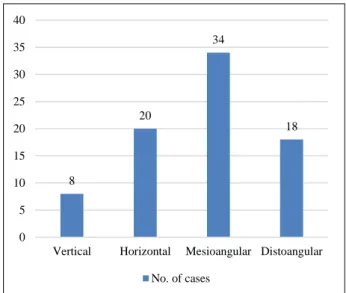
A total of 80 patients were enrolled in this study out of which 52% were female and 48% were male. Their age group ranged between 18 to 50 years with mean age of 26.2 year. Of the 80 impacted mandibular third molars that were surgically extracted, mesioangular (42.5%) was the most common type of impaction (Figure 2) and the mean depth of impaction of all types ranged from 2 to 6.5 mm.
Figure 2: Type of impaction based on angulations.
There was no Inferior alveolar nerve related neurosensory deficits and 6 (7.5%) resulted in Lingual nerve related neurosensory deficits. The remaining 74 patients did not have any neurosensory deficits.
Risk factors of neurosensory deficits
Sex and age
The prevalence of lingual nerve deficit in males and females was 1.3% (1/80) and 6.4% (5/80), respectively. This does not have statistical significance (p = 0.090)
Out of the total 7.5% of LN deficit, the age group of patients between 16-24years, 25-35years and above 35years showed 1.3%, 2.5% and 3.8% respectively. There was no statistical significance between age and LN deficit (p= 0.237)
8
20
34
18
0 5 10 15 20 25 30 35 40
Vertical Horizontal Mesioangular Distoangular
Type and depth of impaction
The incidence of LN deficit for mesioangular, horizontal, distoangular was 1.3%, 3.8% and 2.5% respectively. Type of impaction assumed a mild statistical significance (p = 0.050)
Raising of lingual flap and lingual nerve protection
Lingual flap elevation and lingual nerve protection were done in 60% of cases (48/80), out of which 5% (4/80) cases resulted in LN deficit. It was not done in 40% of cases (32/80) out of which 2.5% (2/80) cases resulted in LN deficit. There was no statistical significance (p= 0.729).
Tooth sectioning
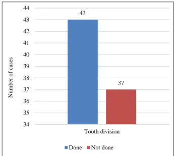
Out of 80 surgical removal, 43 (53.8%) required sectioning of tooth (Figure 3).
Figure 3: Tooth sectioning.
The incidences of Lingual nerve deficit in groups with tooth sectioning were 6.3% (5/80) and without tooth sectioning were 1.3% (1/80). Proportions of extractions that did and did not use tooth sectioning were not statistically significant (p =0.131)
Depth of impaction
The incidence of LN deficit for depth of impaction between (0-3 mm), (4-6 mm) and (>6 mm) were 2%, 4% and 0% respectively, there was no statistical significance (p= 0.249)
DISCUSSION
An Impacted third molar is a developmental anomaly caused by an obstruction in the eruption path or by an ectopic position of tooth.6 The inferior alveolar, lingual,
mylohyoid and buccal nerves lies in close proximity to impacted mandibular third molars. Therefore, during surgical removal of mandibular third molar, each of these nerves are at the risk of damage, particularly inferior alveolar and lingual nerve.
The inferior alveolar nerve runs within the mandibular canal for a considerable distance, surrounded by the neurovascular bundle. Hence, In the event of injury, the severed nerve ends will remain in apposition and will not retract unless obstructed by displaced fragments of bone or root tip. Hence good regeneration would be expected after injury.7
On the contrary, lingual nerve is morphologically different from the inferior alveolar nerve. Adjacent to lower third molar, the lingual nerve is covered by thin layer of soft tissue and mucosa, rather than a bony canal. Hence, if sectioned or adjacent soft tissue is damaged, the cut nerve ends retract apart and may become misaligned or constricted by scar tissue. Apart from the mechano/thermosensitive nerve fibers, the lingual nerve also contains gustatory, vasomotor and secretomotor nerve fibers which makes successful regeneration of the axons less likely.7
Injuries to the inferior alveolar nerve
To some extent, the risk of injury to the IAN may be assessed preoperatively, through examination of the radiographic relationship of the third molar to the inferior alveolar canal. Although standard radiographs only provide a 2-dimensional image of the 3-dimensional anatomies, by using morphological and location characteristics, in some cases the higher likelihood of IAN injury may be anticipated. IAN injury can occur after direct or indirect trauma during surgical removal of third molar. For example, compression of the nerve by apex elevators causing blunt trauma from the elevated roots or after inferior alveolar nerve block injections. Tay AB and Go WS found that if an intact inferior alveolar nerve bundle is observed during third molar surgery, this indicates an intimate relationship with the third molar and has a 20% risk of postoperative paresthesia, with a 70% chance of recovery within 1 year.8 Hence, nerve injury
can occur at any point during the surgical procedure. If the IAN directly traverses the root of the tooth, when the tooth is elevated the IAN may be dissected, resulting in neurotmesis.9 In this study no IAN deficit was elicited.
Injuries to the lingual nerve
The position of the lingual nerve is variable and although all efforts are made to avoid lingual nerve trauma during third molar surgery, lingual nerve damage may sometimes be inevitable. Kiesselbach JE and Chamberlain JG, studied the variable position of the lingual nerve.10 In 17.6% of the dissections, the lingual
nerve was found at the level of the alveolar crest or higher. Of 256 patients, the nerve was visualized above
43
37
34 35 36 37 38 39 40 41 42 43 44
Tooth division
N
u
mb
er
o
f
ca
se
s
the height of the lingual plate of the lower third molar in 12 (4.6%) and in 62% of cases the nerve contacted the lingual plate. As a result, clinicians cannot depend on the lingual plate to act as a protective barrier during third molar surgery. The inconsistent position of the lingual nerve in the region of the retromolar area means that it may be subject to damage throughout the procedure-during incision, buccal flap elevation, flap retraction, tooth sectioning and removal, and suturing.
Risk factors
Several studies have shown that age is associated with an increased risk of nerve damage in third molar surgeries. Bruce RA et al.11 noted that the risk of nerve damage was
significantly higher for patients aged 35 years or older than for those aged 14-24 years. Although age and LN deficit is not statistically significant in present study, 3 out of 6 cases (3.8%) of LN deficit observed were above 35years of age. Increasing age has also been shown to be related to an increasing risk of LN injury. Black concurred that there was a strong association between age and IAN deficit, and recommended removal of third molars before the age of 20 years.12 No IAN deficit was
observed in present study. Some authors have suggested germectomy during adolescence to reduce the risk of nerve damage.
Previous studies have also suggested that nerve dysfunction and postoperative complications are more common in female patients.13 However, other studies
have also not confirmed any association between the prevalence of nerve damage and patient’s gender.14
Although in this study the proportion of female patients suffering from LN deficit was greater, it was not statistically significant. This is contrary with the findings of Tay AB and Go WS, who found that male gender was a risk factor for paresthesia.8
Kipp DP et al, reported horizontally impacted mandibular third molar surgeries have increased risk of lingual nerve damage compared with other types.15 Carmichael FA and
McGowan DA, reported a similar results and proposed that vertical impaction carried a lower risk of lingual nerve damage.16 This study found horizontally impacted
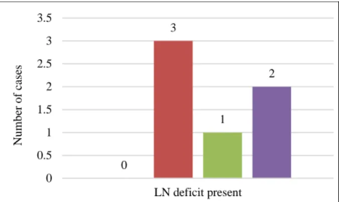
lower third molars being at highest risk of LN deficit (p=0.050), followed by disto -angular (Figure 4).
This could be possibly due to excess amount of distal bone and abutting lingual cortex guttering to surgically remove the tooth, leading to accidental damage to the LN. In contrary, 5 studies with 7256 subjects described the incidence of LN deficit to be high in disto angular impaction (4.0%), followed by horizontal (2.8%), mesio angular (2.4%) and vertical impaction (1.9%). With regard to depth of impaction and nerve injury, Kipp DP et al, and Carmichael FA and McGowan DA, concluded that full bony impaction has the greatest risk of nerve damage.15,16
Figure 4: Lingual nerve deficit in relation to angulation of impaction.
Using winter’s red line as the measurement of depth of impaction, the authors confirmed that the risk of IAN deficit is increased in third molars of greater depth. This is due to poor accessibility of the surgery, as well as the close proximity of the root to the IAN. However, in this study no IAN deficit was elicited, even though 8.8% cases showed>6mm depth from point of elevation. Deeper impacted lower third molars did not pose a significant risk of lingual nerve deficit. This study supports this, since LN deficit and depth were statistically insignificant (p= 0.249).
There has been a continuous argument over the past decade, whether raising a lingual flap/placement of a subperiosteal retractor protects the LN or accidentally damages the Pogrel MA et al, in their prospective study found no risk of permanent LN damage when retraction was used thereby recommending lingual retraction to improve surgical access.17
Pichler J W and Beirne OR, in a systematic review concluded that placing a lingual tissue retractor could induce a higher risk of transient lingual nerve damage than when a retractor was not used; but there was no difference between the two groups in terms of permanent LN damage.18 Leung YY and Cheung LK, in a literature
review of prospective studies in 2011, stated that 16 papers with 10,893 subjects reported LN deficit elicited in 3.1% with lingual flap raised and 1.5% in which the lingual flap was not raised.19 The risk ratio of LN deficit
if the lingual flap was raised was 1.94 times more likely to occur than if it was not. In this study, authors did not find any statistical difference in relation to the incidence of LN deficit from either lingual flap raising or protection of the LN (Figure 5).
Blackburn CW et al, stated that ‘the lesson to be learnt is quite simple, never let the bur enter the tissues on the lingual side of the mandible, whether there is a lingual flap retractor/guard in position or not’.20
0 3
1 2
0 0.5 1 1.5 2 2.5 3 3.5
LN deficit present
N
u
mb
er
o
f
ca
se
s
Figure 5: Lingual nerve deficit in relation to lingual
flap raising and lingual nerve protection.
Different surgical techniques for removing wisdom teeth have been proposed. The lingual split technique described by Ward TG, in 1956 is still used by some surgeons, although it has been abandoned by many centers due to the higher risk of LN injury.21 Buccal approach has been
used mostly for lower third molar removal worldwide. Recently, coronectomy, which intentionally removes the crown of a wisdom tooth without taking out its roots, has become more popular owing to the smaller risk of IDN injury shown in several studies. In 2011 Leung YY, Cheung LK stated, the incidences of LN deficit using the buccal approach, lingual split technique and coronectomy were 2.3%, 9.3% and 0.7% and for IAN 2.5%, 5.7% and 0%, respectively in a literature review of 26 prospective studies.19 In present study all the cases were done by
buccal approach.
CONCLUSION
Neurosensory deficit is a possible complication after lower third molar surgery though its occurrence is uncommon. The incidence of inferior alveolar and lingual nerve deficit was found to be 0% and 7.5%, respectively in present study. Although in this study the proportion of female patients suffering from LN deficit was greater (6.3%) compared to 1.3% for male patients , it was not statistically significant. With regard to the inferior alveolar nerve, this study did not find any factors to be associated with a significantly higher incidence of paresthesia, since there was no IAN deficit. With regard to the lingual nerve, factors found to be associated with a significantly higher incidence of paresthesia include horizontal impaction, followed by distoangular impactions 3.8% and 2.5% respectively. Although these figures are relatively low, they are still of great significance for both patients and clinicians and may have legal implications. All patients must be warned of the risks of possible damage to the inferior alveolar and lingual nerves. This study also highlights the importance of careful preoperative clinical and radiographic
assessment of patients where third molar surgery is planned. The surgical technique of third molar removal is also likely to have great impact on the outcome. Further research into the influencing factors, prevention, assessment, and treatment of postoperative inferior alveolar nerve and lingual nerve paresthesia is necessary.
Funding: No funding sources Conflict of interest: None declared
Ethical approval: The study was approved by the Institutional Ethics Committee
REFERENCES
1. Johansson RS, Trulsson M, Olsson KÅ, Westberg KG. Mechanoreceptor activity from the human face and oral mucosa. Exp Brain Res. 1988;72(1):204-8. 2. Akal ÜK, Sayan NB, Aydoǧan S, Yaman Z.
Evaluation of the neurosensory deficiencies of oral and maxillofacial region following surgery. Int J Oral Maxillofacial Surg. 2000;29(5):331-6.
3. Bui CH, Seldin EB, Dodson TB. Types frequencies and risk factors for complications after third molar extraction. J Oral Maxillofacial Surg. 2003;61(12):1379-89.
4. Cheung LK, Leung YY, Chow LK, Wong MC, Chan EK, Fok YH. Incidence of neurosensory deficits and recovery after lower third molar surgery: a prospective clinical study of 4338 cases. Int J Oral Maxillofacial Surg. 2010;39(4):320-6. 5. Boffano P, Roccia F, Gallesio C. Lingual nerve
deficit following mandibular third molar removal: review of the literature and medicolegal considerations. Oral Surg Oral Med Oral Pathol Oral Radiol. 2012;113(3):e10-8.
6. Alling CC, Helfrick JF, Alling RD. Mandibular third molars. In: Impacted teeth. 1st ed. Philadelphia, PA: WB Saunders; 1993:49-54. 7. Loescher AR, Smith KG, Robinson PP. Nerve
damage and third molar removal. Dent Update. 2003;30(7):375-82.
8. Tay AB Go WS. Effect of exposed inferior alveolar neurovascular bundle during surgical removal of impacted lower third molars. J Oral Maxillofacial Surg. 2004;62(5):592-600.
9. Jerjes W, Upile T, Shah P, Nhembe F, Gudka D, Kafas P, et al. Risk factors associated with injury to the inferior alveolar and lingual nerves following third molar surgery-revisited. Oral Pathol Oral Radiol Endodontol. 2010;109(3):335-45.
10. Kiesselbach JE, Chamberlain JG. Clinical and anatomic observations on the relationship of the lingual nerve to the mandibular third molar region. J Oral Maxillofacial Surg. 1984;42(9):565-7.
11. Lyons CJ, Bruce RA, Frederickson GC, Small GS. Age of patients and morbidity associated with mandibular third molar surgery. J Am Dental Assoc. 1980;101(2):240-5.
2
30
4
44
0 5 10 15 20 25 30 35 40 45 50
LN deficit present No LN deficit
N
u
mb
er
o
f
ca
se
s
12. Black CG. Sensory impairment following lower third molar surgery: a prospective study in New Zealand. New Zeal Dental J. 1997;93(413):68-71. 13. Blondeau F, Daniel NG. Extraction of impacted
mandibular third molars: postoperative complications and their risk factors. J Canad Dental Assoc. 2007;73(4).
14. Gülicher D, Gerlach KL. Sensory impairment of the lingual and inferior alveolar nerves following removal of impacted mandibular third molars. Int J Oral Maxillofacial Surg. 2001;30(4):306-12. 15. Kipp DP, Goldstein BH, Weiss WW. Dysesthesia
after mandibular third molar surgery: a retrospective study and analysis of 1,377 surgical procedures. J Am Dental Assoc. 1980;100(2):185-92.
16. Carmichael FA, McGowan DA. Incidence of nerve damage following third molar removal: A west of Scotland Oral Surg Res Group study. British J Oral Maxillofacial Surg. 1992;30(2):78-82.
17. Pogrel MA, Goldman KE. Lingual flap retraction for third molar removal. J Oral Maxillofacial Surg. 2004;62(9):1125-30.
18. Pichler JW, Beirne OR. Lingual flap retraction and prevention of lingual nerve damage associated with third molar surgery: a systematic review of the literature. Oral Surg Oral Med Oral Pathol Oral Radiol Endodontol. 2001;91(4):395-401.
19. Leung YY, Cheung LK. Risk factors of neurosensory deficits in lower third molar surgery: a literature review of prospective studies. Int J Oral Maxillofacial Surg. 2011;40(1):1-0.
20. Blackburn CW, Bramley PA. Lingual nerve damage associated with the removal of lower third molars. British Dental J. 1989;167(3):103.
21. Ward TG. The split bone technique for removal of lower third molors. Br Dent J. 1956;101:297-304.