Original Research Article
Revisiting conventional adenoidectomy using technology
Kiran Gangadhar Selvam, Sonee Thingujam, Jayita Poduval*
INTRODUCTION
The palatine tonsils, nasopharyngeal tonsil (or) adenoids, lingual tonsils, tubal tonsils and lateral pharyngeal band form the Waldeyer’s ring and are part of the mucosa-associated lymphoid tissue (MALT) system. Tonsils and adenoids are the body’s first line of defense for the protection of the lower airways and the gastrointestinal tract as well as for the development of antigenic memory by the host. Adenoidectomy (alone or in association with tonsillectomy) is one of the oldest and most common ENT procedures performed in children. Adenotonsillar hypertrophy, obstructive sleep apnoea, otitis media with
effusion, recurrent otitis media and nasal obstruction remain the most common indications for adenoidectomy.1
Reduction of adenoidal size, removal of adenoidal tissue in the choanae and clearance of adenoidal tissue around the Eustachian tube openings are the critical aspects in performing an adequate adenoidectomy. This should be easier to achieve with a method that can be performed under direct vision.2
The optimal method for adenoidectomy would facilitate the surgeon’s ability to visualise the adenoid pad and provide the efficient removal of tissue with minimal
ABSTRACT
Background: Adenoidectomy is one of the most frequently applied surgical procedures in the paediatric population, either alone or in conjunction with tonsillectomy and/or insertion of ventilation tubes. The main purpose of the adenoidectomy is to eliminate the nasopharyngeal respiratory pathogens and to remove nasal airway obstruction. Aim of the study was to compare the outcomes of conventional and endoscopic assisted curettage adenoidectomy.
Methods: In this comparative study, 50 patients were divided into 2 groups. Group 1 (25 patients) underwent endoscopic curettage adenoidectomy and group 2 (25 patients) underwent conventional curettage adenoidectomy. Intraoperative time, complications and postoperative pain were recorded.
Results: 72% in group 1 and 64% in group 2 had grade 2 adenoid hypertrophy. 68% in group 1 and 64% in group 2 had a moderate degree of obstruction in lateral view of soft tissue X-ray nasopharynx. The mean time taken for surgery in group 1 was 13.29±3.28 minutes, and in group 2,6.28±2.31 minutes. Minimal loss of blood was recorded in group 1 with less than 20 ml, whereas in group 2, the blood loss was high; 30% of patients had blood loss more than 30 ml. In group 1, the mean VAS was 3.25 and 2.55 in group 2. In group 1, 4% of patients had primary haemorrhage and in group 2, 8% of patients had primary haemorrhage. No patient had velopharyngeal dysfunction in either group.
Conclusions: More operative time but less blood loss were noted in endoscopic adenoidectomy. Intraoperative visualisation of the nasopharynx in endoscopic procedures showed no significant advantage over conventional adenoidectomy.
Keywords: Adenoidectomy, Conventional method, Endoscopic method, Curettage Department of ENT, Aarupadai Veedu Medical College and Hospital, Pondicherry, India
Received: 28 May 2020
Revised: 11 June 2020
Accepted: 12 June 2020
*Correspondence:
Dr. Jayita Poduval,
E-mail: [email protected]
Copyright: © the author(s), publisher and licensee Medip Academy. This is an open-access article distributed under the terms of the Creative Commons Attribution Non-Commercial License, which permits unrestricted non-commercial use, distribution, and reproduction in any medium, provided the original work is properly cited.
blood loss. Complete removal of the adenoids is challenging to determine when performing curette adenoidectomy. The importance of removing laterally based adenoidal tissue when performing adenoidectomy for otitis media with effusion has been well described.2
In the past, conventional methods for removing adenoids involved cold techniques such as transoral cold curettage and the use of the adenectomy generally followed by packing and occasionally electrocautery. After conventional adenoidectomy, there may be transient Eustachian dysfunction and aural fullness caused by probable post-surgery clots and edema in the nasopharynx. Since the early 90’s with the innovations in technology and techniques, some authors have defined excising adenoid tissue with various instruments like microdebrider under visualisation of the adenoidectomy surgical field with a laryngeal mirror, transnasal or transoral endoscope.3,4
More recent methods utilise electrocautery alone, coblation, or microdebrider removal followed by electrocautery touch-up (ME). More current methods of adenoidectomy allow for added precision during resection and decrease blood loss. Rare complications following adenoidectomy with any device can include bleeding, pain, velopharyngeal insufficiency, Eustachian tube stenosis, and nasopharyngeal stenosis, which are difficult to treat once they develop. These sequelae are best prevented by precise resection of the adenoid tissue.5
Using endoscopes to visualise the surgical field provides for the removal of residual adenoid tissue at the upper part of the nasopharynx, the choane and the peritubal region. Also, the probability of harming the Eustachian tube orifice and pharyngeal muscles is reduced, and bleeding detection and treatment can be done smoothly.6
Comparison of conventional and endoscopic adenoidectomy in terms of duration of surgery, and complications during and after the operation were aimed for with the present study.
Aim of the study was to compare the outcomes of conventional and endoscopic assisted adenoidectomy.
METHODS
This comparative study was conducted in patients with adenoid hypertrophy attending ENT OPD in Aarupadai Veedu Medical College and Hospital from October 2017 to September 2019.
Inclusion criteria
Patients between the age group of 6-18 years with symptoms of adenoid hypertrophy and patients with presence of nasal airway obstruction with sleep-disordered breathing, otitis media with effusion or
recurrent otitis media, and chronic or recurrent rhinosinusitis were included.
Exclusion criteria
Patients with significant deviated nasal septum were excluded from the study, patients below the age group of 6 years and above 18 years, patients with congenital deformities of head and neck, patients with benign or malignant tumours of the nasopharynx were excluded.
In this study 50 patients were included, 25 patients in group 1 and 25 patients in group 2. Group 1 underwent endoscopic adenoidectomy and group 2 underwent conventional adenoidectomy.
The blood soiling the nasopharyngeal pack after surgery was measured and the number of packs was counted. Blood loss was noted by the difference in the weight of the pack before and after the surgery.
Endoscopic grading was done as proposed by Clemens et al.7 Preoperative radiographic grading was done as
proposed by Bitar et al.8 The postoperative pain intensity
was done as proposed by Nikanne et al.9 Postoperative
velopharyngeal assessment as proposed by Bispo et al, was by auditory perceptual assessment of resonance and adequate velopharyngeal closure at the speech bulb with good displacement of pharyngeal walls in postoperative nasoendoscopic assessment.10
The collected data were entered and analysed with Microsoft Excel spreadsheet software, and statistically evaluated using SPSS version 21 software.
RESULTS
In this study, 50 patients were included and they were divided into 2 groups. Group 1 underwent endoscopic adenoidectomy and group 2 underwent adenoidectomy; each group consisted of 25 patients who were randomly allocated.
There was no statistical difference noted in gender distribution between groups. In group 1, male patients were 14 and were 11 female patients, in group 2, 12 male and 13 female patients were included.
In this study, aged 6 years to 18 years patients were included. The mean age of group 1 was 9.24±3.28 years and in group 2 was 10.28±2.11 years, which was statistically insignificant.
Figure 1: Site of adenoids.
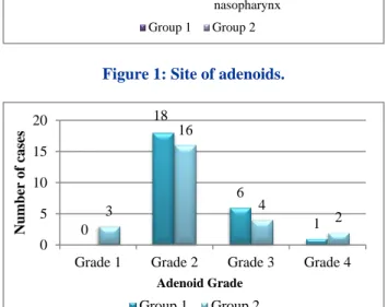
Figure 2: Preoperative grading of adenoids.
72% of patients who underwent endoscopic adenoidectomy were in grade 2, and 64% of patients who underwent conventional adenoidectomy were in grade 2. 24% of patients who underwent endoscopic adenoid-dectomy were in grade 3 and 16% of patients who underwent conventional adenoidectomy were in grade 3 (p=0.278) (Figure 2).
Figure 3: Preoperative nasal obstruction index.
In this study, 36% of patients in endoscopic adenoidectomy had a nasal obstruction index of 2 and 40% of patients in conventional adenoidectomy had an index of 2. There was no statistical difference in the nasal obstruction index between the groups (Figure 3).
In the lateral view of X-ray soft tissue nasopharynx, in an average of 68% of patients showed moderate degree of obstruction, (64% in group 1 and 72% in group 2). There was no statistical difference noted between the groups (Figure 4).
Figure 4: Preoperative radiological grading of adenoid hypertrophy.
Figure 5: Operative time.
The mean time taken for surgery in group 1 was 13.29±3.28 minutes, and in group 2, 6.28±2.31 minutes, which was statistically significant (p<0.0001) (Figure 5).
Figure 6: Intraoperative blood loss.
In this study, minimal losses of blood were recorded in group 1 with less than 20 ml, whereas in group 2, the blood loss was high (30% of patients had blood loss more than 30 ml). The blood loss between groups was statistically significant (p<0.0001) (Figure 6).
7 10 1 7 7 9 1 8 0 2 4 6 8 10 12 Choana Peritubaric region Superior part of the nasopharynx Multiple site Num ber o f Ca ses
Group 1 Group 2
0 18 6 1 3 16 4 2 0 5 10 15 20
Grade 1 Grade 2 Grade 3 Grade 4
Nu
mber
of cases
Adenoid Grade
Group 1 Group 2
3 4 9 6 2 1 2 2 10 5 6 0 0 2 4 6 8 10 12
1 1.5 2 2.5 3 3.5
Nu
mber
of cases
Nasal Obstruction Index Group 1 Group 2
7 16 2 5 18 2 0 5 10 15 20
Low Intermediate High
Nu
mber
of C
ases
Obstruction in X-ray Group 1 Group 2
13.29 6.28 0 5 10 15 20
Group 1 Group 2
Time in minu tes 2 18 5 0 0
0 0 2
11 13 0 5 10 15 20
<5 ml 6-10 ml 11-20 ml 21-30 ml >30 ml
Nu
mber
of cases
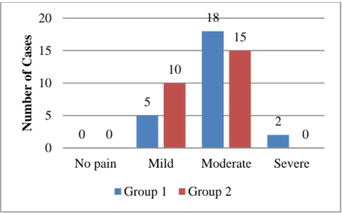
Figure 7: Postoperative pain.
On measurement of postoperative pain intensity was lesser in group 2 (p<0.0001) (Figure 7).
In group 1, the mean VAS was 3.25 and in group 2 the mean VAS was 2.55 which was statistically significant.
Table 1: Complications.
Complication Group 1 Group 2
Primary haemorrhage 1 2
Velopharyngeal dysfunction 0 0
Infection 0 0
In group 1, 4% of patients had primary haemorrhage and in group 2, 8% of patients had primary haemorrhage (Table 1).
The blood soiling the nasopharyngeal pack after surgery was measured and the number of packs was counted. Blood loss was noted by the difference in the weight of the pack before and after the surgery. On measurement of velopharyngeal insufficiency by speech analysis and postoperative nasoendoscopic assessment, no patients had velopharyngeal dysfunction in either groups.
A postoperative nasopharyngeal swab taken on postoperative day 1 showed no growth in either groups.
DISCUSSION
Adenoidectomy is one of the most commonly performed surgeries in children. Various techniques have been proposed to reduce the amount of bleeding during the procedure and to facilitate the easy and safe removal of adenoid tissue. Adenoidectomy can be done using an adenoid curette, bipolar cautery, power-assisted microdebrider, and coblator. Though there are many options, emphasis should be on the most effective technique and postoperative outcome. Recurrence rates following adenoidectomy have never been documented in literature, and thus it has come to be assumed that curettage adenoidectomy could leave residue and cause recurrence, and that endoscopic removal could ensure complete removal and hence no recurrence.
Yearsley et al performed the first adenoidectomy in 1842.11 Conventionally, adenoidectomy is performed
with a curette, without visualising the nasopharynx. The use of a laryngeal mirror to visualise the nasopharynx has been mentioned in the literature; however, the use of an endoscope has revolutionised the technique of adenoidectomy. This was popularised by Cannon et al.12
Multiple other methods have evolved since the inception of curettage adenoidectomy, such as suction diathermy, laser ablation, and radiofrequency ablation, using equipment including a molecular resonance tool, a coblation wand, and a microdebrider. The aim of adenoidectomy should be complete adenoid removal, with minimal morbidity and quick recovery. In 1996, Parsons described the use of powered instruments in the paediatric population and explained the precision of the microdebrider system.13 Given the range of methods
available, there is a quest for the most optimal method for complete adenoid removal and better relief of symptoms, with the least number of postoperative complications.
Conventionally, a curette is used to perform adenoidectomy. Curettage is a blind procedure that mainly targets the central portion of the adenoid mass. The lateral tissue abutting the Eustachian tube opening and the tissue in the most superior part of the nasopharynx remains inaccessible to the curette. Furthermore, the risk of collateral damage is high as curettage is carried out without direct visualisation of the area to be worked on. The ideal procedure to remove the adenoid mass should involve removal of the entire mass under direct visualisation, and should be minimal in terms of operative time, blood loss, damage to surrounding tissues, complications and pain.
In our study, the mean age of the children who underwent endoscopic curettage was 9.24 years, and the mean age of the children who underwent the conventional method was 10.28 years. Adenoid enlargement was mostly seen in age less than 13 years. This was similar to the study conducted by Datta et al, Somani et al and Prakash et al.6,14,15 Preoperative nasal endoscopy revealed that
grade 2 adenoid hypertrophy was the most common followed by grades 3 and 4.
The surgical time was significantly longer for endoscopic assisted adenoidectomy than for the conventional technique. The mean surgical time for endoscopic assisted surgery was 13.29 minutes and 6.28 minutes for the conventional method. Endoscopic-assisted adenoid-ectomy also requires the setting up of additional instruments and more technical skills, and involves the complete removal of adenoids under vision, which takes more time than conventional adenoidectomy. Moreover, removal of the adenoids from all nasopharyngeal areas creates a wider raw area, which bleeds more, and this requires more time to control the bleeding.
The operative time for both the techniques was in accordance with Datta et al.6 Time taken in conventional
0
5
18
2 0
10
15
0 0
5 10 15 20
No pain Mild Moderate Severe
Nu
mber
of C
ases
surgery varied from 22-39 minutes (95% confidence interval (CI) 27.7-30.9) and in endoscopic surgery from 27-55 minutes (95% CI 36.6-41.9) (p<0.05).7 In a study
done by Cannon et al, the median time required to complete the conventional adenoidectomy was 1.67 minutes (range 0.5-7.17 minutes) and the median time required to complete endoscopic assisted was 2.08 minutes (range 0.25-11.0 minutes).12 However, powered
instruments were found to be 58 percent faster in a study by Koltai et al.16 Stanislaw et al reported powered
adenoidectomy to be 20 percent faster than curette adenoidectomy, and Feng et al, and Murray et al, also found that endoscopic assisted powered adenoidectomy was 59% faster.17-19
Ravishankar et al compared endoscopic microdebrider assisted adenoidectomy with the conventional procedure and found that in the conventional group, the surgical time was significantly less compared to the endoscopic method- 20.79 minutes in the endoscopic method and 14.42 minutes in the conventional method. Muniraju et al compared endoscopic assisted adenoidectomy with conventional adenoidectomy.21 The mean time for
surgery in endoscopic assisted adenoidectomy was 34.10 minutes, and in conventional adenoidectomy 22.83 minutes. More blood loss was noted in endoscopic assisted adenoidectomy compared to conventional adenoidectomy, 29.57 ml and 16.67 ml respectively. Mean blood loss in the endoscopic procedure was 31.06 ml and in the conventional group 22.26 ml, which is dissimilar to our results (the maximum blood loss noted with endoscopic procedure was 46 ml and in the conventional method 60 ml, being comparatively higher). These differences could be attributed to the use of powered instrumentation. In a meta-analysis done by Yang et al conventional curettage adenoidectomy caused greater blood loss than endoscopic assisted adenoidectomy.22 This is mainly related to direct
visualisation, treatment of the source of bleeding, the effect of a microdebrider with suction, and time to haemostasis by Vokurka et al. This reduction of blood loss would be highly suitable for children because it reduces the risk of hemorrhage by Al-Mazrou et al.24 The
blood loss in the series by Feng et al.18 was more in the
conventional adenoidectomy group though it was not statistically significant. Stanislaw et al, however reported a significant reduction in blood loss following endoscopic adenoidectomy.17
Adenoidectomy by curettage showed more residual nasopharyngeal adenoid tissue (43.33%) than by endoscopic assisted adenoidectomy (20%), in our study. Also, this study does not show a significant reduction in post-operative pain, probably due to the small number of cases and the fact that only adenoidectomy was done in 15 cases, causing less postoperative pain.
The recovery time after any surgery is difficult to define as different parameters are used for different studies. The question of time taken to return to normal activity
following the surgery is the most important in the post operative follow up period. The recovery period in the endoscopic assisted adenoidectomy was shorter than in conventional adenoidectomy, and this difference was statistically significant in different studies. The Indian scenario presents a situation where the availability of equipment is also a factor in choosing the method of surgery. Though nasal endoscopes are fast becoming basic tools, powered instrumentation like microdebrider is not common. We attempted to analyse whether the endoscope actually helps to improve outcomes.
CONCLUSION
Intraoperative time for endoscopic adenoidectomy being considerably more than for conventional adenoidectomy, intraoperative blood loss for endoscopic adenoidectomy being only slightly lower compared to conventional adenoidectomy, postoperative pain assessment showing no significant difference in either group, and velopharyngeal insufficiency being absent in both groups, it may be concluded that intraoperative endoscopic visualisation of the nasopharynx has no significant advantage over conventional blind curettage adenoidectomy. Better technology therefore does not always translate into better outcomes, though larger and better designed research needs to be undertaken in this regard. Like cold steel tonsillectomy being conclusively proved to be the most cost effective method, conventional adenoidectomy is still a good choice.
Funding: No funding sources Conflict of interest: None declared
Ethical approval: The study was approved by the Institutional Ethics Committee
REFERENCES
1. Thornval A. Wilhelm Meyer and the adenoids. Arch Otolaryngol. 1969;90(3):383-6.
2. Gerhardsson H, Stalfors J, Odhagen E, Sunnergren O. Pediatric adenoid surgery in Sweden 2004-2013: incidence, indications and concomitant surgical procedures. Int J Pediatr Otorhinolaryngol. 2016;87:61-6.
3. Babakurban ST, Aydın E. Adenoidectomy: Current approaches and review of the literature. Kulak Burun Bogaz Ihtis Derg. 2016;26(3):181-90. 4. Anand V, Sarin V, Singh B. Changing trends in
adenoidectomy, Indian J Otolaryngol Head Neck Surg. 2014;66(4):375-80.
5. Randall DA, Hoffer ME. Complications of tonsillectomy and adenoidectomy. Otolaryngol Head Neck Surg. 1998;118(1):61-8.
6. Datta Lt Col R, Singh Col VP. Deshpal Col. Conventional versus Endoscopic powered adenoidectomy: a Comparative Study. MJAFI. 2009;65:308-12.
comparison of postoperative results. Int J Pediatr Otolaryngol. 1998; 43:115-22.
8. Bitar MA, RahiA,Khalifeh M, Madanat LM A suggested clinical score to predict the severity of adenoid obstruction in children. Eur Arch Otorhinolaryngol. 2006;263:924-8.
9. Nikanne E, Kokki H, Tuovinen K. Postoperative pain after adenoidectomy in children. Br J Anaesthesia. 1999;82(6):886-9.
10. Bispo NH, Whitaker ME, Aferri HC, Neves JD, Dutka Jde C, Pegoraro-Krook MI. Speech therapy for compensatory articulations and velopharyngeal function: a case report. J Appl Oral Sci. 2011;19(6):679-84.
11. Yearsley J. On Deafness from Morbid Conditions of the Mucous Membrane of the Stomach, Throat, and Ear. The Effect of Cold, Dyspepsia, Scarlatina, Measles, etc. London, England: Nisbet & Co; 1842. 12. Cannon CR, Replogle WH, Schenk MP.
Endoscopic-assisted adenoidectomy. Otolaryngol Head Neck Surg. 1999;121:740-4.
13. Parsons, DS. Rhinologic uses of powered instrumentation in children beyond surgery. Otolaryngol Clin North Am. 1996;29:105-14. 14. Somani SS, Naik CS, Bangad SV. Endoscopic
adenoidectomy with microdebrider. Indian J Otolaryngol Head Neck Surg. 2010;62(4):427-31. 15. Prakash NS, Mallikarjunappa AM, Samuel HT.
Endoscopic assisted adenoidectomy versus conventional curettage adenoidectomy- A comparative study. National J Otorhinolaryngol Head & Neck Surg. 2013;1(10):10-2.
16. Koltai PJ, Kalathia AS, Stanislaw P, Heras HA. Power-assisted adenoidectomy. Arch Otolaryngol Head Neck Surg. 1997;123:685-8.
17. Stanislaw P, Koltai PJ, Feustel PJ. Comparison of powerassisted adenoidectomy vs adenoid curette
adenoidectomy. Arch Otolaryngol Head Neck Surg. 2000;126:845-9.
18. Feng Y, Yin S. Comparison of the powered-assisted adenoidectomy with adenoid curette adenoidectomy. Lin Chuang Er Bi Yan Hou Ke Za Zhi. 2006;20:54-7.
19. Murray N, Fitzpatrick P, Guarisco JL. Powered partial adenoidectomy. Arch Otolaryngol Head Neck Surg. 2002;128:792-6.
20. Ravishakar C, Killera S. Comparing endoscopic microdebrider assisted adenoidectomy with curettage procedure. Int J Otorhinolaryngol Head Neck Surg. 2018;4:559-64.
21. Muniraju M, Saifulla M. A comparative study of adenoidectomy by microdebrider vs conventional method. Int J Otorhinolaryngol Head Neck Surg. 2018;4:808-14.
22. Yang L, Shan Y, Wang S, Cai C, Zhang H. Endoscopic assisted adenoidectomy versus conventional curettage adenoidectomy: a meta-analysis of randomised controlled trials. Springerplus. 2016;5:426.
23. Vokurka J. Shaver (micro debridor) in otorhinolaryngology. Int Congr Ser. 2003;1240:1411-5.
24. Al-Mazrou KA, Al-Qahtani A, Al-Fayez AI. Effectiveness of transnasal endoscopic powered adenoidectomy in patients with choanal adenoids. Int J Pediatr Otorhinolaryngol. 2009;73(12):1650-2.