1Department of Epidemiology, University of North Carolina at Chapel Hill, Chapel Hill, NC, USA
2Department of Gastroenterology and Hepatology, University of North Carolina at Chapel Hill, Chapel Hill, NC, USA 3Department of Medicine A, University Medicine Greifswald, Greifswald, Germany
4Department of Medicine, Ludwig-Maximilians University, Munich, Germany
5Department of Periodontology, University of North Carolina at Chapel Hill, Chapel Hill, NC, USA
6Institute of Clinical Chemistry and Laboratory Medicine, University Medicine Greifswald, Ernst-Moritz-Arndt-University, Greifswald, Germany 7Institute for Community Medicine, University Medicine Greifswald, Greifswald, Germany, and German Center of Diabetes Research, Site Greifswald,
Germany
8Department of Dental Ecology, University of North Carolina at Chapel Hill, Chapel Hill, NC, USA
9Unit of Periodontology, Department of Restorative Dentistry, Periodontology, Endodontology, and Preventive and Pediatric Dentistry, University
Medicine Greifswald, Greifswald, Germany
10Unit of Periodontology, Department of Restorative Dentistry, Periodontology, Endodontology, and Preventive and Pediatric Dentistry, University
Medicine Greifswald, Greifswald, Germany
A supplemental appendix to this article is available online.
Corresponding Author:
A.A. Akinkugbe, Department of Epidemiology, University of North Carolina at Chapel Hill, 135 Dauer Drive CB 7435, Chapel Hill, NC 27599-7450, USA.
Email: [email protected]
Do Genetic Markers of Inflammation
Modify the Relationship between
Periodontitis and Nonalcoholic Fatty Liver
Disease? Findings from the SHIP Study
A.A. Akinkugbe
1, C.L. Avery
1, A.S. Barritt
2, S.R. Cole
1, M. Lerch
3, J. Mayerle
4,
S. Offenbacher
5, A. Petersmann
6, M. Nauck
6, H. Völzke
7, G.D. Slade
8, G. Heiss
1,
T. Kocher
9, and B. Holtfreter
10Abstract
An association between periodontitis and nonalcoholic fatty liver disease (NAFLD) has been reported by experimental animal and epidemiologic studies. This study investigated whether circulating levels of serum C-reactive protein (CRP) and a weighted genetic CRP score representing markers of inflammatory burden modify the association between periodontitis and NAFLD. Data came from 2,481 participants of the Study of Health in Pomerania who attended baseline examination that occurred between 1997 and 2001. Periodontitis was defined as the percentage of sites (0%, <30%, ≥30%) with probing pocket depth (PD) ≥4 mm, and NAFLD status was determined using liver ultrasound assessment. Serum CRP levels were assayed at a central laboratory, and single-nucleotide polymorphisms previously identified through genome-wide association studies as robustly associated with serum CRP were combined into a weighted genetic CRP score (wGSCRP). Logistic regression models estimated the association between periodontitis and NAFLD within strata of serum CRP and
separately within strata of the wGSCRP. The prevalence of NAFLD was 26.4% (95% confidence interval [CI], 24.6, 28.1) while 17.8% (95%
CI, 16.0–19.6) had ≥30% of sites with PD ≥4 mm. Whereas the wGSCRP was not a modifier (Pinteraction = 0.8) on the multiplicative scale,
serum CRP modified the relationship between periodontitis and NAFLD (Pinteraction = 0.01). The covariate-adjusted prevalence odds ratio
of NAFLD comparing participants with ≥30% of sites with PD ≥4 mm to those with no site affected was 2.39 (95% CI, 1.32–4.31) among participants with serum CRP <1 mg/L. The corresponding estimate was 0.97 (95% CI, 0.57–1.66) for participants with serum CRP levels of 1 to 3 mg/L and 1.12 (95% CI, 0.65–1.93) for participants with serum CRP >3 mg/L. Periodontitis was positively associated with higher prevalence odds of NAFLD, and this relationship was modified by serum CRP levels.
Introduction
In response to an injury or infection, inflammation occurs as a complex series of short-term adaptive responses accompanied by local tissue damage with manifestations that gradually resolves as inflammation abates, leaving little to no permanent damage (Kumar et al. 2014). Inflammation is regulated primar-ily by the innate immune system (Takashiba and Naruishi 2006; Kumar et al. 2014), and it involves a coordinated cas-cade of biological events regulated by specific cells and molec-ular signals (Naitza et al. 2012).
An excessive inflammatory response that occurs upon stim-ulation of the innate immune system has been described as a hyperresponsive trait (Shaddox et al. 2010) that presents sys-temically as a heightened expression of systemic markers of inflammation (Southerland et al. 2006). Population-based genetic studies suggest that natural selection has shaped the evolution of the innate immunity with specific focus on inflam-matory genes that are pivotal in host-pathogen interactions (Barreiro and Quintana-Murci 2010). Inflammatory biomarkers are reported to be highly heritable, with studies among ethni-cally homogeneous groups and twins indicating that about half of interindividual variability in markers of inflammation is genetically determined (Pankow et al. 2001; Dupuis et al. 2005). For instance, the Framingham Heart Study reported age- and sex-adjusted heritability of 25.3%, 25.4%, and 45.2% for C-reactive protein (CRP), interleukin 6 (IL-6), and monocyte chemoattractant protein 1 (MCP-1), respectively (Dupuis et al. 2005), while the National Heart Lung and Blood Institute (NHLBI) Family Heart Study reported heritability ranging from 35% to 40% for CRP, white blood cells, and albumin (Pankow et al. 2001; Dupuis et al. 2005).
Findings among humans and from mice models (Yoneda et al. 2012) suggest a relationship between periodontitis and nonalcoholic fatty liver disease (NAFLD), both of which are chronic health conditions characterized by a heightened inflam-matory burden (Day and James 1998; Haukeland et al. 2006; Targher 2006; Tilg and Moschen 2010; Schenkein and Loos 2013; Gocke et al. 2014). Indeed, individuals with periodontitis present with frequent bacteremia (Schenkein and Loos 2013) that promotes a proinflammatory state, while obesity, a precur-sor for NAFLD, is characterized by a state of chronic low-grade systemic inflammation (Shoelson et al. 2006; Ouchi et al. 2011).
Genome-wide association studies (GWAS) have identified 19 independent loci that are robustly associated with CRP lev-els (Dehghan et al. 2011; Naitza et al. 2012), an acute phase reactant, and a marker of systemic inflammation (Pearson et al. 2003; Raman et al. 2013). Because genetic determinants of inflammatory biomarkers can more accurately indicate life-long inflammatory status (Raman et al. 2013) compared to bio-marker concentrations obtained at a given point in time, polymorphisms in genes regulating inflammatory processes may influence the expression of periodontitis and NAFLD, as well as modify the relationship between these 2 conditions.
The purpose of this investigation was to determine whether CRP-associated genetic loci and serum CRP levels modify the association between periodontitis and NAFLD.
Methods
Data Source
The Study of Health in Pomerania (SHIP) is a population-based cohort sampled from the Western Pomeranian region of northeastern Germany (John et al. 2001; Volzke et al. 2011). The SHIP was designed to provide prevalence estimates for various diseases and disease risk factors, incidence of common risk factors, subclinical disorders, and clinical diseases and to evaluate associations among these factors. From eligible inhabitants of West Pomerania in 1996, 6,265 adults aged 20 to 79 y were invited to participate. A total of 4,308 participated in the baseline examination conducted between 1997 and 2001. Study participants underwent rigorous examinations, and interviewer-administered questionnaires were used to collect information on relevant covariates. Ethics approval for this study was obtained from the institutional review board of the University of North Carolina at Chapel Hill, and this study conformed to the Strengthening the Reporting of Observational Studies in Epidemiology (STROBE) guidelines for reporting observational studies.
Exposure Assessment and Definition
Dental examiners performed periodontal examination on study participants with no medical contraindication. Measurements of probing pocket depth (PD) and clinical attachment level (CAL) were obtained on 4 sites per tooth: distobuccal, mesio-buccal, midmesio-buccal, and midlingual or midpalatal (except the third molars) on 2 quadrants (quadrants 1 and 4 or quadrants 2 and 3). For this investigation, periodontitis was defined as the proportion of periodontal sites with PD ≥4 mm categorized as none (0%), moderate (<30%), and extensive (≥30%). In a sec-ondary analysis, periodontitis was investigated as the propor-tion of sites with CAL ≥3 mm (none, moderate, and extensive) and according to the Centers for Disease Control and Prevention–American Association for Periodontology (CDC-AAP) criteria that define severe periodontitis as ≥2 interproxi-mal sites with CAL of ≥6 mm (not on the same tooth) and ≥1 interproximal sites with PD of ≥5 mm, and moderate periodon-titis as ≥2 interproximal sites (not on the same tooth) with CAL of ≥4 mm or ≥2 interproximal sites (not on the same tooth) with PD of ≥5 mm (Page and Eke 2007). Individuals with mod-erate or severe periodontitis were categorized as having peri-odontitis, while participants not meeting these criteria were categorized as healthy/mild.
Outcome Assessment and Definition
Covariates
Covariates identified as confounders were determined after analyzing a directed acyclic graph (Greenland et al. 1999) and included age modeled with a quadratic term, sex, alcohol con-sumption in the past 30 days, and waist circumference, dichot-omized at ≥88 cm for women and ≥102 cm for men and is indicative of abdominal obesity (Grundy et al. 2005). Diabetes was based on self-reported physician’s diagnosis or study’s measurement of hemoglobin A1c (HbA1c) ≥6.5%. Self-reported smoking status was categorized as never, former, and current. Physical activity was based on self-report of the num-ber of hours per week of moderate physical activity.
Laboratory Measurements
Nonfasting blood samples were drawn from the cubital vein in the supine position. HbA1c was measured by high-performance liquid chromatography (ClinRep HbA1C, Recipe Chemicals Instruments GmbH), while serum levels of high-sensitivity CRP (hs-CRP) were estimated with the Behring Nephelometer II (Dade Behring) (Gocke et al. 2014).
Genotyping
Genomic DNA from blood samples was collected using stan-dardized procedures. Blood aliquots were immediately placed on ice after collection and stored at −80°C in a biobank (John et al. 2001). A total of 4,096 samples were genotyped using the Human SNP 6.0 Array (Affymetrix) with overall genotyping efficiency of 98.6% and imputed to the 1000 Genomes v3 ref-erence panel released March 2012 (ALL ancestries panel, build 37) (Volzke et al. 2011; Teumer et al. 2013).
A total of 19 CRP single-nucleotide polymorphisms (SNPs) from 19 loci, 1 IL-6 SNP, 2 MCP-1 SNPs, and 2 erythrocyte sedimentation rate (ESR) SNPs were identified as genetic markers of inflammation for this investigation. After quality control analysis, genotype data were available for 4,070 par-ticipants. None of the SNPs deviated significantly from Hardy-Weinberg equilibrium (P> 0.003), and call rates were >95% for all SNPs.
CRP-Specific Weighted Genetic Score
To study the cumulative effect of multiple gene loci, a weighted genetic score for CRP was computed for each study partici-pant. A risk allele was defined as the allele associated with a unit increase in log-transformed serum CRP level. The corre-sponding effect sizes from previous GWAS of inflammatory mediators (Naitza et al. 2012) and a meta-analysis of GWAS of CRP levels (Dehghan et al. 2011) were used to weigh the con-tribution of each risk allele within the weighted genetic CRP score (wGSCRP) as previously described (Pharoah et al. 2008) and implemented (Thanassoulis et al. 2012; Xiao et al. 2015). The lead SNP in each identified locus were used in creating the weighted genetic score. The list of SNPs and effect sizes
(β estimates) are presented in Appendix Table 1. The weighted genetic score for CRP was calculated as follows:
wGSCRP=α1 1x +α2 2x + ....αk kx +αn nx ,
where αk is the per-allele β estimate associated with the risk allele for CRP SNP k, and Xk is the number of risk alleles for the same SNP, and n is total number of SNPs used in creating the genetic score. In addition to CRP, weighted genetic scores were also created for IL-6, MCP-1, and ESR.
Exclusions
From 4,308 eligible participants at baseline, participants with no genotype data (n = 238) were excluded. Also excluded were participants who reported excessive alcohol consumption, defined as 70 g of ethanol/wk (equivalent to 1 standard drink/ day) for women and 140 g of ethanol/wk (equivalent to 2 stan-dard drinks/day) for men (n = 970). Participants with no liver ultrasound reading (n = 48), as well as those with no periodon-tal examination (n = 14) or edentulous (n = 563), were also excluded. Also excluded were participants with chronic or autoimmune viral hepatitis as well as those who self-reported use of the following steatosis-promoting medications: tamoxi-fene, methotrexate, and amiodarone. The resulting analytic sample size was 2,481, noting that some participants were ineligible for multiple reasons.
Statistical Analysis
The respective genetic scores, including the wGSCRP and log-transformed serum CRP levels, were modeled as continuous traits in separate linear regression models investigating whether these traits were associated with periodontitis and NAFLD.
Logistic regression models stratified according to the median value for the wGSCRP (<1.98 vs. ≥1.98) assessed the relationship between periodontitis and NAFLD. In separate stratified analyses, a similar association was investigated within strata of low (<1 mg/L), intermediate (1 to 3 mg/L), and high (>3 mg/L) (Pearson et al. 2003) serum CRP levels. Statistical tests were 2-sided, and the test for statistical signifi-cant interaction was set a priori at P< 0.1. Data analysis was conducted in SAS version 9.4 (SAS Institute).
Results
respectively, and 1.11 mg/L (IQR, 0.51– 2.95) for participants with no site with PD ≥4 mm (Table 1). Participants with NAFLD were less likely to report physical activity (33% vs. 49%), equally as likely to con-sume alcohol, and more likely to be men (58% vs. 41%) than participants without NAFLD (Table 1).
Appendix Table 1 shows the full list of SNPs with the corresponding effect sizes used in creating the respective weighted genetic scores. Most of the CRP SNPs were positively associated with serum CRP levels in this study population (Appendix Fig. 1), and the wGSCRP aligns well with serum CRP levels such that participants with a low genetic CRP score had lower mean serum CRP levels compared to participants with a high genetic CRP score (Fig. 1). The median value for the wGSCRP was 1.98 (IQR, 1.82– 2.14). Participants with wGSCRP above the median had a mean (standard deviation [SD]) serum CRP of 3.24 (4.72) mg/L, while participants with wGSCRP at or below
the median had a mean (SD) serum CRP of 2.65 (6.62) mg/L. As expected, serum CRP was associated with periodontitis. Specifically, each unit increase in log-transformed serum CRP was associated with a 23% increase in adjusted prevalence odds of having ≥30% sites with PD ≥4 mm, prevalence odds ratio [POR] = 1.23 (95% CI, 1.09–1.39), while the corresponding esti-mate for having <30% of sites with PD ≥4 mm was POR = 1.16 (95% CI, 1.06–1.26) (Table 2). There was no meaningful associa-tion between individual CRP SNPs and periodontitis (Appendix
Figs. 2–4). Even after combining into a score, the wGSCRP was not associated with the prevalence odds of having <30% sites or ≥30% sites with PD ≥4 mm, POR = 1.02 (95% CI, 0.69–1.51) and 1.01 (95% CI, 0.61–1.69), respectively (Table 2).
As was observed for periodontitis, each unit increase in log-transformed serum CRP was associated with a higher preva-lence odds of NAFLD, with adjusted POR = 1.26 (95% CI, 1.14–1.39) (Table 2). Likewise, most of the individual CRP SNPs had no meaningful effect on the prevalence odds of
Table 1. Distribution of Baseline Factors According to the Proportion of Sites with PD ≥4 mm and Nonalcoholic Fatty Liver Disease Status among Participants of the Study of Health in Pomerania, 1997 to 2001.
Proportion of Sites with PD ≥4 mm NAFLD
Total
(n = 2,481) None (0%) (n = 733) Moderate ((n = 1,307)<30%) Extensive ((n = 441)≥30%) (n = 1,827)No (n = 654)Yes
CRP, mg/L 1.42 (0.64, 3.01) 1.11 (0.51, 2.95) 1.48 (0.68, 3.01) 1.82 (0.82, 3.90) 1.20 (0.55, 2.95) 2.16 (0.99, 4.41) Age, y 47.0 (34.0, 60.0) 36.0 (27.0, 56.0) 47.0 (35.0, 60.0) 55.0 (46.0, 65.0) 42.0 (31.0, 57.0) 56.0 (46.0, 65.0) Waist circumference, cm 88.0 (77.2, 98.1) 83.0 (73.0, 94.0) 89.0 (78.0, 98.0) 94.0 (84.5, 103) 84.0 (74.3, 93.8) 98.5 (90.8, 106) Alcohola 4.10 (1.31, 6.86) 4.56 (1.39, 6.71) 4.09 (1.31, 7.17) 3.92 (0.65, 6.41) 4.08 (1.31, 6.86) 4.28 (0.83, 6.86)
Abdominal obesityb 747 (30.1) 170 (23.2) 389 (29.8) 188 (42.6) 383 (21.0) 364 (55.7)
Physical activity 1,099 (44.5) 388 (53.0) 586 (45.0) 125 (28.5) 881 (48.5) 218 (33.3)
Men 1,116 (45.0) 281 (38.3) 592 (45.3) 243 (55.1) 740 (40.5) 376 (57.5)
Smoking
Never 966 (39.1) 323 (44.1) 515 (39.6) 128 (29.2) 717 (39.4) 249 (38.1)
Former 780 (31.6) 203 (27.7) 434 (33.4) 143 (32.7) 527 (29.0) 253 (38.7)
Current 726 (29.4) 207 (28.2) 352 (27.1) 167 (38.1) 574 (31.6) 152 (23.2)
Diabetes mellitus 465 (18.7) 98 (13.4) 248 (19.0) 119 (27.0) 226 (12.4) 239 (36.5)
NAFLD 654 (26.4) 133 (18.1) 348 (26.6) 173 (39.2) — —
Data are presented as No. (%) or median (lower quartile, upper quartile).
CDC-AAP, Centers for Disease Control and Prevention–American Academy of Periodontology; CRP, C-reactive protein; NAFLD, nonalcoholic fatty liver disease; PD, pocket depth.
aAverage number of standard drinks in the past 30 days.
bSex-specific weight circumference defined as ≥88 cm for women and ≥102 cm for men (Grundy et al. 2005).
NAFLD (Appendix Fig. 5), while each unit increase in the wGSCRP was associated with a 21% increase in the prevalence odds of NAFLD; POR = 1.21 (95% CI, 0.82–1.78) (Table 2).
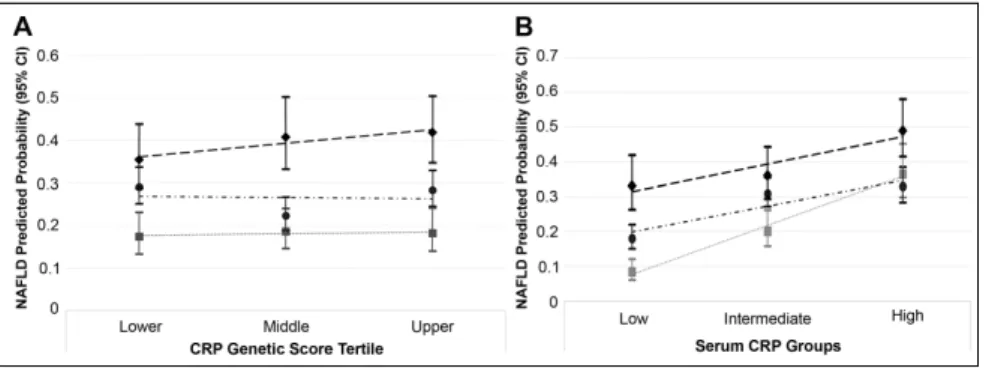
Although participants with periodontitis had a higher pre-dicted probability of NAFLD compared to participants with a healthy periodontium, there was no significant statistical inter-action (i.e., effect measure modification on the multiplicative scale) according to tertile of the wGSCRP (Pinteraction = 0.8) (Fig. 2A). Despite this, estimates stratified at the median value for wGSCRP are presented in Table 3. Participants in the stratum of wGSCRP at or below the median had slightly lower unadjusted and covariate-adjusted prevalence odd ratios for the relation-ship between periodontitis (PD ≥4 mm) and NAFLD compared to participants in the wGSCRP stratum above the median (Table 3).
In contrast, the higher predicted probability of NAFLD for participants with periodontitis (PD ≥4 mm) differed according to serum CRP levels (Pinteraction = 0.01) (Fig. 2B). And in con-trast to the wGSCRP, stratified analyses show a magnitude of unadjusted and covariate-adjusted prevalence odds of NAFLD comparing participants with periodontitis to those without to be highest in the low serum CRP (CRP <1 mg/L) stratum. For instance, the unad-justed and covariate-adunad-justed NAFLD prevalence odds ratio comparing par-ticipants with ≥30% sites with PD ≥4 mm to those with no sites affected in the low-serum CRP (<1 mg/L) stratum were 5.34 (95% CI, 3.18–8.97) and 2.39 (95% CI, 1.32–4.31), respectively, while the corresponding estimates for participants in the high-serum CRP (>3 mg/L) stratum were 1.66 (95% CI, 1.04–2.65) and 1.12 (95% CI, 0.65–1.93), respectively (Table 3).
Contrary to the finding of a significant statistical interaction between serum CRP and PD ≥4 mm, there was no significant interaction with CAL ≥3 mm (Pinteraction = 0.2). This suggests that the significant interaction observed with the CDC-AAP periodontitis classification (Pinteraction = 0.014) appeared to have been driven mostly by PD instead of CAL. Stratified estimates for periodontitis defined as the proportion of sites with CAL ≥3 mm and based on the CDC-AAP criteria are presented in Appendix Tables 2 and 3, respectively.
Table 2. Association between Serum CRP and Genetic Determinants of Inflammatory Mediators with Proportion of Sites with PD ≥4 mm and NAFLD in the Study of Health in Pomerania, 1997 to 2001.
Prevalence Odds Ratio (95% CI) for the Proportion of
Sites with PD ≥4 mm (Reference = 0%) Prevalence Odds Ratio (95% CI) for NAFLD
PD ≥4 mm Crude Adjusteda Crude Adjusteda
Serum CRPb
<30% 1.19 (1.10–1.29) 1.16 (1.06–1.26) 1.49 (1.37–1.61) 1.26 (1.14–1.39)
≥30% 1.40 (1.26–1.56) 1.23 (1.09–1.39)
Genetic scoresb
wGSCRP
<30% 1.02 (0.69–1.51) 1.21 (0.82–1.78)
≥30% 1.01 (0.61–1.69)
wGSIL-6
<30% 0.83 (0.49–1.40) 0.54 (0.32–0.90)
≥30% 0.60 (0.31–1.19)
wGSESR
<30% 1.24 (0.53–2.91) 1.61 (0.69–3.76)
≥30% 0.99 (0.33–3.04)
wGSMCP
<30% 0.88 (0.60 1.29) 1.07 (0.73–1.57)
≥30% 1.09 (0.66–1.80)
CI, confidence interval; CRP, C-reactive protein; NAFLD, nonalcoholic fatty liver disease; PD, pocket depth; wGSCRP, weighted genetic CRP score;
wGSESR, weighted genetic erythrocyte sedimentation rate score; wGSIL-6, weighted genetic interleukin 6 score; wGSMCP, weighted genetic monocyte
chemoattractant protein score.
aAdjusted for age, sex, waist circumference, smoking, physical activity, alcohol consumption, and diabetes. bEstimate is for each unit increase in the respective genetic scores or log-transformed CRP.
Discussion
Findings from this investigation aimed at assessing whether inflammatory burden of which CRP was used as a marker modified the relationship between periodontitis and NAFLD, were consistent with a positive association between serum CRP and NAFLD as well as serum CRP and periodontitis. Under the premise that genetic determinants of inflammatory markers are better able to indicate lifelong inflammatory sta-tus, genetic variants robustly associated with CRP levels were combined into a genetic score that was substituted for serum CRP in this study. Contrary to the findings for serum CRP, the wGSCRP predicted NAFLD to a greater extent than it did for periodontitis. And while serum CRP was a significant modi-fier, the wGSCRP was not a modifier of the association between periodontitis and NAFLD.
NAFLD has a multifactorial etiology with conditions like insulin resistance and obesity identified as risk factors (Farrell and Larter 2006; Angulo 2007). Increased levels of inflamma-tory mediators have also been reported in individuals with NAFLD (Haukeland et al. 2006; Targher 2006). While a formal mediation analysis was beyond the scope of this study because of its cross-sectional nature, investigating the periodontitis-NAFLD association conditioning on or stratifying by markers of inflammatory burden while adjusting for confounders and NAFLD risk factors offers insights into the potential role of other factors besides inflammation in NAFLD etiology.
Despite the high CRP heritability (Pankow et al. 2001; Dupuis et al. 2005), currently identified genetic loci explain
~5% of the variation in serum CRP levels (Dehghan et al. 2011). Thus, currently identified loci may not be sufficiently robust to characterize associations or detect gene-environment interactions, even after combining the lead SNPs into a score as was done in this study. This may in part explain why no significant effect measure modification within strata of the genetic CRP score was detected.
Serum CRP represents a systemic marker of an inflamma-tory response and can be instrumental in detecting effect mea-sure modification. The associations between periodontitis and NAFLD within levels of serum CRP were contrary to expecta-tion. Indeed, the greater magnitude of association in the stra-tum of serum CRP <1 mg/L suggests a contribution of periodontitis to NAFLD burden independent of chronic low-grade systemic inflammation. This association may have been induced via pathways involving an alteration in the gut micro-bial composition by swallowed periodontal pathogens (Arimatsu et al. 2014). Furthermore, participants with serum CRP <1 mg/L were less likely to have established NAFLD risk factors like abdominal obesity and diabetes (Appendix Table 4), consistent with an association of periodontitis with NAFLD independent of these factors.
While periodontitis may also contribute to NAFLD burden in the intermediate- and high-serum CRP strata, these associa-tions in opposite of expectation suggest that the effects of peri-odontitis on NAFLD may be conditioned by a systemic inflammatory response of which CRP is a marker. Alternatively, the higher levels of serum CRP may indicate the presence to a larger extent of competing risk factors for NAFLD (Appendix
Table 3. Stratified Analysis for the Association between the Proportion of Periodontal Sites with Pocket Depth ≥4 mm (0%, <30%, ≥30%) and NAFLD According to Categories of the Weighted Genetic CRP Score and Serum CRP Levels, in the Study of Health in Pomerania, 1997 to 2001.
NAFLD, n Prevalence Odds Ratio (95% CI)
Cases Noncases Crude Adjusteda
wGSCRP≤1.98 (n = 1,222)
No site (0%) 65 292 Reference Reference
Moderate (<30%) 166 479 1.56 (1.13–2.15) 1.08 (0.75–1.57)
Extensive (≥30%) 81 139 2.62 (1.78–3.84) 1.14 (0.72–1.80)
wGSCRP >1.98 (n = 1,259)
No site (0%) 68 308 Reference Reference
Moderate (<30%) 182 480 1.72 (1.26–2.35) 1.33 (0.94–1.89)
Extensive (≥30%) 92 129 3.23 (2.22–4.70) 1.65 (1.07–2.55)
Serum CRP <1 mg/L (n = 972)
No site (0%) 29 313 Reference Reference
Moderate (<30%) 89 402 2.40 (1.53–3.73) 1.62 (1.00–2.61)
Extensive (≥30%) 46 93 5.34 (3.18–8.97) 2.39 (1.32–4.31)
Serum CRP 1 to 3 mg/L (n = 887)
No site (0%) 48 190 Reference Reference
Moderate (<30%) 151 337 1.77 (1.23–2.57) 1.37 (0.90–2.08)
Extensive (≥30%) 58 103 2.23 (1.42–3.50) 0.97 (0.57–1.66)
Serum CRP >3 mg/L (n = 622)
No site (0%) 56 97 Reference Reference
Moderate (<30%) 108 220 0.85 (0.57–1.27) 0.70 (0.45–1.10)
Extensive (≥30%) 69 72 1.66 (1.04–2.65) 1.12 (0.65–1.93)
Interaction P value for the genetic CRP score = 0.6. Interaction P value for serum CRP levels = 0.01. CI, confidence interval; CRP, C-reactive protein; NAFLD, nonalcoholic fatty liver disease; wGSCRP, weighted genetic score for CRP.
Table 4), whose effects likely overshadowed those of periodon-titis. Last, it is also possible that the high CRP levels may have been generated in response to hepatic injury as a result of NAFLD.
In theory, polymorphisms robustly associated with serum CRP levels are expected to predict an increase in the risk of coronary heart disease (CHD) and other cardiovascular disease–related events. However, these polymorphisms are not associated with an increased risk of CHD (Zacho et al. 2008; Elliott et al. 2009; Dehghan et al. 2011; Wensley et al. 2011), while serum CRP levels have been reported in several longitu-dinal studies to be associated with increased risk of CHD and myocardial infarction (Danesh et al. 2004; Lange et al. 2006). The SNPs used in creating the genetic CRP score in this study were robustly associated with CRP levels (Appendix Fig. 1), but these SNPs did not independently predict periodontitis or NAFLD risk (Appendix Figs. 2–5). While the findings for the genetic CRP score are not entirely surprising, it is also likely that the inability to detect effect measure modification by the genetic CRP score was also due to the relatively small effect of the individual SNPs even after combining them into a score.
Strengths and Limitations
Given the modest size, this study may be insufficiently pow-ered to detect effect measure modification, especially for the genetic CRP score. In addition, the cross-sectional design makes it difficult to infer how the modification by serum CRP levels might affect the periodontitis-NAFLD association over time. Last, due to the mostly homogeneous study population, findings may not generalize to other racial/ethnic groups espe-cially given that the genetic architecture of CRP differs by eth-nicity (Carlson et al. 2005). Study strengths include a good characterization of the cohort that enabled the implementation of relevant exclusions of factors like alcohol consumption that might bias findings if not accounted for. Also, the availability of genotype and phenotype data for CRP allowed an investiga-tion of this factor as a potential effect measure modifier.
Conclusion
Serum CRP was a significant modifier of the relationship between periodontitis and NAFLD, and there was a discor-dance of effect measure modification of this association by serum CRP and the weighted CRP genetic score. Given that only a fraction of the variability in CRP is explained by cur-rently identified genetic loci, more research may be needed to identify missing CRP heritability that could provide a more robust picture of genetic loci predictive of CRP levels and inflammatory markers in general.
Author Contributions
A.A. Akinkugbe, contributed to conception, design, data acquisi-tion, analysis, and interpretaacquisi-tion, drafted and critically revised the manuscript; C.L. Avery, A.S. Barritt, G.D. Slade, T. Kocher, B. Holtfreter, contributed to data acquisition, analysis, and inter-pretation, critically revised the manuscript; S.R. Cole, contributed
to data analysis, critically revised the manuscript; M. Lerch, J. Mayerle, S. Offenbacher, A. Petersmann, M. Nauck, H. Völzke, contributed to data acquisition, critically revised the manuscript; G. Heiss, contributed to conception, design, data acquisition, anal-ysis, and interpretation, critically revised the manuscript. All authors gave final approval and agree to be accountable for all aspects of the work.
Acknowledgments
The authors thank the participants of the Study of Health in Pomerania. The Study of Health in Pomerania is part of the Community Medicine Research network of the University of Greifswald, Germany, and funded by the Federal Ministry of Education and Research (grants 01ZZ9603, 01ZZ0103, and 01ZZ0403), the Ministry of Cultural Affairs, and the Social Ministry of the Federal State of Mecklenburg–West Pomerania. Genome-wide data were supported by the Federal Ministry of Education and Research (grant 03ZIK012) and a joint grant from Siemens Healthcare, Erlangen, Germany, and the Federal State of Mecklenburg, West Pomerania. The University of Greifswald is a member of the Center of Knowledge Interchange program of the Siemens AG and the Caché Campus program of the InterSystems GmbH. Support for this work was provided by the National Institutes of Health/National Institute of Dental and Craniofacial Research (grant R03DE025652-01A1). The authors declare no potential conflicts of interest with respect to the authorship and/or publication of this article.
References
Angulo P. 2007. Gi epidemiology: nonalcoholic fatty liver disease. Aliment Pharmacol Ther. 25(8):883–889.
Arimatsu K, Yamada H, Miyazawa H, Minagawa T, Nakajima M, Ryder MI, Gotoh K, Motooka D, Nakamura S, Iida T, et al. 2014. Oral pathobiont induces systemic inflammation and metabolic changes associated with alteration of gut microbiota. Sci Rep. 4:4828.
Barreiro LB, Quintana-Murci L. 2010. From evolutionary genetics to human immunology: how selection shapes host defence genes. Nat Rev Genet. 11(1):17–30.
Baumeister SE, Volzke H, Marschall P, John U, Schmidt CO, Flessa S, Alte D. 2008. Impact of fatty liver disease on health care utilization and costs in a general population: a 5-year observation. Gastroenterology. 134(1):85–94. Carlson CS, Aldred SF, Lee PK, Tracy RP, Schwartz SM, Rieder M, Liu K,
Williams OD, Iribarren C, Lewis EC, et al. 2005. Polymorphisms within the C-reactive protein (CRP) promoter region are associated with plasma CRP levels. Am J Hum Genet. 77(1):64–77.
Danesh J, Wheeler JG, Hirschfield GM, Eda S, Eiriksdottir G, Rumley A, Lowe GD, Pepys MB, Gudnason V. 2004. C-reactive protein and other circulating markers of inflammation in the prediction of coronary heart disease. N Engl J Med. 350(14):1387–1397.
Day CP, James OF. 1998. Steatohepatitis: a tale of two “hits”? Gastroenterology. 114(4):842–845.
Dehghan A, Dupuis J, Barbalic M, Bis JC, Eiriksdottir G, Lu C, Pellikka N, Wallaschofski H, Kettunen J, Henneman P, et al. 2011. Meta-analysis of genome-wide association studies in >80 000 subjects identifies multiple loci for C-reactive protein levels. Circulation. 123(7):731–738.
Dupuis J, Larson MG, Vasan RS, Massaro JM, Wilson PW, Lipinska I, Corey D, Vita JA, Keaney JF Jr, Benjamin EJ. 2005. Genome scan of systemic biomarkers of vascular inflammation in the Framingham Heart Study: evi-dence for susceptibility loci on 1q. Atherosclerosis. 182(2):307–314. Elliott P, Chambers JC, Zhang W, Clarke R, Hopewell JC, Peden JF, Erdmann
J, Braund P, Engert JC, Bennett D, et al. 2009. Genetic loci associated with C-reactive protein levels and risk of coronary heart disease. JAMA. 302(1):37–48.
Gocke C, Holtfreter B, Meisel P, Grotevendt A, Jablonowski L, Nauck M, Markus MR, Kocher T. 2014. Abdominal obesity modifies long-term associations between periodontitis and markers of systemic inflammation. Atherosclerosis. 235(2):351–357.
Greenland S, Pearl J, Robins JM. 1999. Causal diagrams for epidemiologic research. Epidemiology. 10(1):37–48.
Grundy SM, Cleeman JI, Daniels SR, Donato KA, Eckel RH, Franklin BA, Gordon DJ, Krauss RM, Savage PJ, Smith SC Jr, et al. 2005. Diagnosis and management of the metabolic syndrome: an American Heart Association/ National Heart, Lung, and Blood Institute scientific statement. Circulation. 112(17):2735–2752.
Haukeland JW, Damås JK, Konopski Z, Løberg EM, Haaland T, Goverud I, Torjesen PA, Birkeland K, Bjøro K, Aukrust P. 2006. Systemic inflamma-tion in nonalcoholic fatty liver disease is characterized by elevated levels of Ccl2. J Hepatol. 44(6):1167–1174.
John U, Greiner B, Hensel E, Ludemann J, Piek M, Sauer S, Adam C, Born G, Alte D, Greiser E, et al. 2001. Study of Health in Pomerania (SHIP): a health examination survey in an east German region: objectives and design. Soz Praventivmed. 46(3):186–194.
Kumar V, Abbas AK, Fausto N, Aster JC. 2014. Robbins and Cotran pathologic basis of disease, professional edition: expert consult-online. Philadelphia (PA): Saunders Elsevier Health Sciences.
Lange LA, Carlson CS, Hindorff LA, Lange EM, Walston J, Durda JP, Cushman M, Bis JC, Zeng D, Lin D, et al. 2006. Association of polymorphisms in the CRP gene with circulating C-reactive protein levels and cardiovascular events. JAMA. 296(22):2703–2711.
Naitza S, Porcu E, Steri M, Taub DD, Mulas A, Xiao X, Strait J, Dei M, Lai S, Busonero F, et al. 2012. A genome-wide association scan on the levels of markers of inflammation in Sardinians reveals associations that underpin its complex regulation. Plos Genet. 8(1):e1002480.
Ouchi N, Parker JL, Lugus JJ, Walsh K. 2011. Adipokines in inflammation and metabolic disease. Nat Rev Immunol. 11(2):85–97.
Page RC, Eke PI. 2007. Case definitions for use in population-based surveil-lance of periodontitis. J Periodontol. 78(7 Suppl):1387–1399.
Pankow JS, Folsom AR, Cushman M, Borecki IB, Hopkins PN, Eckfeldt JH, Tracy RP. 2001. Familial and genetic determinants of systemic markers of inflam-mation: the NHLBI family heart study. Atherosclerosis. 154(3):681–689. Pearson TA, Mensah GA, Alexander RW, Anderson JL, Cannon RO III, Criqui
M, Fadl YY, Fortmann SP, Hong Y, et al; Centers for Disease Control and Prevention; American Heart Association. 2003. Markers of inflammation and cardiovascular disease: application to clinical and public health prac-tice: a statement for healthcare professionals from the Centers for Disease Control and Prevention and the American Heart Association. Circulation. 107(3):499–511.
Pharoah PD, Antoniou AC, Easton DF, Ponder BA. 2008. Polygenes, risk prediction, and targeted prevention of breast cancer. N Engl J Med. 358(26):2796–2803.
Raman K, Chong M, Akhtar-Danesh GG, D’Mello M, Hasso R, Ross S, Xu F, Pare G. 2013. Genetic markers of inflammation and their role in cardiovas-cular disease. Can J Cardiol. 29(1):67–74.
Schenkein HA, Loos BG. 2013. Inflammatory mechanisms linking periodontal diseases to cardiovascular diseases. J Periodontol. 84(4 Suppl):S51–S69. Shaddox L, Wiedey J, Bimstein E, Magnuson I, Clare-Salzler M, Aukhil I,
Wallet SM. 2010. Hyper-responsive phenotype in localized aggressive periodontitis. J Dent Res. 89(2):143–148.
Shoelson SE, Lee J, Goldfine AB. 2006. Inflammation and insulin resistance. J Clin Invest. 116(7):1793–1801.
Southerland JH, Taylor GW, Moss K, Beck JD, Offenbacher S. 2006. Commonality in chronic inflammatory diseases: periodontitis, diabetes, and coronary artery disease. Periodontol 2000. 40:130–143.
Takashiba S, Naruishi K. 2006. Gene polymorphisms in periodontal health and disease. Periodontol 2000. 40:94–106.
Targher G. 2006. Relationship between high-sensitivity C-reactive protein lev-els and liver histology in subjects with non-alcoholic fatty liver disease. J Hepatol. 45(6):879–881; author reply 881–882.
Teumer A, Holtfreter B, Volker U, Petersmann A, Nauck M, Biffar R, Volzke H, Kroemer HK, Meisel P, Homuth G, et al. 2013. Genome-wide associa-tion study of chronic periodontitis in a general German populaassocia-tion. J Clin Periodontol. 40(11):977–985.
Thanassoulis G, Peloso GM, Pencina MJ, Hoffmann U, Fox CS, Cupples LA, Levy D, D’Agostino RB, Hwang SJ, O’Donnell CJ. 2012. A genetic risk score is associated with incident cardiovascular disease and coronary artery calcium: the Framingham Heart Study. Circ Cardiovasc Genet. 5(1):113–121. Tilg H, Moschen AR. 2010. Evolution of inflammation in nonalcoholic fatty
liver disease: the multiple parallel hits hypothesis. Hepatology. 52(5): 1836–1846.
Volzke H, Alte D, Schmidt CO, Radke D, Lorbeer R, Friedrich N, Aumann N, Lau K, Piontek M, Born G, et al. 2011. Cohort profile: the Study of Health in Pomerania. Int J Epidemiol. 40(2):294–307.
Wensley F, Gao P, Burgess S, Kaptoge S, Di Angelantonio E, Shah T, Engert JC, Clarke R, Davey-Smith G, Nordestgaard BG, et al.; C Reactive Protein Coronary Heart Disease Genetics Collaboration (CCGC). 2011. Association between c reactive protein and coronary heart disease: Mendelian randomi-sation analysis based on individual participant data. BMJ. 342:d548. Williams CD, Stengel J, Asike MI, Torres DM, Shaw J, Contreras M, Landt
CL, Harrison SA. 2011. Prevalence of nonalcoholic fatty liver disease and nonalcoholic steatohepatitis among a largely middle-aged population uti-lizing ultrasound and liver biopsy: a prospective study. Gastroenterology. 140(1):124–131.
Xiao Q, Liu ZJ, Tao S, Sun YM, Jiang D, Li HL, Chen H, Liu X, Lapin B, Wang CH, et al. 2015. Risk prediction for sporadic Alzheimer’s disease using genetic risk score in the Han Chinese population. Oncotarget. 6(35): 36955–36964.
Yoneda M, Naka S, Nakano K, Wada K, Endo H, Mawatari H, Imajo K, Nomura R, Hokamura K, Ono M, et al. 2012. Involvement of a periodontal pathogen, porphyromonas gingivalis on the pathogenesis of non-alcoholic fatty liver disease. BMC Gastroenterol. 12:16.