Case Report
Giant perivascular spaces: two case reports
Yuying Qi, Xiaofeng Han, Shujun Lu, Wenbin Ma, Panpan Chen, Xuezhen Wang*
INTRODUCTION
Virchow-Robin spaces (VRSs) are perivascular spaces that course along the cerebral vessels as they traverse from the subarachnoid space to the brain parenchyma.1
They act as a pathway for the egress of interstitial fluid. VRSs frequently become dilated and can be seen on magnetic resonance images (MRI) of patients across all ages.2 They are usually bilateral, smaller than 5mm, and
asymptomatic.2
However, on rare occasions, extreme dilatation may lead to the formation of giant VRSs. Such giant tumefactive dilated VRSs (dVRSs) can produce pressure symptoms and can easily be confused with other more sinister cystic intracranial conditions.3 Only a few previous cases are
available in literature.
This paper reports two cases and presents a review of literature to explore the etiological factor, pathology, and MRI presentation.
CASE REPORT
Case 1
A 15-year-old male was admitted to the affiliated hospital of Binzhou Medical University. Patient had been suffering from headache and had repeated seizures for 1h. He had a heavy blow on the head one month before his admission. He underwent rehabilitation after 15 days of treatment in the hospital. Half a month after his discharge, he suddenly appeared with headache and repeated tics on his limbs. Patient had no relevant drug history or significant family history. His vital signs and neurological examination and his blood, urine RT, and hepatic and renal functions were all normal. The color Doppler ultrasonography of the abdomen was normal. The MRI findings of the brain shows inequality in the size of multiple cysts in the bilateral cerebral hemisphere, round, oval or curvilinear. Those in the frontal and occipital lobes are the biggest, whereas those in the apical lobe are the smallest.
Department of Neurology, Binzhou Medical University Hospital, Binzhou, Shandong, China
Received: 13 December 2018
Accepted: 07 January 2019
*Correspondence:
Dr. Xuezhen Wang,
E-mail: [email protected]
Copyright: © the author(s), publisher and licensee Medip Academy. This is an open-access article distributed under the terms of the Creative Commons Attribution Non-Commercial License, which permits unrestricted non-commercial use, distribution, and reproduction in any medium, provided the original work is properly cited.
ABSTRACT
Giant perivascular spaces or Virchow-Robin spaces (VRSs) are uniquely inherent developmental malformation and are generally lined by ependymal or leptomeningeal cells. The cerebral hemispheres with VRSs present multiple cysts of curvilinear, round, oval, or layered configuration, which have the same signal intensity as the cerebrospinal fluid (CSF) and represent extremely dilated VRSs. The cortex became extremely thin with a well-defined margin. Herein, we report two cases of true giant perivascular spaces and present a review of pertinent literature. A patient has multiple cysts in the unilateral and bilateral and has polycystic liver. The clinical presentation, image logical features, and diagnosis are discussed.
Keywords: Diagnosis, Giant perivascular spaces, MRI features
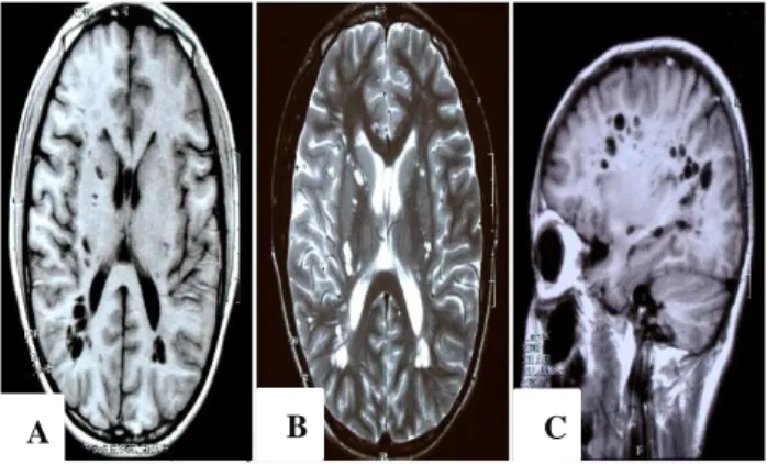
The MRI scan of the cysts demonstrate hypointensity on T1WI (Figure 1A) and hyperintensity on T2WI (Figure 1B), showing isointensity as the cerebrospinal fluid (CSF). The cortex shows a very thin and clear adumbration, and it became the compartment between the cysts (Figure 1C). The cerebellum, cerebral ganglion, brainstem, and cerebral ventricles were normal.
Figure 1: MRI scan of the cysts, demonstrating hypointensity on T1WI (A) and hyperintensity on T2WI (B) and showing isointensity as CSF. The cortex
showed an extremely thin and clear adumbration, (C) The cerebellum, cerebral ganglion, brainstem, and the
cerebral ventricles were normal.
Case 2
A 50-year-old female suffering from headaches for 10 years. She received anesthesia in her left face and the left toes for 1 year. Patient has no significant family history. All blood laboratory data were within normal ranges, including blood RT, hepatic and renal functions, dielectric, and complete set of tumor. A lumbar puncture produced clear acellular CSF with normal opening pressure. CSF RT and biochemistry, OB, and the full set of parasite were all normal. Patient vital signs and neurological examination were normal, and no objective anesthesia was found. The color Doppler ultrasonography of the abdomen showed a polycystic liver. The chest roentgenograms were normal. Patient brain MRI was similar to that in case 1 but her cysts in the left cerebral hemisphere had variable sizes and were round, oval, or curvilinear. Those in the apical and occipital lobes are the biggest, whereas those in the frontal lobe are smaller. The MRI scan of the cysts showed isointensity as CSF (Figure 2A, B, C). The cortex showed extremely thin and clear adumbration. The cerebellum, cerebral ganglion, brainstem, and cerebral ventricles were all normal. The cysts did not show potentialization (Figure 2D, E, F), and the T2 flare did not show hydrops (Figure 2G). The MRA showed that the branches of the left middle cerebral artery decreased with no other abnormalities (Figure 2H, I).
Figure 2: Brain image MRI, showing the isointensity of the cysts as CSF (A-C). The cysts did not show potentialization (D-F). The T2 flare did not show hydrops (G). The MRA showed that the branches of the middle
cerebral artery decreased with no other abnormalities (H, I). The cortex shows a very thin and clear adumbration (A-G).
A B C
A B C
D E F
DISCUSSION
The etiopathogenisis of giant perivascular spaces
The etiology of giant perivascular spaces is currently unknown. A few cases have been reported, including our two cases, which have no family histories. In the case of Li Mao, one of the twins experienced abdominal delivery, while his twin brother experienced fetal death; hence, this disease may be due to the factors, which affected the fetal development in the trimester of pregnancy.4 Our two
cases did not involve abnormalities during pregnancy and were detected occasionally because of other symptoms that were not associated with the cysts.
Different hypotheses have been proposed to explain the dilatation of VRSs, including the impaired drainage of the interstitial fluid into the subarachnoid space, segmental necrotizing angiitis of the cerebral vessels or the increased permeability of vessels, fibrosis, and obstruction of VRSs lead to the impaired fluid drainage and leakage of the interstitial fluid from the intracellular compartment into the VRSs.5-7
The pathology of giant perivascular spaces
VRSs are perivascular spaces in the brain and can be visualized through MRI.8,9 They are extensions of the
subarachnoid into the brain, which accompany the penetrating vessels to the level capillaries. They are lined by a single layer of epithelial cells that correspond to meningina or ependymal cells and are full of CSF.4,10
VRSs have been essential to the maintenance of homogenous intracranial pressure by providing drainage routes for cerebral metabolites.11 Small VRSs smaller
than 2mm are found in all age groups and are considered normal. Aged people with encephalatrophy often have perivascular spaces that are larger than 2mm, and this condition has been thought to be normal. Rarely, VRSs appear strikingly enlarged and cause mass effect and unusual cystic conformations that may be misinterpreted as other pathologic processes, such as a cystic neoplasm. Dilated VRSs are classified into three types according to their locations: type 1, which is found along the perforating medullary arteries in the basal ganglia; type 2, which is found along the lenticulostriate arteries along the cerebral convexities; and type 3, which are found in the spaces in the midbrain.12 Perivascular spaces are usually
observed in individuals with hypertension, arteriosclerosis, diabetes, and SLE. Ectodermal dysplasia or degeneration leads to VRSs.10 Dysplasia of the skin
and teeth can also be present.
Clinical syndrome of giant perivascular spaces
Dysplasia of skin and teeth can occur simultaneously because the central nervous system and the adamant of teeth are all traceable from the ectoderm. Ectodermal
dysplasia, a newly discovered and distinct neurocutaneous syndrome, was observed in the cases of Sener and Zhou Jian, who had thinning hair, dystrophic nails, and dental abnormalities. However, the two cases in our study did not involve ectodermal dysplasia. The patients may show retardation of intelligence development. The case of Li Mao involved suffering from stiffness, hyperspasmia, and hypermyotonia and Babinski sign positive.4 Our two cases did not involve
these symptoms with no related manifestation of giant perivascular spaces. Case 2 had polycystic liver at the same time.
Imageology features of giant perivascular spaces
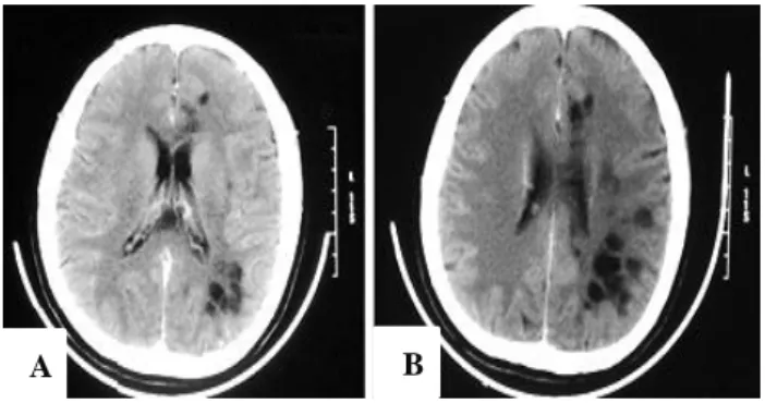
A cranial CT study revealed multiple hypodense lesions with barely visible walls located within the white matter in the entire cerebral hemisphere, as well as the CT of our second case (Figure 3).10 On MRI1, the signal intensity of
these cystic structures was exactly parallel to that of CSF. They were located predominantly in the white matter, including the periventricular area, centrum semiovate, and the U-fibers. The callosum was also involved while the cerebellum, thalamencephal, and the brain stem were unaffected. The cysts did not show potentialization and affected all the cerebral lobes.
In the MRI of the patient of Sener, the anterior and inferior frontal regions were relatively less affected, and the cysts were larger around the lateral ventricles compared with those in the other areas. The case of Li Mao had no normal parenchymatous tissue and has symptom of stiffness.
Figure 3: Brain image of CT, showing multiple hypodense lesions, isointensity as CSF, and no
obvious edema.
The MRIs of our two cases showed the cysts isointensity as CSF. Moreover, the thickness of the cortex considerably decreased with clear adumbration. The cerebellum, cerebral ganglion, brainstem, and cerebral ventricles were unaffected. The cysts did not show perennialization and hydrops. The MRA of case 2 showed that the branches of the middle cerebral artery decreased with no other abnormalities.
Diagnosis and differential diagnosis of giant perivascular spaces
Given that the giant dVRSs can assume bizarre configurations and produce a mass effect, they may be misidentified as other cystic lesions.7 They can easily be
misdiagnosed as cystic neoplasms, parasitic cysts, non-neoplastic neuroepithelial cysts, ventricular diverticula, cystic infarction, and mucopolysaccharidosis, especially because dVRSs are usually periventricular in location.7
When a patient shows intelligence hypoevolutism, stiffness, or no symptoms, the MRI shows multiple curvilinear, round, or oval hypodense lesions isointensity as CSF. Moreover, the cortex is extremely thin and has no parasitic infection and tumor, giant perivascular spaces can be considered.
Neonatal polycystic encephalomalacia is caused by diffuse lesion or viral infection during the circumnatal period.13 Neonatal onset is typical. Seizures, often focal,
are characteristic. Progressive evolution of spastic quadriparesis, dementia, blindness, and deafness occur. The size zone of necrosis in the brain are unequal, and gliocyte proliferation surrounding the cysts occur. The cysts are located in the white or gray matter and not in the periventricular area. The periventricular encephalomalacia is located in the white matter circumambient cerebral ventricle and gliocyte proliferation. The clinical manifestation is cerebral palsy. Our two cases did not involve cerebral palsy and gliocyte proliferation surrounding the cysts.
Neonatal periventricular pseudocyst includes periventricular leukomalacia and ependyma ependyma.14
This is sequela because of hypoxia, toxication, viral infection, or chromosomal abnormality during the perinatal period. Other pseudocysts include parasite, tumor, or wound. It is easily distinguished through clinical examination and MRI.
Adult unilateral periventricular pseudocysts become symptomatic in adulthood and are manifested by ipsilateral headache, contralateral hemi-hypesthesia, depression, collapses, and slight cognitive decline, which are rarely reported in adults.15 Finsterer reported a case of
a 48-year-old woman suffering from right-sided, pulsating headache, and left-sided hemi-hypesthesia. The CT and MRI scans of the brain disclosed right-sided cysts in the white matter. Our case 2 involved cysts unilateral in the periventricular area, but with no other symptoms as listed above. Hence, we considered that her case involved giant perivascular spaces with no PVPC.
CONCLUSION
We report two cases of polycystic brain, a very unique inherent developmental malformation. There are very small population in the available literature. Dilatation of VRS may produce multicystic giant lesions that can easily be confused with other pathologic conditions that
have a completely different prognosis and management. Knowledge about the existence of such entity and its radiologic features is important to the prevention of misdiagnosis and mismanagement in the form of biopsy/excision, which can have devastating consequences.
Funding: No funding sources Conflict of interest: None declared Ethical approval: Not required
REFERENCES
1. Hissah Al Abdulsalam, Abdullah A. Alatar, Sherif Elwatidy. Giant tumefactive perivascular spaces: a case report and literature review. World Neurosurg. 2018;112:201-4.
2. Heier LA, Bauer CJ, Schwartz L, Zimmerman RD, Morgello S, Deck MD: Large Virchow-Robin spaces: MR-clinical correlation. AJNR Am J Neuroradiol. 1989;10:929-36.
3. Kumar A, Gupta R, Garg A, Sharma BS. Giant mesencephalic dilated virchow robin spaces causing obstructive hydrocephalus treated by endoscopic third ventriculostomy. World Neurosurg. 2015 Dec 1;84(6):2074-e11.
4. Li M, Qing YH, Li RF, Liang S. MR diagnosis of polycystic brain. J Guangxi Med University. 2000;17(2):233-4.
5. Homeyer P, Cornu P, Lacomblez L, Chiras J, Derouesne C. A special form of cerebral lacunae: expanding lacunae. J Neurol Neurosurg Psychiatry. 1996;61:200-2.
6. Mascalchi M, Salvi F, Godano U, Nistri M, Taluti R, Tosetti M, et al. Expanding lacunae causing triventricular hydrocephalus: report of two cases. J Neurosurg. 1999;91:669-74.
7. Kumar A, Gupta R, Garg A, Sharma BS. Giant mesencephalic dilated virchow robin spaces causing obstructive hydrocephalus treated by endoscopic third ventriculostomy. World Neurosurg. 2015 Dec 1;84(6):2074-e11.
8. Groeschel S, Chong WK, Surtees R, Hanefeld F. Virchow-Robin spaces on magnetic resonance images: normative data, their dilatation, and a review of the literature. Neuroradiol. 2006 Oct 1;48(10):745-54.
9. Potter GM, Doubal FN, Jackson CA, Chappell FM, Sudlow CL, Dennis MS, et al. Enlarged perivascular spaces and cerebral small vessel disease. Int J Stroke. 2015 Apr;10(3):376-81.
10. Sener RN. Polycystic brain (cerebrum polycystica vera) associated with ectodermal dysplasia: A new neurocutaneous syndrome, Pediatr Radiol. 1994;24(2):116-8.
12. Ahmad M, Narayanasamy S, Siddiqui MA, Ahmad I. Giant perivascular spaces: utility of MR in differentiation from other cystic lesions of the brain. JBR-BTR. 2014 Nov 1;97(6):364-5.
13. Chutorian AM, Michener RC, Defendini R, Hilal SK, Gamboa ET. Neonatal polycystic encephalomalacia: four new cases and review of the literature. J Neurol Neurosurg Psych. 1979 Feb 1;42(2):154-60.
14. Lu JH, Emons D, Kowalewsk I. Differential diagnosis of periventricular pseudocysts in the neonatal period. Klin Padiatr. 1991;203(1):8-14.
15. Finsterer J, Kopsa W. Adult unilateral periventricular pseudocysts with ipsilateral headache. Clin Neurol Neurosurg. 2005;108(1):73-6.