WADE HENDERSON WEISMAN. Toxicity, Mutagenicity and Mutational Spectra of Vinyl Chloride, 2-Chloroethylene Oxide, and Chloracetaldehyde in a Human Lymphoblastoid Line Expressing Cytochrome P450nEl.
(Under the direction of Thomas R. Skopek)
ABSTRACT
Vinyl Chloride Monomer (VCM, CAS # 75-01-4) is one of the few chemicals identified as a human carcinogen (lARC, 1979). In this report, the toxicity and mutagenicity of VCM and its two metabolic products, 2-chloroethylene oxide (CEO) and 2-chloroacetaldehyde (CAA) were studied in a human B-lymphoblastoid line containing cloned cytochrome P450IIE1. Cytochrome P450IIE1 is capable of metabolizing VCM to CEO which non-enzymatically rearranges to CAA. Toxicity was determined by plating cells immediately after exposure and comparing their clone-forming ability to that of untreated cells. Mutagenicity at the hprt locus was determined by plating cells in the presence of the selective agent 6-thioguanine. Delivered doses of 25 mM to 400/iM VCM x 24 hours produced no measureable toxicity but resulted in induced mutation frequencies that ranged from 0.5xl0'"to 5.6x10"". Although increases in mutation frequency were consistently seen, a clear dose-response was not
apparent. Dose dependent increases in toxicity and mutagenicity were observed with both
CEO and CAA. Treatments of 25 mM CEO x 24 hours resulted in survival of 0.82 and
suggest the majority of mutations induced by VCM must be produced by CEO.
Denaturing gradient gel electrophoresis (DGGE) was used to identify unique VCM,
CEO, and CAA mutations in exon 3 of the hprt gene. DGGE banding patterns from CAA
and CEO isolated mutants were compared to the banding patterns from VCM. 118 VCM
mutants analyzed in the low temperature melting domain of exon 3, 8.5% (10 isolates)
produced identical DGGE banding patterns; sequencing of the isolated mutant bands revealed
a G-T transversion at base pair 292. In the high temperature melting domain, 3.4% (4 of 118
isolates) exhibited identical DGGE bands; sequencing revealed a G-»A transition at base pair 197. These sequence changes represent potential VCM mutational "hotspots". The
low-temperature "hotspot" banding pattern was evident in less than 2% (1 isolate) of the
spontaneous mutants in the VCM experiment. However, it was present in 5.6% (11 isolates)
TABLE OF CONTENTS
Page LIST OF FIGURES AND TABLES...v ACKNOWLEDGEMENTS...vi
FOREWORD...vii
SPECinCAIMS...X
Chapter
L TOXICOLOGY OF VCM...1 n. HPRT MUTATION ASSAY: THEORY AND DESIGN...18 III. MOLECULAR ANALYSIS OF MUTATIONS:
THE POLYMERASE CHAIN REACTION, DENATURING GRADIENT
GEL ELECTROPHORESIS AND DNA SEQUENCING...28
IV. TOXICITY AND MUTAGENICITY OF VCM, CEO AND CAA...36
V. MOLECULAR ANALYSIS OF MUTANTS INDUCED BY
VCM, CEO, AND CAA...60 VL CONCLUSIONS AND SUMMARY...79
Figure 1.1: Metabolic Activation of VCM...5
Figure 1.2: VCM Adducts and Inferred Point Mutations...17
Figure 2.1: HPRT Metabolic Pathway...27
Figure 4.1: Mutation Assay Exposure Summary...52
Figure 4.2: CAA Survival...53
Figure 4.3: CEO Survival...54
Figure 4.4: VCM Survival...55
Figure 4.5: CAA Growth Delay...56
Figure 4.6: CAA Induced Mutant Fraction...57
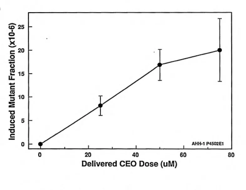
Figure 4.7: CEO Induced Mutant Fraction...58
Figure 4.8: VCM Induced Mutant Fraction...59
Figure 5.1: Mutation Analysis Summary...76
Table 5.1: PCR and Sequencing Primers...77
VI
ACKNOWLEDGEMENTS
As an undergraduate student I could never imagine continuing to a master's program,
let alone completing a project the scope and complexity of this one. This would not have
been possible but for the patience and willingness to help from the entire group of peoplewho are Dr. Skopek's Lab. I am especially indebted to Teresa Craft whose help 'when there
was just not enough time left' made the difference.As for the direction and incredible planning abilities to get me done "on-time", I know
I could not have worked for a better investigator, thank-you Tom.Whatever significance may have been attached to this document will quickly pass. For
this reason I dedicate it to someone whose work has infinitely more significance than mine
ever will, my patient and understanding wife Debbie, whose job in training up our two boys
FOREWORD
The identification of biomarkers that can serve as indicators of human exposure to
specific chemicals is an important goal of the study of carcinogenesis. The use of biomarkers
takes into account a variety of factors affecting a compound's toxicity, including differences
in route of uptake, the metabolism to activate or detoxify the compound, the ability to excrete the compound or its metabolites and the ability to repair damage (Wogan, et al., 1987).Biomarkers include a broad range of analytical targets that can be grouped in two general
categories: 1) measurements of concentrations of chemicals or their derivatives in body fluids or excreta, and 2) measurement of biological reaction products such as DNA adducts,mutation, chromosomal abberations or sister chromatid exchanges produced in the tissues of
exposed individuals (Wogan, et al., 1987). Two important elements of a biomarker are 1) its
relevance to the exposure of concern (correlation of the measured biomarker to an adverse human health effect); and 2) the sensitivity of the biomarker, (whether it can detectdifferences between exposed and non-exposed individuals).
DNA adduct formation is an endpoint relevant to exposure to genotoxic agents.
Furthermore, it can be measured with sufficient sensitivity to differentiate between exposed
and non-exposed individuals and can function over a wide range of doses (Swenberg et al,
1990). The molecular dose of a compound is the actual amount of chemical that reacts with
macromolecular targets, such as DNA. This dose is very small in comparison to the
vm
dose, at exposure concentrations which are much less than those used in bioassays (Swenberg
etal., 1990).DNA adducts do not always lead to mutations. Replication of the DNA is required to convert the adducts formed (promutagenic lesions) to actual mutations (Swenberg et al.,
1990). DNA repair systems can repair some of the adducts before replication of DNA thus avoiding mutation. Also, many adducts are intrinsically nonmutagenic while others are very promutagenic. Some sites on DNA called, "hot spots", are more susceptible than other locations to the effects of chemical agents (Benzer, 1961 and Briscoe et al., 1984). Many mutagens exhibit characteristic patterns of mutation in the DNA that can be distinguished by comparison to pattems of mutation induced by spontaneous processes or by other mutagens
(Fuchs, 1981; Skopek, 1982; Duane, 1986; Schaaper, 1986; Drobetsky, 1987; Richardson,
1987; Giroux, 1988; Vrieling, 1988; Liber, 1989). The pattems of these mutations are termed mutational spectra. Within these spectra, the active locations or "hot spots" can readily be discriminated by the location, type and frequency of the mutation in relation to other mutational events. Because of this mutational specificity, the base pair changes produced by the mutagen can imply additional information about the DNA adducts and the molecular events that produce the mutations (Fuchs, 1981; Skopek, 1982; Duane, 1986;Schaaper, 1986; Drobetsky, 1987; Richardson, 1987; Giroux, 1988; Vrieling, 1988; Liber,
1989). The study of DNA adducts and mutational spectra can help to determine causal
relationships and may improve extrapolation of risk for cancer from experimental animals to
humans. Measurement of DNA adducts and mutational spectra may also be used inSPECIFIC AIMS
The primary goal of the planned work was to study the frequency and nature of mutations induced by VCM and its metabolites, CEO and CAA, in a human cell Mne. This information would then be used to conclude which metabolite is mainly responsible for the mutagenic activity of VCM. Specifically, the following two aims were proposed:
1. To determine the toxicity and mutation frequency at the hprt gene induced by VCM, CEO, and CAA in a human lymphoblastoid cell line expressing cytochrome P450IIE1.
Vinyl Chloride is a synthetic chlorinated monomer manufactured from petrochemical feedstock and chlorine. The compound is a colorless, explosive gas with a boiling point of -14" C at atmospheric pressure and is slightly water soluble (EPA, 1975). About 96% of the VCM manufactured worldwide is used in the production of vinyl chloride homopolymer and copolymer resins. One of the major commercial products made with VCM is polyvinyl chloride (PVC) which is then used in the manufacture of a myraid of products (Sax, 1988). The estimate for vinyl chloride production in the U.S. for 1993 is approximately 11.0 billion pounds (ATSDR, 1992). The Occupational Safety and Health Administration (OSHA) has set a regulatory standard for VCM exposure in the workplace of 1 part per million (ppm) weighted over an 8-hour time period. Other OSHA regulations prohibit operations which result in VCM coming in contact with the skin. The National Institute of Occupational Safety and Health (NIOSH) has recommended the lowest limit of detectability as the limit for a ten-hour averaged exposure (Sax, 1988). A NIOSH survey conducted from 1981-1983 estimated that 81,000 U.S. workers are potentially exposed to VCM in the workplace (NOES, 1990). The major route of VCM exposure in an industrial setting is through inhalation; however, skin absorption is possible in the cleaning of polymerization ovens. In the 1970's the acute effects seen in VCM polymerization workers were believed to be the only hazardous properties of vinyl chloride. Monitoring of vinyl chloride at this time was directed primarily toward maintaining levels below the lower explosive limit (LEL) of 4% (Sax, 1988). Because of the low level of concern and the high levels permitted, VCM workers (especially those who cleaned the polymerization ovens), could have been exposed
exposure to VCM in levels over 10,000 ppm can cause central nervous system depression, dizziness, lightheadedness, nausea, dulling of the senses and headache (Lester et al., 1963).
Additionally, two fatalities had been reported in 1960 due to extremely high acute exposures
(Danziger, 1960). Chronic adverse health effects are also seen from VCM exposure. These effects include: degenerative bone changes, circulatory disturbances, thrombocytopenia, spleenomegaly, hepatomegaly and hepatic fibrosis (La Dou, 1990). More central to the focus of this research is the confirmation that angiosarcoma of the liver, a rare form of liver cancer, is caused almost exclusively by VCM exposure and is an often studied endpoint for chronic
exposures (Purchase et al., 1987). Since 1974 there have been 118 cases worldwide of
angiosarcoma of the liver reported to a central registry. Most of these individuals were
workers involved in the cleaning of ovens used for VCM polymerization. This registry,
along with numerous epidemiological studies estimate an additional 200-350 deaths over the next 30 years associated with past VCM exposure (Forman et al., 1985). The level ofexposure to VCM capable of causing angiosarcoma has not been definitively shown to cause
any other carcinogenic effects in man, as indicated by epidemiological studies with endpoints
other than angiosarcoma (Purchase et al., 1987). This is another reason why most cohort
studies of VCM workers use angiosarcoma of the liver as a definitive endpoint. Manyfound in most of these studies was due to an excess of angiosarcoma. One retrospective cohort study of 1151 workers at four vinyl chloride production facilities with over 12,000 person years at risk demonstrated that of the fourteen cases of biliary and liver cancer observed, eleven were cases of angiosarcoma (Sax, 1988). While the preponderance of epidemioligical data strongly indicate that exposure to vinyl chloride increases a persons risk to a variety of cancers, angiosarcoma of the liver is the only cancer type that can be
attributed unambiguously to VCM exposure.
Environmental sources of VCM contamination include product manufacturing and hazardous waste sites (EPA, 1985, Stephens et al., 1986). VCM was identified in at least 10% of the hazardous waste sites listed on the Environmentnal Protection Agency's National Priorities' List (NPL). Vinyl chloride eminating from VCM and PVC processing faciUties accounts for the majority of VCM entering the environment. VCM has a half-life in the environment of only 1-2 days as sunlight will cause the monomer to polymerize. People who live near production facilities are the only segment of the non-occupationally exposed public to receive any measureable exposure. Their estimated daily intake of VCM is from 0-2,100 Mg/person/day (ATSDR, 1992). Other potential routes of non-occuaptional VCM exposure is from ingestion of food and water contaminated with VCM through leaching of packaging material and pipes. A study on bottled water estimated the average daily intake of VCM at 120ng VCM/person/day if bottled water were the only source of drinking water. Many pipes that supply homes drinking water are made from PVC and the potential exists that the
Vinyl chloride is metabolized through an NADPH-dependent cytochrome P450IIE1 mixed function oxidase system to the reactive species 2-chloroethylene oxide (CEO), which non-enzymatically rearranges to form 2-chloroacetyldehyde (CAA). Both CEO and CAA are highly reactive and are capable of modifying DNA. Consequently, the carcinogenic effects of VCM have been attributed to these electrophillic metabolites rather than the parent compound (Miller, 1976). The general reaction and rearrangement for this process is given in Fig. 1-1:
DNA
CI
CyP450
HC=CCIor-H,C^^CHg-z-CHJCHO
^ LI NADPH ^ \ / ^3 ^ '
" \/ I CI
(From Purchase et al., 1987 and Zajedela et al., 1980)
following Michaelis-Menten kinetics. Using ^^C labeled VCM, they measured total
radioactivity in animals sacrificed zifter equal exposure time to various concentrations of VCM. They discovered that the amount of VCM metabolized reached a plateau between 9 and 25 parts per million (ppm). They also concluded from this study that VCM-induced angiosarcoma is a function of the metabolic activation rather than the exposure concentration (Gehring et al., 1978).
A large number of in vivo animal studies as well as in vitro studies have been carried out to examine the mechanism of action of VCM. An extensive listing which summarizes these studies is provided in Sax (1988).
Work by C. MalaveUle et al. (1975), using histidine reversion in S. typhimurium as an endpoint, demonstrated the need for metabolism in VCM mutagenesis. They exposed bacteria to gaseous VCM, both with and without S-9 liver fractions from humans, rats and mice. They demonstrated a mutagenic effect from CAA and an even greater effect from CEO in bacterial strains which revert by base pair substitution. No mutagenic activity was
demonstrated in strains reverted by a frameshift (Maleville, et al., 1975). In the same study,
using the 4-(p-nitrobenzyl)pyridine assay with CEO or CAA, they determined that CEO possess a greater alkylating activity than CAA. In a review of in vitro studies involving VCM exposure to microorganisms and VCM exposures to Chinese hamster ovary cells,
Bartsch et al.,(1976) concluded that both CAA and CEO are mutagenic in these test systems,
l,N"-ethenoadenosine. 1,N°- Ethenoadenosine was not produced by CEO when epoxide hydrolase was present, but formation did occur in the presence of alcohol dehydrogenase. They concluded through this and earlier studies that CEO is sufficiently reactive to alkylate DNA efficiently, but is not so reactive as to bind solely to P450 proteins (Guengerich, et al., 1979).
L.M. Gwinner et al., ( 1983) compared alkylation produced by VCM and 2,2'-dichlorodiethylether (BCME) in hepatocytes of male Wistar rats. The latter compound is metabolized completely to CAA (partly through an intermediate). They concluded that CAA was responsible for protein alkylation while the VCM (and therefore CEO) was mainly responsible for binding to DNA. This study was unique in its use of BCME which is
oxidized to CAA and 2-chloroethanol (CE), which in tum is completely metabolized to CAA. Unlike VCM, 2,2'-dichlorodiethyl ether does not pass through a reactive epoxide;
consequently, effects from the epoxide and the aldehyde could be differentiated in vivo, something heretofore only possible in vitro (Gwinner, et al., 1983).
Zajdela et al. administered CEO subcutaneously to eight-to ten-week- old male and female XVUnc/Z mice. The same was done with BCME, a compound that is a direct acting human and animal carcinogen, similar in structure to CEO. Tumor formation from CEO was comparable to BCME. CAA was also included in this study, but was used in an
12-8
O-n-tetradecanoylphorbol-13-acetate was used as a promoter with all three chemicals. Both CEO and BCME produced skin tumors, while CAA did not. These results led them to conclude that CEO was the reactive metabolite of VCM and therefore, responsible for its carcinogenic activity. (Zajdela et al., 1980).
In conclusion, CAA and CEO were demonstrated to react with DNA in vitro causing mutations, however CEO was more strongly mutagenic. Additionally, CEO was
carcinogenic in vivo while CAA was not. This brief summary of the growing body of evidence in numerous in vivo and in vitro test systems strengthens the following conclusions about VCM: 1) VCM requires metabolic activation to exert its mutagenic and carcinogenic effects, 2) CEO and CAA, two reactive products of metabolism manifest their effects in different ways, 3) CAA, although the more cytotoxic compound, shows less mutagenic potential in vitro and less carcinogenic potential in vivo compared to CEO.
DNA Adduct Formation and Relevance
adduct and the function and availability of various DNA repair enzymes (Singer, 1985). The production of DNA adducts shows specificity for certain sites on the DNA
(Richardson 1989, Warpehoske 1988). The propensity of sites to react is affected by the base
adducted and usually the neighboring base or bases (5' or 3'). The differences in reactivity no doubt contribute to "hot spots" and cold spots of base substitutions (Cooper, 1990). The frequency of mutations occurring at different modified bases may be influenced by several factors including differences in repair efficiencies and differences in the rate of base misinsertion, both of which are expected to be influenced by local DNA sequence context.
Briscoe and Cotter conducted a study in 1984 to determine whether there was any effect of cytosines or guanines as nearest neighbors upon the alkylation of a guanine residue in DNA. N-methyl-N-nitrosourea (MNU) was reacted with a synthetic polynucleotide. They
concluded that the alkylation pattern of guanine was dependent on the neighboring bases and this finding would be relevant in defining hot spots of mutational activity within a genome (Briscoe et al., 1984). Richardson et al., (1989) investigated the formation of
10
and the distribution was only evident in the dsDNA oligonucleotide tested, not the ssDNA. These results suggest a sequence dependent mutation site that is also dependent on the secondary DNA structure (Richardson et al., 1989).
In vivo and in vitro studies by Laib et al., (1981) concluded that the DNA adduct N -(2-oxoethyl) guanine (7-OEG) was the major product of alkylation from the reactive VCM
metabolites. This was true both for rats exposed to [1,2-^^C] vinyl chloride and in vitro
studies with rat liver microsomes, an NADPH-regenerating system, DNA and [ C] vinyl chloride (Laib, et al., 1981). It was proposed that this adduct in the cyclic hemiacetal form could be read by the polymerase as adenine and therefore cause mispairing. However numerous studies have shown that modification at the N7 position of guanine does not prompt mispairing. Alkylation at the N7 position does promote depurination which can also occur spontaneously or through the action of glycosylases. Apurinic sites have been shown to be promutagenic but their production and conversion to mutations is expected to be inefficient relative to other adducts formed by VCM (see below). Therefore, even though 7-OEG accounts for up to 98% of the modified bases in DNA, it was predicted that it would contribute little to induction of mutagenicity by vinyl chloride or its metabolites (Barbin et al., 1985; Laib et al., 1981; Swenberg et al., 1990). Furthermore, it has a half-life of
approximately 62 hours in DNA and therefore is not extremely persistent (Fedtke et al., 1990,
Swenberg et al., 1990).
Different methods of adduct detection and quantification revealed an additional three adducts formed by the interaction of DNA with the metabolites of VCM. These are:
recently identified VCM adduct N-^,3-ethenoguanine (N^,3eG) (Singer, 1983). These
adducts and the point mutations which have been inferred to result upon DNA replication are pictured in figure 1.2.
The biological importance of these adducts in mutagenesis and carcinogenesis remains to be determined. As stated above, 7-OEG is found in vivo and in vitro in the greatest
concentrations relative to other VCM adducts, but no studies have demonstrated the
mutagenic properties of this adduct. Laib et al., (1985) identified the presence of N'^,3 eG in
VCM-exposed fourteen-day-old rats. Singer et al., (1987) went on to determine that this adduct can act like a guanine or adenine (in reference to recognition by DNA polymerase) causing GC-»AT transitions approximately twenty percent of the time. Their study involved the synthesis of N'',3 eG, and its copolymerization with cytosine diphosphate (CDP) into an oligonucleotide. They then measured the rate of incorporation of incorrect nucleosides using the oligonucleotide as a template. Based on these results, they proposed that N'^.S eG is an important promutagenic adduct of vinyl chloride (Singer et al., 1987). Fedtke, et al., (1990)
'y
Studied the formation and persistence of both 7-OEG and N'^,3 eG in target and non-target tissues of preweanling Sprague-Dawley rats exposed 5 days to gaseous VCM. The ratio of
/y
12
Eberle et al., (1989) raised monoclonal antibodies to edAdo and edCyd to permit their quantification in vivo. Young Sprague-Dawley rats were exposed by inhalation to 2000 ppm
of VCM for ten days and the levels of edAdo and edCyd were quantified in the lung and
liver tissues through the use of the Mabs. Both adducts were found in lung and liver tissues, with greater concentrations of both in the lung. Additionally, the levels of edCyd were 2.5 times and 3.2 times higher in the lung and liver, (respectively), than the levels of edAdo. These results demonstrated that both of these adducts were formed efficiently in vivo during VCM exposure (Eberle, et al., 1989).
To determine whether CEO or CAA is primarily responsible for the formation of these minor, yet persistent and promutagenic adducts Guengerich et al., (1992) reacted these compounds directly with calf thymus DNA (Guengerich et al. 1992). They found that the etheno adenine and guanine adducts were formed much more effectively by CEO than CAA (by at least an order of magnitude). The key to their study was the conditions of metabolism of vinyl chloride. l,N"-ethenoadenosine (edAdo) formation was almost completely blocked (from either DNA or adenosine) when purified epoxide hydrolase was included in the
reaction, but when the CAA destroying enzyme (alcohol dehydrogenase) was added, there was little effect on edAdo formation. The in vitro study indicated formation of adducts from
CEO exposure at the following relative levels : 7-OEG » edAdo > N^,3 eG. The lack of
N ,3 eG and edAdo are formed in DNA primarily through the reactive epoxide versus the aldehyde.
As mentioned previously, 7-OEG is not expected to demonstrate any miscoding properties in DNA. This is not the case for the other three eidducts, all of which have
demonstrated miscoding properties. edAdo was found to be a miscoding lesion during RNA transcription, resulting in AT-»GC transitions and AT->CG and AT-»TA transversions
(Spengler and Singer, 1981, de los Santos et al.,1991). edCyd was found to cause errors during both DNA replication and RNA transcription, resulting in CG-»AT transversions, CG-»TA transitions, and, at a much lower frequency, CG->GC transversions (Spengler and
Singer, 1981; Singer and Spengler, 1986). As mentioned previously, the adduct, N'',3eG
causes GC->AT transitions during replication.
Because of the efficiency of formation of these etheno DNA adducts of VCM, their promutagenic properties and persistence in DNA, exposures to even very low concentrations of VCM could pose a significant health risk (Swenberg, 1991). The work with VCM and its metabolites in this research will provide additional information on the role of CEO and CAA in VCM mutagenesis in human cells and contribute to the knowledge of the molecular nature of VCM-induced mutations.
Mutagenicity
A mutation in DNA can be defined as a stably inherited change in the ordering of nucleotide bases in the genome. An organism with a genomic change is called a mutant (for that change) and the same organism without the change is referred to as wild type. The
14
(MF) and represents the fraction of mutants in the population. Mutations can be classified in
several categories. A point mutation affects only a single base in the DNA. The most
common form of point mutations are substitutions, of which there are two types. Transitions substitute one pyrimidine base for another or one purine base for another purine (AT->GC, CG-»TA) and transversions exchange a purine for a pyrimidine or a pyrimidine for a purine (AT-+CG,TA; GC->TA,CG). Point mutations can lead to three outcomes in the transcription of the changed sequence. There may be no change at all in the amino acid sequence from the wild type to the mutant sequence; this is due to the degenerate nature of the code. The change can result in a "missense" mutation, in which a different amino acid is inserted. This can affect a unique enzymatic function of the cell resulting in reduced or negUgible enzyme activity, or possibly a new function. If the change in sequence results in the production of a stop codon, then premature termination of protein synthesis will occur. This type of change is called a "nonsense" mutation. Rearrangements usually effect large portions of the gene and are manifested through the loss (deletions) or gains (insertions) of nucleotides into the DNA. Mutations of this kind can result in shifts of the reading frame of the DNA sequence due to the fact that information is stored in codons consisting of three nucleotides (Lewin,
1990).
Mutations can also be classified as to their general causative source. "Naturally-arising" mutations are usually called spontaneous mutations and the level at which they occur for an organism (or cell line) is termed the background mutant frequency (MF). This level of
mutation can be determined from the zero treatment controls used for the mutation studies.
chapter II) of 3.0x10"" (or three mutants out of one million wild type cells with HPRT activity) (Gentest, 1990). These spontaneous events can arise due to a variety of causes including the processes of DNA replication and DNA repair. Giroux et al., (1988) in a study to determine the DNA sequence of 196 spontaneous mutants in the SUP4-0 gene of S.
cerevisiea identified all possible types of base pair substitutions, deletions, and complex alterations involving multiple changes, as well as insertions and transpositions. Their work led them to support the hypothesis that many spontaneous mutations are actually the result of DNA sequence-directed events (Giroux et al., 1988). Similar results were obtained by Schaaper et al., (1986) in their analysis of the spectmm of spontaneous mutation in the Escherichia coli lac I gene. They analyzed 174 spontaneous mutations and determined that the spectrum consisted of base pair substitutions, frameshifts, deletions, dupUcations, and transpositions. These results led them to conclude that the spontaneous mutations occurring in the organism studied were the result of a variety of endogenous mutational mechanisms (Schaaper, et al.,1986). Mutations induced by chemical or physical damage can be
ascertained in a test system when the level of mutations induced is significantly greater than the level of spontaneous mutations. The level of MF increase over background determines the strength of mutagenic compounds and allows for their comparison. Many mutagenic compounds exert their effect by covalently binding to (adducting) a particular base or intercalating (Lewin, 1990). If this bound chemical/DNA complex is stable it can be
processed through the repair, replication, and recombination systems of the cell, and may be converted to a mutation (Fuchs, et al., 1981).
16
Vinyl chloride was shown to be mutagenic in many test systems including bacteria (Escherichia coli K12, Salmonella typhimuriwn TA1530, TA1535, 646) yeast
I HCH
I II >
HjN—C^ ___C---"'
I II >
7-(2-OXOETHYL)GUANINE N,3-ETHENOGUANINE
^.
HC
II >
1,N-ETHENOADENINE
//
HC
\ /^ \
CH
o*^\„,^
1 ,N -ETHENOCYTOSINE
REACTIONS WITH DNA IN VITRO MISCODING PROPERTIES CEO CAA
V 7-(2-OXOETHYL)GUANINE NONE
X
X N:^3-ETHEN0GUANINE G—AX
X
1,N*-ETHEN0ADENINE A^T A -*G A—CX
X
1,N*-ETHEN0CYT0SINE C—A C-i^T C -i^Fig 1.2. Identified DNA adducts from in vitro and in vivo exposures to VCM and its metabolites along with inferred base pair changes. Compiled from: Barbin et al., 1985; Laib et al., 1981; Swenberg et al., 1990; Spengler and Singer, 1981; de los Santos et al., 1991; Singer
CHAPTER n
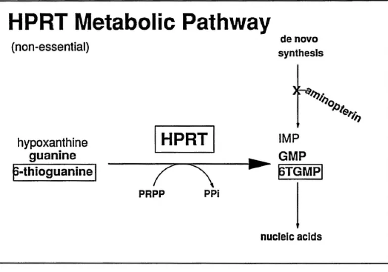
The hypoxanthine phosphoribosyl transferase (hprt) gene was the genetic target used in all the mutation assays reported here. HPRT (protein) is a purine salvage enzyme that
catalyzes the condensation of free hypoxanthine or guanine with
5-phosphoribosyl-l-pyrophosphate (pp-ribose-p). Although non-essential for cells growing in culture, the lack of
HPRT can have serious effects in man. Complete lack of HPRT activity results in the genetic disease known as Lesch-Nyhan syndrome (Holden et al., 1978). Less severe deficiencies can result in gouty arthritis (Kelly et al., 1967). HPRT enzyme activity has an influence directly or indirectly on cellular processes related to purine synthesis. This is known primarily through examination of people with gout who have been shown to exhibit elevated rates of purine synthesis (Caskey, 1979).
Hprt is located on the long arm of the X chromosome (Holden et al., 1978) and consists of nine exons and eight introns which total 57,000 base pairs in length. The molecular weight of HPRT is 24,300, and it exists in vivo as a tetramer (Holden, 1978). Since hprt is located on the X-chromosome, males possess only one copy and females possess only one active copy due to X-chromosome inactivation. Consequently, hprt is effectively hemizygous in both males and females and only a single mutational event is required to render a cell phenotypically hprt". HPRT substrates include: hypoxanthine, guanine, 6-mercaptopurine, and 6-thioguanine (6TG) (Krenitsky et al., 1969).
The key advantage for the use of hprt in gene mutation studies is the existence of
stringent selective conditions for both hprt"*" and hprt' phenotypes. Hprt"*" cells can be
20
salvage pathways (hprt, tk) for survival. Hprt"'' ceUs can phosphoribosylate and utilize
hypoxanthine in HAT media and grow, while hprt" cells cannot (Thomas, 1979; see figure
2.1.) The process of treating a cell culture in HAT media prior to a mutation study reduces
the level of background mutants (those caused by means other than the test substances) and
increases the sensitivity of the assay.
When grown in the presence of 6TG a cell with active HPRT will metabolize 6TG to
the ribonucleoside monophospate 6-thioGMP (6TGMP). 6TGMP is further metabolized,
incorporated into nucleic acid and kills the cell. Consequently, hprt"^ cells are killed in the
presence of 6TG (Nelson, 1975). However, cells without HPRT activity (hprt*) cannot
phosphoribosylate 6TG zmd can survive and grow in its presence. The ability to select hprt'
mutants provides a straight-forward means to detect mutation induction resulting from a
chemical exposure.
Cells mutated at the hprt locus no longer produce functional hprt mRNA; however, for
a period of time they still possess HPRT enzymatic activity due to the presence of preexisting
hprt mRNA and HPRT proteins Prior to 6TG-mediated selection, the cells must pass through
several cell cycles (without selection) to ensure that newly formed hprt' cells lose their
residual HPRT activity. This length of time is known as the phenotypic expression period
and for most cells is greater than five days (Crespi, 1984).
The hprt locus has been widely studied and the types of mutations fi"om different
chemicals reported. A review of the hprt locus by Caskey in 1979 indicated studies at that
time had demonstrated missense mutants (point mutations), frameshifts, deletions, and
Subsequent studies verified and added to the location, type and frequency of mutations at
hprt in different systems. Cariello et al., (1988) identified a point mutation in hprt in a
patient with gout, utilizing denaturing gradient gel electrophoresis and direct sequencing as
analytical tools (Cariello et al., 1988). Deletions primarily of exon 2 and 3 were identified by
McGinniss et al (1989) in their analysis of in vivo hprt mutations in human new bom
T-lymphocytes (McGinniss et al., 1989) A study by Recio et al., (1990) of spontaneous in vivo
hprt mutations in human T-lymphocytes identified single base substitutions, insertions, small
deletions and large deletions of entire exons. The large deletions suggested mutations in hprt sequences that regulate splicing of hprt mRNA. The growing amount of scientific data onhprt is significant. Currently a data base is being compiled of all mutations at hprt produced
by chemicals and physical agents in a wide variety of model systems (Cariello et al., 1992).The truly unique capability of the cell line used in this study is their ability to express cytochrome P450nEl activity. Most mamalian cell lines used in mutation studies lack the ability to metabolize xenobiotic compounds and convert them to their mutagenic form. To
circumvent this deficiency, some form of exogeneous activating enzymes must also be
provided to metabolize promutagenic chemicals to their active form. This limitation also demands that the metabolites are produced outside of the plasma membrane and must betransported across to interact with the genetic target (Gonzalez et al., 1991).
Since cell lines can be stably transfected with extrachromosonal vectors, the
opportunity exists to transform the cells with cloned P450 activity needed to metabolize the
compounds of interest (Gonzalez et al., 1991). The AHH-1 cell line have within their
22
responsible for the immortalization of the cell line and is necessary for the cells' continued
propagation in culture. The cytochrome introduced extrachromosomally into AHH-1 cells, is
IIEl (Gonzalez, 1991). This P450 is capable of oxidizing many compounds including VCM
(Guengerrich et al., 1991). The vector used to transfect the cDNA with P450nEl in this cell
line was pEBVHistk. This vector contains the origin of replication (P element) of EBV and
allows replication of the extrachromosomal plasmid carrying the P450nEl cDNAs (Sugden
et al., 1985 and Yates et al., 1984). The selectable marker in the vector is the Escherichiacoli HisD (histidinol dehydrogenase) gene which confers resistance to 1-histidinol.
Histidinol is a potent protein synthesis inhibitor. Histidinol dehydrogenase can convert the
histidinol present in the cell to the essential (and non-toxic) amino acid L - histidine. Cells
that have lost all copies of the extrachromosomal vector (or have lost enough copies so there
is not sufficient histidinol dehydrogenase activity present to convert the histidinol) will not
survive. Therefore, maintaining cells in media containing 1- histidinol provides adequate
selective pressure to ensure cells in culture contain the transfected plasmid and thus the
desired cytochrome P450nEl enzymatic capabihty (Crespi,1990). Constant selection
maintains approximately 40 extrachromosomal copies per cell. This methodology of
selection in mammalian cell culture was originally reported by Hartman (Hartman et al.,
1988).
Since induction of the cytochrome P450nEl is not possible in these cells, the level of
metabolic activity is determined by the plasmid copy number per ceU. This vector is stable
A final important aspect of the cell line is its lack of microsomal epoxide hydrolase
activity (Crespi, 1990). One would expect that epoxide hydrolase activity near thecytochrome P450 system would catalyze the detoxification of CEO and essentially abolish
the alkylating ability of CEO (Guengerich, et al., 1979 and Amdur et al., 1986).
Toxicitv
Defining toxicity at the cellular level is less complicated than for an entire organism,
but is still not trivial. Toxic effects in a cell can be observed at many levels, with cell deathbeing the most easy to identify. Toxicity at the cellular level can also include growth delays
due to destruction of intracellular macromolecules, alterations in metabolic processes, changes in cellular or nuclear membranes, disruption of the cells replicative cycle, orinteractions with proteins, DNA, and RNA and the processes responsible for synthesis and
repair of these cellular molecules (Grisham, 1984).
Observations of toxic effects in human cellular systems provide little information on
the ultimate toxic effect in humans. However, it is an important and useful parameter to
measure in cell systems. First, within a given cell system, it can be used to measure the
relative reactivity of closely related test compounds (such as in the present study). More
importantly, toxicity determinations are a necessary element in demonstrating that mutation
experiments are correctly carried out and contain statistically significant number of surviving
24
mutants after exposure serves to decrease the variability between repeat experiments and
increases the sensitivity of the protocol (Penman and Crespi, 1987, and Gentest, 1990).
There are two factors which can be altered to ensure that enough mutant cells are present in culture after treatment; one is to treat a higher concentration (or total number) of cells, and the other is to ensure the level of toxicity is within the range mentioned previously. Anexample will more clearly represent the importance of maintaining the specified range of
toxicity in the experimental design: if 3x10' cells total are treated in culture and the
mutation frequency is 3x10' and there is no measureable toxicity, then there will be 90 total
mutants in the cell population after treatment (3x10' x 3x10'"), with a standard deviation ofthe mutants of about 10%. However, if in the same treatment, the relative survival was only
10%, then there would be only 9 mutants total with a 33% standard deviation (from Gentest,
1990). Additionally, careful consideration of the number of surviving mutants is also critical
in obtaining mutational spectra (as in the present study). In the latter example, the mutant
population analyzed would have been the progeny of only nine different cells, and the
analysis of the resultant spectra would have limited significance.Quantification of cellular toxicity from exposure to VCM, CAA, and CEO is an
important aspect of this study. Trypan blue exclusion observed through microscopic
examination has been determined to be an un-reliable measure of toxicity in general, and
most definitely not a good measure of toxicity for the AHH-1 cell line used in this study
(Grisham, 1984 and Gentest, 1990). Trypan blue only measures plasma membrane
breaching, and as discussed above, there are more subtle ways in which a cell can be killed.
exposures is through the determination of relative macroscopic colony forming ability or
plating efficiency (PE) in 96 well microtiter plates. The plating assay is based on the abiUty
of the plated cells to survive treatment and progress through a sufficient number of cycles in
a given tin^ period to be visible for counting (scoring). The length of time required for
colonies to become visible varies between different cell lines. Fifteen days incubation time has proven to be sufficient for the cell line used in this study.
Another measure of cellular toxicity is growth delay. Cells are counted each day after
treatment and the length of time prior to the resumption of logarithmic growth with normal
doubling time is determined. This growth delay time may be due to inhibition of cell growth
due to the exposure, or to actual cell kilUng. In the latter case the growth lag time results
from the time required by the small number of Uving cells to repopulate the culture.
Comparison of growth delay and PE can distinguish between the two possibilities.
Control populations of untreated cells are a crucial part of the study. Even untreated
cells do not form colonies at 100% efficiency. For this reason, untreated, control cells which
were handled in a manner identical to the treated cells (less the chemical exposure) are used
as comparison when the weU count data is processed to ascertain the level of toxicity. The
distribution of colony forming units (CPU's) is across a large number of possible locations
(96 wells on a single microtiter plate), and is expected to follow the Poission distribution
(Furth et. al., 1981). Because of this distribution, it is possible to calculate the average
number of CPU's that were distributed to the 96 wells by observing the number of positive
and negative wells. According to Poission statistics, the fraction of negative wells P(0) is
26
plated for toxicity at limiting cell density such that both positive and negative wells are seen.
The plating efficiency (PE) of a culture can be calculated as:
PE = CPU per well/number of cells per well
The number of cells per well is based on physical cell counts and the preparation of known
dilutions. The PE calculated for the zero treatment controls can be compared to the PEobtained for different levels of exposure and a percent relative survival can be calculated :
Relative Survival = PE treated cultures/PE controls
Consequently a culture displaying the same PE as the control has a relative survival (RS) of
1.0; cultures with a lower PE have a RS <1.0. For the cells used in this protocol, average
values for PE ranged from 30%-60%. This methodology provides a quantitative measure of toxicity in the cells at different levels of exposure to VCM and its metabolites.Mutagenicity
The plating efficiency of a culture in the presence of 6TG can also be determined in the same
fashion described in the previous section. Of course, the resultant colonies are 6TG^, hprt"
mutants. To convert the PE of the culture in the presence of 6TG to actual mutation frequency, the 6TG PE is divided by the PE of the culture in the absence of 6TG:
HPRT Metabolic Pathway
(non-essential)
de novo
synthesis
)Ha
hypoxanthine
guanine
HPRT
'^.
'^».
'^/^.,
<5a>»
^-thioguanine
IMP
GMP
$TGMP
PRPP PPi
nucleic acids
Fig 2.1. The HPRT metabolic pathway. This diagram illustrates the two selection
CHAPTER m
MOLECULAR ANALYSIS OF MUTATIONS:
THE POLYMERASE CHAIN REACTION,
DENATURING GRADIENT GEL ELECTROPHORESIS,
•
Polymerase Chain Reaction
The practical analysis of mutations in defined regions of DNA has been made possible through the advent of the polymerase chain reaction (PCR). PCR is an in vitro method for
the amplification of defined DNA sequences. Two oligonucleotide primers are used that hybridize to the regions on the DNA flanking the section to be amplified (Erlich, 1989). The flanking sequences of the target DNA must be known in detail so that primers can be
designed to anneal specifically at those regions. In general, the reaction mixture consists of
DNA (which does not need to be purified but only include the desired sequence to be copied), an excess of the two primers, the four different deoxyribonucleoside triphosphates, and DNA polymerase (MuUis, 1987). This reaction mixture is subjected to a specified number of thermal cycles, each of which consists of a high temperature melting step to denature the DNA (94-96' C), a lower temperature, primer-annealing step (42-47' C) followed by an intermediate extending step (72 * C) where the deoxyribonucleosides are incorporated into a "new" strand of DNA by polymerase. The reaction produces an exponentially increasing number of dsDNA molecules of the length defined by the two primers (MuUis, 1987). A significant improvement in the PCR process was the discovery of a thermostable DNA polymerase isolated from Thermus aquaticm bacteria (Taq) by
Gefland and Stoffel. The use of Taq as the DNA polymerase has resulted in an automated PCR process, since Taq polymerase can withstand the high temperatures needed to denature
the DNA and therefore does not have to be added at each cycle. Additionally, since Taq also
has a higher optimized temperature, higher annealing and extending temperatures can be
30
resulting from primer annealing at incorrect locations (Saki, 1988, and Rychilik, 1990). PCR
also allows the incorporation of new base pairs at the ends of amplified molecules through the construction of primers containing the new sequences. This is possible because
mismatches between the 5' end of the oligonucleotide primer and the initial flanking regions of DNA are tolerated in the PCR reaction and the production of new strands of specified DNA become incorporated into all subsequent copies produced. After several cycles, the vast majority of product contains the entire primer sequence, including the added base pairs (Erlich, 1989). This aspect of PCR is exploited for the analysis of mutant DNA by allowing the incorporation of sections rich in G-C pairs (G-C clamps) and the incorporation of bases that act as recognition sequences for the fluorescent sequencing primers; both of which will be discussed in the following sections.
Denaturing Gradient Gel Electrophoresis
Denaturing gradient gel electrophoresis (DGGE) allows the resolution of DNA
fragments that differ in sequence by as little as a single base pair (Fisher and Lerman, 1983). This procedure can translate a minor difference in sequence into significant differences in position on a polyacylamide denaturing gel (Lerman, 1986). These separations are brought
about by differences in melting temperatures (Tm) of DNA, which is criticallly dependent on
the DNA sequence. Melting of the DNA is defined as an "equilibrium between two well-defined states for each base pair - that of the double helix and that of a more nearly random chain in which bases are neither paired nor stacked on adjacent bases in an orderly way"
(Myers et al., 1987). Describing melting domains based on their temperature is analogous to
DGGE system (urea and formamide) (Myers et al., 1987). The DNA melts in discrete
segments or domain of 25 to several hundred base pairs in length, and for each domain the
melting is cooperative across the domain, at the Tm (Erlich, 1989). These domains are clearly demarcated from one another and a base pair change in one domain, affecting its melting temperature, will usually have little or no effect on the surrounding domain(s)(Myers et al., 1987). In exon three of human hprt, there are two well defined domains. The
low temperature melting domain is located from base pair 214-319, of exon 3, while thehigh-temperature domain is bp 135-213.
The central principle of DGGE is that DNA molecules that are partly helical and partly melted (caused by the melting of a single domain) will migrate at a much slower rate in a
polyacrylamide gel than the fully helical molecule and the rate will decrease more as the melted portion increases (Lerman, 1986). In practice, a DNA fragment migrates through a polyacrylamide gel that contains a linear gradient concentration, increasing from top to bottom. As the DNA reaches the denaturant concentration where the lowest melting domain denatures (Tm) the DNA is in a partly melted, partly helical state and its mobility is greatly decreased (Erlich, 1989). Staining of the DNA with ethidium bromide or labeling with
radioactivity permits the location of the DNA band to be determined.
In its unmodified form, DGGE is only partly successful in separation of all mutants in a
DNA fragment of interest. This is due to the fact that DGGE cannot separate fragmentswhere the base pair changes are in the highest melting domain since the mobility of the DNA
has already been gready reduced when the low temperature domain separated, and melting of
32
al., 1985). To overcome the inherent inability to resolve mutants in the high temperature melting domain, the melting characteristics of the DNA fragment can be altered through the attachment of a length of nucleotides rich in guanine and cytosine. This attached sequence is termed a GC clamp (Myers et al, 1985a). With a GC-clamp, the entire fragment of interest can be melted as it passes through the denaturant, but the GC clamp remains duplexed. This
significant alteration in the melting temperature of the molecule was hypothesized to enable detections of base changes in all melting domains of a DNA fragment (Myers et al., 1985b). In the first studies with this procedure (in the jj-globin promoter fragment) the fi-action of all substitution that could be detected by DGGE increased from 40 to 95% with the addition of a GC clamp (Myers et al., 1985b). The first clamps utilized in these studies were long (300bp) and were not 100% G+C. It was later determined that a 100% G+C clamp of only 40 base pairs was needed to resolve base substitutions in all of the melting domains of DNA fragment
(Sheffield etal., 1989).
The process of altering the melting temperature of a DNA fragment through the clamp, was combined with PCR technology as the means to attach the clamped fragment. This technique increased the ability to detect most base substitutions in all of the melting domains from a very small quantity of initial, undamped fragment as the starting template (Sheffield et al., 1989). This meant that DGGE technology could be used to detect mutations in human genomic DNA from a starting point of less than 5 ng of DNA (Sheffield et al., 1989).
Heteroduplex DNA, in which there is a mismatch at a single base, always migrates at a much slower rate than either the mutant:mutant or wild-type homoduplexes. This
The use of heteroduplex DNA in DGGE analysis is advantageous since their physical
separation on the gel from the wild type homoduplex is always greater than that of mutant
homoduplexes. This allows for the identification of mutants in cases where the mutant
homoduplex does not resolve from the wild-type homoduplex (Erlich, 1989). To form heteroduplexes, mutant and wild-type homoduplexes are denatured and allowed to reanneal. Heteroduplex formation insures the resolution of all mutants in the denaturing gel.
The ability of DGGE to distinguish mutant DNA from wild-type DNA is limited by fidelity of the DNA polymerase used in the PCR reactions. Cariello et al., (1990) analysed
complex human cell populations containing mutants induced by the alkylating agent MNNG
or the intercalating agent ICR-191. In the study they determined that mutants comprising at
least one percent of the total population of cells can be distingushed from wild type DNA as a
distinct band or set of bands on a denaturing gradient gel. If the mutant population was less than one percent, the background noise on the gel (visuaUzed by a smear) caused by DNA
polymerase infidelity would prevent the identification of unique bands (Cariello et al., 1990).
In this present study, all mutants analyzed are hprt", but not all mutations reside in
exon 3, the target of study. We are concentrating on exon 3 since this is the only exon which can be examined in the in vivo mouse model in our laboratory. Also, exon 3 contains 28% of the coding frame in a continuous length and is the exon believed to code for both catalytic sites of the enzyme (Wilson et al., 1983). EXjGE analysis of DNA is used to 1) identify mutants with mutations in exon 3 and 2) purify mutant from wild type DNA (Cariello, et al.,
34
Automated DNA Sequencing
Sequencing of the amplified mutant DNA is the analytical endpoint for mutants that resolved as unique bands on the denaturing gradient gel. Direct visual comparison of the mutant sequence with the wild type sequence will indicate the exact nature and location of the mutation. The frequency of occurrence of each of the mutations is calculated and a mutational spectrum with mutational "hot spots" constructed.
The automated sequencing is based on the dideoxy termination method of DNA
sequencing. In this enzymatic approach, ssDNA template is copied by a polymerase using a
specific primer. The reactions are carried out in four separate tubes, each containing ssDNA template, primer, dNTP's and a small amount of a specific dideoxy nucleotide (lacking the 3'-OH). A different dideoxy is added to each of the four reaction mixtures. Incorporation of the dideoxy nucleotide into the elongating dsDNA causes termination at the base specified by the dideoxy ribose. Each of the four reaction tubes will contain varying lengths of DNA and in a given tube all DNA molecules will end in the same dideoxy nucleotide (Lewin, 1990). When the samples are run on a denaturing polyacrylamide gel, the DNA size-fractionates and the sequence pattern can be read horizontally across the gel with the next base in the
in real time near the bottom of the gel as the data is stored in computer memory for further
CHAPTER IV
TOXICIY AND MUTAGENICITY
OF
MATERIALS AND METHODS
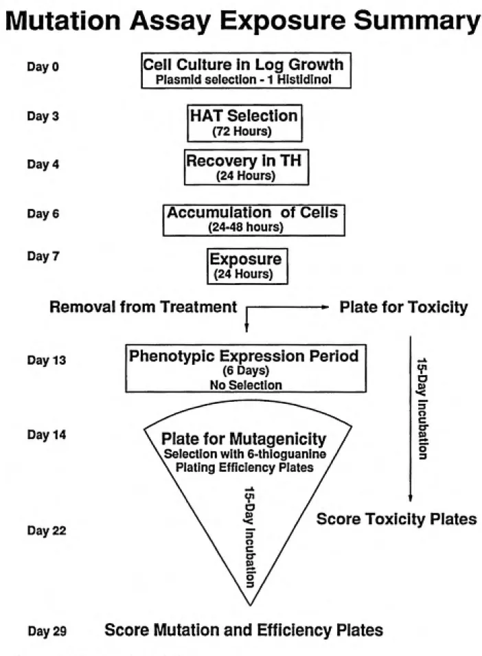
An overview in the form of a flow chart of the mutation assays done for this study is provided in figure 4.1.
Chemicals, Enzymes and Media Compounds:
Materials for the toxicity and mutation assays were obtained from the following sources: RPMI 1640 (Ix with L-glutamine), Dulbecco's phosphate buffered saline (PBS) pH
7.5, 8.0 mg/ml NaCl, 0.2 mg/ml KCl, 1.15 mg/ml Na2Hpo4, and 0.2 mg/ml KH2PO4 and
penicillin and streptomycin (lOOOx) were obtained from Lineberger Cancer Research Center,
Tissue Culture Facility. L-glutamine; 200mM as 29.2 mg/ml in 0.85M NaCl were from Gibco, Life Technologies Grand Island, NY. Bovine Calf Serum, Lots 21512008 and 21511064, from HyClone Laboratories, Logan UT. Aminopterin, hypoxanthine (6-hydroxypurine), thymidine (l-[2-deoxy a-D-ribofuranosyl]-5-methyl uracil) 99-100%, deoxycytidine, 6-thioguanine - (2 amino-6-mercaptopurine) 98%, and L-histidinol were purchased from Sigma Chemical Company, St. Louis, MO. Vinyl chloride and ethylene oxide (>99.5%) were obtained from Fluka Chemica, Switzerland. CAA was obtained as a
50% w/w solution in H2O from Aldrich Chemical Company, Milwaukee WI. CEO was
38
Safety Considerations
Safety in handling chemical carcinogens, selective agents, and the cells in culture was always paramount in experimental design.
The cell line used in this study contains Epstein Barr Virus DNA which necessitates the handling in accordance with NIH guidelines for research with recombinant DNA molecules (Gentest 90-1). EBV transfected cells do have the potential to shed the active
virus, although this is highly unlikely. All regular cell culture work was accomplished in a
Health and Safety certified biological safety hood with vertical laminar an- flow and extemal exhaust. In addition, latex gloves and a laboratory coat were worn when handling cells in culture. The selective agents aminopterin and 6TG are both toxic compounds and were handled in the biological safety hood only after they were placed in solution. Solutions of these compounds were prepared in an approved chemical laboratory hood.
under pressure. Consequendy, the compound was handled very cautiously when it was in the gaseous state and all sources of ignition were removed form the hood. When the stock gas cylinder was not in use, it was stored inside the chemical lab hood. All contaminated media and disposable plasticware were kept in the chemical laboratory hood until it was packaged for disposal through the Health and Safety office. Material safety data sheets for the compounds used were available in the work area to provide additional safety guidelines. Cell Culture
The stocks were maintained at -70' C in DMSO until ready for use. Cells were quickly thawed at 37 * C, diluted in 50 ml of prewarmed RPMI 1640,10% bovine calf senim without antibiotics (stock media), pelleted with centrifugation (lOOOg 3 min) and
resuspended in 50 ml of stock media. The cytoprotective agent, DMSO inhibits cytochrome P450IIE1 activity and must be removed from the media (Yoo et al., 1987). Selective
pressure for the IIEl plasmid is begun immediately after resuspending the cell pellet in fresh media and is accomplished by the addition of 3mM L-histidinol. L-histidinol was stored as 300x stock in 0.500 ml aliquots in cryogenic vials at -20' C and thawed individually. (Repeated freezing and thawing of L-histidinol in solution (sterile PBS) is not appropriate
according to the technical department of Sigma Chemical Company.) 7.5%NaHC03 was
added 0.250 ml/ 50 ml whenever L-histidinol was added to buffer the alkalinity of histidinol. Histidinol was added to the cell culture after each dilution (three days per week) up through
the treatment with HAT. Cells were maintained in a 37 ° C incubator with 6% CO2,100%
humidity in 75 cm sterile tissue culture flasks. Cells were enumerated approximately every
40
prewarmed (37 * C stock media) to a concentration of 0.5 - 4.0 x 10^ cells/ml. Cells were
never grown at cell concentrations exceeding 1.2x10 cells/ml. Cell cultures were tested periodically and found negative for mycoplasma contamination. Cell doubling time was calculated periodically using the formula:
r = [ln2(T)]/[ln(N/No)
Where t is the doubling time in hours, T the difference in hours between the time the cells
were last diluted and the current cell count, N is the current cell concentration (cells/ml) and N_ is the cell concentration when the cells were last counted. Average doubling time for this cell line is 22 hours; longer doubling times (in excess of 30 hours) were viewed as suspect for possible culturing problems. Cell culture was maintained during and after treatment as described, but the cultures were supplemented with penicillin (100 units/ml) and streptomycin (100 ng/ml).
Pretreatment of cell stocks in HAT media to reduce the level of background mutants was carried out prior to each exposure to the test substance. The HAT treatment consisted of
2x10 M hypoxanthine, 8x10"' M aminopterin and 3.5x10"^ M thymidine. Stock media
was used through HAT treatment. HAT was prepared as 333x HT in 0.3N HCl, 0.2 nm filter
sterilized and 250x A in 0. IN NaOH, 0.2 fim filter sterilized and stored at -20' C in a
through the HAT treatment in a single culture. Cells were counted and diluted after 48 hours and the HAT concentrations re-established for an additional 24 hours. Cells were then
centrifuged in an lEC Centra 8 table top centrifuge for three minutes at 1000 rpm, 25 * C.
The supernatant was carefully removed and the cell pellet resuspended in pre-warmed stock media, and HT (no aminopterin) added in the same concentrations as in the HAT treatment
for recovery. Cells were allowed to grow for two to three days after the 24 hour recovery in HT to achieve adequate cell numbers for treatments. Treatments were accomplished in 50 ml
cell suspension, (4xl0^/ml) in 75 cm"^ tissue culture flasks for 24 hours. The media used was
RPMI1640,10% BCS with penicillin and streptomycin (referred to as treatment media). During treatment, flasks were sealed to prevent carcinogen contamination of the atmosphere
in the incubator. While sealed, the CO2 in the incubator could not reach the media to
maintain pH; therefore the treatment media was supplemented with 29 mM HEPES buffer during the 24 hour treatment period. After treatment, the entire volume of cells or a fraction thereof (depending on whether toxicity was anticipated or not) was pelleted, resuspended in fifteen ml prewarmed treatment media in a 50 ml centrifuge tube, pelleted again and
resuspended in 50 ml of treatment media in 75 cm"^ flasks. The cell density was determined
and two plates were seeded per treatment concentration for toxicity determination. The time at which this initial density was determined was the starting point for evaluating growth delay. The counts from the two plates were pooled to calculate PE. Cells were carried for six days to allow phenotypic expression at the hprt phenotype; during this time the cell
cultures were diluted daily to 4x10^ cells/ml. Cell density was ascertained on day seven and
42
depending on the treatment and anticipated MF) were used for each treatment and the counts of all the plates were pooled to calculate the MF. Three independent cultures were exposed and carried through MF determination for each concentration of compound tested. Actual number of flasks (and consequently number of cells) initially treated varied according to toxicity. Four control flasks were maintained in every experiment.
Plating
Cells at a determined density were plated in 96-well, flat bottom microliter plates (well capacity 0.35 ml, surface area of 0.28 cm , Costar Corp). The volurrw plated per well was 0.2 ml in all cases. The media used for plating was RPMI1640 plus 15% bovine calf serum,
IX penicillin and streptomycin, and 1.0 mM L-glutamine. Plates were maintained
undisturbed in a water jacket incubator at 6% CO2 for 15 days before colonies were scored.
Because of the ability of this cell line to form rather large colonies (2 mm in diameter and larger, both mutant colonies and wild type) the plates were scored by counting colonies from the back side of the plate without magnification. Contaminated or empty wells were
excluded from the calculation to determine plating efficiency or mutant fraction. Plating efficiencies ranged from 20-60%. Cells for the purpose of determining toxicity or plating efficiency were plated at a density of two cells per well. The only exception was in plating cells after exposure to higher doses of CAA where toxicity required that four cells/well (16mM exposure) and 30 cells/well (24 mM exposure) be plated. To determine the frequency of hprt" mutants generated by exposure to the test substances, cells were plated at an
agent 6-thioguanine (6TG). Plating efficiencies (PE) and mutant fraction (MF) were calculated as described previously.
Determination of Treatment Concentrations
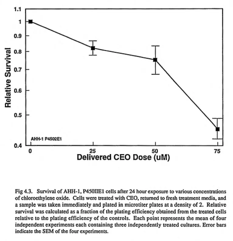
Initial cytotoxicity studies were accomplished with the three compounds over a broad range of test substance concentrations. Ten milliliter cultures at 4x10-^ cells/ml in 25 cm'' sterile tissue culture flasks were used. Preliminary results from the VCM toxicity and mutagenicity experiments indicated that 24 hour treatments would be preferable versus shorter periods due to the limited response seen. Therefore, for consistency, all cell exposures were conducted for 24 hours. The preliminary results from the VCM studies indicated that VCM solubility would determine the upper treatment concentration. This level was determined as 500mM. Dose ranges of 8-24/xM for CAA and 25-75 mM for CEO were selected; the upper level of these doses approached 10% survival.
When the first mutation determinations were made, a very high background MF was observed, (9 - 75 x 10""). High background levels of mutants can be due to: artifacts from low plating efficiencies, improper aminopterin concentrations, exposure to some other mutagen, or an inadequate selective agent (6TG) (Gentest, 1990). These possibilities were tested and eliminated. We then demonstrated that the high background was dominated by an
unusual 6TG^, HAT^ population. Although never tested, it was assumed that this was an
44
fifteen days incubation. Cultures with very low background MF (3 x 10' or less) were considered to be mutant free and aliquots cryo-preserved. These "mutant free" cell stocks were then used for all mutation assays.
VCM Treatment
volume accurately delivered with a Ice tuberculin syringe. Treatment flasks contained 50
ml of 4x10^ cells/ml. Flasks were sealed with sterile rubber septum stoppers which were
sealed with vinyl tape. VCM/MeOH stocks and treatment solutions were maintained as cold as possible during transfers. Vials containing these solutions were kept in dry ice/acetone (stock solutions) or near freezing on dry ice (treatment solutions). Appropriate volumes of treatment solution were measured in a syringe by holding the syringe against dry ice as the solution was drawn into it. Doses delivered into the treatment flasks were always injected through the cell suspension with the opening of the treatment flask pointing down and the mixture gently agitated before setting the flask down. This arrangement ensured: 1) efficient delivery of the treatment solutions with minimal losses, 2) the delivered dose remained in the tissue culture flask (in solution or in the head space) 3) no contamination of either the
incubator or the laboratory with VCM. After treatment, the rubber septums were cut off and held for appropriate disposal. Three cultures were treated for each exposure concentration and three MF plates were prepared from each culture (nine plates for each dose). Two MF plates were prepared for each control culture and two plates for each treatment concentration for both PE and toxicity plates. Two consecutive treatments were accomplished at 300 and 500 mM to induce greater mutant fractions and allow for the collection of mutants for analysis. Since VCM toxicity was never observed, the second treatment was administered the same day the cells were removed form the first treatment.
CAA Treatment
CAA treatment stock was prepared as 100 mM in PBS. This was diluted to 1 mM in
46
was prepared from the purchased CAA stock for each treatment The 1 mM stock was added
to 50 ml cultures at 4.5x10^ cells/ml at a final concentration of 8,16, or 24 nM. Cultures
were tightly capped, gently mixed and placed in the 37' C incubator for 24 hours. Controls were also capped tightly and contained HEPES buffer at the same concentration. Four replicate control cultures, three replicate cultures at 8 pM, 6 cultures at 16/iM, and 15 cultures at 24 mM were treated. After growth resumed in the flasks, the 16 and 24 nM treatments were pooled into three cultures. When cells were plated for MF, there was a total
of three cultures for each treatment concentration and four controls.
The following number of plates were used for each treatment concentration to determine MF: two plates per each of the three treatment cultures for S/iM, six plates per each of the three pooled treatment cultures at 16mM and ten plates for each of the three pooled treatment cultures at 24mM. Two plates for each control were plated. To obtain cells for DGGE analysis, two consecutive treatments were carried out at 16mM; the second
treatment started after the cells had recovered from the first treatment (i.e. log growth with
approximately 22 hour doubling times). Attempts were made for bulk selection of mutants in
large spinner cultures (900 ml, 4x10^ cells/ml - 1.0x10" cells/ml in 1 liter jars on a magnetic
stir plate) (Carriello et al.,1991). However, the combined effects of toxicity from the CAA exposure and the propensity to grow poorly in bulk spinner cultures (Gentest, 1990) resulted in forgoing this method of isolating mutants for analysis.
CEO Exposure
the fourth treatntent. Purity was determined based on nuclear magnetic resonance (NMR) recorded at the time of synthesis. CEO treatment stocks were prepared in acetone from the pure CEO immediately before treatment. The appropriate volume of acetone for treatment solutions was aliquoted into 20ml amber serum vials and kept covered in dry ice/acetone. The CEO was quickly thawed, the volume of CEO needed removed with a pipette, and delivered to the serum botde which was then sealed with a rubber septum and collar. The CEO was quickly returned to the dry ice acetone and cell cultures were treated as described
fortheVCM. Treatments of 100mM,75mM, 50/iM and 25mM were conducted. CEO was
delivered in acetone at a final level of 0.04% in each culture including controls, (acetone produces signs of toxicity in these cells at 0.5% (Gentest, 1990). Triplicate cultures were treated at each dosing concentration and ultimately three MF plates were prepared from each treatment flask (nine plates per treatment dose).
RESULTS
Toxicitv