THE EFFECT OF TEMPERATURE AND SALINITY
ON THE DEPURATION OF HEPATITIS A VIRUS
AND OTHER MICROBIAL INDICATORS FROM CLAMS
5^
JULIE C. MURRAY. Effect of Temperature and Salinity on
the Depuration of Hepatitis A Virus and Other Microbial
Contaminants in Clams.
(Under the direction of Dr. Mark D. Sobsey)
Depuration or the process of self-purification has been used
for many years to reduce the enteric microbial contaminants
from shellfish harvested from sewage polluted waters.
Little is known however about the effectiveness of this
process in eliminating enteric viral pathogens. This study
compared the reduction of hepatitis A virus (HAV) to that of
poliovirus 1, bacteriophage MS2, Escherichia coli. and
Streptococcus faecalis^ from experimentally contaminated
clams fMercenaria mercenaria). The clams were subjected
standard depuration conditions in a model, laboratory-scale
system. The effect of temperature (12, 18, 25 C) and
salinity (8, 18, 28ppt) on virus and bacteria depuration
over period of 4-5 days was studied. The viruses were not
depurated as effectively as the bacteria. Elimination rates
of test organisms was independent of temperature.
Depuration rates were reduced at lower salinity (8 and 18
ppt) . The results indicate that the current the practices
and conditions of depuration are ineffective in eliminating
enteric viruses from clams. These studies further suggest
that coliform bacteria is an inadequate indicator of virus
ACKNOWLEDGEMENTS
I would like to thank Dr. Mark D. Sobsey for his
guidance and support throughout my study in Environmental
Science and Engineering. I am also grateful to Dr. Frederic
K. Pfaender, Dr. Morris Shiffman, and Dr. Alvis Turner for
their time and positive criticism during the preparation of
this report.
A special heartfelt thanks is deserved by Greg Lovelace
for his invaluable assistance and technical support. I am
indebted to Doug Wait for is many ideas and to Dorthy
Thompson for her work in cell culture preparation.
Special thanks is also due my colleague, Soumaya
Alouini, for her friendship and work on this project.
Finally, and most importantly, I appreciate my family
and friends, without whose support and encouragement this
m
I. INTRODUCTION 1
II. OBJECTIVES 4
III. LITERATURE REVIEW 5
A. Test organisms 5
A.l. Viruses 5
A.1.1. Hepatitis A virus 6
A. 1.2. Poliovirus-1 9
A.2. MS2 bacteriophage 10
A.3. Bacteria 11
A.3.1. E^ coli 11
A.3.2. S^ Faecalis 11
B. Enteric Bacteria and Viruses in the
Aquatic Environment 11
B.l. Sources of Contamination 11
B.2. Contamination of Shellfish 17
B.3. Factors Influencing Survival
and Persistence 20
C. Epidemiology of Shellfish-Associated
Viral Disease (Disease transmission) 23
D. Clams 27
D.l. Introduction 27
D.2. Filter Feeding 29
E. Accumulation of Enteric Bacteria and
Viruses by Shellfish 31
E.2. Concentration of Bacteria and Viruses 32
E.3. Factors Affecting Acciamulation 34
F. Elimination and Depuration of Enteric Bacteria
and Viruses by Shellfish 39
F.l. Mechanisms of Elimination 39
F.2. Factors Affecting Rates of Elimination 41
F.3. Commercial Depuration 50
IV. Materials and Methods 57
A. Media and Components 57
B. Cultivation and Assay Systems 57
B.l. Cell Culture 57
B.2. Propagation and Cultivation
of Test Organisms 58
B.2.1. Hepatitis A virus 58
B.2.2. Poliovirus-1 58
B.2.3. Bacteriophage MS2 59
B.2.4. Bacteria 60
B.3. Assay of Test Organisms 60
B.3.1. Plaque Assay 60
B.3.1.1. HAV 60
B.3.1.2. Polio 62
B.3.2. MS2 Bacteriophage Assay 63
B.3.3. Bacteria Plate Counts 63
C. Clams 63
D. Seawater 63
E. Test Organism Recovery 66
F. Depuration Experimental Methods 66
G. Uptake Experimental Methods 70
H. Statistical Methods 70
IV. Results 73
A- Depuration 73
A.l. Effect of Temperature 73
A.2. Effect of Salinity 88
B. Uptake 104
V. Discussion 111
A. Depuration 111
A.l. Effect of Temperature 115
A.2. Effect of Salinity 116
B. Uptake 119
C. Factors Contributing to Variation 121
D. Implications for Commercial Depuration 123
VI. Conclusions 126
VII. Future research 127
Appendix A-Media and Components
Appendix B-Data
LIST OF TABLES
VIRUS REMOVAL IN WASTEWATER TREATMENT PROCESSES...15
NATIONAL SHELLFISH SANITATION PROGRAM REGULATIONS...19
FACTORS AFFECTING ENTERIC BACTERIA AND VIRUS
PERSISTENCE IN THE ENVIRONMENT...21
SHELLFISH-ASSOCIATED HAV OUTBREAKS...25
POSSIBILITIES TO PREVENT FUTURE SHELLFISHBORNE
DISEASE OUTBREAKS...28
FACTORS AFFECTING BACTERIA AND VIRAL
ACCUMULATION IN SHELLFISH TISSUE...35
FACTORS AFFECTING BACTERIA AND VIRAL ELIMINATION
FROM SHELLFISH TISSUE...42
IMPORTANT EDIBLE SHELLFISH...52
ELIMINATION OF TEST ORGANISMS AT TEMPERATURE 12°C
(% OF INITIAL REMAINING)...74
ELIMINATION OF TEST ORGANISMS AT TEMPERATURE 18°C
(% OF INITIAL REMAINING)...75
ELIMINATION OF TEST ORGANISMS AT TEMPERATURE 25°C
(% OF INITIAL REMAINING)...76
SUMMARY OF LINEAR REGRESSION FOR
TEMPERATURE EXPERIMENTS...86
SUMMARY OF EFFECT OF TEMPERATURES
ON TEST ORGANISMS DEPURATION...87
SUMMARY OF EFFECT OF TEMPERATURES ON EACH ORGANISM...88
ELIMINATION OF TEST ORGANISMS AT SALINITY 8 ppt
(% OF INITIAL REMAINING)...90
ELIMINATION OF TEST ORGANISMS AT SALINITY 18 ppt
(% OF INITIAL REMAINING)...91
ELIMINATION OF TEST ORGANISMS AT SALINITY 28 ppt
(% OF INITIAL REMAINING)...92
SUMMARY OF LINEAR REGRESSION FOR SALINITY EXPERIMENTS..101
SUMMARY OF EFFECT OF SALINITIES ON
UPTAKE OF TEST ORGANISMS (PFU/ml)...105
RELATIVEUPTAKE OF TEST ORGANISMS
(% DETECTED / GRAM TISSUE RELATIVE TO INITIAL WATER
CONCENTRATION)...106
LIST OF FIGURES
MODEL DEPICTING FATE OF VIRUSES IN COASTAL SYSTEMS...12
SOURCES OF VIRUSES IN WATER...13
FILTER FEEDING. ...---30
BACTERIAL STOCK PROCEDURE...61
PLATING PROCEDURE FOR MS2 COLIPHAGE...64
PLATING PROCEDURE FOR E^ colj and S^ faecalis...65
CLAM PREPARATION FOR TEST ORGANISM RECOVERY... . 67
DEPURATION OF ORGANISMS AT 3 TEMPERATURES
(ORGANISMS / GR CLAM TISSUE)...77
DEPURATION OF ORGANISMS AT 3 TEMPERATURES
(% OF INITIAL REMAINING)...78
DEPURATION OF ORGANISMS AT 3 TEMPERATURES
(% OF INITIAL REMAINING LINEAR REGRESSION LINES)...79
DEPURATION AT 3 TEMPERATURES OF ORGANISMS
(ORGANISMS / GR CLAM TISSUE)...81
DEPURATION AT 3 TEMPERATURES OF ORGANISMS
(% OF INITIAL REMAINING)...82
DEPURATION AT 3 TEMPERATURES OF ORGANISMS
(% OF INITIAL REMAINING LINEAR REGRESSION LINES)...83
DEPURATION OF ORGANISMS AT 3 SALINITIES
(ORGANISMS / GR CLAM TISSUE)...93
DEPURATION OF ORGANISMS AT 3 SALINITIES
(% OF INITIAL REMAINING)...94
DEPURATION OF ORGANISMS AT 3 SALINITIES
(% OF INITIAL REMAINING LINEAR REGRESSION LINES)...95
DEPURATION AT 3 SALINITIES OF ORGANISMS
(ORGANISMS / GR CLAM TISSUE)...97
DEPURATION AT 3 SALINITIES OF ORGANISMS
(% OF INITIAL REMAINING)...98
DEPURATION AT 3 SALINITIES OF ORGANISMS
(% OF INITIAL REMAINING LINEAR REGRESSION LINES)...99
(% DETECTED/ GRAM TISSUE RELATIVE
INTRODUCTION
The pollution of estuaries and other coastal marine
waters is threatening an important food source, edible
bivalve molluskan shellfish. Shellfish resources are
increasingly being closed to harvesting because of the public health risks associated with consumption of
contaminated shellfish. Due to expanding populations and development in coastal areas and man's disregard for his environment the economic effects of this loss are great.
Primary sources of sewage pollution include sewage treatment plant effluents and sludges, septic tank seepage, boat waste discharges, and land runoff. Shellfish filter feed in these polluted water and retain suspended
particulate matter including pathogenic microorganisms.
Thus, shellfish harvested from a fecally contaminated area
may contain enteric viruses and bacteria. Shellfish may then transmit disease if eaten raw or partially cooked.
The current standards for shellfish and harvesting
waters were developed over 40 years ago and are based on the presence of total or fecal coliform bacteria. Enforcement of this standard has protected consumers from bacterial infection but the ability of coliforms to indicate viral
from consumption of contaminated shellfish, some harvested
from 'approved' beds. Therefore, there is reason for
concern that shellfish may accumulate and retain HAV and
other enteric viruses more efficiently than they do
indicator bacteria.
Depuration is a process in which contaminated shellfish
are placed in a clean flowing system and allowed to
naturally eliminate or purge thenselves of accumulated
contaminants. However, levels of enteric microorganisms may
persist in shellfish for a period of time after they have
been transferred to clean water; reports of adequate time
requirements vary with respect to the environmental
conditions of depuration. If the depuration process and the
factors which affect it can be understood, commercial scale
depuration or relaying could alleviate some of the economic
loss of closed marginally polluted harvesting areas and
ensure pathogen free shellfish.
Microbial information on depuration is limited, with
very little known about the elimination of HAV and most
other pathogenic enteric viruses. Hepatitis A virus is
probably the most serious viral disease transmitted by
contaminated shellfish. Studies are therefore needed to
evaluate and characterize the behavior of HAV compared to
other enteric viruses, indicator bacteria, and other
studies are needed to determine the effect of environmental
factors such as temperature and salinity on depuration to
identify the optimum conditions needed for the elimination
To evaluate the reduction of hepatitis A virus compared to
poliovirus-1, bacteriophage MS2, Escherichia coli B, and
Streptococcus faecalis. from the hardshell clam, Mercenaria
mercenaria, at different conditions of temperature and
salinity in a lab-scale depuration system.
-To determine the rates of depuration of high levels of
HAV, polio, MS2, Ej. coli. and S_i. faecalis at
temperatures of 12, 18, 25oC.
-To determine the rates of depuration of high levels of
HAV, polio, MS2, Ej. coli, and S^ faecalis at
salinities of 8, 18, 28ppt.
To determine the comparative time period for maximum uptake
and concentration from water of bacteria (Ej^ coli B and S^.
faecalis) and viruses (HAV, poliovirus, and bacteriophage
A.l. Viruses
Viruses are the smallest infectious agents, ranging in
size from 20-300 nm in diameter. They are obligate
intracellular parasites which are capable of replication
only in a living host cell.
Viruses contain either single or double stranded
nucleic acid, either RNA or DNA and either in one
polycistronic molecule or in a different segments. The
genome is encased in a protein shell capsid which is
specific to the virus. The shell may then be surrounded by
a lipid containing membrane.
Viruses have one of three general shapes; spherical
(icosahedral), helical (rod-shaped), and complex.
The host range for a specific virus may be broad or
extremely limited. Viruses are known to infect unicellular
organisms such as mycoplasma, bacteria, and algae, as well
as eukaryote cells and multicellular organisms such as
plants and animals.
Enteric viruses
Enteric viruses are transient inhabitants of the human
alimentary tract. Enteroviruses lack a lipid envelope, and
consequently they are stable in the digestive tract
""^^Z^/^S^^'--&
exposure to acids at pH levels as low as 3.0. Viruses
multiply within the gut and are excreted in the feces in
large numbers. The titer of some enteric viruses may be as
high as 1x10-^^ particles per gram/feces.
Disease transmission is by the fecal oral route. This
mode of transmission is facilitated through person to person
contact and through virus contamination of water and food.
There are more than a hundred types of enteric viruses,
these including the enteroviruses, adenoviruses,
rotaviruses, reoviruses, Norwalk and Norwalk-like viruses,
calciviruses, astroviruses, and hepatitis viruses. The most
commonly known enteroviruses are the three types of
poliovirus (types 1-3). Other enteroviruses include: 34
types of echoviruses; 30 types of coxackieviruses A + B; and
hepatitis A virus, which is now classified as enterovirus
72.
Enteric virus infections are asymptomatic most of the
time especially in infants and young children. In some
cases, however, they may cause symptoms such as vomiting,
diarrhea, gastrointestinal illness, malaise, hepatitis,
jaundice, aseptic meningitis, and severe paralysis.
A.1.1. HAV
Based on recent studies HAV, a member of the
picornaviridae family, is classified with the enteroviruses
suggest that it may more appropriately belong in a seperate
genus of this family (Melnick, 82). Like enteroviruses,
HAV remains stable after exposure to ether and acid, but is
more resistant to heating at high temperatures (60°c for 1
hour) and to common disinfectants. Therefore, extra
protection is needed in dealing with hepatitis A patients
and their products. Similar in morphology to other
picornaviruses, HAV is a 27 to 32 nm spherical (icosohedral)
particle with cubic symmetry. It's physical and chemical
characteristics are also like those of enteroviruses, with a
linear single stranded RNA genome, having a molecular weight
of about 2.25x10 , positive polarity, a 5'- terminal
protein(VPG), and a 3' poly (A) tail. The capsid is
composed of four different structural proteins (VP1-VP4; 60
copies each), and 32 capsomeres.
Studies have shown that HAV is persistent in soil,
sewage, and the water environment (Sobsey et al, 1987). The
most likely mode of transmission of HAV is the fecal-oral
route, and both person to person contact and fecally
contaminated common-source vehicles, such as water and food
are implicated in disease outbreaks. A documented cause of
Hepatis A virus disease is the consumption of raw or
partially cooked bivalve mollusks (e.g. oysters, clams,
mussels, ect.) obtained from fecally contaminated water.
Hepatitis A, or infectious icterus, is a viral
influenza-like illness with an abrupt onset of fever,
headache, malaise, fatigue, anorexia, and nausea, followed
usually by vomiting, abdominal pain, dark urine, and
jaundice. The liver and spleen may be enlarged.
The incubation period of HAV ranges from 15-40 days
with a large amount of hepatitis A virus excreted in the
stool during the latter part of the incubation before outset
of the disease.
The disease is generally more severe and prolonged in
adults than in children. Hepatitis A is worldwide in
distribution but there is a high endemic prevalence in
developing countries.
The wild type hepatitis virus doesn't replicate well in
cell culture and this may be due to a defect in its
replicative cycle (Locarini et al, 1981). However, adapted
strains of HAV have been obtained by multiple passages of
virus through cell culture (Provost and Hillman, 1979). One
of these serially passaged HAV strains (HM-175), produced a
lytic response in two persistently infected cell lines:
FRHK-4,and BSC-1 (Cromean et al, 1986). Although such
strains don't reflect accurately the responses of wild type
HAV, they help simplify the development of methods for the
concentration and detection of HAV from environmental
samples of water and shellfish as well as for vaccine
A.1.1. Poliovirus-1
Poliovirus is a member of the picornavirus family. The
particles are small (28 nm) in diameter and nonenveloped.
They contain a single strained RNA genome having a positive
polarity and have a molecular weight of 2.5x10 . The virus
is acid stable down to pH 3.0 and has a buoyant density in
cesium chloride of about 1.34 g/ml.
Poliovirus has a very restricted host range. Most
strains can only be grown on primary or continuous cell line
cultures derived from a variety of human or monkey tissues.
There are three antigenic types of poliovirus 1, 2, and
3. They are transmitted via the fecal-oral route through
direct or indirect human contact as well as through water
and food contamination.Poliovirus causes poliomyelitis, which occurs worldwide
usually in children. Poliomyelitis is caused by anyone of
the three serotypes of poliovirus, and the disease occurs
generally one of two forms. In the abortive form,
poliomyelitis is a minor illness with rapid recovery without
paralysis (the nonparalytic form), it is also called aseptic
meningitis. The other form causes paralytic meningitis.
In this form spinal or meningeal symptoms develop, often
accompanied by fever, malaise, nausea or vomiting. Pain in
13.
Due to the success of polio vaccine and improvement in
sanitation, there are currently few outbreaks of
.poliomyelitis in the U.S.A. and other developed countries.
The widespread use of polio vaccine has lead to
presence of high concentration of poliovirus in almost all
sewage contaminated water (post vaccinal fecal excretion).
Poliovirus is one of the easiest viruses to grow and
detect in cell culture. These two characteristics make
poliovirus a useful model indicator of virus associated
fecal contamination in the environment.
A.2. MS2 Bacteriophage
Bacteriophages are associated with almost all bacterial
genera. MS2 phage is host specific to male (F+) or pili
producing strains of E. coli.
MS2 is a RNA phage with a particle weight of about 4 x
10 . It is composed of 180 molecules of a single coat
protein forming an icosohedral shell. The shell encloses a
single molecule of RNA of 3500 to 4700 nucleotides that are
plus sense and fully competent to cause infection and
production of virus progeny. The viruses replicate quickly,
causing lysis of the host cell in 30 to 40 min and yield a
very high level of progeny particles, approximately 10,000
A. 3. Bacteria
E. coli and S^ fecaelis are generally nonpathogenic
bacteria that are part of the natural flora of the human
intestinal tract. They are excreted in large numbers in
human sewage and therefore are adequate indicators of fecal
contamination by pathogenic bacteria.
A.3.1. Escherichia coli
The concept of using Ej. coli as an indicator was
introduced in 1885. The coliform group is defined as
bacteria that are aerobic or facultatively anaerobic,
Gram-negative, nonspore forming, rod-shaped, and which ferment
lactose with acid and gas production within 48 hr. at 35°C.
A.2.2. Streptococcus fecaelis
Classification of Sj^ fecaelis, on of the fecal
streptococci (Lance field group), is dependent on the
bacteria's ability to grow in 6.5% NaCl broth in 0.1%
methylene blue milk, at pH 9.6, at 10 to 45°C, and to
survive at 60°C for 30 min.
B. Enteric Bacteria and Viruses in the Aquatic Environment
B.l. Sources of Contamination
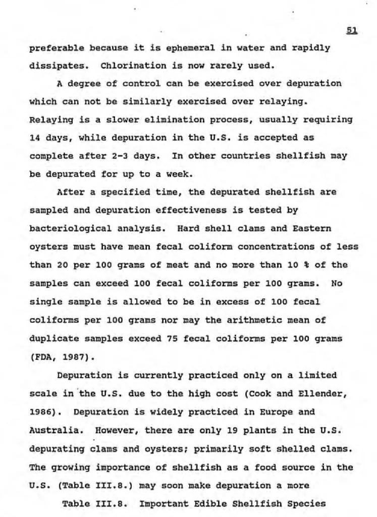
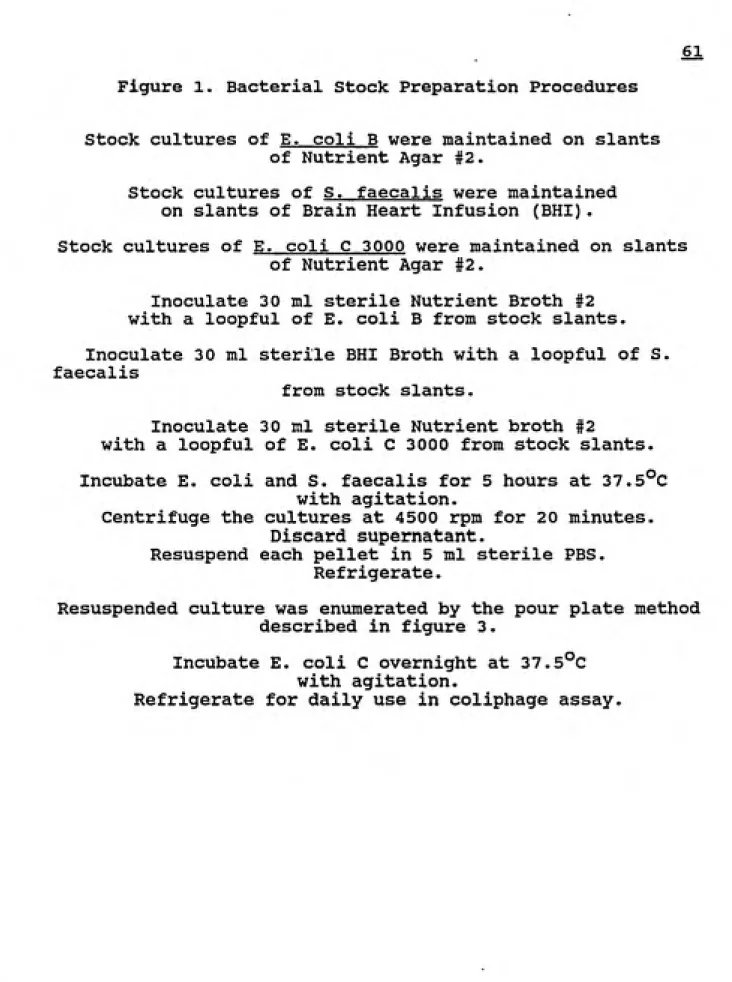
The principle source of enteric viruses in the aquatic
environment is the worldwide disposal of sewage and other
forms of fecal excreta to surface waters (Figure III.l).
Figure 1. Model Depicting Fate of Viruses in Coastal Systems
SEWAGE OUTFALL
AEB0S0LI2ATI0S
BY BREAKING WAVES
^-^t^
I
VIRUS ASSOCIATION WITH SUSPENDED SOLIDS (actt to prolong virul survival)
RESUSPENSION BY RAIN WAVE ACTION TIDES.
DREDGING, ETC
<. M
v^-^>;j^
ACCUMULATIO"; BY SHELLFISH DURING FEEDING
UPTAKE BY CRUSTACEA AND BOTTOM FEEDING FISH
ACCUMULATION IN SEDIMENTS
(virusrt occur in highc eoncenitationj in (edimeni than the overlaying waif'
Excreta from Man
Solid Waste Landfills
Sewage
Land Runoff
t
Rivers and Lakes
Groundwater
Oceans and Estuaries Irrigation
Recreation Water Supply Shellfish
Aerosols
Man
Fig. 2. Routes of enteric virus transmission [Melnick et al., 1978].
transmission. Enteric viruses are excreted in the feces of
infected individuals at levels as high as 10^^ particles per
gram (Flewett and Woode, 1978). Different wastewater
treatment processes remove viruses to varying degrees. The
efficiencies of each method are sximmarized in Table III.l.
A combination of these processes can be very effective
depending on the extent of treatment used.
The conventional waste water treatment processes
comprise primary sedimentation, secondary or biological
treatment, and disinfection. Tertiary treatment is also
practiced in some cases.
Primary treatment removes the suspended solids from
sewage by sedimentation. This step removes up to 50% of
bacteria and viruses from sewage; removal is by adsorption
of microorganisms to solids with subsequent settling (Gerba
and Goyal, 1982).
Secondary or biological sewage treatment utilizes
microbial degradation of soluble organics and solids.
Trickling filters, activated sludge, and oxidative ponds
remove up to 90% of the microbes by inactivation or
adsorption and physical removal in sludge (Gerba, 1981).
If treatment is continued through the tertiary stage a
high percentage of removal can be expected. This process is
facilitated by a coagulant such as aluminum or lime,
TABLE 1
VIRUS REMOVAL IN WASTEWATER TREATMENT PROCESSES
TREATMENT EXPECTED REMOVALr%^
Primary
Primary Sedimentation 0-75
Chlorination of Primary Sedimentation
effluent 50
Secondary
Trickling Filter 50
Chlorination of Trickling Filter
effluent 50
Activated Sludge 40-90
Chlorination of Activated Sludge
effluent 50-90
Tertiary
ͣ
Coagulation-Flocculation
and Sedimentation 90-99.99
Activated Carbon Adsorption 10-50
Chlorination of Tertiary effluent 99-99.99
1&
contact time, and quality of influent can succeed in up to
99.99% virus reduction (Gerba, 1981).
Tertiary treatment is rarely used and even conventional
primary and secondary treatment followed by disinfection is
not always employed. Approximately 5% of the U.S.
population still discharge untreated sewage directly via
ocean outfalls (Rao & Melnick, 1986). Bitton (1980)
estimates that four billion gallons of sewage with only
secondary treatment are discharged per day into coastal U.S.
waters. These discharges contain an estimated 380 virus
PFU/gal in the U.S. (Metcalf, 1987). Although the
concentrations of pathogens are supposed to be reduced by
dilution and natural degradation, enteric bacteria and
viruses have been detected greater than 8 miles from
discharge sites (Metcalf, 1974; Dahling and Safferman,
1979) .
Sewage sludge disposal also contributes to the presence
of enteric pathogens in the marine environment. Bacteria
and viruses are concentrated into the sludge during
treatment but are not rendered inactive (Goyal, 1984).
Although sewage effluent may meet coliform, suspended
solids, BOD, and other quality control standards, treatment
processes are ineffective in removing all viral
contaminants. It is primarily from these sources,
arises.
B.2. Contamination of Shellfish
Pollution of estuaries and other shellfish habitats by
enteric pathogens leads to the contamination of bivalve
mollusks. Enteric viruses have been isolated from many
edible bivalves including clams, oysters, and mussels (Gerba
and Goyal, 1978). Viral contaminants have been found in
shellfish harvested from both closed and approved areas
(Metcalf and Stiles, 1968b; Goyal et al, 1979; Vaughn et al;
1979b; Ellender et al, 1980; Wait et al, 1983). In many of
the above cases no viruses were detected in the overlaying
waters.
Shellfish harbor viruses in their tissues and passively
transmit them to humans who ingest raw or inadequately
cooked shellfish. When survival after typical cooking
methods was examined, somewhat better inactivation was
observed with steaming and stewing than baking or frying (Di
Girolamo, 1970), but overall virus inactivation was not
appreciable. After 8 min of stewing 10% of the initial
polio remained; 7% remained after 30 min of steaming, while
13% remained after 10 min of frying and 20 min of baking at
121°C. Mazanti (1987) showed that even under pasteurization
shellfish, it is necessary to eliminate the pathogenic
microbes before they reach market.
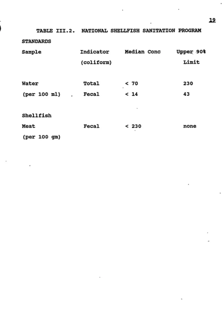
The U.S. has tried to achieve a pathogen free shellfish
market by establishing a coliform standard designed to
indicate sewage and fecal contamination. The National
Shellfish Sanitation Program (NSSP) established these
national standards over 40 yr ago. They are based on total
and fecal coliform bacteria levels enumerated by the most
probable number method. The NSSP standards are summarized
in Table III.2. Many shellfish areas have been closed due
to failure to meet these standards.
Coliform testing is an inexpensive, relatively easy,
and often reliable method of indicating fecal contamination.
Enforcement of NSSP standards has successfully limited the
number of bacterial disease outbreaks. However, the ability
of coliforms to indicate viral contamination of shellfish
and harvesting waters has been questioned due to enteric
virus isolation from shellfish and overlaying waters in
approved areas (Gerba and Goyal, 1978; Morris and Waite,
1981; Larkin and Hunt, 1982; Fugate et al, 1975; Goyal et
al, 1979; Vaughn et al, 1980; Portnoy et al, 1975).
The virological quality of shellfish and overlying
water is not adequately indicated or controlled by current
standards. Other indicators such as poliovirus and other
TABLE III.2. STANDARDS
Sample
NATIONAL SHELLFISH SANITATION PROGRAM
Indicator Median Cone Upper 90%
(coliform) Limit
Water Total < 70
(per 100 ml) Fecal < 14
Shellfish
Meat Fecal < 23
(per 100 gm)
230
43
20.
bacteriophages, have been considered but have proven
unreliable (Morris and Waite, 1981). It seems that
currently, the only valid determination of contamination is
direct detection of the specific pathogen of interest.
Unfortunately, many of the viral pathogens of interest are
difficult or impossible to detect and quantify and for those
that can be detected, the methods are unreliable,
technically difficult, expensive, and slow to yield results.
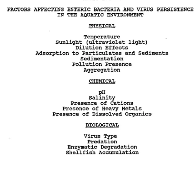
B.3. Factors Influencing Survival and Persistence of
Enteric Bacteria and Viruses in the Aquatic Environment
The fate of bacteria and viruses in the environment is
controlled by physical, chemical, and biological factors.
The factors of interest are listed in Table III.3. Rhodes
and Kator (1988) showed that enteric microbe survival
potential was a function of interacting biological and
physical factors.
Temperature, a physical factor, plays the largest role
in microbial inactivation. Using membrane dialysis chambers
in situ, O'Brien and Newmann (1977) showed the rate of virus
inactivation was exponential and affected primarily by water
temperature. Additionally, temperature predisposes enteric
microorganisms to biological actions such as predation and
21
TABLE III.3.
FACTORS AFFECTING ENTERIC BACTERIA AND VIRUS PERSISTENCE
IN THE AQUATIC ENVIRONMENT PHYSICAL
Temperature
Sunlight (ultraviolet light)
Dilution Effects
Adsorption to Particulates and Sediments
SedimentationPollution Presence
Aggregation
CHEMICAL
pH
Salinity
Presence of Cations
Presence of Heavy Metals
Presence of Dissolved Organics
BIOLOGICAL Virus Type
Predation
Enzymatic Degradation
-#
showed that microbial dieoff was inversely related to water
temperature.
Numerous studies have demonstrated that adsorption to
sediments can protect bacteria and viruses and increase
their persistence in the aquatic environment (Gerba and
McLeod, 1976; Kapuscinski and Mitchell, 1980; LaBelle and
Gerba, 1980). Adsorption to particulates and sediments
shields microorganisms from potentially harmful ultraviolet
light and facilitates settling out. Greater than 99% of
enteric viruses rapidly adsorb to estuarine sediment
(LaBelle and Gerba, 1980; Bitton, 1980), thereby causing
pathogens to concentrate in the bottom sediments. The
viruses can later be resuspended into the water by
turbulence. The concentrated microorganisms then have an
enormous infection potential, since viruses in sediment
fully retain their ability to initiate infection (Berg,
1983; Bitton, 1980; LaBelle and Gerba, 1980).
An important chemical factor affecting microbial
inactivation is heavy metals. The metals compete for
adsorption sites on particulates and dissolved organics
thereby limiting the protection these agents afford enteric
microbes.
Biological factors also contribute to microbial
persistence. Bacterial cells, living and dead, act as a
haven by providing adsorption sites for viruses (Mitchell,
m
bacteriophages can reduce the numbers of enteric bacteria
and viruses. Shuval gt al (1971) isolated a marine bacteria
capable of diminishing poliovimis-l 1000-fold in seven days.
This antiviral action is closely associated with the marine
bacteria's metabolic activity (Bitton, 1980; Shuval, 1971;
Mitchell, 1971). Oliver and Hermann (72) found that some
enteric viruses are susceptible to proteolytic enzymes.
Ward et al (1986) confirmed this; their experiments showed
proteolytic bacterial enzymes inactivated echovirus in fresh
water by cleaving the protein capsid, exposing the viral RNA
to nuclease digestion.
The type of microbe plays a large role in survival.
Enteric viruses generally survive longer in seawater than do
coliform bacteria (Melnick and Gerba, 1980). Further
studies with hepatitis A have shown that it is capable of
surviving longer than other enteric viruses (Bosch and
Shields, 1987).
C. Epidemiology
Contaminated shellfish pose a public health risk due to
the accumulation and persistence of pathogenic microbes in
their tissues. Transmission of pathogens by shellfish was
demonstrated before the turn of the century (Metcalf, 1987).
Large scale shellfish-associated disease outbreaks, such as
the New York state outbreak reported by Morse et al (1986),
of illness and at least 10 cases of hepatitis A or
infectious hepatitis.
Between 1900 and 1984, 11600 cases of shellfishborne
disease have been documented in the U.S.. Although there
are more than 100 known enteric viruses, only a few have
been shown to be transimitted by shellfish: hepatitis A,
non-A non-B hepatitis, Norwalk, Snow Mountain agent,
astrovirus, caliciviruses, and small round viruses (Gerba,
1988).
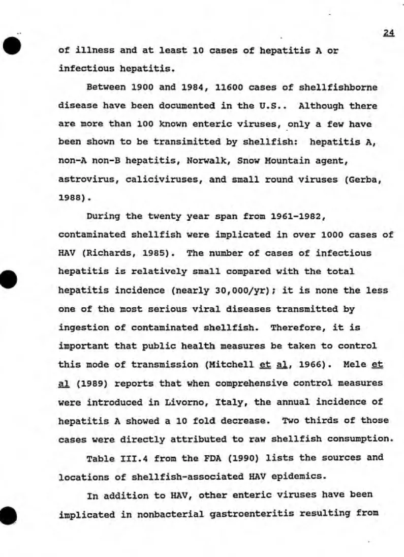
During the twenty year span from 1961-1982,
contaminated shellfish were implicated in over 1000 cases of
HAV (Richards, 1985) . The number of cases of infectious
hepatitis is relatively small compared with the total
hepatitis incidence (nearly 30,000/yr); it is none the less
one of the most serious viral diseases transmitted by
ingestion of contaminated shellfish. Therefore, it is
important that public health measures be taken to control
this mode of transmission (Mitchell et al, 1966). Mele et
al (1989) reports that when comprehensive control measures
were introduced in Livorno, Italy, the annual incidence of
hepatitis A showed a 10 fold decrease. Two thirds of those
cases were directly attributed to raw shellfish consumption.
Table III.4 from the FDA (1990) lists the sources and
locations of shellfish-associated HAV epidemics.
In addition to HAV, other enteric viruses have been
Table 4.
III.4. Shellfish-associated HAV Outbreaks
YEAR 1961 1961 1961 1961 1962 1963-6 1964 1964 1964 1964 1964 1966 1966 1966 1967 1968 1969 1969 1971 1971 1972 1973 1973 1973 1973 1977 1979 1982 1983 1983 1985 1988 CASES 84 459 15 31 3 46 123 249 3 43 3 4 3 4 3 3 6 13 5 3 2 263 15 37 1 17 10 11 01 04 01 51 SOURCE oysters clams clams oysters clams oysters/clams clams clams oysters clams clams clams clams " clams
oysters/clams
clams clams oysters clams clams clams oysters oysters oysters clams shellfish oysters clams several species clams clams oysters T.OCATION Alabama and Mississippi New Jersey,and New York Connecticut Alabama
New York
Mass.
Conn, and RI
Pennsylvania
N.C. New York Wash.,D.C. New Jersey Mass. New Jersey Texas New York New York Florida Mass. R.I. Florida,Mass Texas Georgia Louisiana Minnesota Washington Alabama,Fla. New York New York New York FloridaCompiled from FDA Sanitation Program Technical Report.
Shellfish Borne Disease Outbreaks. Dr. S. Rippy. February,
2&
shellfish consximption. Norwalk and rotavirus have been
confirmed as the causative agent in many gastroenteritis
outbreaks in the U.S. (Richards, 1985). It is also unclear
in some cases whether or not viruses were the cause of
disease outbreaks, but viruses were the likely etiologic
agent in most instances (Richards, 1985).
It is also, apparent that the high number of cases in
areas such as New York is attributable to increased
awareness and better reporting practices concerning
shellfishborne disease. It is thus likely that the actual
number of shellfish related disease cases nationwide is
grossly underestimated, due to lax surveillance and
reporting. This is further compounded by the fact that mild
cases may go untreated or not recognized as shellfish
related.
Many factors have led to current occurrences of enteric
disease transmission by shellfish. Current bacteriological
standards are inadequate for determining viral contamination
in shellfish and overlying waters. Illegal poaching has
certainly led to some untraceable cases.
Finally improperly classified growing and harvesting waters
has resulted in open but contaminated areas.
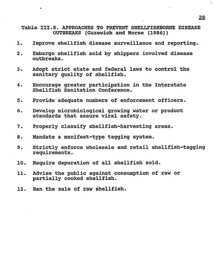
Because many outbreaks are due to shellfish harvested
from 'approved areas' as well as from shellfish that have
purged themselves to levels that met current bacterial
adequate standards and to perfect techniques to ensure
pathogen free shellfish. Other approaches to prevent
shellfishborne disease outbreaks are listed in Table III.5.
D. Clams
D.l. Introduction
Mercenaria mercenaria or the hard shell clam is a
member of the Pelecypoda, a class within the phylum
Mollusca. This type of shellfish is set apart from other
mollusks by their use of a filter feeding process to obtain
food materials. Besides the hard shelled clam, other
commercially important bivalves in the U.S. include the
Eastern oyster, Crassostrea virqinica. the Pacific oyster,
Crassostrea gigas. the Olympia oyster, Ostrea lurida, the
softshell clam, Mya arenaria. the Manila clam, Tapes
iaponica. and the blue mussel, Mytilus edulis (Werner,
1983). These clam, oyster, and mussel bivalve forms
represent the shellfish most often involved in transmission
of virus-caused diseases in the U.S.A. (Metcalf, 1980).
Clams are found in estuaries a few inches below the
water-sediment interface. The soft fleshy body is enclosed
within two hinged shells called valves. Beneath the
calcified valves a mantle structure encloses the body. The
clam has fused mantle lobes with siphon structures. The
siphons inhale up to 19 1 water/hr/oz of tissue (Metcalf,
28
Table III.5. APPROACHES TO PREVENT SHELLFISHBORNE DISEASE
OUTBREAKS (Guzewich and Morse (1986))
1. Improve shellfish disease surveillance and reporting.
2. Embargo shellfish sold by shippers involved disease
outbreaks.
3. Adopt strict state and federal laws to control the
sanitary quality of shellfish.
4. Encourage greater participation in the Interstate
Shellfish Sanitation Conference.
5. Provide adequate numbers of enforcement officers.
6. Develop microbiological growing water or product
standards that assure viral safety.
7. Properly classify shellfish-harvesting areas.
8. Mandate a manifest-type tagging system.
9. Strictly enforce wholesale and retail shellfish-tagging
requirements.
10. Require depuration of all shellfish sold.
11. Advise the public against consumption of raw or
partially cooked shellfish.
virus pollution of shellfish is often related to
association of a virus with a solid. Clams are more likely
to take up particles settled onto the uppermost layer of the
bottom sediments (Metcalf, 1978; Landry, et al, 1983).
Clays, part of the sediment, are among the most important
inorganic substances with which viruses associate (Metcalf,
1980).
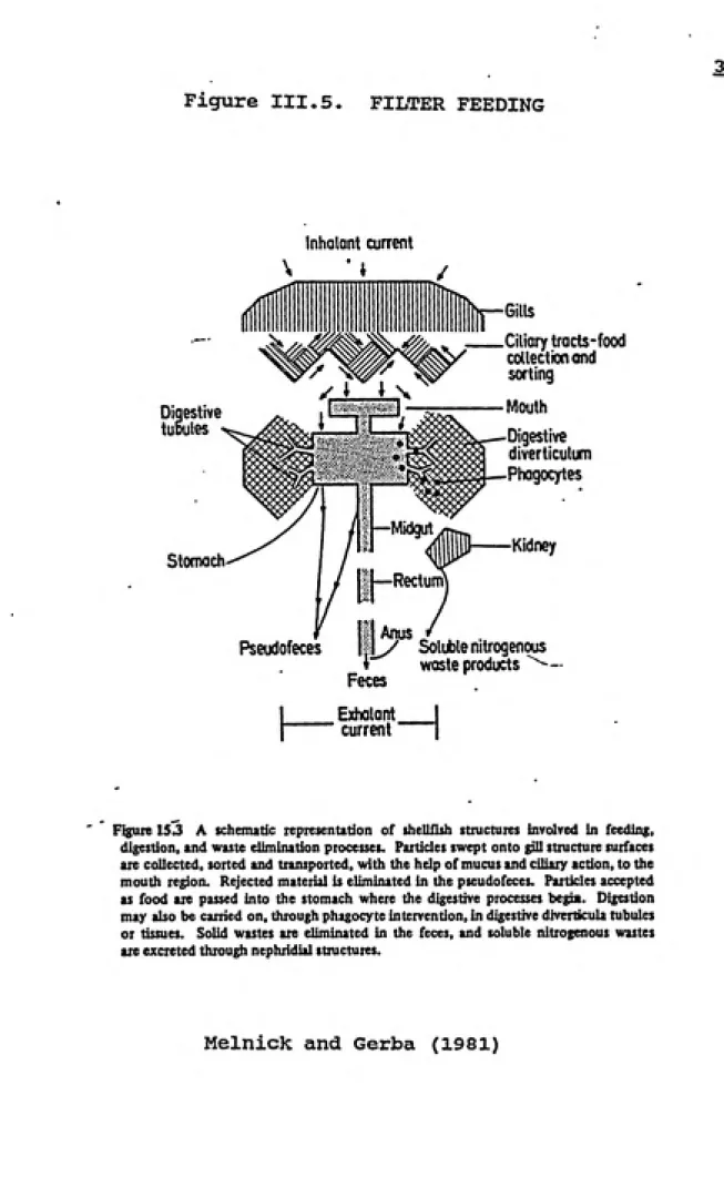
D.2. Filter feeding
Filter feeding, outlined in figure III.5, is initiated
during the pumping of water through the gill slits.
Particulate matter is removed by mucus sheets secreted by
the gills, with transport to the mouthparts facilitated by
ciliary action. The mouth accepts particulates as food based
on weight, size, and shape. Clams feed on suspended algae,
nanoplankton, and bacteria. Particles rejected as food
sources are eliminated from the mantle cavity in the form of
pseudofeces (Metcalf, 1980).
Bivalve feeding is influenced by several factors.
Water temperature, salinity, pH, turbidity and dissolved
oxygen are among the most important factors influencing
2Sl
Figure III.5. FILTER FEEDING
Inhalant current
Digestive
tuDutes
Stomach
—Midgut
11—Rectum
Gills
Ciliary tracts-food
collection ond
sorting
Mouth
Digestive
diverticulum
Phagocytes
Kidney
Pseudofeces
i Anus „
. ly Soluble nitrogenous
i waste products
"^^-Feces Exhalont
current
H
Figure 153 A schematic representation of shellfish structures involved in feeding,
digestion, and waste elimination processes. Particles swept onto gill structure surfaces
ate collected, sorted and transported, with the help of mucus and ciliary action, to the
mouth region. Rejected material is eliminated in the pseudofeces. Particles accepted
as food are passed into the stomach where the digestive processes begia. Digestion
may also be carried on, through phagocyte intervention, in digestive diverticula tubules
or tissues. Solid wastes are eliminated in the feces, and soluble nitrogenous wastes
are excreted through nephridial structures.
E. Accumulation of Enteric Bacteria and Viruses by
Shellfish
E.l. Mechanism of Uptake
Bacteria and viruses enter the shellfish cavity with
the currents of water pumped during feeding. A microbe can
either be taken up as a free suspended agent or adsorbed to
a suspended solid. Two possible mechanisms exist to explain
microbial accumulation in the bivalve's tissue.
Di Girolamo et al (1977) investigated the mechanism of
viral uptake by the shellfish mucous. He found that
"viruses become attached to secreted mucus and are ingested
by the shellfish during feeding." This study indicated that
the probable mechanism is one of ionic binding to the
mucopolysaccharide fraction of the mucus layer. However,
the influence of salinity and pH on uptake indicates that
ionic bonding is not the only means of attachment.
A second mechanism of uptake involves the particle to
which the microbe is adsorbed. Viruses may be retained on
the cilia of the gill surface. Differences in the surface
charges of viruses may cause them to accumulate at different
rates. According to this theory, viruses with the strongest
positive charge should bind most efficiently to the
shellfish mucus (Duff, 1967). They will then be transported
along the gills to the mouth and enter the digestive system
(Metcalf, 1987).
•22
Although there is no direct correlation of the level of
viruses in shellfish to the level in the overlying water
(Ellender et al, 1980, Gerba et al, 1979), this comparison
is often used to express the level or 'concentration' of
viruses in the animals tissues. The extent to which the
shellfish is contaminated can be expressed by this
terminology (Meinhold, 1982).
Investigators have reported that shellfish can
'concentrate' microbes in their tissues many times above the
level in the overlying water (Duff, 1967; Hoff and Becker,
1969; Di Dirolamo et al, 1975). Shellfish can accumulate
bacteria 10-3 0 times higher than the surrounding water
(Mitchell, 1966). Virus concentrations up to 60 times
greater than overlaying water have been reported (Metcalf,
1987; Mitchell,1966). There appears to be a threshold level
of microbial concentration. The most logical explanation is
that after a certain microbial titer is reached, elimination
balances accumulation. Thus, it appears that this is a
dynamic process rather than simple filtration (Mitchell,
1966). In some cases, viral accumulation did not exceeded
the exposure level and was several orders of magnitude less
(Canzonier, 1971; Hedstrom and Lycke, 1964). It has been
suggested that the low level of accumulation observed in
these studies was due to the suboptimal conditions for
metabolic activity (Hamblet et al, 1969).
The highest concentration of viruses is found in the
digestive tract, in the stomach-intestine, and diverticula
tissue. This is followed by the mantle fluid,
mouth-esophagus, and gills (Metcalf, 1987; Canzonier, 1971). Liu
(1966) found that over 90% of poliovirus accumulated by the
hard shell clam was concentrated in the gastrointestinal
tract. Early reports by Metcalf and Stiles (1965) studying
the Eastern oyster, Crassostrea virqinica. suggested that
the mantle fluid contained the highest concentration of
virus with the PFU values equalling that of the seawater.
However Meinhold (1982) found that in the Eastern oyster
polio 1 was found in the highest concentrations in the
digestive tract tissue. In the soft shell clam, Mva
arenaria. polio was concentrated in the siphons and
digestive diverticula (Metcalf et al, 1979). It is
therefore logical to postulate that the area of maximal
concentration of viruses in tissues and organs may be
specific to the types of shellfish.
There appears to be a threshold level below which
viruses are not accumulated. Landry (1982) noted viral
accumulation only when water column concentrations exceeded
0.10 PFU/ml. At concentrations below this level, viruses
were seldom detected in clams or oysters. Evidence
indicated that the lack of accumulation was not due to
presented evidence that an uptake-elimination equilibrium
was present at 0.10 PFU/ml.
Enteric viral replication in shellfish has never been
demonstrated (Metcalf, 1980; Chang et al, 1985). Metcalf's
work showed each virion detected was taken up from the
surrounding seawater and did not originate within the
shellfish (Metcalf, 1987).
E.3. Factors Affecting Accumulation
The extent of uptake and accumulation of enteric
microbes by edible shellfish in their natural habitat and in
the laboratory is affected by various factors (see Table
III.6). The level of contamination in the water column
affects the accumulation of bacteria and viruses;
specifically the initial concentration of microbes in the
water plays a major role in concentrating these organisms in
shellfish tissue (Metcalf and Stiles, 1965). A low
concentration of virus will be adsorbed and accepted into
the shellfish tissue but will reach equilibrium with the
elimination process. Shellfish can harbor a low but
consistent concentration of indigenous virus (Landry et al,
1982). However, earlier studies by Canzonier (1971)
contradict this observation. Examining the uptake of
Coliphage S-13 by Mercenaria mercenaria using low levels of
viruses, he found the virus level was 2 to 1000 times the
level of virus in the surrounding water after 24 hours of
TABLE III.6. FACTORS THAT AFFECT ACCUMULATION AND
CONCENTRATION OF ENTERIC BACTERIA AND VIRUSES
POLLUTION CONCENTRATION LEVEL EXPOSURE PERIOD
VIRUS SURFACE PROPERTIES
ASSOCIATION WITH PARTICULATES AND SUSPENDED SOLIDS
EXCESSIVE TURBIDITY
TEMPERATURE
SHELLFISH INTERSPECIES DIFFERENCES
DISSOLVED OXYGEN FOOD AVAILABILITY METABOLIC WASTE DILUTION
PH
uptake increased with increasing concentrations of viruses.
Research by Bedford et al (1978) indicated that a maximum
concentration level is reached by shellfish. Their work
with Rock Oysters indicated that saturation is achieved at 4
10
X 10"^ reovirus particles per oyster.
The virus surface properties also play a role in virus
concentration. Duff (1967) found that the attachment of
viruses to the mucus of the shellfish gills was due to ionic
bonding of the virus to the negatively charged sulfate
radicals of the mucus. The strongest positively charged
particles should bind most efficiently. Investigators have
implied that oysters have a large but finite number of
adsorption sites (Bedford et al, 1982). Surface
characteristics also affect the binding of viruses to
particulates and sediments, thereby affecting the likelihood
of virus transfer into the shellfish system (Canzonier,
1971),
Virus association with solids present in water has been
shown to increase the extent of viral uptake by shellfish
(Landry, 1982; Canzonier, 1971; Hamblet, 1969). Hoff and
Becker (1969) reported that cell-associated microbes were
accumulated 40-60 times greater in Pacific oysters and
Manila clams than was free virus. Metcalf et al (1979)
similarly showed that feces- and solids associated
accumulation. First, the effect is the direct result of the
virus adsorbed particles being accepted as food, and second,
that the particulate matter stimulates the pumping and
feeding process with the uptake of free or solid-associated
viruses as an indirect result (Werner,1983).
The presence of excess turbidity or suspended
particulate matter can inhibit microbial uptake. Hamblet
(1969) demonstrated that shellfish subjected to low
turbidity water (16-24 ppm) for 24 hours accumulated
approximately three times as much virus as shellfish
subjected to high turbidity (54-77 ppm). The excessive
turbidity clogs the gills and palps, thereby interfering
with pumping, feeding, and filtration (Lovelace, personal
communication; Hamblet, 1969).
Feeding and microbial accumulation normally increase
with temperature within the physiological tolerance of
shellfish. Meinhold (1982) found that the maximum uptake of
poliovirus by the Eastern oyster occurred in 5 hours at 6°C,
2-3 hours at 17°C, and 1-3 hours at 28°C. Shellfish have
not been shown to accumulate detectable virus at all in cold
water. Metcalf and Stiles (1968) showed oysters do not
accumulate enteroviruses below 7°C. These observations are
supported by earlier research on the physiological
activities of oysters at varying temperatures. The pumping
23.
action decreases at 4-6°C with no feeding below 4°C
(Loosanoff and Nomejko, 1958; Nelson, 1923).
Interspecies differences among shellfish have been
associated with varying bioaccumulation rates. Olympia
oysters accumulated 86% of the poliovirus in seawater in 12
hours, while Pacific oysters required 48 hours to attain an
equal accumulation level (Di Girolamo et al, 1975).
In the laboratory the use of a static system as opposed
to a flow through system dramatically affects the level of
accumulation. Hamblet et al (1969) notes that differences
in experimental observations of accumulation levels (Metcalf
and Stiles, 1965; Hedstrom and Lycke, 1964) relate
principally to the design of the experimental seawater
supply system, i.e., static versus flow-through systems.
The use of continuously flowing seawater simulates the
natural shellfishes environment and is therefore more
conducive to feeding. Static systems do not provide optimal
conditions such as adequate dissolved oxygen, food
availability, and metabolic waste dilution and therefore
inhibit natural feeding and virus accumulation (Hamblet et
al, 1969).
Increases in ionic concentrations (salinity) or
alteration of pH weakens the virus bond to the mucus of the
shellfish gill. Di Girolamo et al observed that decreasing
salinity from 28 to 14 ppt caused a 10% increase in viral
cation concentration. This may be due to competition
between cations and viral capsid coats for mucus anions
(Di Girolamo et al, 197 5).
F. Elimination and Depuration of Enteric Bacteria and
Viruses by Shellfish
F.l. Mechanisms of Elimination and Reduction
Shellfish have the ability to get rid of accumulated
bacteria and viruses when placed in noncontaminated water.
The shellfish replace the microbes with a food source. The
bacteria and viruses are then eliminated in feces and
pseudofeces in the normal digestive and excretion process.
Elimination by this method is closely related to the degree
of physiological vigor of the bivalve (Metcalf, 1987).
Virus elimination through the intestinal tract, the usual
method, is due to the virus being firmly enclosed in the
fecal bolus consisting of waste products, undigested
materials, and mucus (Perkins, 1980; Metcalf, 1987). A
fully infectious virion is eliminated unaffected by the
shellfish's digestive processes (Metcalf, 1987).
A second method of microbial elimination is through
physical inactivation (Canzonier, 1971; Perkins, 1980).
Canzonier (1971) suggests that this reduction is a result of
the influence of temperature and other physical factors
prevailing during depuration, canzonier (1971) further
ͣ
4fl
virus persisted for periods commensurate with virus
inactivation in seawater at the temperature of his
experiments. In this case the stability of the virus
appears to be the dominant factor in virus persistence
(Canzonier, 1971). "Exceptions to this process occur when
viruses are phagocytized and the phagocytes pass through the
cell membranes" (Metcalf, 1978). Some removal of the
microbes is therefore due to the phagocytic action of the
hemolymph. The phagocytes act as a means of intracellular
digestion or to protect the cells from foreign substances.
Phagocytes eliminate bacteria through enzymatic digestion or
exportation to the surrounding water through the epithethial
borders. The extruded phagocytes are carried away in the
mucus or feces by the water stream set up by the shellfish's
natural pumping action (Hartmond and Timoney, 1979).
In contrast to bacteria, phagocytes may transfer
viruses to tissue far removed from the normal elimination
processes, where they can remain for long periods of time
(Canzonier, 1971). Cook and Ellender (1986) offer the
explanation that free viruses become entrapped in mucus and
are sequestered in the digestive gland and hemolymph. In
this tissue, they are refractory to the mechanisms
responsible for elimination (Canzonier, 1971). These
particles are not dislodged easily and may remain, under
ideal conditions for days to weeks (Canzonier, 1971). Fries
can phagocytize a 60 nm algae DNA viirus within two hours of
exposure. Others studies have repeatedly confirmed that
viruses can be found in the hemolymph (Liu et al, 1966; Di
Girolamo, 1975; Metcalf et al, 1979; Metcalf et al, 1980).
F.2. Factors Affecting Elimination Rates
The rate of microbial elimination is dependent on the
factors listed in Table III.7. These factors affect the
metabolic activity of the shellfish such as the rate of
pumping, feeding, and elimination.
A static depuration plant design has resulted in
relatively inefficient virus elimination (Hedstrom and
Lycke, 1964). A gradual linear decrease of virus was seen
but some viruses were detected even after a 100 hours of
elimination. Mitchell et al (1966) found that a static
system limits the essential factors for shellfish activities
such as dissolved oxygen, food, and dilution of metabolic
waste. A flow-through system more closely simulates the
natural environment. A more suitable environment for
natural activities is thus provided and a more rapid and
efficient viral elimination can be expected and is observed
(Di Girolamo et al, 1975; Hamblet, 1969; Mitchell et al,
1966). Di Girolamo et al, (1975) compared poliovirus
Lsc-2ab depuration in Western oysters using both a stationary
and a free-flow seawater system. In the static system 15%
42
TABLE III.7. FACTORS AFFECTING ELIMINATION RATES
DEPURATION PLANT DESIGN SHELLFISH CHARACTERISTICS
LEVEL OF CONTAMINATION TYPE OF CONTAMINATION
VIRUS SURFACE CHARGE DISSOLVED OXYGEN CONTENT
PH
TURBIDITY
ASSOCIATION WITH PARTICLES TEMPERATURE
SALINITY
the free-flow system had a reduction of 99% after only 72
hours. Other researchers have also observed rapid
elimination of virus using flow-through systems. Mitchell
et al (1966) demonstrated that poliovirus was reduced
greater than 99.9% within 24 hours in Crassostrea virqinica.
Liu et al (1967b) found polio was not detectable after 3-4
days of elimination by Mercenaria mercenaria. Metcalf et al
(1979) reported that feces-associated polio was reduced by
98-100% after 6 days of elimination by Mj. arenaria using a
free flow system.
The biological characteristics of the shellfish are
important in the design of the depuration plant and they
also affect elimination. Each type of shellfish has its own
unique limiting conditions. These parameters define the
most suitable conditions of effective purification. They
include temperature, salinity, dissolved oxygen, number of
organisms per tank, water volume per unit mass of shellfish,
and the depth of water over the shellfish (Metcalf, 1987).
This can be observed by varying the time necessary for
different types of shellfish to depurate to acceptable
levels under the same conditions. According to Hoff and
Becker (1969), the Eastern oyster can eliminate polio to
undetectable levels in 24 hours while 48 hours is necessary
for the soft shelled clam.
Additionally, individual activities of shellfish
M
(1968) noted "there is no doubt that the majority of
shellfish are capable of cleansing themselves when they are subjected to an ideal and clean environment. On the other hand a small number appear not to be functioning well. Thus, a few shellfish still harbor virus after 48 to 72 hr of depuration." Factors such as the age, sex, and size of the shellfish as well as physical injury (e.g. damage to the shell) can influence their activity and thus their
elimination rate.
The level and type of contamination appear to play a
major role in the efficiency of elimination. Little
information is available on comparative viral and bacterial depuration. The relative patterns and rates of elimination of three organisms were studied by Power and Collins (1989).
The logarithms of reduction for polio, E^. coli, and
coliphage were 1.86, 2.9. and 2.16, respectively, within 52
hr of depuration. The differences in the rates of
depuration under ideal conditions of poliovirus, E^ coli,
and a 22-nm icosohedral coliphage suggest that they are
eliminated from mussels by different mechanisms. Thus the
type of contaminant plays a major role in the efficiency of
depuration.
The level of contaminants in the water is generally related to the accumulation level of microorganisms in
shellfish. Thus, when the pollutant concentration
Shellfish (Hedstrom and Lycke, 1964; Metcalf and Stiles,
1965). The efficiency of depuration is heavily dependent on
the initial contamination level. Less time is required for
virus elimination from lightly contaminated shellfish (Liu
et al, 1967b; Metcalf and Stiles, 1965; Janssen, 1973; Cook and Ellender, 1986; Metcalf et al, 1979; Perkins et al. 1980; Canzonier, 1971). Liu et al (1967b) found hard
shelled clams, contaminated with 10 PFU of polio/g of meat,
required 24 hr to eliminate to nondetectable levels, while
3
clams contaminated with 10 PFU required 72 hr. No apparent
difference in the rate of elimination was seen. Many
explanations for this occurrence have been postulated.
Metcalf et al (1979) stated that depuration effectiveness
was dependent upon the number of viruses bioaccumulated and
whether these viruses were solid-associated. The associated
particles would be quicker to depurate since they were not
sequestered in the tissue. Seraichekas et al (1968)
proposed that the residual virus in some shellfish are the
result of physiological inactivity due to the high
contamination level reached. Canzonier (1971) and Mesquite
(1988), had slightly contradictory results. They both found
that at low initial titers, accumulated over an extended
period of time, the retention of virus can be quite
prolonged and independent of clam activity as indicated by
bacterial elimination. This may mean that the rapid
factors such as temperature. Thus the thermal stability of
the virus may play a major role in virus persistence.
Mesquite (1988) further found that exposure to high titers for a short time resulted in rapid reductions down to a certain level and that some of the bacteia were always
retained.
Differences in virus surface charge may cause viruses
to eliminate at different rates. Viruses with a strong positive charge may attach more effectively to the
negatively charged sulfate radical of the shellfish mucus and therefore depurate more slowly (DiGirolamo et al, 1975; Duff, 1967). Poliovirus depuration occurs quickly with 80-99% removal in 48 hr (Hoff and Becker, 1969; Liu et al.
1967b; Metcalf et al, 1979; Davis, 1986). After the initial
drop, the low levels of polio may persist as long as 6 days
(Hoff and Becker, 1969). HAV depurates much slower than other viruses studied. Sobsey et al (1987) found Eastern oysters reduced HAV less than 90% under most test conditions even after 5 days.
Elimination of microbes is directly related to the
degree of physiologic vigor shown by the bivalve. This
vigor is in turn related to environmental factors such as
temperature, salinity, pH, dissolved oxygen, turbidity and
particulate concentration. Ideal conditions help to
il


![Fig. 2. Routes of enteric virus transmission [Melnick et al., 1978].](https://thumb-us.123doks.com/thumbv2/123dok_us/8335918.2212733/24.1203.169.1119.53.801/fig-routes-of-enteric-virus-transmission-melnick-al.webp)