Novel Colorimetric Method for the Detection of Pathogenic
Vibrio vulnificus
Alexandra Lorentz, Brett Froelich, Rachel Noble
University of North Carolina at Chapel Hill Institute of Marine Sciences
Abstract: Vibrio vulnificus infections are expected to continue to increase with climate change
yet current detection methods are either too expensive for wide spread use or unable to
distinguish between strains likely to cause disease and those that will not. This study
investigates the use of serum and mannitol to develop a culture-based detection method for
pathogenic strains. Two genes correlated with virulence were used as indicators of
pathogenicity, vcg and pilF. The medium developed contains 30% serum and 70% mannitol
broth, and has accuracies in detecting pathogenic strains of V. vulnificus as high as 74% when
pilF is used as the indicator of pathogenicity.
1. Introduction
1.1 Vibrio spp. and Vibrio Vulnificus
Vibrio spp. are Gram negative bacteria that are found in coastal and estuarine waters. There are over 50 species of Vibrio that can survive in a wide range of salinities and temperatures. Of these, there are 12 species that are known to cause human infections, most notably Vibrio cholera that has long been considered a pathogen of medical importance. While Vibrio cholera has been of medical concern previously in the United States and continues to be of concern in much of the developing world, other Vibrio species including Vibrio vulnificus are considered emerging pathogens (Tantillo 2004). The Centers for Disease Control and Prevention (CDC) reports have shown that while most foodborne diseases have decreased by about 22% between 1996 and 2012, Vibrio infections have increased by 116% (CDC). The emergence of a pathogen can be contributed to many factors including ecological and environmental changes, changes in human demographics and behavior, and an increase in the size of the susceptible population (Tantillo 2004). Temperature however is the largest contributing factor to distribution and abundance of pathogenic V. vulnificus. The highest concentrations are seen between 20°C and 30°C and the bacteria can only be found in sediments or in a viable but not culturable (VBNC) state when the water column temperature is below 10°C (Tantillo 2004). The increase in disease incidence has been related to climate change, and as warming continues, the likelihood of disease from V. vulnificus may continue to increase (Paz 2007). Climate change leading to warmer waters will not only allow V. vulnificus to exist in higher concentrations in places currently at the lower end of its range, but it could also expand the geographic locations in which V. vulnificus is found and lengthen the season in which V. vulnificus in shellfish is common.
concentration at 7 CFU/ml. Some strains of V. vulnificus have been known to cause diseases such as wound infections, gastroenteritis, and primary septicemia. Primary septicemia is a blood infection that also results in lesions and necrotic areas (Horre 1996). Primary septicemia, as well as gastroenteritis, can result from eating raw or undercooked shellfish. Shellfish such as oysters are frequently consumed raw and thus 93% of primary septicemia caused by V. vulnificus are from the consumption of raw oysters (Oliver 2012). 95% of all seafood related deaths are attributed to V. vulnificus (Oliver 2005,2012). Septicemia almost always requires hospitalization and results in death for approximately 50% of patients (Mead 1999). Infections almost always occur in patients with pre-existing conditions such as liver disease or those with compromised immune systems (94% of cases), since these diseases cause elevated iron concentrations in the blood which has also been linked to the bacteria’s ability to cause infection (Linkous 1999, Wright 1981, Oliver 2012).
Not all strains of V. vulnificus pose the same risk of infection. The species has a large range of genetic variability, and only certain genes are associated with pathogenicity. While there are many genes correlated with virulence, the virulence correlated gene (vcg) is commonly used to distinguish strains of possible virulence. The gene has two genotypes, vcgC and vcgE which often correlated with clinical and environmental strains, respectively (Rosche 2005). Since 90% of vcgC strains have been isolated from clinical samples, they are of the greatest concern (Oliver 2012). However the risk of V. vulnificus infection is relatively low in comparison to total Vibrio vulnificus concentrations. This is partially contributed to low percentage of vcgC strains found in shellfish. About 50-85% of total V. vulnificus found in oysters are the lower virulence vcgE strains (Froelich 2013). One survey of 85 oysters taken from waters in North Carolina and Florida found that 84% of the nearly 900 isolates collected were vcgE strains and that only two of the 85 oysters had higher concentrations of vcgC strains.
Other indicators of pathogenicity include variations of the pilF gene. Certain variations of the pilF gene have been shown to indicate survivability in human serum and thus contributes to virulence (González 2012). There are other mutations of the pilF gene that may also reduce the bacteria’s ability to attach to host cells. There is likely not one gene that controls virulence and while pilF may contribute to virulence it is not indicative of virulence (Jones 2009). The pilF virulent variations may be more inclusive than vcg since E-type strains with the possibility of causing infection (primarily wound infections) are also included.
Current FDA regulations do not have set limits on V. vulnificus concentrations, but do require action plans when harvesting shellfish under certain conditions. This generally includes refrigeration directly after harvest to prevent further growth of the bacteria (FDA). There may however still be high enough concentrations of the bacteria in the oysters to cause illness, especially in those with other pre-existing conditions. Regular and direct testing of V. vulnificus in oysters by the scientific community could further decrease the risk of illness.
Current technologies for detection of V. vulnificus include both culture-based and molecular techniques. Culture-based methods such as Chromagar VibrioTM only detect total vulnificus, and thereby cannot distinguish between stains that are likely pathogenic and those that are not. Medias such as Chromagar VibrioTM also have low accuracies: approximately 40% (Williams 2013). Molecular techniques, while accurate and effective, require expensive equipment and extensive training. This makes the likelihood of molecular techniques being implemented by small shellfish harvesters or fishermen extremely low. A medium for the detection of pathogenic strains of V. vulnificus that could be easily used by non-technical professionals could reduce the risk of illness from shellfish and seafood related deaths.
ATP binding cassette transport or ABC transport is a type of secondary active transport. Protein pores provide selective passage for molecules across cell membranes. These pumps use ATP as the source of energy input and only transport specific molecules. Transporters are specific to the particular substrate and can transport a wide range of substrates including sugars such as mannitol. ATP binding and hydrolysis drive conformational changes that open a passage way for the substrate (Pollard 2008). Since transporters are selective, not all cells have the ability to transport particular substances including mannitol. Only some strains of V. vulnificus possess the mannitol fermentation operon that is required to ferment mannitol. The mannitol fermentation operon has been associated with the more virulent C-genotype strains of V. vulnificus and in a study conducted by Froelich and Oliver found that all 38 vcgC strains tested had the mannitol operon genes while only 8 of 20 vcgE strains had the operon (Froelich 2011). This means that the ability to ferment mannitol is primarily seen in vcgC strains and while there are some vcgE strains that can ferment mannitol, strains that cannot ferment mannitol are of low virulence.
1.3 Role of Serum in Immune Response and Pathogenicity
Serum has been thought to distinguish between pathogenic and non-pathogenic strains of bacteria since it plays an important role in early immune system response. Innate immune responses are the earliest defenses against microbes including bacteria such as Vibrio vulnificus (Abbas 2015). Complement is one of the blood proteins that is vital to the innate immune system. Complement responds to any foreign bacteria in the body and is not a learned response. It acts directly to lyse the bacteria (Abbas 2015). In Gram negative bacteria such as Vibrio vulnificus, lipopolysaccharides on the outer membrane activate the alternative complement pathway (Abbas 2015). There is always a background concentration on the complement protein 𝐶3 in serum that can undergo spontaneous hydrolysis. Once 𝐶3 is hydrolyzed
it undergoes a conformational change that allows it to then interact with Factor B. Factor B and 𝐶3 are
then cleaved by Factor D into Bb and Ba. 𝐶3𝑏𝐵𝑏 is the 𝐶3 converting enzyme that cleaves additional 𝐶3
into 𝐶3𝑎 and𝐶3𝑏. The process is stabilized by the properdin. The process of cleaving additional 𝐶3 can only
take place on activator surfaces such as cell membranes of bacteria. Non-activation surfaces such as the host’s cells displace Factor B from 𝐶3𝑏 via Factor H (Sullivan).
Once additional 𝐶3 proteins are cleaved, it becomes part of the next enzymatic complex that leads
to the terminal components of the membrane attack complex. The membrane attack complex is the process that causes complement-mediated lysis in bacteria. Gram negative bacteria such as V. vulnificus are particularly susceptible to complement mediated lysis. The enzymatic complex 𝐶3𝑏2𝐵𝑏, or 𝐶5
convertase, causes the cleavage of 𝐶5. The terminal components 𝐶5, 𝐶6, 𝐶7, and 𝐶8 bind on the lipid
membrane of the bacteria and begin to cause the membrane to leak. The addition of the final terminal component 𝐶9 also binds, a pore forms on the membrane and unless the cell is able to repair the
membrane its contents will leak out (Sullivan). Use of serum in a media can help distinguish pathogenic strains of bacteria since many pathogenic bacteria have evolved to resist innate immune responses including complement mediated lysis (Abbas 2015).
As rates of V. vulnificus infections continue to rise, the need for a fast and inexpensive test method that could be used in by both the shellfish industry and medical professionals is needed. The goal of this study was to use known distinguishing features of pathogenic V. vulnificus to develop a culture based media that can detect which strains are pathogenic. Focus on optimization of the components was to reduce cost of the media to make it widely affordable. The speed and user
friendliness will allow wide use even among those with limited experience in microbiological methods.
2.1 Mannitol Fermentation
Initial stages of development for this medium began with testing and optimization of the components. Nine initial strains of V. vulnificus were tested in Mannitol/ Phenol Red broth. The broth was made by mixing 15 g of phenol red broth base with 10 grams of mannitol. Approximately 4 mL aliquots of broth were placed into test tubes, capped, and autoclaved at 121°C for 15 minutes. All of the stains used in these experiments are listed in Table 1. with their codes. Phenol Red was used as an indicator of mannitol membrane transport since the fermentation of mannitol sugar by the bacteria produces acid, turning the broth yellow. Mannitol sugar was used in this media since the ability to ferment it is often correlated with virulent stains of V. vulnificus (Drake 2010).Strains known to be vcgE and known to ferment mannitol were used. Tubes of mannitol/ phenol red broth were inoculated with 100 uL of broth cultures of V. vulnificus and monitored for color change overnight. Positive samples were considered those that turned yellow. Negative samples were orange to red in color.
2.2 Serum resistance
The concentration of serum that the vcgC strains could survive in was also optimized. A serum concentration that allowed for growth of only vcgC and limited the need for a high amount of serum was used. The goal of optimizing serum concentration was to limit the cost of the medium while maintaining the highest possible accuracy. Serum is a key component in distinguishing between pathogenic and non-pathogenic strains, but it is also the mostly costly component. Minimizing the use of serum will reduce the cost of the medium. Three different experiments were conducted to optimize the serum concentration used. The first tested 10% and 50% serum concentrations with and without heart infusion broth (HI). HI broth is a non-selective medium that promotes the growth of microorganisms (Difco™ & BBL™ Manual, 2nd Edition). This experiment was conducted using strains VV1 (vcgC) and VV5 (vcgE). Strains were grown up from freezer stock on HI plates at 37° overnight. The relative concentrations were measured using the spectrophotometer with the lowest concentration assumed to be ~109 cfu/ml. Broth cultures were diluted to approximately 106 cfu/ml with phosphate-buffered saline (PBS) solution. 10 μL of diluted broth cultures were added to the mixtures shown in Table 2. The samples were incubated in the human serum mixes overnight at 37°C and then diluted 10-fold. 100 uL of each diluted broth culture was plated on Thiosulfate-citrate-bile salts-sucrose agar (TCBS) plates and again grown overnight at 37°C.
In the second serum experiment, serum concentrations of 20, 30, 40, and 50% were tested in mannitol-phenol red broth with of two clinical strains (VV1,VV2) and two environmental strains (VV4, VV5). Strains were again grown up from freezer stock on HI plates incubated at 37°C. Colonies were then picked from the HI plates and grown over night at 37°C in HI broth. Again the relative concentrations were measured using the nanodrop with the lowest concentration assumed to be 109. Broth cultures were diluted to approximately 106 with PBS solution. 10 uL of diluted broth cultures were added to the mixtures shown in Table 3. After overnight incubation at 37°C, relative concentrations were measured using the nanodrop.
Experiment three used similar methods for growing initial broth cultures, but only tested survivability in serum of one environmental (VV8) and one clinical (VV1) strain. 10 uL of broth culture was added to each combination of mannitol and serum in Table 3. Both VV1 and VV8 have the ability to ferment mannitol. The tubes of culture were incubated at 37°C and monitored for color change every hour until all samples were positive.
2.3 Optimal Incubation Time Testing
the mix with broth cultures grown in HI broth at 37°C for 3-4 hours and observing color change every 30 minutes for 5 hours. The time-point was developed from a minimum of six replicates from each strain.
2.4 Final Formula Testing with Additional Strains
Once a time point was developed using the first nine stains, the time point was tested with an additional 10 strains. Strains were grown in HI broth at 37°C for 4 hours and 10 uL of broth culture was inoculated into 1 mL of the standard medium mix. At 2.25 hours, color change was observed.
3. Results
3.1 Mannitol Fermentation and Serum Resistance
Table 4 shows the strains positive for color change in mannitol broth. All strains known to be vcgC were positive whereas 3 vcgE stains were positive and 3 were negative. The use of mannitol did eliminate some non-virulent strains, but not all. Serum concentrations were also manipulated. The first serum experiment results are shown in Table 5. Both the environmental and clinical strain survived in 10% serum, but neither grew in 50% serum. The second experiment showed that at 30% serum there was still some growth occurring in both vcgC and vcgE strains and while there may have been growth at 50% the cell concentrations were lower. Table 6 shows the average absorbance of light at 600 nm wavelength (A600 value), a measure of cell density, in 30% and 50% serum after three hours for each strain tested. Experiment 3 results in Table 7 show the time of color change for two strains in serum and mannitol broth at different concentrations of serum. Both strains changed colors at the same time at all concentrations of serum despite VV8 being a vcgE whereas VV1 is a vgC.
3.2 Time Point Development and Final Formula Testing
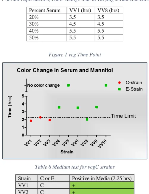
A time point of 2.25 hours was developed to distinguish between vcgC and vcgE strains in the medium. Figure 1. shows this time point for color change in the detection of vcgC. At 2.25 hours, all vcgC stains are positive (yellow) while there is only a single positive vcgE strain (VV8). When all 19 strains were tested at the 2.25 time point, 16 of 19 (68%) strains were correctly identified as either vcgC or vcgE. There were no false negatives. The results for each strain are shown in Table 8.
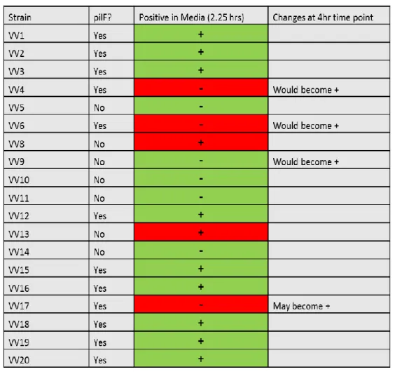
A time point was also developed to distinguish between strains with pilF genes correlated with virulence and those that do not. The time point was increased from 2.25 hours to 4 hours to include all pilF positive strains shown in Figure 2. This new time point had an accuracy of 78% with the initial nine strains of identifying those with genes correlated with pathogenicity and still contained no false negatives. Additional strains were not tested at the extended time point due to time constraints. However including those strains that exhibited a positive color change within 2.25 hours still had an accuracy rate of 74% with the possibility of 84% accuracy with an extended time point. This test does have a single false negative when all 19 strains are considered, but the extension of the time point may eliminate it. The results for individual strains are shown in Table 9.
an extended time of 4 hrs however, there is a difference between positive and negative pilF strains and whether or not they produce a positive result.
4. Cost and Accuracy Comparisons
The cost of the novel colorimetric media is primarily driven by the cost of pooled human serum and thus the optimization of the percentage of serum used for the media was vital to creating an affordable method for detection. The total cost per sample for the new method is $0.65 assuming that only 1 mL of media is needed. The cost for six 100 mL bottles of pooled human serum from Fisher Science is $1,307.06 or $0.65 per sample. Mannitol purchased from VWR International costs $473.85 for 5 kg. When purchased at this large quantity, the cost of mannitol for the medium is about $0.03 per 100 samples. Phenol Red Broth Base from Thermo Scientific™ costs $130.53 when purchased from Fisher Science, or $0.18 per 100 samples. The primary startup cost for the novel medium would be the purchase of a microbiological incubator. Fisher Scientific™ Isotemp™ Microbiological Incubators can be purchased for $2,600. Similar startup costs would be required for all culture-based methods including Chromagar Vibrio. By comparison, traditional PCR costs $1.17 per sample and takes at least 5 hours compared to the 2.5 hours required for the new method (Williams 2013).
5. Discussion
In the United States there has only been national surveillance of Vibrio vulnificus infections since 2007. Previously, infections were only reported the CDC from Gulf States. On average, there are 95 cases of Vibriosis caused by V. vulnificus reported every year with 85 cases leading to hospitalization and 35 cases leading to death. The number of cases is likely underreported (CDC). Although the number of infections are low, the cost of a hospitalization and death is high. The total cost associated with the average V. vulnificus infection is $2,792,171 per case and an average of 268 million dollars per year is spent on all cases (Scharff 2011). Improved methods of detection and increased surveillance have the potential to greatly decrease these costs and decrease loss of life.
With the rates of Vibrio infections expected to continue to increase with climate change and a higher number of susceptible people, the need for a rapid, easy and affordable method of detection becomes increasingly critical. The ability to distinguish V. vulnificus strains of the greatest concern will also help to make more informed public health decisions. The combination of human serum and mannitol with phenol red as an indicator has been shown in these experiments to detect pathogenic strains of V. vulnificus with high accuracy. The test can also be adjusted to meet the needs of the specific application. The medium could potentially be modified to detect pathogenic strains of V. vulnificus in shellfish such as oysters, water, and clinical samples.
Serum is an important aspect of the medium since it is known to be an important aspect of the human immune system. Complement is the bactericidal component of serum that has been shown to lower survivability of V. vulnificus in serum, especially vcgE type strains (Williams 2014). vcgC strains are also able to survive in blood with much lower iron concentrations than vcgE type strains (Bogard 2007). Keeping the concentration of serum as low as possible while still distinguishing between strains was vital to keeping the cost of the medium low.
septicemia and almost all cases of septicemia are caused by vcgC strains. The time point for pilF could also be used if the goal was to be as protective of public health as possible. There are vcgE strains that may have the ability to cause primary septicemia although their main route of infection is through wounds. For the inclusion of risk of wound infections especially, pilF time point would be the more accurate than vcg.
Polymorphisms in pilF have been associated with serum resistance and has also been shown that it may be a better measurement of pathogenicity than vcg (González 2012). It is a broader measurement since it will also include vcgE strains that have possibility to cause infection. While the vcg test may be sufficiently protective for oyster tissue testing, pilF should be used for water samples if wound infections are also of concern. It would be a better predictor of Vibriosis than vcg since any vcgE strain that may be causing illness would not be detected.
While there are many genes correlated with virulent strains of V. vulnificus, those that are known to be correlated with resistance to serum such as pilF as commonly used. These experiments have shown that there is also potential to develop a medium with serum to detect pathogenic strains. In comparison to other mediums that detect total V. vulnificus the accuracy of this medium is quite high. The further development of such a medium that could be used for both environmental and clinical samples could be used on a wider scale and would be protective of public health.
Tables and Figures
Table 1 Strain Identifications
Strain abbreviation Strain
vcg
E or
vcg
C
pilF
VV1
VV MO6
C
+
VV2
VV C7184
C
+
VV3
VV YJO16
C
+
VV4
VV JY1701
E
+
VV5
VV JY1305
E
-
VV6
VV E64MW
E
+
VV7
VV ENV1
E
?
VV8
VV SREL 106
E
-
VV9
VV SREL 89
E
-
VV10
VV SREL 46
E
-
VV11
VV AB6-307
?
-
VV12
VV SREL 190
C
+
VV13
VV SREL 119
E
-
VV14
VV SREL 118
E
-
VV15
VV LSU 1014
E
+
VV17
VV SREL 28
E
+
VV18
VV SREL 54
E
+
VV19
VV SPRC 10113
E
+
VV20
VV LSU 2098
E
+
Table 2 Serum Experiment 1 Broth Mixtures
10 % w/ 10 % w/o 50 % w/
50% w/o
+ control
-
control
PBS (uL)
800
900
400
500
900
1000
HI broth (uL)
100
0
100
0
100
0
Human Serum
(uL)
100
100
500
500
0
0
Table 3 Serum Experiment 2 and 3 Broth Mixtures
Serum (uL)
Mannitol (uL)
200
800
300
700
400
600
500
500
Table 4. Color change in Mannitol Broth
Strain
Positive in Mannitol
VV1
Yes
VV2
Yes
VV3
Yes
VV4
No
VV5
No
VV6
No
VV8
Yes
VV9
Yes
VV10
Yes
Table 5 Serum Experiment 1
VV1
VV5
Control -
NG
NG
Control +
++
++
10% serum + HI
+
+
10% serum
NG
NG
50% serum
NG
NG
NG= no growth ++=lawn += some growth
Table 6 Serum Experiment 2, Cell growth after 3 hours
Strain
A600 (30% serum)
A600 (50% serum)
VV1
0.3005
0.0055
VV2
0.0804
0.0215
VV4
0.051
0.0135
VV5
0.2475
0.188
Table 7 Serum Experiment 3, color change time at varying serum concentrations
Percent Serum
VV1 (hrs)
VV8 (hrs)
20%
3.5
3.5
30%
4.5
4.5
40%
5.5
5.5
50%
5.5
5.5
Figure 1 vcg Time Point
Table 8 Medium test for vcgC strains
Strain
C or E
Positive in Media (2.25 hrs)
VV1
C
+
VV2
C
+
VV3
C
+
Figure 2 pilF Time Point
VV5
E
-
VV6
E (clinical)
-
VV8
E
+
VV9
E
-
VV10
E
-
VV11
?
-
VV12
C
+
VV13
E
+
VV14
E
-
VV15
E
+
VV16
C
+
VV17
E
-
VV18
E
+
VV19
E
+
Table 9 pilF Medium Results
References
The biology of vibrios2006. , eds. American Society for Microbiology., , J. G. Swings, , Fabiano Lopes Thompson and . Washington, D.C.: ASM Press, http://search.lib.unc.edu?R=UNCb4905636.
Accuracy of HIV test is high but not perfect. 1994. AIDS Alert, 09; 2015/3, 1994, sec 9.
http://ic.galegroup.com/ic/scic/NewsDetailsPage/NewsDetailsWindow?failOverType=&query=&pr
odId=SCIC&windowstate=normal&contentModules=&display-query=&mode=view&displayGroupName=News&limiter=&currPage=&disableHighlighting=false& displayGroups=&sortBy=&search_within_results=&p=SCIC&action=e&catId=&activityType=&sca nId=&documentId=GALE%7CA15836520&source=Bookmark&u=unc_main&jsid=98ed749740d2e 1a9daa2862234b60b55.
- Morrison, Shatavia S., Tiffany - Williams, Aurora - Cain, Brett - Froelich, Casey - Taylor, Craig - Baker-Austin, David - Verner-Jeffreys, Rachel - Hartnell, James D. - Oliver, and Cynthia J. - Gibas. - Pyrosequencing-based comparative genome analysis of vibrio vulnificus environmental isolates- Public Library of Science.
Abbas, Abul K. 2015. Cellular and molecular immunology electronic resource], eds. Andrew H. Lichtman, , Shiv Pillai and . Philadelphia, PA: Elsevier Saunders,
http://search.lib.unc.edu?R=UNCb7925737; Full text available via the UNC-Chapel Hill Libraries (http://VB3LK7EB4T.search.serialssolutions.com/?V=1.0&L=VB3LK7EB4T&S=JCs&C=TC0001294 092&T=marc).
Amaro González, Carmen, Craig Baker-Austin, Elizabeth Lemm, Rachel Hartnell, James Lowther, Richard Onley, James D. Oliver, and David Lees. 2012. pilF polymorphism-based real-time PCR to distinguish vibrio vulnificus strains of human health relevance. Food Microbiology, 2012, Vol.30, Num.1, p.17-23.
Bogard, R. W., and J. D. Oliver. 2007. Role of iron in human serum resistance of the clinical and environmental vibrio vulnificus genotypes. Applied and Environmental Microbiology 73 (23) (Dec): 7501-5.
Centers for Disease Control and Prevention (CDC). 2007. Preliminary FoodNet data on the incidence of infection with pathogens transmitted commonly through food--10 states, 2006. MMWR.Morbidity and Mortality Weekly Report 56 (14) (Apr 13): 336-9.
Chatzidaki-Livanis, M., M. A. Hubbard, K. Gordon, V. J. Harwood, and A. C. Wright. 2006. Genetic distinctions among clinical and environmental strains of vibrio vulnificus. Applied and
Environmental Microbiology 72 (9) (Sep): 6136-41.
Drake, Stephenie L., Brooke Whitney, Jay F. Levine, Angelo DePaola, and Lee-Ann Jaykus. 2010. Correlation of mannitol fermentation with virulence-associated genotypic characteristics in vibrio vulnificus isolates from oysters and water samples in the gulf of mexico. Foodborne Pathogens and Disease 7 (1): 97-101.
Froelich, B., B. Froelich, and J. Oliver. 01. Orientation of mannitol related genes can further
differentiate strains of vibrio vulnificus possessing the vcgC allele. Advanced Studies in Biology 3 (4) (/01/2011): 151; 151,160; 160.
Froelich, Brett, and James D. Oliver. 2013. The interactions of vibrio vulnificus and the oyster crassostrea virginica. Microbial Ecology 65 (4): 807-16.
Garrido-Maestu, Alejandro, María-José Chapela, Elvira Peñaranda, Juan M. Vieites, and Ana G. Cabado. 2014. In-house validation of novel multiplex real-time PCR gene combination for the simultaneous detection of the main human pathogenic vibrios (vibrio cholerae, vibrio
parahaemolyticus, and vibrio vulnificus). Food Control 37 (0) (3): 371-9.
———. 2014. In-house validation of novel multiplex real-time PCR gene combination for the simultaneous detection of the main human pathogenic vibrios (vibrio cholerae, vibrio parahaemolyticus, and vibrio vulnificus). Food Control 37 (0) (3): 371-9.
Horré, Regine, Günter Marklein, and Klaus Peter Schaal. 1996. Vibrio vulnificus, an emerging human pathogen. Zentralblatt Für Bakteriologie 284 (2–3) (7): 273-84.
Linkous, Debra A., and James D. Oliver. 1999. Pathogenesis of vibrio vulnificus. FEMS Microbiology Letters 174 (2): 207-14.
Oliver, JamesD. 2013. Vibrio vulnificus: Death on the half shell. A personal journey with the pathogen and its ecology. Microbial Ecology 65 (4) (05/01): 793-9, http://dx.doi.org/10.1007/s00248-012-0140-9.
———. 2005. Vibrio vulnificus. In , eds. Shimshon Belkin, RitaR Colwell, 253-276Springer US,
http://dx.doi.org/10.1007/0-387-23709-7_10 .
Paz, S., N. Bisharat, E. Paz, O. Kidar, and D. Cohen. 2007. Climate change and the emergence of< i> vibrio vulnificus</i> disease in israel. Environmental Research 103 (3): 390-6.
Pfeffer, C. S., M. F. Hite, and J. D. Oliver. 2003. Ecology of vibrio vulnificus in estuarine waters of eastern north carolina. Applied and Environmental Microbiology 69 (6) (Jun): 3526-31.
Pollard, Thomas D. (Thomas Dean), 1942-. 2008. Cell biology electronic resource], eds. William C. Earnshaw, , Jennifer Lippincott-Schwartz and . Philadelphia: Saunders/Elsevier,
http://search.lib.unc.edu?R=UNCb7437858; Full text available via the UNC-Chapel Hill Libraries (http://VB3LK7EB4T.search.serialssolutions.com/?V=1.0&L=VB3LK7EB4T&S=JCs&C=TC0000386 388&T=marc).
Rosche, Thomas M., Yutaka Yano, and James D. Oliver. 2005. A rapid and simple PCR analysis indicates there are two subgroups of vibrio vulnificus which correlate with clinical or environmental isolation. Microbiology and Immunology 49 (4): 381-9.
Scharff, Robert L. 2012. Economic burden from health losses due to foodborne illness in the united states. Journal of Food Protection® 75 (1): 123-31.
Sullivan,Kathleen E. and Jerry A. Winkelstein. 186: genetically determined disorders of the complement system. In The online metabolic and molecular bases of inherited disease.
Tantillo, G. M., M. Fontanarosa, A. Di Pinto, and M. Musti. 2004. Updated perspectives on emerging vibrios associated with human infections. Letters in Applied Microbiology 39 (2): 117-26.
US Food and Drug Administration. 2003. National shellfish sanitation program. guide for the control of molluscan shellfish. Center for Food Safety and Applied Nutrition, US Food and Drug
Administration, Washington, DC.
Williams, Tiffany C., Mesrop Ayrapetyan, Heather Ryan, and James D. Oliver. 2014. Serum survival of vibrio vulnificus: Role of genotype, capsule, complement, clinical origin, and in situ incubation.
Pathogens 3 (4): 822-32.